EXAMINATION OF THE KNEE - c.ymcdn.com · 4/24/15 2 • Knee Anatomy • Articular Cartilage •...
20
4/24/15 1 Evaluation and Treatment of Knee Injuries in Primary Care by Bethany Reed, MSN, AGPCN-BC EXAMINATION OF THE KNEE DECLARATIONS There has been no commercial support or sponsorship for this program. The planners and presenters have declared that no conflicts of interest exist. The program co-sponsors do not endorse any products in conjunction with any educational activity. OBJECTIVES • At the end of this session, the participant will be able to: • 1. Identify common knee injuries seen in primary care. • 2. Review the anatomy of the knee joint. • 3. Review physical assessment and diagnostic evaluation of the knee. • 4. Discuss treatment recommendations, rehabilitation protocols, and when to refer.
Transcript of EXAMINATION OF THE KNEE - c.ymcdn.com · 4/24/15 2 • Knee Anatomy • Articular Cartilage •...

4/24/15
1
Evaluation and Treatment of Knee Injuries in Primary Care by
Bethany Reed, MSN, AGPCN-BC
EXAMINATION OF THE KNEE
DECLARATIONS
There has been no commercial support or sponsorship for this program. The planners and presenters have declared that no conflicts of interest exist. The program co-sponsors do not endorse any products in conjunction with any educational activity.
OBJECTIVES • At the end of this session, the participant will be able to: • 1. Identify common knee injuries seen in primary care.
• 2. Review the anatomy of the knee joint.
• 3. Review physical assessment and diagnostic evaluation of the knee.
• 4. Discuss treatment recommendations, rehabilitation protocols, and when to refer.
4/24/15
2
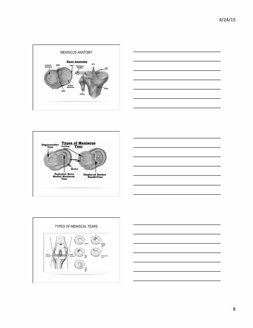
• Knee Anatomy
• Articular Cartilage
• Meniscal Cartilage
• Ligamentous Anatomy
• Patello-femoral Joint
• General Principles
• Early examination sometimes difficult due to guarding
• Mechanism of Injury
• Effusion
• Able to bear weight after the injury?
PHYSICAL EXAM - KNEE HISTORY
• Age
• Occupation
• Chief complaint: Pain
• Mechanical symptoms (catching, locking, instability)
• Clicking, popping
• Location
• Onset
• Injury
• Precipitating factors
• Previous treatments: surgeries, medications, PT, injections
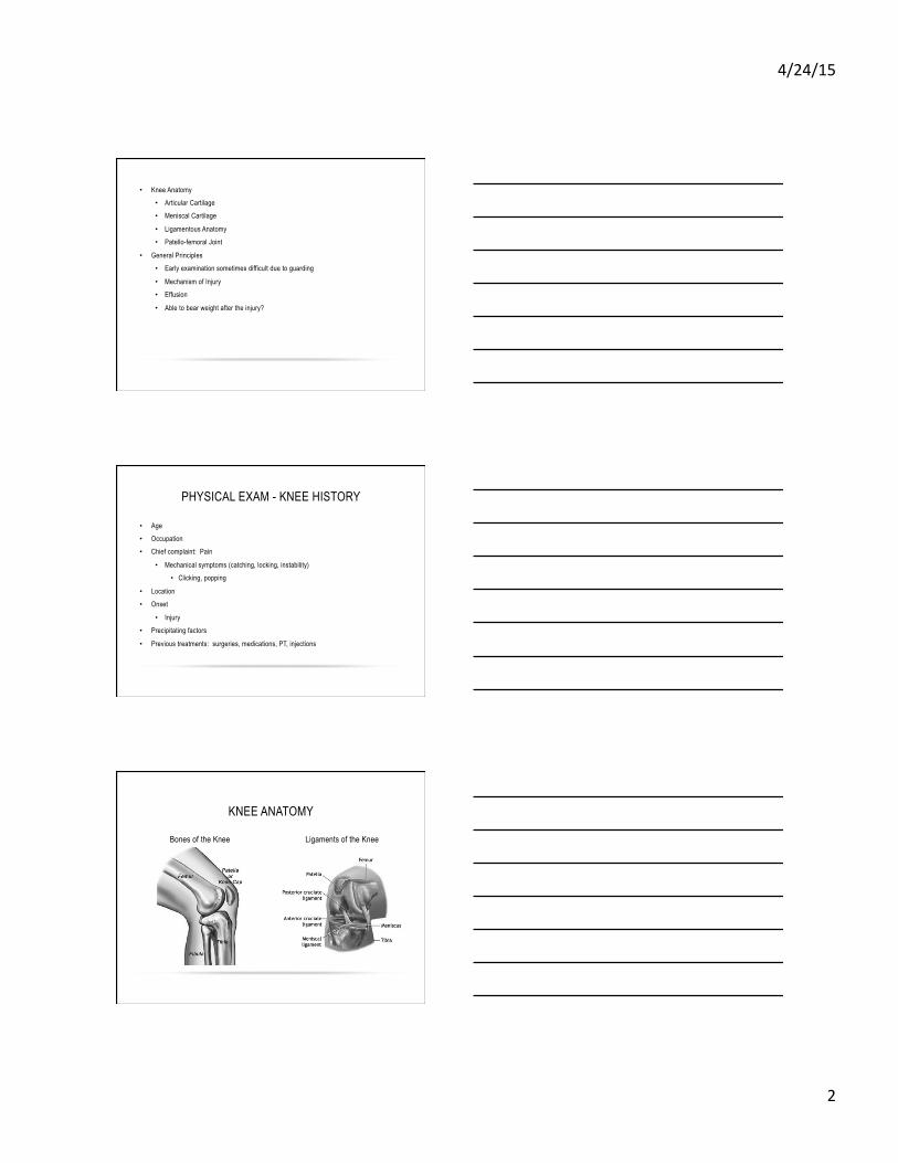
KNEE ANATOMY
Bones of the Knee Ligaments of the Knee
4/24/15
3
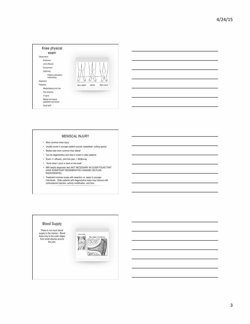
Knee physical exam
Observation
Erythema
Joint effusion
Ecchymosis
Deformity
Patella subluxation maltracking
Alignment
Palpation
Medial/lateral joint line
Pes anserine
IT band
Medial and lateral patellafemoral facets
Quad girth
MENISCAL INJURY • Most common knee injury • Usually acute in younger patient (soccer, basketball, cutting sports) • Medial side more common than lateral
• Can be degenerative and slow in onset in older patients
• Exam +/- effusion, joint line pain, + McMurray
• “Hurts when I pivot or twist on the knee”
• MRI helpful diagnostic test (NOT NECESSARY IN OLDER FOLKS THAT HAVE SIGNIFICANT DEGENERATIVE CHANGES ON PLAIN RADIOGRAPHS.)
• Treatment involves scope with resection vs. repair in younger individuals. Older patients with degenerative tears may improve with corticosteroid injection, activity modification, and time.
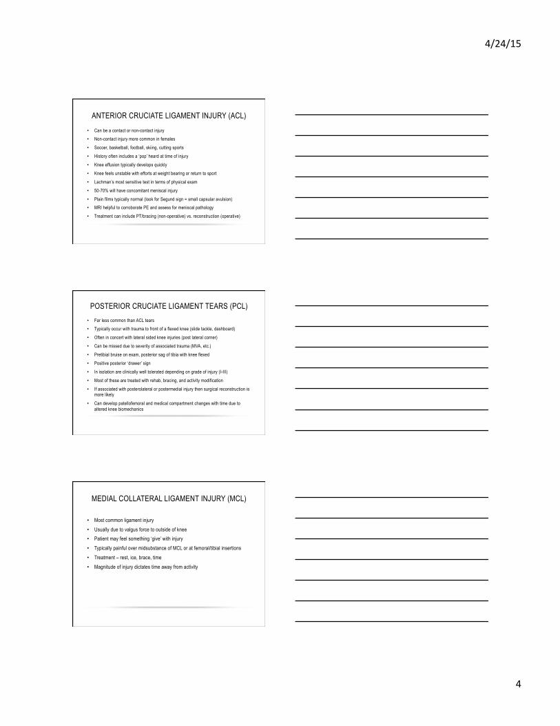
Blood Supply There is not much blood
supply to the menisci. Blood flows only to the outer edges
from small arteries around the joint.
4/24/15
4
ANTERIOR CRUCIATE LIGAMENT INJURY (ACL) • Can be a contact or non-contact injury • Non-contact injury more common in females
• Soccer, basketball, football, skiing, cutting sports
• History often includes a ‘pop’ heard at time of injury
• Knee effusion typically develops quickly
• Knee feels unstable with efforts at weight bearing or return to sport
• Lachman’s most sensitive test in terms of physical exam
• 50-70% will have concomitant meniscal injury
• Plain films typically normal (look for Segund sign = small capsular avulsion) • MRI helpful to corroborate PE and assess for meniscal pathology
• Treatment can include PT/bracing (non-operative) vs. reconstruction (operative)
POSTERIOR CRUCIATE LIGAMENT TEARS (PCL) • Far less common than ACL tears • Typically occur with trauma to front of a flexed knee (slide tackle, dashboard)
• Often in concert with lateral sided knee injuries (post lateral corner)
• Can be missed due to severity of associated trauma (MVA, etc.)
• Pretibial bruise on exam, posterior sag of tibia with knee flexed
• Positive posterior ‘drawer’ sign
• In isolation are clinically well tolerated depending on grade of injury (I-III)
• Most of these are treated with rehab, bracing, and activity modification
• If associated with posterolateral or postermedial injury then surgical reconstruction is more likely
• Can develop patellofemoral and medical compartment changes with time due to altered knee biomechanics
MEDIAL COLLATERAL LIGAMENT INJURY (MCL)
• Most common ligament injury • Usually due to valgus force to outside of knee • Patient may feel something ‘give’ with injury
• Typically painful over midsubstance of MCL or at femoral/tibial insertions
• Treatment – rest, ice, brace, time
• Magnitude of injury dictates time away from activity

4/24/15
5
PATELLOFEMORAL INSTABILITY/PATELLA DISCLOCATION
• Most common in adolescent female (gymnastics, soccer, etc.) • Often results from sudden quadriceps contraction combined with valgus knee • Many patients will sublux and spontaneously relocate • May present to office days to weeks later with swollen knee. Pain primarily over medial
retinaculum (MPFL region). • True dislocations that require reduction will have large effusion/hemarthrosis. Difficulty even
bending knee. • Patients can avulse cartilage/bone from patella (typically as the knee cap reduces back into the
trochlea from lateral position). Small fragments can sometimes be visualized on plain radiographs. With large, tense hemarthrosis, aspiration for pain control is sometimes helpful.
• MRI with large effusions may help to assess cartilage damage to patella and point out cartilaginous or osteocartilaginous loose bodies.
• Most 1st time dislocations can be treated with immobilization, rest, physical therapy, and bracing.
• Recurrent dislocations may require re-alignment procedure depending on pre-existing risk factors and level of injury.
QUADRICEPS CONTUSION
• Direct blow to the front of thigh (helmet hit in football) • Often very sore with swelling ‘hidden’ in the soft tissues • Knee flexion is painful and avoided
• Be alert for potential compartment syndrome
• Ice and ? of immobilization in flexion
• Myositis Ossificans is largest long term concern
• More common with immobilization • Larger hematoma
• Role of NSAID’s in prevention is debated
ILIOTIBIAL BAND SYNDROME (RUNNER’S KNEE)
• Often comes on slowly. Runners will say that it comes on after 20 minutes or after certain mileage.
• Pain is lateral, above the level of the joint.
• Rarely associated with much swelling.
• Pain dissipates with relative rest ( ‘if I walk it goes away’ ).
• Tight iliotibial band is often the etiology. May have trochanteric symptoms at their hip as well.
• Rest, ice, NSAID’s, iliotibial band stretching, foam rolling, formal PT.
• Difficult to get runners to take the necessary break from training to heal.
4/24/15
6
Knee Anatomy • Patellofemoral joint
• Articular cartilage
• Tibiofemoral joint
• Articular cartilage
• Meniscus
• Ligaments
• Anterior cruciate ligament (ACL)
• Medial collateral ligament (MCL)
• Posterior cruciate ligament (PCL)
• Lateral collateral ligament (LCL)
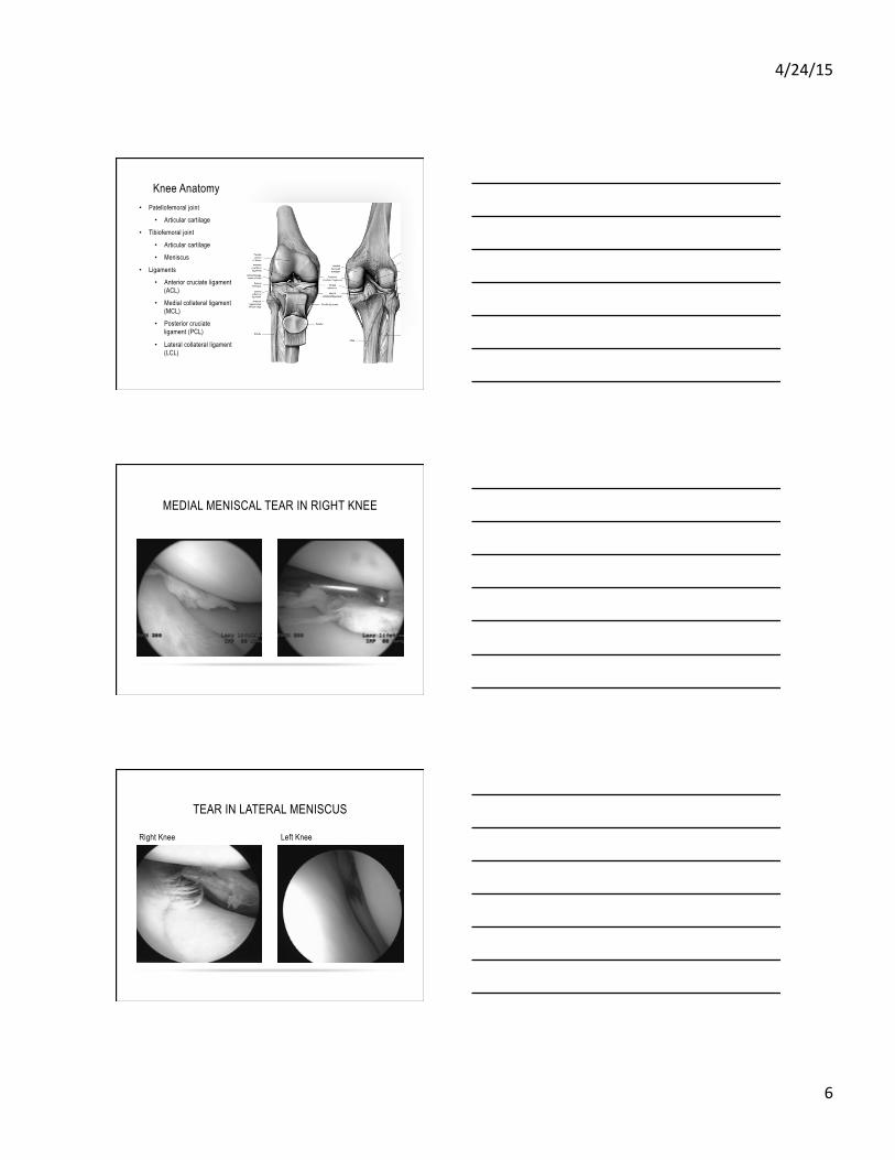
MEDIAL MENISCAL TEAR IN RIGHT KNEE
TEAR IN LATERAL MENISCUS
Right Knee Left Knee

4/24/15
7
Subluxed bucket handle tear of medial meniscus causing right knee ‘locking’
Cartilage wear in the center of the femoral trochlear groove
ACUTE KNEE INJURIES
• When evaluating, timing is important • Earlier is better • Muscle spasm and swelling can make examination difficult • Mechanism of injury • Location of pain • Effusion
MENISCUS INJURY
MRI of intact meniscus MRI of torn meniscus
4/24/15
8
MENISCUS ANATOMY
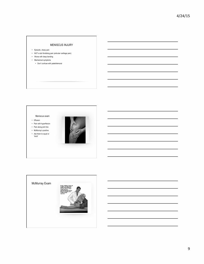
TYPES OF MENISCAL TEARS
4/24/15
9
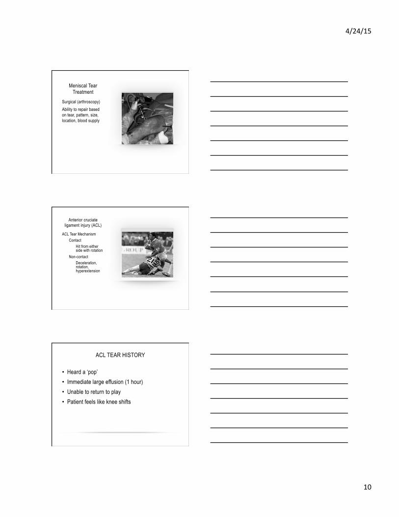
MENISCUS INJURY • Episodic, sharp pain • NOT a dull throbbing pain (articular cartilage pain) • Worse with deep bending
• Mechanical symptoms
• Don’t confuse with patellofemoral
Meniscus exam • Effusion • Pain with hyperflexion • Pain along joint line
• McMurray’s positive
• Ask them to squat or ‘duck’
McMurray Exam
4/24/15
10
Meniscal Tear Treatment
Surgical (arthroscopy) Ability to repair based on tear, pattern, size, location, blood supply
Anterior cruciate ligament injury (ACL)
ACL Tear Mechanism Contact
Hit from either side with rotation
Non-contact Deceleration, rotation, hyperextension
ACL TEAR HISTORY
• Heard a ‘pop’ • Immediate large effusion (1 hour) • Unable to return to play • Patient feels like knee shifts

4/24/15
11
ACL Tear Examination
• Often limited by pain and guarding
• Effusion
• Hemarthrosis
• Lachman • Anterior drawer
• Pivot shift
ACL Examination The Lachman
• Most sensitive • Knee in 30 degrees of
flexion • Hang heel off side of bed
• Stabilize femur with one hand
• Translate tibia with other hand
ACL Examination Anterior Drawer • Knee flexed to 80
degrees • Hamstrings are palpated • Proximal tibia moved
anteriorly • Compare to contralateral
knee
4/24/15
12
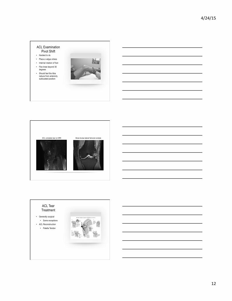
ACL Examination Pivot Shift
• Hardest to do • Place a valgus stress • Internal rotation of foot
• Flex knee beyond 30 degrees
• Should feel the tibia reduce from anteriorly subluxated position
ACL complete tear on MRI Bone bruise lateral femoral condyle
ACL Tear Treatment
• Generally surgical • Some exceptions
• ACL Reconstruction
• Patella Tendon
4/24/15
13
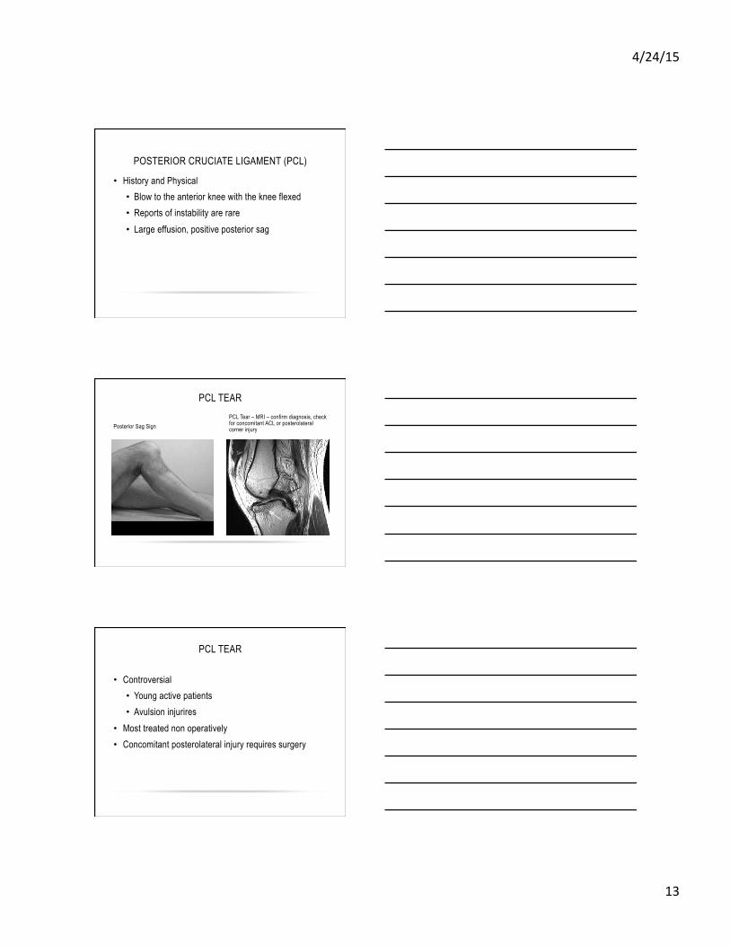
POSTERIOR CRUCIATE LIGAMENT (PCL)
• History and Physical • Blow to the anterior knee with the knee flexed • Reports of instability are rare • Large effusion, positive posterior sag
PCL TEAR
Posterior Sag Sign PCL Tear – MRI – confirm diagnosis, check for concomitant ACL or posterolateral corner injury
PCL TEAR
• Controversial • Young active patients • Avulsion injurires
• Most treated non operatively • Concomitant posterolateral injury requires surgery
4/24/15
14
MEDIAL COLLATERAL LIGAMENT INJURY (MCL) • Most common ligament injury
• Usually caused by hit to the outside of knee (valgus force)
• Patients often describe feeling something ‘give’ but not a true ‘pop’
• Painful to flex the knee
• Ask them to point to the location of the pain – often exquisitely tender of medial epicondyle
• On physical exam no or small effusion appreciated
• Tenderness medial epicondyle
• Usually torn from femur
• Valgus stress testing
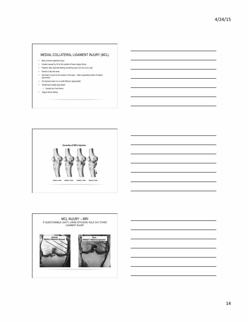
MCL INJURY – MRI IF QUESTIONABLE LAXITY, LARGE EFFUSION, RULE OUT OTHER
LIGAMENT INJURY
4/24/15
15
MCL INJURY - TREATMENT
• Almost always non-surgical • 95% will heal with support • With concomitant ACL injury usually do not need to fix MCL
• Treatment based on severity
• Grade 1: no brace, early rehabilitation, full return to activity 5 days
• Grade 2: brace 2-3 weeks, full return to activity 17 days
• Grade 3: brace 4-6 weeks, full return to activity 33 days
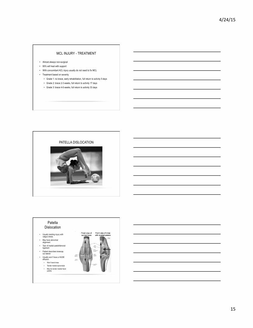
PATELLA DISLOCATION
Patella Dislocation
• Usually twisting injury with valgus stress
• May have abnormal alignment
• Tear of medial patellofemoral ligament
• Patient describes kneecap out lateral
• Usually won’t have a HUGE effusion • Won’t bend knee • Tender medial epicondyle • May be tender medial facet
patella

4/24/15
16
Patella Dislocation • Apprehension Test • Treatment
• RICE • Brace or tape to hold
patella • PT for quadriceps
training • Return to play after
completing functional rehabilitation/running program
Patella Dislocation Treatment
• 1st time dislocation treat non-operatively • 80% will heal
• MRI to look for loose fragments in the knee if persistent effusion
• Multiple dislocations will require operative treatment
• Each time patella dislocates, the articular cartilage can be injured on the femur or patella
QUADRICEPS CONTUSION • Direct blow to the thigh (quad) • Hematoma in muscle • Painful knee flexion • Watch for compartment syndrome! • Grade based on knee flexion at 24 hours
• Grade 1: >90 degrees • Grade 2: 45-90 degrees • Grade 3: <45 degrees
• Treatment • Ice, keep knee maximally flexed, +/- compression wrap, NSAID’s controversial • Dreaded complication is myositis ossificans in quad muscle
• 9% incidence. Higher when poor initial ROM, delayed treatment • Immobilization increases chance of it
• Early ROM
4/24/15
17
Iliotibial Band (ITB) Friction
Syndrome • Overuse injury • Worse with increased
activity • Dull, throbbing pain
• No effusion
• Pain over lateral epicondyle
• Tight ITB
• Differentiate between popliteus tendonitis, LCL sprain, lateral meniscus tear
Pes Anserine Bursitis
• Hamstring tendonitis • Chief Complaint with
sleeping and walking
• Pain on palpation of the knee and hamstring contracture
• Treatment includes:
• Ice
• PT • Eccentric hamstring
strenthening, ham/quad/ITB flexibility, hip strengthening
• Corticosteroid injections
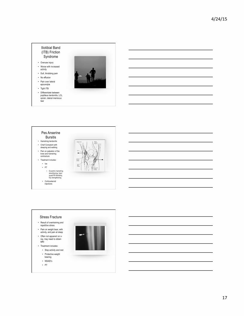
Stress Fracture • Result of overtraining and
repetitive stress • Pain on weight bear, with
activity, and pain at sleep • Often not apparent on x-
ray, may need to obtain MRI
• Treatment includes:
• Stop activity and rest
• Protective weight bearing
• NSAID’s • PT
4/24/15
18
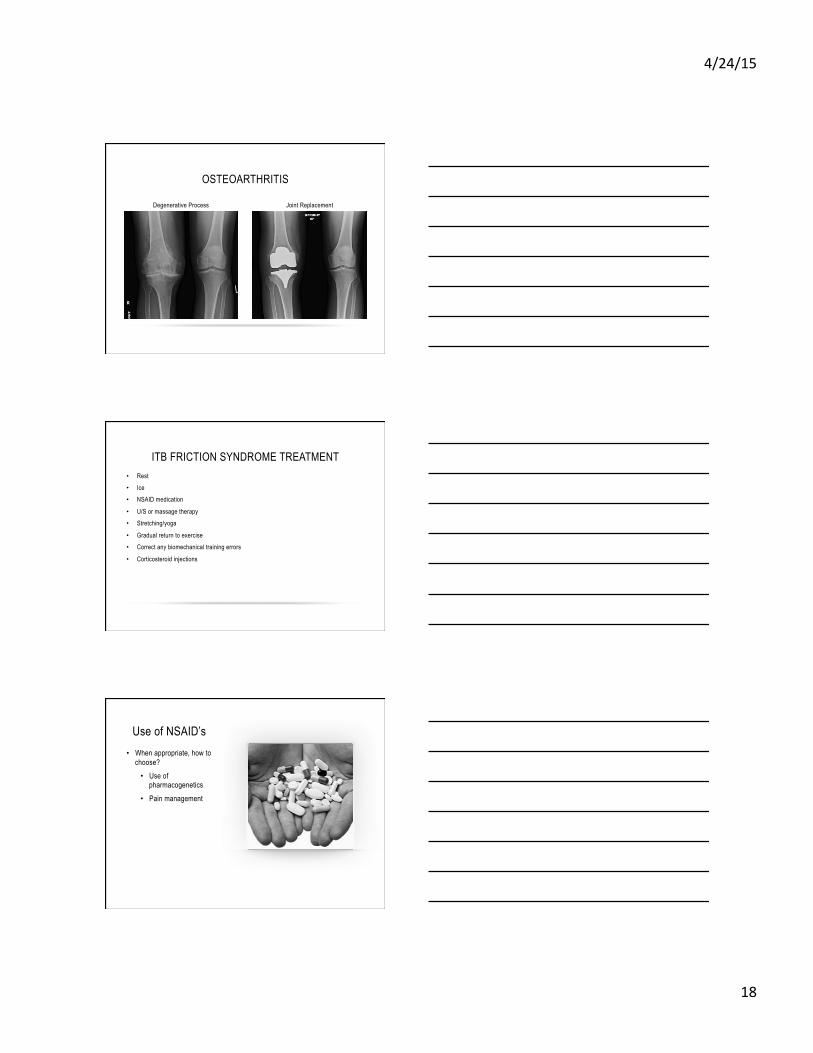
OSTEOARTHRITIS
Degenerative Process Joint Replacement
ITB FRICTION SYNDROME TREATMENT • Rest
• Ice
• NSAID medication
• U/S or massage therapy
• Stretching/yoga
• Gradual return to exercise
• Correct any biomechanical training errors
• Corticosteroid injections
Use of NSAID’s • When appropriate, how to
choose? • Use of
pharmacogenetics • Pain management
4/24/15
19
PHARMACOGENETICS & PAIN MANAGEMENT
• Ruth is a healthy, active 59 year old retired executive administrative assistant
• Suffers from daily right knee joint stiffness and pain with minimal exertion.
• Acetaminophen 4 grams daily provides only partial relief.
• Physical exam WNL
• Knee painful with squatting, rising from sitting position, knee flexion/extension
• Standard blood chemistry and lipid profiles WNL
• Radiography of knee reveals irregular joint space narrowing and slightly increased radiologic density of subchondral bone, signs consistent with OA.
• No GI complications
• Drug of choice for pain management?
CASE STUDY
HOW IT WORKS
4/24/15
20
THANK YOU
E-mail: [email protected] LinkedIn: www.linkedin.com/in/TravelNP
Skype: malaproject
BETHANY REED, MSN, AGPCN-BC