Examination of Smad2 and Smad4 copy-number variations in skin cancers
5
Abstract Background Smad2 and Smad4 transcription factors were identified as the signalling mediators of transforming growth factor ß (TGF ß) pathway. Copy number variations (CNVs) have been discovered to have phenotypic conse- quences and be associated with various types of cancers. CNVs of Smad2 and Smad4 were found to be associated with cancer pathogenesis in the recent array-based study. However, no such study has been performed in skin cancer yet. In this study, we aim to examine the CNVs of Smad2 and Smad4 in skin samples. Methods A total of 195 paired samples including basal cell carcinoma (BCC), squamous cell carcinoma (SCC) and actinic keratosis (AK) were included. Real-time PCR was used for the quantification of Smad2 and Smad4 copy numbers. Results CNVs of Smad2 showed statistical differences between cancer samples (both SCC and BCC) and normal tissues (p<0.05). For Smad4, statistical difference was ob- served only in SCC samples (p=0.014), but not in BCC and AK samples (p=0.173 and 0.314, respectively). Association analysis showed that the frequencies of Smad2 and Smad4 CNVs were correlated with the severity of skin abnormali- ties (p=0.002 for Smad2 and p=0.029 for Smad4). Conclusions CNVs of Smad2 are associated with SCC and BCC, while CNVs of Smad4 are associated with SCC but not BCC. Keywords Smad2 · Smad4 · Copy number variations · Basal cell carcinoma · Squamous cell carcinoma · Actinic keratosis Introduction Smad transcription factors were identified as the signal- ling mediators of the transforming growth factor ß (TGF ß) superfamily, which consists of three major subfamilies: TGFß, bone morphogenetic proteins (BMPs) and activins/ inhibins [1]. The TGFß superfamily directly activates the Smad signalling pathway, in addition to other Smad- independent pathways. The Smad family of proteins can be divided into three functional groups: the receptor-activated *These authors contributed equally to this work. § These authors contributed equally to this work. Y. Shao · J. Zhang · J. Wan · W. Zhang () · B. Yu Shenzhen Key Lab for Translational Medicine of Dermatology Shenzhen-PKU-HKUST Medical Center 1120 Lian-Hua Road, Fu-Tian District Shenzhen, Guangdong 518036, China e-mail: [email protected] Y. Shao · J. Zhang · J.Wan · B. Yu Biomedical Research Institute Shenzhen-PKU-HKUST Medical Center Shenzhen, Guangdong, China Y. Shao · J. Zhang · B. Yu Department of Dermatology Shenzhen Hospital Peking University Shenzhen, Guangdong, China R. Zhang Department of General Surgery Taizhou Municipal Hospital Zhejiang Province, China J. Wan · W. Zhang Division of Life Science Hong Kong University of Science and Technology Hong Kong, China W. Zhang JNU-HKUST Joint Lab Ji-Nan University Guangdong, China Clin Transl Oncol (2012) 14:138-142 DOI 10.1007/s12094-012-0773-7 RESEARCH ARTICLES Examination of Smad2 and Smad4 copy-number variations in skin cancers Yong Shao* · Jie Zhang* · Richu Zhang · Jun Wan · Wei Zhang § · Bo Yu § Received: 9 February 2011 / Accepted: 21 May 2011
Transcript of Examination of Smad2 and Smad4 copy-number variations in skin cancers

AbstractBackground Smad2 and Smad4 transcription factors were identified as the signalling mediators of transforming growth factor ß (TGF ß) pathway. Copy number variations (CNVs) have been discovered to have phenotypic conse-quences and be associated with various types of cancers. CNVs of Smad2 and Smad4 were found to be associated
with cancer pathogenesis in the recent array-based study. However, no such study has been performed in skin cancer yet. In this study, we aim to examine the CNVs of Smad2 and Smad4 in skin samples.Methods A total of 195 paired samples including basal cell carcinoma (BCC), squamous cell carcinoma (SCC) and actinic keratosis (AK) were included. Real-time PCR was used for the quantifi cation of Smad2 and Smad4 copy numbers.Results CNVs of Smad2 showed statistical differences between cancer samples (both SCC and BCC) and normal tissues (p<0.05). For Smad4, statistical difference was ob-served only in SCC samples (p=0.014), but not in BCC and AK samples (p=0.173 and 0.314, respectively). Association analysis showed that the frequencies of Smad2 and Smad4 CNVs were correlated with the severity of skin abnormali-ties (p=0.002 for Smad2 and p=0.029 for Smad4).Conclusions CNVs of Smad2 are associated with SCC and BCC, while CNVs of Smad4 are associated with SCC but not BCC.
Keywords Smad2 · Smad4 · Copy number variations · Basal cell carcinoma · Squamous cell carcinoma · Actinic keratosis
Introduction
Smad transcription factors were identifi ed as the signal-ling mediators of the transforming growth factor ß (TGF ß) superfamily, which consists of three major subfamilies: TGFß, bone morphogenetic proteins (BMPs) and activins/inhibins [1]. The TGFß superfamily directly activates the Smad signalling pathway, in addition to other Smad-independent pathways. The Smad family of proteins can be divided into three functional groups: the receptor-activated
*These authors contributed equally to this work.§These authors contributed equally to this work.
Y. Shao · J. Zhang · J. Wan · W. Zhang (�) · B. YuShenzhen Key Lab for Translational Medicine of DermatologyShenzhen-PKU-HKUST Medical Center1120 Lian-Hua Road, Fu-Tian DistrictShenzhen, Guangdong 518036, Chinae-mail: [email protected]
Y. Shao · J. Zhang · J.Wan · B. YuBiomedical Research InstituteShenzhen-PKU-HKUST Medical CenterShenzhen, Guangdong, China
Y. Shao · J. Zhang · B. YuDepartment of DermatologyShenzhen Hospital Peking UniversityShenzhen, Guangdong, China
R. ZhangDepartment of General SurgeryTaizhou Municipal HospitalZhejiang Province, China
J. Wan · W. ZhangDivision of Life ScienceHong Kong University of Science and TechnologyHong Kong, China
W. ZhangJNU-HKUST Joint LabJi-Nan UniversityGuangdong, China
Clin Transl Oncol (2012) 14:138-142DOI 10.1007/s12094-012-0773-7
R E S E A R C H A R T I C L E S
Examination of Smad2 and Smad4 copy-number variations in skincancers
Yong Shao* · Jie Zhang* · Richu Zhang · Jun Wan · Wei Zhang§ · Bo Yu§
Received: 9 February 2011 / Accepted: 21 May 2011

Clin Transl Oncol (2012) 14:138-142 139
Smads (R-Smads), common mediator Smads (Co-Smads) and the inhibitory Smads (I-Smads). The R-Smads include Smad1, Smad2, Smad3, Smad5 and Smad8. Smad6 and Smad7 are I-Smads, while Smad4 is the only mammalian Co-Smad identifi ed thus far which mediates signals from both the TGFß/activin and BMP signalling pathways [2].
The TGFß signalling pathway plays an important role in tumour suppression, primarily via growth inhibition, apop-tosis and maintenance of differentiation. Aberrations in components of the TGFß signalling pathway were observed in the majority of human epithelial cancers (>85%) includ-ing pancreatic, colon, breast, prostate and lung [3]. For skin cancer, expression of Smad2 was found to be lost in almost all human skin squamous cell carcinomas (SCCs) exam-ined, suggesting that Smad2 plays a tumour suppressive role [4]. Likewise, deletion of Smad4 in multiple murine tissues results in spontaneous cancers including skin cancer [5, 6]. It also has been reported that copy number variations (CNVs) of SMAD2 and Smad4 were associated with can-cer pathogenesis in the recent array-based study [7, 8].
CNVs were originally defi ned by the presence of vari-able numbers of copies of large, multi-kilobase genomic regions in the genomes of different individuals [9]. How-ever, recent high-resolution genome maps have revealed smaller CNVs among healthy humans [9, 10], thus extend-ing the defi nition of CNVs to the length of regions being as short as several hundred bases. Several methodologies, such as the most commonly used array-based comparative genomic hybridisation (aCGH), were utilised for genome-wide CNV detection and genotyping. CNVs have been discovered to have phenotypic consequences and have been associated with various types of cancers over the past few years [11]. However, few CNV studies were performed in skin malignancies [12, 13].
Skin cancers are divided into melanoma and non-mela-noma. Non-melanoma, which is about 20 times more com-mon than melanoma, includes basal cell carcinoma (BCC), SCC and other types of skin cancer. Actinic keratosis (AK) is considered the earliest stage in the development of skin cancer and has the potential to progress to SCC. In our
study, we examined the CNVs of Smad2 and Smad4 in 195 paired samples including BCC, SCC and AK. We found that CNVs of Smad2 are associated with SCC and BCC, while CNVs of Smad4 are associated with SCC but not BCC.
Materials and methods
Controls and patient samples
Surgically resected tumour tissues and adjacent normal tissues were collected from 67 SCC, 85 BCC and 43 AK patients. The study was approved by the ethical committee of Peking University Shenzhen Hospital. The individuals gave their written informed consent. The investigations were conducted according to the Declaration of Helsinki principles.
DNA extraction and quantifi cation of copy numbers
Genomic DNA was isolated from the tissues using the Ge-nomic DNA Extraction Kit (Innogent, Shenzhen, China) according to the manufacturer’s instruction. Quantitative PCR was performed through BioRad Chromo4 real-time PCR system. Average copy numbers of RNAse P in nor-mal candidates (copy numbers=2) were used as the control [14]. The copy numbers of Smad2 and Smad4 were calcu-lated by using the comparative C(T) method [15]. Cut-off values of 0.33, 0.67 and 1.33 were used to defi ne the copy numbers as 0, 1 and 2 respectively. The primers for initial quantifi cation are listed in Table 1. Standard curves of the primers are shown in Fig. 1 and calculation of primer ef-fi ciency is shown in Table 2. The primers for validation of Smad2 and Smad4 are listed in Table 3. Statistical analysis was performed using chi-square test or Fisher’s exact test. Association analysis was performed using the linear-by-linear association test. P-values less than 0.05 were consid-ered statistically signifi cant.
Table 1 Primers for initial quantifi cation of copy numbers

140 Clin Transl Oncol (2012) 14:138-142
Results
Table 4 shows CNVs of Smad2 in skin samples. A total of 195 paired samples were examined. Statistical differences were observed in SCC and BCC samples as compared with the normal tissues (p<0.05). However, there was no signifi cant difference between AK samples and controls (p=0.152). For Smad4, statistical difference was observed only in SCC samples (p=0.014), but not in BCC and AK samples (p=0.173 and 0.314, respectively) (Table 5). The initial quantifi cation of Smad2 and Smad4 was further vali-dated by two additional sets of primers and similar results were obtained (Table 6).
For both Smad2 and Smad4, The highest frequencies of CNVs were observed in SCC samples, while the lowest frequencies were observed in AK samples. Since SCC is known to be more malignant than BCC, while AK belongs to skin pre-malignancy, we then performed the association analysis among different types of abnormalities (Table 7). Statistical differences were observed for Smad2 (p=0.002) and Smad4 (p=0.029), indicating that the frequencies of CNVs may be correlated with the severity of skin abnor-malities.
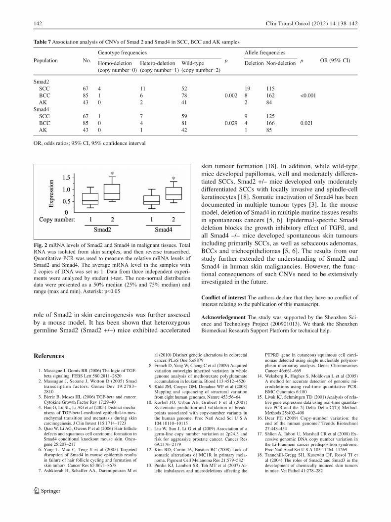
Next, we checked whether the mRNA levels of Smad2 and Smad4 were positively correlated with their copy numbers. Representative samples from the malignant tis-sues were divided into two groups: copy numbers=2 and copy number=1. As shown in Fig. 2, expressions of Smad2 and Smad4 in the samples with two copies of DNA were signifi cantly higher than those with one copy of DNA, sug-gesting that CNVs have phenotypic consequences.
Discussion
CNVs have been clearly shown to have the potential to di-rectly or indirectly infl uence a healthy individual’s suscep-tibility to cancer, for example by varying the gene dosage of tumour suppressors or oncogenes [16, 17]. It is suggest-
Fig. 1 Standard curves of the primers
Table 2 Calculation of primer effi ciency
Genes Dilutions Log Ct Slope Effi ciency
1 1 32.63 10 2 29.72 Smad2 100 3 25.59 -3.35 0.98841698 1000 4 22.86 10000 5 19.31 1 1 34.12 10 2 30.73 Smad4 100 3 27.48 -3.254 1.02914964 1000 4 24.37 10000 5 21.03 1 1 31.75 10 2 28.37 RNAse P 100 3 25.34 -3.296 1.01093512 1000 4 21.93 10000 5 18.49
Table 3 Primers for validation of copy numbers of Smad2 and Smad4
Forward Reverse
Smad2 (exon 1) CAGTTCCGCCTCCAATCGC GGGACCTTTTGTTCCTTCCTCTTSmad2 (exon 11) GCTACCACCTGCCACT AGCCCAAACATAGACCTTASmad4 (exon 1) GCTCAGTGGCTTCTCGACAAGTT TCCCCTCACCCGCTCCCASmad4 (exon 12) AGAGGAAGGGATGAAAC GAAATACCACCACCAAA

Clin Transl Oncol (2012) 14:138-142 141
ed that the genes present in very small regions of CNVs are excellent candidates for evaluation in cancer pathogenesis. Examination of the CNVs for such genes helps to under-stand the functional consequences of these CNVs. Previ-ous studies have shown that CNVs of SMAD2 and Smad4 were associated with cancer pathogenesis [7, 8]. However, in skin cancer, no such study has yet been performed.
In our study, we found that CNVs of Smad2 are as-sociated with SCC and BCC, while CNVs of Smad4 are associated with SCC but not BCC. The significance of these fi ndings was supported by some previous studies. It is reported that Smad2 was lost in almost all human skin SCCs examined [4]. The loss of Smad2 in human cancers suggests that Smad2 plays a tumour suppressive role. The
Table 4 CNVs of Smad2 in skin samples
Population No.
Genotype frequencies
p
Allele frequencies
p OR (95% CI) Homo-deletion Hetero-deletion Wild-type Deletion Non-deletion (copy number=0) (copy number=1) (copy number=2)
Total Cancer samples 152 5 17 130 <0.001 27 277 <0.001 29.5 (4.0–218.8) Normal tissues 152 0 1 151 1 303 SCC Cancer samples 67 4 11 52 <0.001 19 115 <0.001 22.0 (2.9–166.7) Normal tissues 67 0 1 66 1 133 BCC Cancer samples 85 1 6 78 0.026 8 162 0.004 –a
Normal tissues 85 0 0 85 0 170 AK Abnormal 43 0 2 41 0.152 2 84 0.155 –a
Normal 43 0 0 43 0 86
OR, odds ratios; 95% CI, 95% confi dence intervalaOR and 95% CI cannot be calculated because the value of one weight variable was zero
Table 5 CNVs of Smad4 in skin samples
Population No.
Genotype frequencies
p
Allele frequencies
p OR (95% CI) Homo-deletion Hetero-deletion Wild-type Deletion Non-deletion (copy number=0) (copy number=1) (copy number=2)
Total Cancer 152 1 11 140 0.022 13 291 0.001 13.5 (1.8–104.1) Normal 152 0 1 151 1 303 SCC Cancer 67 1 7 59 0.014 9 125 0.002 –a
Normal 67 0 0 67 0 134 BCC Cancer 85 0 4 81 0.173 4 166 0.176 4.1 (0.5–36.8) Normal 85 0 1 84 1 169 AK Abnormal 43 0 1 42 0.314 1 85 0.316 –a
Normal 43 0 0 43 0 86
OR, odds ratios; 95% CI, 95% confi dence intervalaOR and 95% CI cannot be calculated because the value of one weight variable was zero
Table 6 Compatibility between different sets of primers
Smad2 Smad4
Initial primers Exon 1 Exon 11 Initial primers Exon 1 Exon 12
Exon 1 0.993 — — 0.994 — —Exon 11/12 0.991 0.988 — 0.995 0.992 —
tissues tissues
tissues
tissues
tissues
tissues tissues
samples
samples
samples
142 Clin Transl Oncol (2012) 14:138-142
role of Smad2 in skin carcinogenesis was further assessed by a mouse model. It has been shown that heterozygous germline Smad2 (Smad2 +/–) mice exhibited accelerated
skin tumour formation [18]. In addition, while wild-type mice developed papillomas, well and moderately differen-tiated SCCs, Smad2 +/– mice developed only moderately differentiated SCCs with locally invasive and spindle-cell keratinocytes [18]. Somatic inactivation of Smad4 has been documented in multiple tumour types [3]. In the mouse model, deletion of Smad4 in multiple murine tissues results in spontaneous cancers [5, 6]. Epidermal-specifi c Smad4 deletion blocks the growth inhibitory effect of TGFß, and all Smad4 –/– mice developed spontaneous skin tumours including primarily SCCs, as well as sebaceous adenomas, BCCs and trichoepitheliomas [5, 6]. The results from our study further extended the understanding of Smad2 and Smad4 in human skin malignancies. However, the func-tional consequences of such CNVs need to be extensively investigated in the future.
Confl ict of interest The authors declare that they have no confl ict of interest relating to the publication of this manuscript.
Acknowledgement The study was supported by the Shenzhen Sci-ence and Technology Project (200901013). We thank the Shenzhen Biomedical Research Support Platform for technical help.
Table 7 Association analysis of CNVs of Smad 2 and Smad4 in SCC, BCC and AK samples
Population No.
Genotype frequencies
p
Allele frequencies
p OR (95% CI) Homo-deletion Hetero-deletion Wild-type Deletion Non-deletion (copy number=0) (copy number=1) (copy number=2)
Smad2 SCC 67 4 11 52 19 115 BCC 85 1 6 78 0.002 8 162 <0.001 AK 43 0 2 41 2 84 Smad4 SCC 67 1 7 59 9 125 BCC 85 0 4 81 0.029 4 166 0.021 AK 43 0 1 42 1 85
OR, odds ratios; 95% CI, 95% confi dence interval
Fig. 2 mRNA levels of Smad2 and Smad4 in malignant tissues. Total RNA was isolated from skin samples, and then reverse transcribed. Quantitative PCR was used to measure the relative mRNA levels of Smad2 and Smad4. The average mRNA level in the samples with 2 copies of DNA was set as 1. Data from three independent experi-ments were analyzed by student t-test. The non-normal distribution data were presented as a 50% median (25% and 75% median) and range (max and min). Asterisk: p<0.05
References
1. Massague J, Gomis RR (2006) The logic of TGF-beta signaling. FEBS Lett 580:2811–2820
2. Massague J, Seoane J, Wotton D (2005) Smad transcription factors. Genes Dev 19:2783–2810
3. Bierie B, Moses HL (2006) TGF-beta and cancer. Cytokine Growth Factor Rev 17:29–40
4. Han G, Lu SL, Li AG et al (2005) Distinct mecha-nisms of TGF-beta1-mediated epithelial-to-mes-enchymal transition and metastasis during skin carcinogenesis. J Clin Invest 115:1714–1723
5. Qiao W, Li AG, Owens P et al (2006) Hair follicle defects and squamous cell carcinoma formation in Smad4 conditional knockout mouse skin. Onco-gene 25:207–217
6. Yang L, Mao C, Teng Y et al (2005) Targeted disruption of Smad4 in mouse epidermis results in failure of hair follicle cycling and formation of skin tumors. Cancer Res 65:8671–8678
7. Ashktorab H, Schaffer AA, Daremipouran M et
al (2010) Distinct genetic alterations in colorectal cancer. PLoS One 5:e8879
8. French D, Yang W, Cheng C et al (2009) Acquired variation outweighs inherited variation in whole genome analysis of methotrexate polyglutamate accumulation in leukemia. Blood 113:4512–4520
9. Kidd JM, Cooper GM, Donahue WF et al (2008) Mapping and sequencing of structural variation from eight human genomes. Nature 453:56–64
10. Korbel JO, Urban AE, Grubert F et al (2007) Systematic prediction and validation of break-points associated with copy-number variants in the human genome. Proc Natl Acad Sci U S A 104:10110–10115
11. Liu W, Sun J, Li G et al (2009) Association of a germ-line copy number variation at 2p24.3 and risk for aggressive prostate cancer. Cancer Res 69:2176–2179
12. Kim RD, Curtin JA, Bastian BC (2008) Lack of somatic alterations of MC1R in primary mela-noma. Pigment Cell Melanoma Res 21:579–582
13. Purdie KJ, Lambert SR, Teh MT et al (2007) Al-lelic imbalances and microdeletions affecting the
PTPRD gene in cutaneous squamous cell carci-nomas detected using single nucleotide polymor-phism microarray analysis. Genes Chromosomes Cancer 46:661–669
14. Weksberg R, Hughes S, Moldovan L et al (2005) A method for accurate detection of genomic mi-crodeletions using real-time quantitative PCR. BMC Genomics 6:180
15. Livak KJ, Schmittgen TD (2001) Analysis of rela-tive gene expression data using real-time quantita-tive PCR and the 2(-Delta Delta C(T)) Method. Methods 25:402–408
16. Dear PH (2009) Copy-number variation: the end of the human genome? Trends Biotechnol 27:448–454
17. Shlien A, Tabori U, Marshall CR et al (2008) Ex-cessive genomic DNA copy number variation in the Li-Fraumeni cancer predisposition syndrome. Proc Natl Acad Sci U S A 105:11264–11269
18. Tannehill-Gregg SH, Kusewitt DF, Rosol TJ et al (2004) The roles of Smad2 and Smad3 in the development of chemically induced skin tumors in mice. Vet Pathol 41:278–282