ewAIZM - Defense Technical Information Center Considerations and Design ... progressively lost...
16
Approved for public release; distributon S. NEOM NING OGAIZON ROR N bM OFIC SYBO S. MONOCRIMN ORITIN REPOT t NUMER 68 wORGAN EROMN G ewAIZM 6b a1EM~BOL 7.NMEO OITRN)RGNZTO Src0RS 12y (fanAC117. ADOESS (City State, and ZIP Cod. 0 SUC O NIN UU PROGRAM PROJCT ITI -- WORK UNIT Washington, DC 20305 ELEMEN [No. N.JACCESN NO0 ii TITLE (/ncda, Seewwy Olawfcjtjo,) (see reprint)345 1 2. PERSONAL. AUTHOR(S) Wu: . A., BRabir, Jo .,Isephr, J. A. , Da ton, T. K., Mury, W. .,an';t~e'~.3A 13a. TYPE OF REPORT 3ib. TIME COVERED 1.oAr~gw RtrosT (Yw, mwdI. aey) f!5. PAGE COUNT Reprint PRO oO 16 SUPPLEMENTARY NOTATION 17COSATI CODES It. SUBJECT TERMS (Conaluiai. on wvern if necenau'y and 4lonfy by bWock iurricer( FIELD GROUP SU"-GOUP '9 A43STRACT (Coftne on rovors if mecewry and 'don"f'~ by bdk wmb 20 DISTRIGUTiON IAVAILAITY Of AISTRACT j21. ABSTRACT SECURITY CLASSIFICATION OuNLASwio~ium~toC3SAME As RP?. Q oriC USaRS 22. NAME OP RESPONSIBLE INOIVICUA. 22b. TELEPHONE (kiiud. Are@ COO) 22C. OFFICE SYMBOL Gloria Ruggier o 1 72 9-071 I 00 FORM 1473. 84 MAR 63 APR eition Maly be uSed untIi xayrted, SECUR!1"F CLAS1SIiCATIC-4 OF ' S -74LG All other editiori SIC OtcI#. UNCLASSIFIED . ..... . ....
Transcript of ewAIZM - Defense Technical Information Center Considerations and Design ... progressively lost...

Approved for public release; distributon
S. NEOM NING OGAIZON ROR N bM OFIC SYBO S. MONOCRIMN ORITIN REPOT t NUMER
68 wORGAN EROMN G ewAIZM 6b a1EM~BOL 7.NMEO OITRN)RGNZTO
Src0RS 12y (fanAC117. ADOESS (City State, and ZIP Cod. 0 SUC O NIN UU
PROGRAM PROJCT ITI -- WORK UNITWashington, DC 20305 ELEMEN [No. N.JACCESN NO0
ii TITLE (/ncda, Seewwy Olawfcjtjo,)
(see reprint)345
1 2. PERSONAL. AUTHOR(S)Wu: . A., BRabir, Jo .,Isephr, J. A. , Da ton, T. K., Mury, W. .,an';t~e'~.3A
13a. TYPE OF REPORT 3ib. TIME COVERED 1.oAr~gw RtrosT (Yw, mwdI. aey) f!5. PAGE COUNTReprint PRO oO
16 SUPPLEMENTARY NOTATION
17COSATI CODES It. SUBJECT TERMS (Conaluiai. on wvern if necenau'y and 4lonfy by bWock iurricer(
FIELD GROUP SU"-GOUP
'9 A43STRACT (Coftne on rovors if mecewry and 'don"f'~ by bdk wmb
20 DISTRIGUTiON IAVAILAITY Of AISTRACT j21. ABSTRACT SECURITY CLASSIFICATIONOuNLASwio~ium~toC3SAME As RP?. Q oriC USaRS
22. NAME OP RESPONSIBLE INOIVICUA. 22b. TELEPHONE (kiiud. Are@ COO) 22C. OFFICE SYMBOLGloria Ruggier o 1 72 9-071 I
00 FORM 1473. 84 MAR 63 APR eition Maly be uSed untIi xayrted, SECUR!1"F CLAS1SIiCATIC-4 OF ' S -74LG
All other editiori SIC OtcI#. UNCLASSIFIED
. ..... . ....

AKWO fORCES RAOIOIOWOLGIISARCH NO iNTITUlI
acideISigc RWOWSR89-1 7
From! TERRESTRIAL SPACE RADIATION AND ITS BIOLOGICAL EFFECTS
Edited by Percival D. McCormack, Charles E. Swenberg,
and Horst Bucker
(Plenum Publishing Corporation, 1988)
EFFECTS OF IRON PARTICLES ON BEHAVIOR AND BRAIN
FUNCTION: INITIAL STUDIES
Walter A. Hunt, Bernard M. Rabin, James A. Joseph,
Thomas K. Dalton, Warren E. Murray, Jr., and Sherrie A. Stevens
Behavioral Sciences Department
Armed Forces Radiobiology Research Institute
Bethesda, MD 20814-5145
INTRODUCTION
Successful operations in space depend in part on the performance
capabilities of astronauts and littlc is known about potential consequences of
exposure to ionizing radiation on behavior and the brain during manned
space flights. This possible threat has not been given much attention, since
all manned mission have been located in low equitorial orbit and radiation
there has not been considered hazardous. Future missions in space will
probably involve polar orbits, long-term space travel beyond the Earth, and
extended periods during which astronauts are operating outside their space
craft. Since exposure to radiation increases under these conditions because of
the absence of the Earth's normally protective transpolar magnetosphere,
astronauts may be placed at consicerable additional risk. An understanding
of this risk may be vital to the survival and effective performance of future
missions in space. Therefore, it is desirable to understand the medical and
operational risks to personnel, including an assessment of possible behavioral
and neurobiological deficits.
Radiation hazards outside the protection of the Earth's magnetic shield
arise from solar flares and intragalactic cosmic rays. Intragalactic cosmic rays
are composed of protons, alpha particles, and particles with high charge and
537

energy (HZE), such as iron. Although the hazards of exposure to cosmic
rays are often minimized, they can destroy existing cells. Unless their effects
can somehow be reduced, the effects on the various organs of the body,
including the brain, by their continuous bombardment by radiation during
long space flights could be disastrous. In some instances, it has been
suggested that the effects of cosmic rays on space travelers could result in
symptomatology resembling those of Alzheimer's or Parkinson's disease or
advancing age, including significant cognitive aLd/or motor impairments.
Such impairments could jeopardize the successful completion of a mission
and have long-term consequences on the health of astronauts. Presently, little
research has been done to address these issues.
Considerable advances have been over the last 20 years in the study of
behavior and their neurobiological correlates. Specific paradigms are being
used to investigate the effects of ionizing radiation on behavior (Mickley et
al., 1988), as well as neurochemical and neurophysiological endpoints that
underlie behavior under study. By combining these analyses with very
sensitive behavioral assessments that fan measure specific aspects of cognitive
or motor performance following HZE irradiation, more information can be
obtained concerning important biochemical and behavioral relationships that
will eventually aid in predicting and controlling possible performance deficits
occurring during manned space flight.
AFRRI RESEARCH IN SPACE BEHAVIORAL NEU7RORADIOBIOLOGY
General Considerations and Design
In order to gain some insight into the possible behavioral and
neurological risks of irradiation in space, investigators at the AFRRI initiated
a broad research effort using a variety of approaches. More specifically, the
general research program is designed to gain information on possible
alterations in behavior and the brain after exposure to heavy particles, such
as those that might be encountered during space travel. The objectives are
to describe and characterize radiation-induced behavioral deficits, determinetheir underlying causes, and develop approaches to minimize such deficits.
In May, 1987, the research group spent 3 weeks at the Lawrence
Berkeley Laboratory (LBL) in Berkeley, CA, assessing the effects of 600 MeV
538

5 6Fe particles delivered by their Bevalac after doses of 50, 100, or 500 rads
on several behavioral and neurochemical endpoints (Table 1). These
measurements were performed at various times after irradiation (Table 2) in
an effort to obtain preliminary information on the potential hazards of space
travel outside the protective shield of the Earth's magnetosphere. The effects
of the three different doses of radiation were studied in rats at five different
times after irradiation (3 days to 6 months). Although all of the data have
not been fully analyzed (6-month groups), several important observations
were made. Presented here are the results of studies examining the induction
of a conditioned taste aversion by 5 6 Fe particles and the actions of these
particles on sodium channels in synaptosomal membranes. Motor responses of
animals exposed to 56Fe particles and the mechaiisns in the brain
underlying them can be found in the paper of Joseph et al. (1988) in this
volume.
In the basic experimental design of these experiments, several
assumptions were made. First of all, in spite of the complexities of the
radiation environment in space, it is presumed that conditions in space can
be simulated on Earth under controlled conditions. Biological experiments in
space are very expensive, yield too little information when sensitive systems
have not yet been identified, and are generally impractical. Secondly, since
little is known about what space radiation might do to behavior and the
brain, effects found with other qualities of radiation are presumed likely to
be found after exposures to space radiation. This approach provides a
starting point for the design of appropriate experiments. Finally, since long-
term, low-level irradiations are impractical with the sources available, the
effect of a single dose over time is assumed to provide useful insights into
how space radiation might affect the behavior and the brain. This
assumption may be especially useful when studying the brain, since the brain
does not significantly repair itself after damage.
The irradiations were performed at the Lawrenc, 3crkeley Laboratory
(LBL) with the remainder of the experiments completed he AFRRI. Male
Sprague-Dawley rats were irradiated by 600 MeV 56Fe particles delivered bythe LBL Bevalac. Iron particles were chosen because of their high LET and
the difficulty to shield against them. Doses of 50, 109, and 500 rads were
used to reflect the maximum exposures expected. Measurements were made
at five time intervals after irradiation ranging from 3 days to 6 months in
order to look for acute and delayed effects.
539
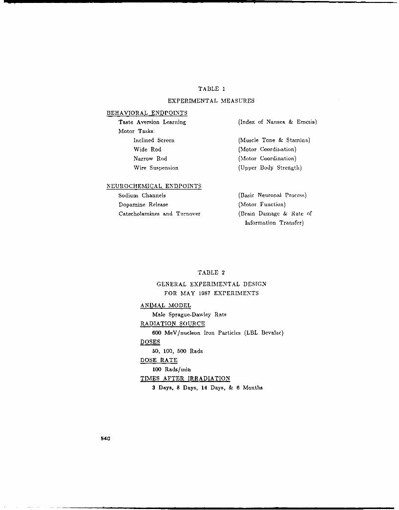
TABLE 1
EXPERIMENTAL MEASURES
BEHAVIORAL ENDPOINTS
Taste Aversion Learning (Index of Nausea & Emcsis)
Motor Tasks:
Inclined Screen (Muscle Tone & Stamina)
Wide Rod (Motor Coordination)
Narrow Rod (Motor Coordination)
Wire Suspension (Upper Body Strength)
NEUROCHEMICAL ENDPOINTS
Sodium Channels (Basic Neuronal Process)
Dopamine Release (Motor Function)
Catecholamines and Turnover (Brain Damage & Rate of
Information Transfer)
TABLE 2
GENERAL EXPERIMENTAL DESIGN
FOR MAY 1987 EXPERIMENTS
ANIMAL MODEL
Male Sprague-Dawley Rats
RADIATION SOURCE
600 MeV/nucleon Iron Particles (LBL Bevalac)
DOSES
50, 100, 500 Rads
DOSE RATE
100 Rads/min
TIMES AFTER IRRADIATION
3 Days, 8 Days, 14 Days, & 6 Months
540

General Methods
Five behavioral and three neurochemical endpoints were assessed in these
experiments (Table 1). These endpoints were chosen because of their
sensitivity to other qualities of radiation. The behavioral endpoints include
the conditioned taste aversion (CTA), an index of nausea and vomiting, and
bc',:era! motor tasks, measures of muscle tone, stamina, coordination, and
upper body strength. The neurochemical endpoints include the movement of
s".dium ions through channels, a basic neuronal process; dopamine release, a
regulator of motor activity; and catecholamine levels and turnover, a rough
estimate of brain damage and the rate of information transfer in the brain.
Male Sprague-Dawley Crl:CD(SD)BR rats (Charles River Breeding
Laboratories, Kingston, NY) weighing 200-300 g were used in these
experiments. Rats were quarantined on arrival and screened for evidence of
disease by serology and histopathology before being released from quarantine.
The rats were housed individually in polycarbonate isolator cages (Lab
Products, Maywood, NJ) on autoclaved hardwood contact bedding ('Beta
Chip' Northeastern Products Corp., Warrensburg, NY) and were provided
conimercial rodent chow ('Wayne Rodent Blok' Continental Grain Co.,
Chicago IL) and acidified water (pH 2.5 using HCl) ad libitum. Animal
holding rooms were kept at 21 + 10 C with 50 + 10% relative humidity
on a 12-hr light:dark lighting cycle with no twilight.
The rats were irradiated with high-energy 56Fe particles (600 MeV) in
the Bevalac at the LBL. The animals were irradiated in well-ventilated
Plexiglas holders with one of three doses, including 50, 100, and 500 rads.
at a dose-rate of 40-140 rad/min. Dosimetric support was provided by
personnel at the Bevalac. Animals irradiated with other radiation sources
were exposed to a single dose of 50, 100, or 500 rads of gamma photons
from a 60Co source at a rate of 40 rads/min or high-energy electrons (18.6
MeV) from a linear accelerator. Radiation dosimetry was performed us'ng
paired 50-ml ion chambers. Delivered dose was expressed as a ratio of the
dose measured in a tissue-equivalent plastic phantom enclosed in a
restraining tube, to that measured free in air.
General Observations after 56Fe Irradiation
Although not a specific part of the experimental design, any unusual
541

reactions by the animals were noted. Based on subjective observations, the
animals appeared normal after irradiation with 56Fe particles. However, after
exposure to 500 rads, several changes were observed. The exposed rats
progressively lost weight, totaling about 20% of body weight over a 14-day
period. In addition, they experienced a reduction in muscle tone, a hind
limb tremor, and hypothermia (animals cool to the touch) 3 days after
irradiation, effects that had disappeared by 8 days after irradiation.
CONDITIONED TASTE AVERSION LEARNING AFTER IRRADIATION
Characteristics of the Conditioned Taste Aversion
Animals have developed over the course of evolution mechanisms to help
prevent accidental poisoning, the best-known one being the emetic response.
Ernesis can occur as a result of consuming presumably tainted food that is
then expelled from the stomach. In addition to emesis, animals are also
capable of avoiding potentially toxic substances after a single ngestion of
quantities less toxic than those required to induce vomiting. This is done
through a process called the conditioned taste aversion (CTA). A CTA
develops when the animal associates the taste of novel tasting food with a
physiological response, possibly illness, and then subsequently avoids further
ingestion of that food. In a laboratory setting, a CTA is typically induced
by pairing a normally preferred but novel tasting fluid with exposure to a
toxin. The animal will then avoid drinking the fluid when presented again.
The conditioned taste aversion (CTA) paradigm in the rat can be used
as a model system to study the mechanisms by which exposure to non-lethal
levels of ionizing radiation can produce changes in the behavior of an
organism (Rabin and Hunt, 1986). Because the functional effects of emesis
and taste aversion learning are similar, in the sense that they limit the
intake and/or absorption of toxic substances, a number of investigators have
argued that the CTA paradigm represents a model system for the study of
radiation-induced nausea and emesis (Garcia et al., 1985; Rabin and Hunt,
1986). Therefore, the CTA provides an index of the probability that nausea
and emesis will occur.
542

The CTA paradigm offers a number of advantages over emesis models.
The paradigm can be used with rats, inexpensive and easily used animals.
They are small enough that more uniform fields of radiation can be obtained
with particle accelerators than with larger animals. Because a CTA can be
easily induced in a relative large number of animals, a great deal of
information can be accumulated quickly as well as the characterization of
any responses. Since the mechanisms underlying the CTA and emesis appear
to be similar, this approach will allow for the formulation of more specific
hypotheses that could be applied eventually to emesis models.
The CTA induced by ionizing radiation has been extensively studied and
a clearer idea of how it develops has been emerging. The most important
discovery is the involvement of a specific nucleus in the brain stem, the area
postrema. The area postrema has been demonstrated to play an critical role
in the development of CTAs induced by a broad range of unrelated toxins.
This part of the brain is sufficiently important that if the area postrema is
lesioned, the development of a CTA is blocked. These toxins include not
only ionizing ra/iatioiL (Ossenkopp, 1983; Rabin et al., 1983), but also
lithium chloride (Ritter et al., 1980; Rabin et al., 1v83), copper sulfate
(Rabin et al., 1985), Bluminum chloride (Rabin and Hunt, unpublished
observation), paraquat (Dey et al., 1987), angiotensm II (Rabin et al., 1986),
amphetamine (Rabin et al., 1987), WR-2721 (Rabin et al., 1986), and
cisplatin (Rabin and Hunt, unpublished observation). In addition, other
evidence indicates that the area postrema is also required for the
development of emesis (Wang et al. 1958; Brizzee, 1970; Harding et al.,
1985; Rabin et al., 1986). Not all toxic drugs induce CTAs through the area
postrema. For example, ethanol- and morphine-induced CTAs are not blocked
by lesions of the area postrema (Hunt et al., 1987; Rabin and Hunt,
unpublished observation).
Since many unrelated toxins induce CTAs through the area postrema, it
has been suggested that a common mechanism may underlie all these effects
(Rabin and Hunt, 1986). Also, since toxins are generally foreign substances,
specific receptors for each possible toxin are not likely to have evolved.
Consequently, an intermediary mechanism in the induction of CTAs and
emesis involving one or more secondary mediators has been postulated (Hunt
et al., 1965; Rabin and Hunt, 1986). If these mediators interact with
receptors in the area postrema, they may then activate the neural circuits
that evoke CTAs.
543

Induction of a CTA after Exposure to 56Fe Particles
Research was initiated to determine whether high-energy iron particles
could induce a CTA similar to other qualities of ionizing radiation, such as
gamma photons. The first experiments were designed to find the doses of5 6 Fe particles that would induce a CTA and compare the sensitivity of the
animals to those irradiated with gamma photons or high-energy electrons.
Conditioned taste aversions were produced by first placing the rats on a
23.5-hr water deprivation schedule for 5 days during which water was
available for only 30 min daily during the early light phase of the diurnal
cycle. On the conditioning day (day 6), the rats were presented with a
solution of 10% sucrose, after which the intake of the fluid was recorded.
Immediately following the drinking period, rats were irradiated with the
doses stated above. On the following day (test day), 10% sucrose was
presented again and the consumption during a 30-min period was recorded.
A CTA existed when the amount of fluid consumed on the test day was
significantly less than that consumed on the conditioning day.
25
20----- T T M COND
SucroseIntake Wm)
cotol 5 100 500Dose (rad)
Fig. 1. Co.idition-d taste aversions after different doses of 56Fe ions.
Sucrose intake was significantly reduced after all doses
studied. The maximum effect of the radiation is presumed
to be < 50 rads.
544
140
120 - 2 0 56Fe
10-. o , 60 Co% Conditioning _ - e-
Day Sucrose 8Intake
60 I
40-
201-
0sham 50 100 500
Dose (rad)
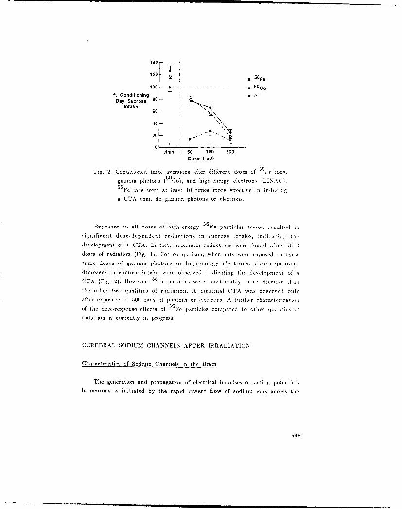
Fig. 2. Conditioned taste aversions after different doses of 56 Fe ion,.
gamma photons (6 0 Co), and high-energy electrons (LINAC).5 6 Fe ions were at least 10 times more effective in inducing
a CTA than do gamma photons or electrons.
Exposure to all doses of high-energy 56Fe particles tested resulted in
significant dose-dependent reductions in sucrose intake, indicating tile
development of a CTA. In fact, maximum reduct-:ns were found after all 3
doses of radiation (Fig. 1). For comparison, when rats were exposed to these
same loses of gamma photons or high-energy electrons, dose-dependent
decreases in sucrose intake were observed, indicating the development of a
CTA (Fig. 2). However, 56Fe particles were considerably more effective than
the other two qualities of radiation. A maximal CTA was observed only
after exposure to 500 rads of photons or electrons. A further characterization
of the dose-response effects of 56Fe particles compared to other qualities of
radiation is currently in progress.
CEREBRAL SODIUM CHANNELS AFTER IRRADIATION
Characteristics of Sodium Channels in the Brain
The generation and propagation of electrical impulses or action potentials
in neurons is initiated by the rapid inward flow of sodium ions across the
545
neuronal plasma membrane (Hodgkin and Huxley, 1952). In the resting state
a neuron maintains a resting membrane potential that results from the
unequal distribution of sodium, potassium, and chloride ions across the
membram tKoester, 1981a,b). When neurons are electrically excited, sodium
ior, fiow inward on their concentration gradient until the membrane
potential is reversed (Koester, 1981c). The movement of potassium ions out
of the neuron proceeds until the neuron has repolarized and the neuron is
again in the resting state.
Sodium ions enter the neuron through pores in the membrane called
channels. These channels are specific to sodium and traverse the neuronal
plasma membrane. They are glycoproteins containing multiple subunits
(Catterall. 1982) and have an absolute requirement for lipids for normal
functioning (Tamkin et al., 1984). At least three functional sites within
sodium channels have been identified based on the actions of specific
neurotoxins (Catterall, 1980). Site I, located on the external surface of the
neuronal membrane, binds tetrodotoxin and saxitoxin, drugs that block the
generation of action potentials. Site II. located in the lipid core of the
membrane, binds batrachotoxin and veratridine, lipid-soluble drugs that
activate sodium channels. And site III, located on the membrane surface but
with projcctions down to site II, binds scorpion and sea anemone toxiis that
c..lhanre the actions of toxins on site II but have no intrinsic activity of
their own. Neurocheinically, the functioning of the sodium channel can bc
studied with a synaptosomal (pinch-off nerve endings) preparation (Krueger
and Blaustein, 1980: Tamkin and Catterall, 1981). The rate of uptake of22Na can be measured after exposure to the neurotoxins batrachotoxin or
veratridine. thereh y assessing what would occur under normal physiologicai
conditions.
Sodium Channel Function after Exposuire to 5 6Fe Particles
The rate of 22Na uptake was determined in synaptosomes from the
cerebral cortex as detailed previously (Mullin et al., 1986). A crude
mitochondrial preparation containing synaptosomes was prepared from a
cortical homogenate. The final pellet was resuspended in ice-cold in.'.ubationm
buffer (8-10 ml/brain) containing 5.4 mM KCI, 0.8 mM MgSO 4 , 5.5 mM
glucose, 130 mM choline chloride, and 50 mM N-2-hydroxyethyl-piperazine-
N'-2-ethanesulfonic acid (HEPES), with the pH adjusted to 7.4 with Tris
base. Thp uptake of 22Na was measured as follows. Aliquots (50 sul) of the
synaptosomal suspension were incubated for Z rain at 360 C. The neurotoxin
546
veratridine was then added, and the incubation was continued for an
additional 10 min. The samples were tien diluted with 300 p1 of uptake
solution containing 5.4 mM KCI, 0.8 mM MgSC 4 , 5.5 mM glucose, 128 mM
choline chloride, 5 mM ouabain, 2 mM NaCl, 1.3 pCi 2 2 Na, 100 #M
veratridine, and 50 mM HEPES (pH adjusted to 7.4 with Tris). After a
5-sec incubation, uptake was terminated by adding 3 ml of ice-cold wash
solution containing 163 mM choline chloride, 0.8 mM MgSO 4, 1.7 imM
CaCl 2 , I mg/ml of bovine serum albumin, and 5 mM HEPES (pH adjusted
to 7.4 with Tris). The mixture was rapidly filtered under vacuum through a
cellulose filter with 0.45-pin pores, and the filters were washed twice with 3
ml of wash solution. Radioactivity was determined by liquid scintillation
spectrophotometry. The data were expressed as specific uptake determined hy
subtracting nonspecific uptake (samples containing I M tetrodotoxin) fron
total uptake.
MControl111111, 50 rad
4 15010 adS-500 rad4 _i_
3nmoes, mg
protein2
1
038 14 6 months
days
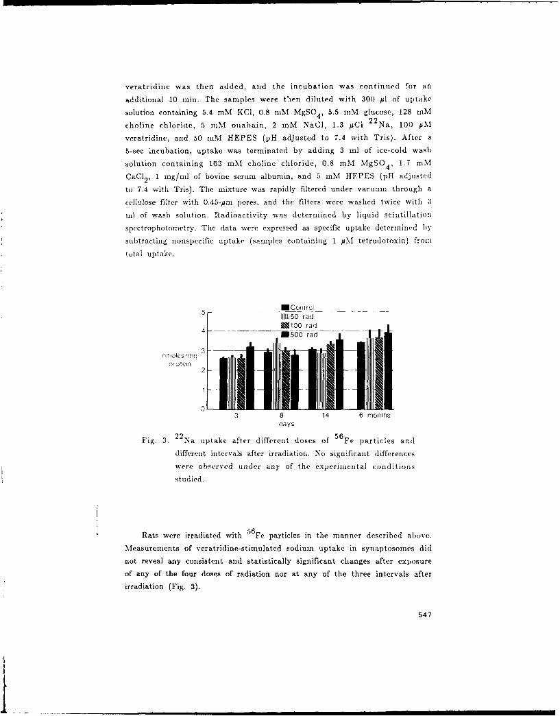
Fig. 3. 2 2 Na uptake after different doses of 56Fe particles and
different intervals after irradiation. No significant differences
were observed under an, of the experimental conditions
studied.
56Rats were irradiated with Fe particles in the manner described above.
Measurements of veratridine-stimulated sodium uptake in synaptosomes did
not reveal any consistent and statistically significant changes after exposure
of any of the four doses of radiation nor at any of the three intervals after
irradiation (Fig. 3).
547
I -

GENERAL DISCUSSION OF RESULTS
The experiments completed to date demonstrate that 56Fe particles can
induce a CTA that may represent a state of illness, possibly nausea. These
results along with those presented by Joseph et a]. (1988) suggest that the
behavioral effects of exposure to 56Fe particles may be at least 10 times
greater than those observed following exposure to gamma photons or high-
energy electrons. They also may reflect a greater chance for the occurrence
of nausea and emesis in astronauts exposed to a space radiation environment
during longer term missions.
The successful completion of missions in space depends in part on
behavioral and neural integrity of the astronauts. Given the potential
significance of the data presented, it is important to seriously pursue an
active program of research into the possible behavioral and neural deficits
that might occur in space as a result of exposure to radiation. By
combining sensitive behavioral assessments that can measure cognitive or
motor performance with neurochemical analyses following HZE irradiation
(e.g. Joseph et al., 1988), more information can be obtained concerning
important behavioral and biological relationships that will aid in predicting
and controlling possible performance deficits (luring manned space flight.
ACKNOWLEDGEMENTS
The authors wish to acknowledge the assistance of Drs. E. John
Ainsworth, Patriciq Durbin. and Bernhard Ludewigt and the staff at the
Lawrence Berkeley Laboratory without whose help these studies could not
have been undertaken. This research was supported by the Armed Forces
Radiobiology Research Institute, Defense Nuclear Agency, under work units
B4098, B4123, and B4157. View's presented in this paper are those of the
authors; no endorsement by the Defense Nuclear Agency has been given or
should be inferred. Research was conducted according to the principles
enunciated in the 'Guide for the Care and Use of Laboratory Animal
Resources, National Research Council.'
REFERENCES
Brizzee, 1. ., 1970, Effect of localized brain stem lesions and
supraciaph-gmatic vagotomy on X-irradiation emesis in the monkey,
Am. J .1. 187:567.
548

Catterall, W. A., 1980, Neurotoxins that act on voltage-sensitive sodium
channels in excitable membranes, Annu. Rev. Pharmacol. Toxicol. 20:15.
Catterall, W. A., 1982, The emerging molecular view of the sodium channel,
Trends Neurosci. 5: 303.
Dey, M. S., Krueger, R. I., and Ritter, R. C., 1987, Paraquat-induced, dose-
dependent conditioned taste aversions and weight loss mediated by the
area postrema, Toxicol. AppI. Pharmacol., 87:212.
Garcia, J., Lasiter, P. S., Bermudez-Ratoni, F., and Deems, D. A., 1985, A
general theory of taste aversion learning. Ann. NY Acad. Sci. 443:8.
Harding, R. K., Hugenholz, H., Keany, M., and Kucharczyk, J., 1985,
Discrete lesions of the area postrema abolish radiation-induced emesis in
the dog, Neurosci. Lett. 53:95.
Hodgkin, A. L. and Huxley, A. F., 1952, A quantitative description of
membrane current and its application to conduction and excitation in
nerve. J. Physiol. (London ) 117:500.
Hunt, E. L., Carroll, H. W., and Kimeldorf, D. J., 1965, Humoral mediation
of radiation-induced motivation in parabiont rats, Science, 150:1747.
Hunt, W. A., Rabin, B. M., and Lee, J., 1987, Ethanol-induced taste
aversions: Lack of involvement of acetaldehyde and the area postrema.
Alcohol 4:169.
Joseph, J. A., Hunt, W. A., Rabin, 13. M., and Dalton T. K., 1988,
Correlative motor behavioral and striatal dopaminergic alterations
induced by 56 Fe, in: 'Terrestrial Space Radiation ard its Biological
Effects', P. D. McCormack, C. E. Swenberg, and H. Bicker, eds.,
Plenum Press, New York.
Koester, J., 1981a, Resting membrane potential, in: 'Principles of Neural
Science,' E. R. Kandel and J. H. Schwartz, eds., Elsevier/North Holland.
Koester, J., 1981b, Passive electrical properties of the neuron, in: 'Principles
of Neural Science,' E. R. Kandel and J. H. Schwartz, eds.,
Elsevier/North Holland.
549

Koester, J., 1981c, Active conductances underlying the action potential, in:
'Principles of Neural Science,' E. R. Kandel and J. H. Schwartz, eds.,
Elsevier/North Holland.
Krueger, B. K., 1980, Sodium channels in presynaptic nerve terminals:
Regulation by neurotoxins. J. Gen. Physiol. 76:287.
Mickley, G. A., Bogo, V., Landauer, M. R., and Mele, P. C., 1988, Current
behavioral radiobiology research at the Armed Forces Radiobiology
Research Institute. 'Terrestrial Space Radiation and its Biological
Effects', P. D. McCormack, C. E. Swenberg, and H. Bcker, eds.,
Plenum Press, New York.
Mullin, M. J., Hunt, W. A., and Harris, R. A., 1986, Ionizing radiation
alter the properties of sodium channels in rat brain synaptosomes. J.
Neurochem. 47:489.
Ossenkopp, K. -P, 1983, Taste aversion conditioned with gamma radiation:
Attenuation by area postrema lesions in rats. Behav. Brain Res. 7:295.
Rabin, B. M., and Hunt, W. A., 1986, Mechanisms of radiation-induced
conditioned taste aversion learning. Neurosci. Biobehav. Rev. 10:55.
Rabin, B. M., Hunt, W. A., and Lee, J., 1983, Attenuation of radiation-
and drug-induced conditioned taste aversions following area postrema
lesions in the rat. Radiat. Res. 93:388.
Rabin, B. M., Hunt, W. A., and Lee, J., 1985, Intragastric copper sulfate
produces a more reliable conditioned taste aversion in vagotomized rats
than in intact rats. Behav. Neural Biol. 44:364.
Rabin, B. M., Hunt, W. A., Bakarich, A. C., Chedester, A. L., and Lee, J.,
1986, Angiotensin II - Induced taste aversion learning in cats and rats
and the role of the area postrema. Physiol. Behav. 36:1173.
Rabin, B. M., Hunt, W. A., and Lee, J., 1986, Effects of area postrema
lesions on the behavioral toxicity of WR-2721 in the rat. Neurobehav.
Toxicol. Teratol. 8:83.
550

Rabin, B. M., Hunt, W. A., Chedester, A. L., and Lee, J., 1986, Role of
the area postrema in radiation-induced taste aversion learning and emesis
in cats. Physiol. Behav. 37:815.
Rabin, B. M., Hunt, W. A., and Lee, J., 1987, Interactions between
radiation and amphetamine in taste aversion learning and the role of the
area postrema in amphetamine-induced conditioned taste aversions.
Pharmacol. Biochem. Behav. 27:677.
Ritter, S., McGlone, J. L. and Kelly, K. W., 1980, Absence of lithium-
induced taste aversion after area postrema lesion. Brain Res. 201:501.
Tamkin, M. M. and Catterall, W. A., 1981, Ion flux studies of voltage-
sensitive sodium channels in synaptic nerve endings. Mol. Pharrnacol.
19:78.
Tamkin, M. M., Talvenheimo, J. A., and Catterall, W. A., The sodium
channel from rat brain: Reconstitution of neurotoxin-activated ion flux
and scorpion toxin binding from purified components. J. Biol. Chem.
259:1676.
Wang, S. C., Renzi, A. A., and Chinn, H. I., 1958, Mechanisms of emesis
following X-irradiation, Am. J. Physiol. 193:335.
Accession For
N'T -3 7, AAEl
r or av o
,D5st5
551