
evolution of the tapetum
14
Trans. Am. Ophthalmol. Soc. Vol. 100, 2002 187 EVOLUTION OF THE TAPETUM BY Ivan R. Schwab, MD, Carlton K. Yuen, BS (BY INVITATION), Nedim C. Buyukmihci, VMD (BY INVITATION), Thomas N. Blankenship, PhD (BY INVITATION), AND Paul G. Fitzgerald, PhD (BY INVITATION) ABSTRACT Purpose: To review, contrast, and compare current known tapetal mechanisms and review the implications for the evolution of the tapetum. Methods: Ocular specimens of representative fish in key piscine families, including Acipenseridae, Cyprinidae, Chacidae; the reptilian family Crocodylidae; the mammalian family Felidae; and the Lepidopteran family Sphingidae were reviewed and compared histologically. All known varieties of tapeta were examined and classified and compared to the known cladogram representing the evolution of each specific family. Results: Types of tapeta include tapetum cellulosum, tapetum fibrosum, retinal tapetum, invertebrate pigmented tapetum, and invertebrate thin-film tapetum. All but the invertebrate pigmented tapetum were examined histologically. Review of the evolutionary cladogram and comparison with known tapeta suggest that the tapetum evolved in the Devonian period 345 to 395 million years ago. Tapeta developed independently in at least three separate orders in inver- tebrates and vertebrates, and yet all have surprisingly similar mechanisms of light reflection, including thin-film inter- ference, diffusely reflecting tapeta, Mie scattering, Rayleigh scattering, and perhaps orthogonal retroreflection. Conclusion: Tapeta are found in invertebrates and vertebrates and display different physical mechanisms of reflection. Each tapetum reflects the wavelengths most relevant to the species’ ecological niche. With this work, we have hypothe- sized that the tapetum evolved independently in both invertebrates and vertebrates as early as the Devonian period and coincided with an explosion of life forms. Trans Am Ophthalmol Soc 2002;100:187-200 INTRODUCTION The tapetum lucidum (shining carpet in Latin) is a catop- tric device found in the eye of many vertebrates and inver- tebrates, which serves to increase the amount of light absorbed by the photoreceptors. The tapetum is proximal to the photoreceptors and may be located in either the choroid or deep retina in vertebrates and proximal to the retinular cells in invertebrates. The tapetum reflects the photons that were not initially absorbed after they passed through the photoreceptors. These reflector mechanisms provide the photoreceptors a second chance at absorbing the (reflected) light, thereby enhancing an organism’s visual sensitivity. This device is often a layer, and it is espe- cially useful in lower light conditions. Animals use a wide range of materials and techniques to provide tapetal reflectance, including guanine, riboflavin, triglycerides, pteridine, cholesterol, zinc, astaxanthin, and collagen. 1 Apparently, tapeta have evolved several times with these different mechanisms and represent convergent evolution of function. METHODS Ocular specimens that are representative of fish in key piscine families, including Acipenseridae (Acipenser medirostris, the green sturgeon), Cyprinidae (Cyprinus carpio, the common carp), and Chacidae (Ictalurus punc- tatus, the channel catfish), were obtained from a local fish market. The ocular specimens were removed from the deceased fish. An ocular specimen of a member of the reptilian family Crocodylidae (Alligator mississippiensis, the American alligator) was obtained from Dennis Brooks, DVM, PhD, of the University of Florida in Gainesville. A fixed ocular specimen of the mammalian family Felidae (Felis domesticus, the domestic housecat) was obtained from Leslie Lyons, PhD, and David Maggs, BVS (hons), DACVO, of the University of California, Davis, School of Veterinary Medicine. A specimen of the Lepidopteran From the Department of Ophthalmology, University of California, Davis, Medical Center (Dr Schwab); the University of California, Davis, School of Medicine (Dr Schwab, Mr Yuen, Dr Blankenship, Dr Fitzgerald); and the University of California, Davis, School of Veterinary Medicine (Dr Buyukmihci). Supported in part by an unrestricted grant from Research to Prevent Blindness, Inc, New York, New York, and by grant P30EY12576 from the National Institutes of Health.
Transcript of evolution of the tapetum

Trans. Am. Ophthalmol. Soc. Vol. 100, 2002 187
EVOLUTION OF THE TAPETUM
BY Ivan R. Schwab, MD, Carlton K. Yuen, BS (BY INVITATION), Nedim C. Buyukmihci, VMD (BY INVITATION), Thomas N. Blankenship, PhD (BY INVITATION), AND Paul G. Fitzgerald, PhD (BY INVITATION)
ABSTRACT
Purpose: To review, contrast, and compare current known tapetal mechanisms and review the implications for the evolution of the tapetum.
Methods: Ocular specimens of representative fish in key piscine families, including Acipenseridae, Cyprinidae,Chacidae; the reptilian family Crocodylidae; the mammalian family Felidae; and the Lepidopteran family Sphingidaewere reviewed and compared histologically. All known varieties of tapeta were examined and classified and compared tothe known cladogram representing the evolution of each specific family.
Results: Types of tapeta include tapetum cellulosum, tapetum fibrosum, retinal tapetum, invertebrate pigmented tapetum, and invertebrate thin-film tapetum. All but the invertebrate pigmented tapetum were examined histologically.Review of the evolutionary cladogram and comparison with known tapeta suggest that the tapetum evolved in theDevonian period 345 to 395 million years ago. Tapeta developed independently in at least three separate orders in inver-tebrates and vertebrates, and yet all have surprisingly similar mechanisms of light reflection, including thin-film inter-ference, diffusely reflecting tapeta, Mie scattering, Rayleigh scattering, and perhaps orthogonal retroreflection.
Conclusion: Tapeta are found in invertebrates and vertebrates and display different physical mechanisms of reflection.Each tapetum reflects the wavelengths most relevant to the species’ ecological niche. With this work, we have hypothe-sized that the tapetum evolved independently in both invertebrates and vertebrates as early as the Devonian period andcoincided with an explosion of life forms.
Trans Am Ophthalmol Soc 2002;100:187-200
INTRODUCTION
The tapetum lucidum (shining carpet in Latin) is a catop-tric device found in the eye of many vertebrates and inver-tebrates, which serves to increase the amount of lightabsorbed by the photoreceptors. The tapetum is proximalto the photoreceptors and may be located in either thechoroid or deep retina in vertebrates and proximal to theretinular cells in invertebrates. The tapetum reflects thephotons that were not initially absorbed after they passedthrough the photoreceptors. These reflector mechanismsprovide the photoreceptors a second chance at absorbingthe (reflected) light, thereby enhancing an organism’svisual sensitivity. This device is often a layer, and it is espe-cially useful in lower light conditions. Animals use a widerange of materials and techniques to provide tapetal
reflectance, including guanine, riboflavin, triglycerides,pteridine, cholesterol, zinc, astaxanthin, and collagen.1
Apparently, tapeta have evolved several times with thesedifferent mechanisms and represent convergent evolutionof function.
METHODS
Ocular specimens that are representative of fish in keypiscine families, including Acipenseridae (Acipensermedirostris, the green sturgeon), Cyprinidae (Cyprinuscarpio, the common carp), and Chacidae (Ictalurus punc-tatus, the channel catfish), were obtained from a local fishmarket. The ocular specimens were removed from thedeceased fish. An ocular specimen of a member of thereptilian family Crocodylidae (Alligator mississippiensis,the American alligator) was obtained from Dennis Brooks,DVM, PhD, of the University of Florida in Gainesville. Afixed ocular specimen of the mammalian family Felidae(Felis domesticus, the domestic housecat) was obtainedfrom Leslie Lyons, PhD, and David Maggs, BVS (hons),DACVO, of the University of California, Davis, School ofVeterinary Medicine. A specimen of the Lepidopteran
From the Department of Ophthalmology, University of California,Davis, Medical Center (Dr Schwab); the University of California, Davis,School of Medicine (Dr Schwab, Mr Yuen, Dr Blankenship, DrFitzgerald); and the University of California, Davis, School of VeterinaryMedicine (Dr Buyukmihci). Supported in part by an unrestricted grantfrom Research to Prevent Blindness, Inc, New York, New York, and bygrant P30EY12576 from the National Institutes of Health.

188
Schwab et al
family, Sphingidae (Arctonotus lucidus, the Pacific greensphinx moth), was obtained with the help of JohnDebenedictis, PhD, staff entomologist, Bohart Museumof Entomology, University of California, Davis, and thecompound eyes were dissected from the specimen. Eachof these ocular specimens was sectioned and stained withhematoxylin-eosin (HE), periodic acid–Schiff (PAS), ortoluidine blue (TB), examined, and compared with oneanother histologically. The evolutionary relationship ofthe ocular specimens was examined and the cladistic posi-tion of each species established, evaluated, and compared.
RESULTS
The eye of A medirostris, the green sturgeon, was foundto contain a tapetum cellulosum. Although it resembles atapetum fibrosum in some respects when stained (PAS), ithas a cellular structure within the choroid immediatelyproximal to the pigment epithelium with from three tofive cells aligned somewhat irregularly (Figure 1), espe-cially when compared with the more regularly and defi-nitely formed feline tapetum cellulosum. When stainedwith HE, the sturgeon eye revealed guanin granules in thesuperficial choroid (Figure 2). The retinal pigmentepithelium (RPE) was generally clear, as is typical for ani-mals with a choroidal RPE, but regularly spaced RPEcells contained a dense concentration of melanin withintervening clear RPE cells (Figures 1 and 2).
The tapetum lucidum of C carpio, the common carp,was reviewed and confirmed to be a retinal tapetum com-posed of guanin in an occlusible pattern. The light-adaptedeye shows pigment drawn into the more inner portions ofthe retinal pigment epithelium and obscuring the guaninof the retinal tapetum (Figure 3).
The eye of I punctatus, the channel catfish, shows anocclusible retinal tapetum. A light-adapted retina showspigment drawn up into the broad retinal pigment epithe-lial cells in a manner similar to the carp (Figure 4).
The eye of Allig mississippiensis, the American alliga-tor, revealed fine opaque crystalline granules in the apex ofthe pigment epithelial cells, and represents a retinal tape-tum. These granules were present in the dorsal half of theretina, although the stain (TB) has made the rodlike struc-tures of the granules appear black (Figures 5A and 5B).
The eye of F domesticus, the domestic housecat,shows a regular tapetum cellulosum. This cellular struc-ture consists of 6 to 12 flat and well-organized cellsarranged in a regular distribution, resembling precisionbrickwork (Figure 6).
The compound eye of Arct lucidus, the pacific greensphinx moth, revealed a tapetum of modified tracheoleswith chitin layers alternating with layers of air. The parallellayers of chitin and air have a long axis that is perpendicular
to the long axis of the rhabdom. These chitinous layers areseparated by a standard distance from rhabdom to rhab-dom (Figure 7).
DISCUSSION
TYPES OF TAPETUM
Tapeta can be classified according to location in verte-brates and mechanism in invertebrates (Table I).
Vertebrate Choroidal TapetaChoroidal tapeta are the most common and are furtherclassified as tapetum fibrosum and tapetum cellulosum.
Histologically, the simplest type is the tapetum fibro-sum, which is found principally in mammals, includingherbivorous ungulates (eg, elephants, horses, goats)(Figures 8 and 9), cetaceans (whales, dolphins), somemarsupials (Tasmanian devil), and a rodent (Cuniculuspacas).2,3 The tapetum fibrosum consists of extracellularcollagen fibrils stacked in an orderly manner with themajority of the fibers running horizontally.4 The numberof layers of fibrils varies between species and can be up toseveral hundred thick.2
The most studied tapetum fibrosum among the ungu-lates is found in the cow. In this species, the tapetum islocated posteriorly and dorsally. It is of variable thickness,increasing in thickness posteriorly. In species with achoroidal tapetum, the retinal epithelium in the area ofthe tapetum is unpigmented, allowing light to pass to besubsequently reflected by the tapetum. This contrastswith the pigmented retinal epithelium cells found in thesame animal in nontapetal regions.4
The tapetum cellulosum is found in cartilaginous fishes(eg, sharks, dogfishes), sturgeons (Figure 1), lobe-finnedfishes (coelacanths and lungfishes), seals, prosimians (eg,bush babies [Figure 10], lemurs), and most mammaliancarnivores (eg, cats, dogs [Figures 6 and 11]).1,5-7 This tape-tum is composed of regular cells in layers of variable thick-ness, containing a variety of reflective material.3,8,9
In the coelacanth (Latimeria chalumnae), the tape-tum cellulosum consists of hexagonal constant-thicknessstacks of guanine crystals within the individual cells. Thistapetum emits a greenish-yellow luminescence in the livespecimen, with a broad maximum wavelength of reflec-tion peaking at 476 nm, which corresponds to the peak ofthe wavelength of light that penetrates to depths of 7,500feet, where the animal is found. This choroidal tapetumunderlies the entire retina and appears bright silvery upondissection. Elasmobranchs also have tapeta composed ofguanine, but their tapeta differ from those of the coela-canth and others in that they are occlusible. The tapetalcells alternate with, and are separated from, one anotherby melanocytes that extend beyond the tapetal cells to

189
Evolution of the Tapetum
FIGURE 1Sturgeon choroid stained with periodic acid-Schiff (x20). Retinal pig-ment epithelium (RPE) can be seen at top of photograph. Note perio-dicity of pigment in RPE. Intervening RPE cells contain no pigment.
FIGURE 3Carp retina stained with hematoxylin-eosin (x20). Note broadly distributedmelanin throughout most of retina. This light-adapted retina exhibits anocclusible retinal tapetum. Cell bodies with nucleus of retinal pigmentepithelium (RPE) can be seen at bottom of photograph. Note how littlepigment is seen near base of RPE cell.
FIGURE 5A
Light-adapted American alligator retinal pigment epithelium (RPE) in anontapetal region with pigment granules visible at tips of RPE stainedwith toluidine blue (x40). This retina is only partially occlusible and hasprobably lost the ability to be truly occlusible but does show some pig-ment migration in light adaptation. Pigment in the RPE is present innontapetal regions of the eye.
FIGURE 2Sturgeon choroid stained with hematoxylin-eosin (x20). Note guaninecrystals in superficial choroid immediately beneath retinal pigmentepithelium (RPE). These crystals are seen as a grey coloration below pig-mented deposits of RPE. Note periodicity of pigment in RPE.
FIGURE 4Light-adapted catfish retina stained with hematoxylin-eosin (x10). Noteocclusible tapetum with the melanin pigment drawn nearly to outernuclear layer, obscuring both tapetum and rod outer segments.
FIGURE 5B
Light-adapted American alligator retinal pigment epithelium (RPE) intapetal region stained with toluidine blue (x40). Note black, flat platelikecrystals in tips of RPE cells in contrast to granules of pigment seen in 5A.Many crystals were lost in preparation of specimen.

190
Schwab et al
intervene between the tapetal cells and incoming light.The melanocytes migrate in conditions of increased lightto occlude the elasmobranch tapetum.5
The RPE and the choroidal tapetum of the sturgeonshow another interesting feature. The regular deposits ofpigment may serve a heretofore unrecognized purpose. Theregular spacing of pigment would create a grating thoughwhich light could be channeled upon reflection, preventingextraneous photons from being scattered to adjacent pho-toreceptors. This would assist in the elimination of the glareof the reflection from the tapeta, which would otherwiselead to the degradation of the image owing to the scatter ofthe extraneous rays, and would be an alternative to anocclusible tapetum as seen in the elasmobranchs.
Vertebrate Retinal TapetaThe retinal tapetum has been found in some lampreys,certain bony fishes, crocodiles, goatsuckers, the Virginiaopossum (Didelphys virginiana) (Figure 12), gar fishes,some old-world fruit bats, and many teleosts (Figures 3and 4).1 The retinal tapeta of fish have been further cate-gorized as those that contain small particles in spheres orcubes, which are classified as diffuse reflectors, and thosecontaining layered crystals, classified as specular reflectors.Specular reflectors function much like a mirror, whereasdiffuse reflectors are more like the reflections from arough surface and may use Mie scattering as their mecha-nism.1,9 Diffuse tapeta can be found in many teleosts,including certain carp and cusk eels. Specular tapeta canbe found in some lantern fish and other abyssal fish.9
Some species of lamprey have both diffuse and spec-
ular mechanisms. The reflecting material in teleostspheres includes nonpigmented materials (such as gua-nine, uric acid, and purines) and pigmented materials(including pteridine, lipid, astaxanthin, and melanoidcompounds).1 This represents a subclassification of thesmall-particle or diffuse reflectors.
In reptiles, only the crocodile and alligator have atapetum, and it is found in the retinal pigment epithelium.This tapetum consists of several layers of crystallineplatelets of guanine arranged in parallel. The tapetumappears to be somewhat occlusible in certain species, suchas the American alligator, and this could be explained bythe diurnal and nocturnal potential of that species.10 Thiscontrasts with another crocodile, the Caiman sclerops, anocturnal animal, which does not have an occlusible tape-tum. The tapetum of the C sclerops also consists of gua-nine crystals diffusely scattered within the RPE.11
The retinal tapetum of the opossum (D virginiana) isa semicircular area in the dorsal fundus. The reflectingmaterial consists of lipoidal spheres scattered throughoutthe epithelial cells.12 The tapeta of the crocodile and theopossum function as diffuse reflectors.
Amphibians, apparently, do not have tapeta, althougha bright reflex is found in many of these species. Thesource of this bright reflection is unknown at present butdoes not conform to the currently understood mecha-nisms of any tapeta.
INVERTEBRATE TAPETA
Pigmented TapetaThe invertebrate tapeta can be categorized as light-scat-tering pigments and those using thin-film interference.
FIGURE 6Cat choroid stained with toluidine blue (x20). Note regular tilelike cellularstructure beneath retinal pigment epithelium (RPE) and photoreceptorouter segments. These cells in tapetum cellulosum are filled with manysmaller platelike structures or rodlets seen with ultrastructural investigation.
FIGURE 7Compound eye of Pacific green sphinx moth stained with toluidine blue(x40). Larger tracheoles can be seen to right of photograph as clear spaces.As tracheoles become smaller (going toward left on photograph), they canbe seen as small, clear circles. Tracheoles that become tapetum can beseen in about the middle of photograph as fine striations. Larger blue ovalstoward left of photograph are undulations of rhabdoms of individualommatidia.

Evolution of the Tapetum
191
FIGURE 8Funduscopic photograph of left eye of 6-year-old female African elephant. The elephant has a tapetum fibrosum.
FIGURE 9Funduscopic photograph of right eye of 6-month-old castrated
male mixed-breed goat. The goat has a tapetum fibrosum.
FIGURE 10Funduscopic photograph of a galago.
Bush babies have a tapetum cellulosum.
FIGURE 11Funduscopic photograph of left eye of an adult female mixed-breed of dog. Dogs have a tapetum cellulosum.
FIGURE 12Funduscopic photograph of right eye of adult male
American opossum with retinal tapetum.

192
Schwab et alTA
BL
EI:
TA
PETA
INT
HE
AN
IMA
LK
ING
DO
M
NO
CH
OR
OID
AL
RE
TIN
AL
LIG
HT-
TH
IN-F
ILM
TAPE
TAR
EF
LE
CT
ING
AN
IMA
LK
ING
DO
MTA
PET
UM
TAPE
TATA
PETA
SCAT
TE
RIN
GTA
PETA
MAT
ER
IAL
FIB
RO
SUM
CE
LL
UL
OSU
MTA
PETA
UN
PIG
ME
NT
ED
PIG
ME
NT
ED
Jaw
less
Lam
prey
s✓
Fish
esH
ag F
ishes
✓
Shar
ks/D
og F
ishes
+G
uani
neJa
wSt
urge
ons
+G
uani
neF
ishes
Gar
pike
s+
Ast
haxa
thin
eB
ony
Fish
es?
++
Seve
ral
Seve
ral
Mat
eria
lsM
ater
ials
Coe
laca
nths
+G
uani
neL
ung
Fish
es+
Not
N
ot
Rep
orte
dR
epor
ted
Sala
man
ders
✓
Fro
gs &
Toa
ds✓
Cro
codi
les
and
+G
uani
neA
lliga
tors
Mos
t rep
tiles
✓
Goa
tsuc
kers
+
Lip
id(N
ight
jars
)M
ost B
irds
✓
Mon
otre
mes
✓
Mar
supi
als
Opo
ssum
s+
Lip
id (B
irefr
inge
nt)
Tasm
ania
n W
olve
s+
Col
lage
nPl
acen
tals
Wha
les
& d
olph
ins
+C
olla
gen
Ele
phan
ts &
Mos
t Hoo
ved
Ani
mal
s+
Col
lage
nC
arni
vore
s (C
ats
& D
ogs)
+Zi
nc p
rote
inH
orse
s+
Col
lage
nL
ower
(Bus
h B
abie
s &
Lem
urs)
+R
ibof
lavi
nA
otus
Mon
keys
✓
All
Oth
er H
ighe
r Pr
imat
es✓
Cun
icul
us P
acas
+C
olla
gen
Mos
t Rod
ents
✓
Seal
s+
Bel
ieve
d to
be
Zin
cF
ruit
Bat
s+
Phos
phol
ipid
(bire
frin
gent
)Sc
allo
ps+
Gua
nine
Cru
stac
eans
Lob
ster
s &
Shr
imp
+L
ipid
Ara
chni
dsSc
orpi
ons
✓
Spid
ers
+G
uani
neIn
sect
sD
rago
nflie
s+
Chi
tin
Fishes Am
phib
ians
Rep
tiles
Bird
s
Primates Rodents
Higher
Mam
mal
s
Mar
supi
als
Plac
enta
ls
Mol
lusk
s
Art
hrop
ods
Ara
chni
ds
Jaw
Fish
es
Jaw
less
Fish
es
Lun
g F
ishes
Bon
y F
ishes
Lam
prey
sH
ag F
ishes
Seal
s

Evolution of the Tapetum
193
In the eyes of crustaceans, light is reflected back tothe rhabdoms by white or lightly colored pigments,including purines or pteridines. The reflection is less useful over shorter wavelengths but is effective over muchof the spectrum and can almost double light capture.9,13
The tapeta of mesopelagic shrimps vary greatly betweenspecies, depending on the depth at which they live, andprobably corresponding to individual ecological niches andthe specific wavelengths required. Histologic examination ofthe tapetum shows that it is incomplete dorsally in somespecies from the upper mesopelagic zone (Sytellaspic debilis,Oplophorus spinosus), with the amount of reflecting materi-al increasing ventrally. Not surprisingly, peak transmission oflight in oceanic species is in the blue-green region of thespectrum (450 to 500 nm). Visual pigments in crustaceansshow absorbence between 470 and 490 nm, and reflectingmaterials appear white. The tapetum is located between therhabdom and basement membrane.14 The tapetum is com-plete in some deep-water species (Systellaspis crsitata,Acanthephyra kingsleyi, Acanthomysis pelagica) and isbelieved to be beneficial because of the lower levels of ambi-ent light at greater depths. Acanthephyra purpurea has athick tapetum peripherally but no tapetum in the central partof the eye. This is thought to interfere with the ability ofpredators to detect this shrimp because of decreased eyeshine centrally. The tapetal adaptations of these species arebelieved to increase sensitivity to the dim upwelling irradi-ance and to bioluminescence.14
Thin-Film Invertebrate Tapeta Thin-film interference is used by some invertebrates toconstruct a reflective tapetum. The best examples of thismechanism are found in the lycosid spiders, scallop(Pecten maximum), ostracod crustaceans, cockle(Cardium edule), and rotifers.13,15-18 In the ctenid spider, atype of lycosid, the tapetum is located proximal to therhabdomeres in the secondary eyes, while the primaryanteromedial eyes do not have a tapetum. The secondaryeyes are built around a “gridiron” tapetum, consisting ofparallel strips of reflecting material forming a double lad-der–like array.15 The reflecting material in lycosid spidershas been reported as guanine.
Tapeta have also been described in only a few mol-lusks. In the scallop (P maximum), for example, squareguanine crystals have been described as a tapetum.16
Nocturnal moths and diurnal butterflies both possessa tapetum constructed of modified tracheoles, or airwaycells.17 The tapeta in nocturnal moths consist of air layersnext to chitin, providing a large difference in refractiveindices, which provides for a higher percentage of reflectedlight. The tapetum of these nocturnal moths appears blue-green, whereas in diurnal butterflies each rhabdom pos-sesses its own tapetum. In many diurnal butterflies, the
tapetum consists of up to 40 overlying layers, which areregularly spaced cuticular plates, each separated from itsneighbors by air spaces. Because the tapetum varies inthickness, and relies on constructive interference, dorsally it tends to reflect blue light, whereas ventrally ittends to reflect red.13,18,19
MECHANISMS OF THE TAPETUM
Although much is known about the structure and bio-chemical composition of the tapetum, there is consider-ably less information on the spectral reflectance charac-teristics and mechanisms.1,13 Thin-film constructive anddestructive interference probably plays a prominent rolein the reflective mechanisms of the tapeta of many ofthese animals. Layers of crystals, or other structures withdiffering indices of refraction at a thickness of λ/4 of themaximal wavelength of reflected light, support the theorythat constructive interference is involved. Theoretically,15 to 20 layers at λ/4 should provide nearly 100% reflec-tion of light at a wavelength of λ.17
Diffusely reflecting tapeta have been described inwhich reflection off a rough surface occurs and enhancesthe sensitivity of the retina, but this cannot be constructiveinterference. Mie scattering has been proposed as a mech-anism for diffusely scattering tapeta, but this has not beenstudied in detail.20,21 Rayleigh scattering (scattering by par-ticles smaller than the wavelength of light with preferentialscattering of the shorter wavelengths, similar to the phe-nomena of our atmosphere creating a blue sky), may alsobe involved, especially for the tapetum fibrosum.20,22
Some butterflies employ a unique form of orthogonalretroreflection that juxtaposes yellow next to blue regions,synthesizing a green color in their wings. A concave surfacecomposed of flat and inclined regions appears yellow andblue, respectively. The juxtaposition of these colorsappears green because these regions produce individualstreams of photons too small to be resolved individually.23,24
Orthogonal retroreflection has never been studied as apotential mechanism for tapetal reflection, but could beinvolved, especially in invertebrate thin-film tapeta.1 In thevertebrate world, the duplex retina of Lestidiops, amesopelagic deep-sea teleost, has an anatomical configura-tion consistent with this form of reflection.25 Other animalsthat may have employed orthogonal retroreflection includethe bigeye fish (Hybopsis amblops) and cat (F domesticus).The tapetum of bigeye fish is actually a mosaic of different-colored spots, where measurements of minute, singly col-ored areas give spectral curves with restricted bands andexpected side oscillations. The spectrum of the cat tapetais variable between animals from green to yellow, and thecolor varies within the eye. The cat’s tapetum is composedof bundles of rodlets, when viewed microscopically, andthere appear to be different colors at varying depths.1

194
PIGMENTED COLORS FORMING A TAPETUM
The mechanism of individual colors produced by the tapetum has not been well studied. Constructive interfer-ence provides structural color, involving layers having thethickness of 1/4λ (where λ is the wavelength of light to bereflected), and has been proposed as a common mecha-nism. Some tapeta, however, contain pigmented reflectingcrystals. In such animals, the appearance of these tapetacorrelates with the pigmented color of these reflecting crys-tals. For example, the galago tapetum is composed of isoal-loxazine or riboflavin crystals possessing a yellow hue.26
The tapetum of the garpike has also been reported as beingmade up of yellow pigments.27 A red pigmented tapetumcan be found in characin fish and garfish.28,29 The pigment isfound enclosed in spheres in the retinal pigment epitheli-um. The reflecting material of characin fish, catfish tapetalpigment, consists of oligomers of 5,6-dihydroxindole-2-car-boxylic acid combined with decarbox-ylated S-adenosylme-thionine.30 The pigmented tapetum has conferred anadvantage to deep-sea fish that exist in an environmentwhere the predominant wavelength of light is 475 nm.Most animals at these depths have photo-receptors thatpredominantly absorb light at this wavelength, since that isthe only wavelength that penetrates to those depths.
A mesopelagic species of fish, Malacosteus niger, hasevolved with a mechanism that allows it to take advantageof this downwelling blue light as well as possessing redlight-emitting organs (photophores) located ventrally andsub-orbitally. These red photophores emit red light that isnot recognized by the photoreceptors of most deep-sea ani-mals. The photophores are larger than most photophores,suggesting that they may assist the M niger in illuminatingprey or signaling other individuals of the same species. It isbelieved that M niger, and not other deep-sea animals, isable to absorb the red light emitted by its own photophoresbecause it possesses a pigmented, diffused, scarlet-redtapetum that reflects the red light emitted by the photo-phores. This may allow the M niger to effectively visualizeprey undetected, as well as communicate with others oftheir species, without alerting their predators.28,30,31
EVOLUTION OF THE TAPETUM
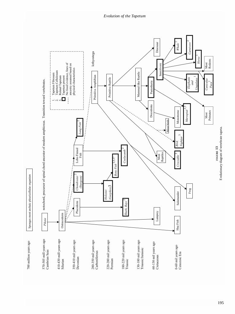
The timing of the evolution of the tapetum will probablynever be determined exactly; however, we provide amodel for the possible development of the tapetumaccording to existing evidence (Figures 13 and 14).Vertebrates are believed to have evolved from the pikaia,a primitive invertebrate and ancestor to the modern-dayamphioxus. The pikaia existed in the Precambrian era,approximately 570 million years ago.32 In the Silurian per-iod (410 to 430 million years ago), fossils of the ostraco-derm, the ancestor to the modern-day agnatha, had beendiscovered. Tapeta do not occur in amphioxus or agnatha;
therefore we make an assumption that tapeta did not existin ostracoderms. Though fossil history does not exist,Placoderms, Chondrostei, and lobe-finned fish are allassumed to have the ostracoderm as a common ancestor,on the basis of physical characteristics.
In the Devonian period (345 to 395 million yearsago), all three orders may have developed tapeta inde-pendent of each other, based on their modem progeny,namely, sharks (Placoderm), sturgeons (Chondrostei), andcoelacanths (lobe-finned fish). These three orders of fishall possess a tapetum cellulosum, suggesting that thistapetum may have been the first type of tapetum to evolvein vertebrates. These species have similar enough tapetathat they may have had a common ancestor with a tape-tum developing at approximately the Devonian period or,at the earliest, the very late Silurian. It is unlikely that thetapetum appeared earlier, because hagfish and most lam-preys do not have tapeta and it is believed these speciesseparated in the Silurian period. The development of thetapetum independently in fish might have occurred toallow them to explore deeper depths of the ocean, wherelight was not as prevalent. Conquering the depths of theocean may have provided sources of food not previouslyaccessible, such as detritus.
The lobe-finned fish ancestor, closely related to thecoelacanth and lungfish, is believed to be the predecessorto amphibians on the basis of physical characteristics.32-34
While amphibians may have possessed a tapetum at onetime, none has been reported in modern species.35 Itwould appear that, through evolution, either amphibianslost the ability to produce a tapetum or their lobe-finnedfish ancestors did not possess one. Amphibians are com-monly accepted as the ancestors of reptiles. The Eryopsgenus is believed to have been primitive reptiles thatevolved from amphibians, dating back to the Permianperiod (220 to 280 million years ago). The crocodile,believed to have descended from these primitive reptiles,evolved retinal tapetum composed of guanine. This sug-gests that crocodiles independently evolved or re-evolveda similar retinal tapetum employing guanine, welldescribed in jawed fishes. Guanine as a reflecting mater-ial is found widely in animals with tapeta. The crocodilealso uses guanine as its reflecting material, as do manyfish, the wolf spider, and many other unrelated species.The crocodile tapetum is located dorsally and temporally.It has been hypothesized that the location of the tapetumin the crocodile correlates with the need to improve theanimal’s ability to see in murky waters below it.20
Mammals evolved from mammallike reptiles, whichpresumably did not have tapeta. This is supported bymonotremes, which evolved independently from othermammals and belong to the order Docodonta. They haveeyes that are much more like reptilian eyes than mam-
Schwab et al
Evolution of the Tapetum
195
�������
����������
��������� �
�����������������
�����������
�����
���������
���������
������
�
���������
���
��������
���������
����
!� ����������
�����"�����
�������������
#��������$�
���������
#%�������$
�
����������
������
FIG
UR
E13
Evo
lutio
nary
dia
gram
of v
erte
brat
e ta
peta
.
196
Schwab et al
�
� �
� �
�
�
��"��"��#�������$
�
FIG
UR
E14
Evo
lutio
nary
dia
gram
of i
nver
tebr
ate
tape
ta.
Evolution of the Tapetum
197
malian eyes, and they do not have tapeta.13 With theexception of monotremes, all other mammals belong tothe order Pantotheria, which gave rise to marsupials andinsectivores, from which all other mammals were prob-ably derived. There are numerous examples of marsupialtapeta, but there are some interesting differences. TheVirginia opossum has a retinal tapetum with lipoid reflect-ing material, while the Tasmanian devil has a tapetumfibrosum, suggesting independent evolution, and no com-mon ancestor with a tapetum. Many other, but not all,mammals have tapeta, including ungulates, cetaceans,carnivores, rodents, and prosimians.1,13,18,35 It seems thatthe tapetum was probably not present in the mammal-likereptile, but appeared later in mammals in the Cenozoicera, no more than 60 million years ago, when the the tape-tum seems to have evolved independently once again.Mammalian tapeta include tapetum fibrosum, cellulosum,and retinal tapeta, similar to yet different from previouslyevolved tapeta. Somewhat unexpectedly, tapeta are alsofound in strictly diurnal mammals, such as the Indianmongoose, ungulates, and dogs,1,36 although dogs andungulates may be functionally nocturnal.
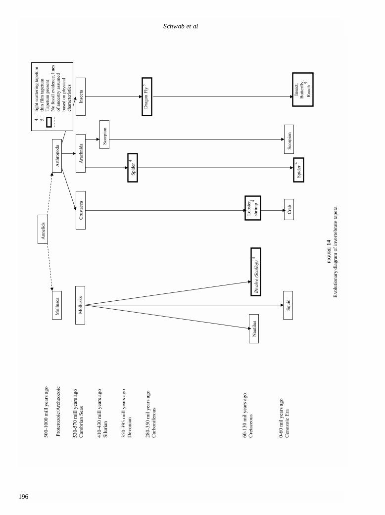
The tapetum has been reported in only two phyla ofinvertebrates, Mollusca and Arthropoda (Figure 14). On thebasis of physical characteristics, both phyla are assumed tohave evolved from Annelids in the Precambrian era (1,000million to 570 million years ago). The common ancestor tomollusks and arthropods–annelids–do not have, and proba-bly never did have, a tapetum. Among only a few mollusks,a guanine tapeta appears to have evolved independently ofthe tapeta of arthropods with different mechanisms. Amongthe mollusks, cephalopods possess some of the mostadvanced eyes among the invertebrates, and even have“camera-style” eyes like most vertebrates, but they do notpossess tapeta.36
Tapeta have been reported in three classes of arthro-pods: Arachnida. Insecta, and Crustacea. These classesappear to have evolved as separate lineages during thePrecambrian era. The tapetum in invertebrates may alsohave evolved in the Devonian period. Spiders are cred-ited as one of the first predators on land and can be tracedback 395 million years ago, according to the fossilrecord.32,33 This was prior to the evolution of flying insectsthat spiders commonly entrap in their webs.32 Hence,prehistoric spiders probably hunted their prey much likelycosid spiders of today. Although the invertebrates andvertebrates both probably developed tapeta in theDevonian period, the solutions were very different forsimilar results. As opposed to fish, which probably devel-oped tapeta to explore the depths of the ocean, spidersprobably developed tapeta to allow them to take advan-tage of nocturnal conditions or to protect themselvesagainst predators. Like sharks, the lycosid spider employs
guanine as its reflecting material organized into a thin-film reflector. In the class Insecta, dragonfly fossils havebeen dated to 350 million years ago, in the late Devonianto early Carboniferous period, and probably represent theearliest insect tapeta. Tapeta have been reported in drag-onfly ocelli.6 Because their common ancestor did not havetapeta, it appears that the dragonfly and the arachnidsevolved tapeta independently of each other. Crustaceans,such as the lobster and shrimp, have pigmentary tapeta,appear to have evolved independently of insects andarthropods, and have different mechanisms.23,24
CONCLUSIONS
Tapeta are found in both vertebrates and invertebrates.Not surprisingly, it appears that tapeta have a tendency toreflect wavelengths most relevant to the animal. The tapetain vertebrates are located in either the choroid or deepretina. Choroidal tapeta are further classified as tapetumcellulosum and tapetum fibrosum, according to theirappearance. The tapetum cellulosum is composed ofreflecting cells stacked in depth, like tile work. The cellscontain numerous refractile bodies with an orderlyarrangement. The tapetum fibrosum is acellular and com-posed of stacks of densely packed collagen fibrils. Retinaltapeta are found in the form of small granules in spheresand cubes or regularly arranged stacked thin platelets.These tapeta reflect light by diffuse reflection and specu-lar reflection, respectively.
A variety of reflecting material has been reported ininvertebrates and vertebrates, including nonpigmentedmaterial such as uric acid, guanine, chitin, and collagen,and pigmented material such as cholesterol esters, lipids,pteridine, and astaxanthin.1 Nonpigmented reflectingcrystals appear to produce colors structurally, while thecolor produced by pigmented reflecting crystals may be aresult of their pigmentation.
Mechanisms by which tapeta reflect light are incom-pletely understood. Constructive interference appears tobe a common mechanism by which some tapeta reflectlight; it does not appear, however, to be the only mecha-nism involved in all tapeta. In addition to thin-film inter-ference, diffuse reflection and pigmented color granulesare probably involved in tapetal reflection. Orthogonalretroreflection, a newly discovered mechanism of reflec-tion in butterfly wings, has never been studied as a poten-tial mechanism in butterfly tapeta and may be a morecommon mechanism than currently recognized.
We hypothesize that the tapetum may have arisenindependently in both invertebrates and vertebrates asearly as the Devonian period (390 to 345 million yearsago). In vertebrates, the guanine choroidal tapetum mayhave arisen in sharks, sturgeon, and lobe-finned fish inde-

pendent of each other, or if there were a common ances-tor, it originated in the Devonian era. This coincides withan explosion in the evolution of many different types ofmarine life. It appears that the choroidal tapetum was thefirst type of tapetum to evolve in vertebrates, with retinaltapeta appearing independently in other tetrapods.
The invertebrate tapetum appears to have evolvedfirst in spiders, in the Devonian period, and consists of adiffusely reflecting guanine tapetum. All of these tapetaemploy unpigmented guanine as their reflecting crystals,and guanine was probably the first reflecting materialemployed. The evolution of the tapetum appears to behighly convergent, but often with subtle differences indevelopment. Tapeta probably arose separately in inverte-brates and vertebrates and even within these broad groups,the tapetal mechanisms appear to have distinctly and sep-arately evolved, yet with surprisingly similar mechanisms.
REFERENCES
1. Nicol JAC. Tapeta lucida of vertebrates. In: Enoch MJ,Tobey FL, eds. Vertebrate Photoceptor Optics. Berlin:Springer-Verlag; 1981:401-431.
2. Bellairs R, Harkness ML, Harkness RD. The structure ofthe tapetum of the eye of the sheep. Cell Tissue Res1975;157:73-91.
3. Duke-Elder S. The eye in evolution. In: System ofOphthalmology, Vol 1. London: Henry Kimpton, 1958.
4. Braekevelt CR. Fine structure of the bovine tapetum fibro-sum. Anat Hist Embryol 1986;15:215-222.
5. Braekevelt CR. Fine structure of choroidal tapetumlucidum in the Port Jackson shark (Heterodontus phillipi).Anat Embryol 1994;190:591-596.
6. Collier LL, King JK, Prieur DJ. Tapetual degeneration incats with Chediak-Higashi syndrome. Curr Eye Res1985;4(7):767-773.
7. Heath AR, Hindman HM. The role of cyclic AMP in thecontrol of elasmobranch ocular tapetum lucidum pigmentgranule migration. Vision Res 1988;28(12):1277-1285.
8. Walls GL. The Vertebrate Eye and Its Adaptive Radiation.New York: Hafner; 1963.
9. Miller WE. Ocular optical filtering. In: Autrum H, ed.Handbook of Sensory Physiology. Vol II/6A. Berlin:Springer; 1981:69-143.
10. Dieterich CE, Dieterich HJ. Electron microscopy of reti-nal tapetum (Caiman crocodilus). Arch Klin ExpOphthalmol 1978;208:159-168.
11. Braekevelt CR. Fine structure of the retinal epithelium ofthe spectacled caiman (Caiman sclerops). Acta Anat1977;97(3):257-265.
12. Braekevelt CR. Fine structure of the retinal epithelium ofthe opossum. J Morphol 1976;150:213-217.
13. Douglas RH, Marshall NJ. A review of the vertebrate andinvertebrate ocular filters. In: Archer, ed. AdaptiveMechanisms in the Ecology of Vision. Dordrecht: KluwerAcademic Publishers; 1999:95-162.
14. Gaten E, Shelton PM. Regional morphological variations inthe compound eyes of certain mesopelagic shrimps in rela-tion to their habitat. J Marine Biol Assoc 1992;72: 61-75.
15. Land MF. The quality of vision in the ctenid spiderCupiennius salei. J Exp Biol 1992;164:227-242.
16. Land MF. Image formation by a concave reflector in theeye of the scallop, Pecten maximum. J Physiol 1965;179:138-153.
17. Land MF. Optics and vision in invertebrates. Handbook ofSensory Physiology. Vol II/6B. Berlin: Springer-Verlag;1981;201-286.
18. Nilsson DE, Howard J. Intensity and polarization of theeyeshine in butterflies. J Comp Physiol 1989;166:51-56.
19. Miller WH, Bemard GD. Butterfly glow. J Ultra Res 1968;24:286-294.
20. Lockett NA. Adaptations to the deep-sea environment. In:The Visual System in Vertebrates. Berlin: Springer-Verlag;1977:68-192.
21. Nicol JAC. Studies on the eyes of fishes: structure andultrastructure. In: Vision in Fishes. New York: PlenumPress 1975;579-608.
22. Goodman LJ. Organization and physiology of the insectdorsa ocellar system. In: Handbook of Sensory Physiology.Berlin: Springer-Verlag; 1981:201-286.
23. Vukusic P, Sambles JR, Lawrence CR. Color mixing in thewing scales of a butterfly. Nature 2000;404:457.
24. Burgess DS. Butterfly’s wings produce colors structurally.Biol Int 2000;7:34-36.
25. Munk O. Duplex retina in the mesopelagic deep-seateleost Lestidiops affinis. Acta Zool 1989;70:143-150.
26. Braekevelt CR. Fine structure of the retinal epithelium inthe bush baby. Acta Anat 1980;107:276-285.
27. Ito S, Thurston EL, Nicol JAC. Melaniod tapeta lucida inteleost fishes. Proc R Soc Lond B Biol Sci 1975;194:369-385.
28. Bowmaker JK, Dartnall HJ, Herring PJ. Longwave-sensi-tive visual pigments in some deep-sea fishes: segregrationof paired rhodopsins and porphyropsins. J Com Physiol A1988;163:685-698.
29. Nicol JAC, Arnott HJ. Studies of gars (Lepisosteidae) withspecial reference to the tapetum lucidum. Can J Zool 1973;51:501-508.
30. Somiya H. Yellow lens eyes of a stomiatoid deep-sea fish,Ialacosteus niger. Proc R Soc Lond B Biol Sci 1982;215:481-489.
31. Angela P, Angela A. The Extraordinary Story of Life onEarth. New York: Promethius Books; 1996.
32. Storer TI, Usinger RL, Nybakken JW, et al. Elements ofZoology. 4th ed. New York: McGraw-Hill; 1977.
33. Fascinating World of Animals: A Unique “Safari” ThroughOur Strange and Surprising Animal Kingdom.Pleasantville, NY: Reader’s Digest Association; 1971.
34. Pirie A. The chemistry and structure of the tapetumlucidum in animals. In: Aspects of ComparativeOphthalmology. Oxford, England: Pergamon Press;1965:57-87.
35. Nellis DW, Sivak JG, McFarland WN, et al. Characteristicsof the eye (Herpestes auropunctatus). Can J Zool 1989;67:2814-2820.

Evolution of the Tapetum
199
36. Denton EJ, Land MF. Mechanism of reflexion in silverylayers of fish and cephalopods. Proc R Soc London B BiolSci 1971;78(50):43-61.
DISCUSSION
DR RALPH C. EAGLE, JR. Tapeta are mirrorlike structuresin the choroid or outer retina that have evolved to sub-serve vision in low levels of light. Essentially, they reflectphotons back from the eyewall, thereby increasing theprobability of capture by the photoreceptors. Tapeta char-acteristically are found in nocturnal animals like the rac-coon and fish or marine mammals like the whales that fre-quent the ocean depths. The authors’ studies and litera-ture review indicate that tapeta have evolved conver-gently in both vertebrates and invertebrates and have atendency to reflect the wavelengths that are most relevantto the animals’ environment. They hypothesize that theymay have arisen as early as the Devonian period.
People probably are most familiar with the tapetalreflex or eye shine of cats. Electron microscopy of thefeline tapetum cellulosum discloses myriad rodlets ofosmiophilic material thought to be a zinc cysteine or tau-rine compound in the cytoplasm of its cells, which arestacked like brickwork. The rodlets are arranged in anexquisitely regular fashion that is reminiscent of the spac-ing of collagen fibrils in the corneal stroma, but the diam-eter of the tapetal rodlets is greater than that of cornealcollagen (120 nm versus 22 to 35 nm) and they are spacedfurther apart (2 to 300 nm versus 42 nm).1 Presumably,the size and spacing of the rodlets and fibrils are consis-tent with constructive interference and reflection in thefeline tapetum and with destructive interference andtransparency in the cornea.
Although tapeta occur in nocturnal prosimians likethe bushbaby, they normally are not found in healthyhigher primates, including man. Abnormal fundus reflex-es that have been likened to tapeta do occur in severalocular diseases, however. Leber applied the termtapetoretinal dystrophy to a variety of heredodegenera-tive retinal diseases including retinitis pigmentosa andfundus flavimaculatus. According to Duke-Elder, thisterm is derived from the tapetum nigrum or black carpet,an archaic term for the retinal pigment epithelium.2
Leber thought that primary defects in the RPE wereresponsible for such disorders.
Shiny reflective fundus reflexes reminiscent of tapetado occur in patients who have some of these heritable dis-orders, including Oguchi’s disease,3,4 X-linked cone dystro-phy,5 and the female carrier state of X-linked retinitis pig-mentosa.6,7 Oguchi’s disease is a form of stationary nightblindness caused by mutation in the gene for arrestin, amolecule involved in the recovery phase of light
transduction. Ophthalmoscopy discloses a shiny goldenfundus reflex in light-adapted patients with Oguchi’s dis-ease. This golden reflex disappears after the patient hasbeen kept in the dark for several hours, and this is calledthe Mizuo-Nakamura phenomenon. The eye shine inOguchi’s disease might be considered a paradoxical tape-tum, for it is present in the light and disappears in dark-ness. I am unaware if patients with Oguchi’s disease havebeen observed to have abnormal eye shine under non-clinical conditions.
A classic example of a human tapetal reflex occurs inchildren who have retinoblastoma. In 1767, Hayes initiallynoted that the pupil in retinoblastoma had “a bright look,something resembling a cat’s eye in the dark.”8 The“amaurotic cat’s eye reflex” is an older alternative term forleukocoria.
On a lighter note, red, glowing eyes purportedlyoccur in a variety of creatures that are legendary or ofquestionable authenticity. The latter include the JerseyDevil, the Chupacabra or goatsucker of Puerto Rico, theMothman of Point Pleasant, West Virginia, and theSasquatch or Bigfoot of the Pacific Northwest and itsFlorida relative the Myakka “skunk ape.”8 Because higherprimates lack tapeta, the presence of a tapetal reflex inBigfoot, would seem to cast doubt on the authenticity ofthis humanoid unless one postulates yet another exampleof convergent evolution. A photograph said to depict theMyakka skunk ape is posted on a cryptozoological Website on the Internet.9 The hairy creature in the photo doeshave glowing eyes.
Various sources on the Internet also indicate thatcreatures from the infernal regions have red glowing eyes.In fact, red eye shine, often transitory in nature, hasbecome a ubiquitous cinematic convention for portrayingdevils, demons, and demonic possession in the movies.One might speculate that the latter association might stemfrom the eye shine of cats, which were thought to beagents of the devil in medieval Europe. Images in our col-lective racial memory of large feline predators lurking inthe shadows around our ancestors’ campfires might be acontributing factor.
The association of red eye shine with Satan anddemonic possession probably is a major factor behind thegeneral population’s revulsion with the common artifact offlash photography called “red eye”. Our repugnance withthis unnatural appearance has led to the development ofcameras equipped with repetitive flashes designed toreduce or eliminate this photographic artifact by inducingpupillary miosis before photos are taken. Computer imageprocessing software programs such as Adobe Photoshopare also touted for their ability to correct red-eye digitally.
Red-eye reduction in amateur photography theoreti-cally could have adverse medical consequences by delay-

200
Schwab et al
ing the diagnosis of retinoblastoma. Not infrequently,parents of affected children initially detect leukocoria as adifference in character of the “red eye” in their child’sphotos. Photographic pseudoleukoria may occur inhealthy children, however, if the flash fortuitously hap-pens to illuminate the optic disk in an appropriatelyadducted eye. I am aware of such an incident of photo-graphic pseudoleukocoria that involved an ocular oncolo-gist’s child, prompting expedient ophthalmoscopy.
REFERENCES
1. Sturman JA, Wen GY, Wisniewski HM, et al. Histochemicallocalization of zinc in the feline tapetum. Effect of taurinedepletion. Histochemistry 1981; 72(3):341-350.
2. Duke-Elder S, Dobree JH. The tapeto-retinal dystrophies.In Duke-Elder S, ed. Diseases of the Retina. Vol X. Systemof Ophthalmology. London: Henry Kimpton. 1967:574.
3. Bergsma DR Jr., Chen CJ. The Mizuo phenomenon inOguchi disease. Arch Ophthalmol 1997;115:560-561.
4. Nakazawa M, Wada Y, Fuchs S, et al. Oguchi disease: phe-notypic characteristics of patients with the frequent1147delA mutation in the arrestin gene. Retina 1997;17:17-22
5. Heckenlively Jr, Weleber RG. X-linked recessive cone dys-trophy with tapetal-like sheen. A newly recognized entity with Mizuo-Nakamura phenomenon. ArchOphthalmol 1986;104:1322-1328.
6. Berendschot TT, DeLint PJ, van Norren D. Origin oftapetal-like reflexes in carriers of X-linked retinitis pigmen-tosa. Invest Ophthalmol Vis Sci 1996;37:2716-2723.
7. Cideciyan AV, Jacobson SG. Image analysis of the tapetal-like reflex in carriers of X-linked retinitis pigmentosa. InvestOphthalmol Vis Sci 1994;35:3812-3824.
8. Hayes. Medical Observations and Inquires. London, 1767;3:120.
9. Coleman L. The Myakka Skunk Ape Photos. Available at:www.lorencoleman.com/myakka.html
DR ALFREDO A. SADUN. I am fascinated by the strategiesthat the tapetum use. If the tapetum were to be a quarter
wavelength in thickness, like a lens coating, then a quarterin, quarter out means that the light’s going to be half awavelength out of phase and you have destructive inter-ference. But that’s only for that given wavelength; forevery place you have destructive interference, 25% longeror 25% shorter wavelengths will have constructive inter-ference. So the trade-off is always choosing what you aregoing to construct and what you are going to destruct. Somy question for Dr Schwab is, Were the shifts along thestrategies of various species such that one animal like thecat is doing constructive interference at yellow anddestructive interference at blue and probably destructiveat the infrared? Were there shifts that reflected the ani-mal’s behavior and needs?
DR TERRY J. ERNEST. Where do these extraordinarily dif-ficult crystal structures in biology work, what cells makethem, where do they come from? What’s evolution doingto these crystals? The last thing you want to do is put amirror inside the eye. If you want to get better vision, yougo to the standard, the eagle, and you add a fovea, but youdon’t put a mirror in there, which would cause terriblereflections everywhere regardless of thickness.
DR IVAN R. SCHWAB. It is difficult to know the processthat selects tapeta as we realize that evolution isn’t a forceas you think of a choice; evolution is a random process—it works by mistakes and time. So, in answer to the ques-tion, I don’t know the answer, but my guess would be tomaximize photons in darkness for prey capture. What arethe strategies and why the different colors? That is cer-tainly unclear, especially since certain species, such ascats, may have different colors or no color at all, depend-ing on the species, depending on the breed; in otherwords, colors can be bred out. So in answer to that firstquestion, the strategy isn’t clear because it’s even differentfrom one cat to another. But, as I say, the strategy almostcertainly is for activity in darkness.


![Transcription Factor OsTGA10 Is a Target of the MADS ... · Transcription Factor OsTGA10 Is a Target of the MADS ProteinOsMADS8andIsRequiredfor Tapetum Development1[OPEN] Zhi-Shan](https://static.fdocuments.us/doc/165x107/60609b31dc150f2d883bbe44/transcription-factor-ostga10-is-a-target-of-the-mads-transcription-factor-ostga10.jpg)

