Evolution of Insect Eyes: Tales of Ancient Heritage ... · lenses, is the basic organization of...
15
ORIGINAL SCIENTIFIC ARTICLE Evolution of Insect Eyes: Tales of Ancient Heritage, Deconstruction, Reconstruction, Remodeling, and Recycling Elke K. Buschbeck & Markus Friedrich Published online: 18 October 2008 # Springer Science + Business Media, LLC 2008 Abstract The visual organs of insects are known for their impressive evolutionary conservation. Compound eyes built from ommatidia with four cone cells are now accepted to date back to the last common ancestor of insects and crustaceans. In species as different as fruit flies and tadpole shrimps, the stepwise cellular patterning steps of the early compound eye exhibit detailed similarities implying 500 million years of developmental conservation. Strikingly, there is also a cryptic diversity of insect visual organs, which gives proof to evolution’ s versatility in molding even the most tenacious structures into something new. We explore this fascinating aspect in regard to the structure and function of a variety of different insect eyes. This includes work on the unique compound–single-chamber combination eye of twisted- winged insects and the bizarre evolutionary trajectories of specialized larval eyes in endopterygote insects. Keywords Evolution of development . Eye development . Visual system . Stemma . Eyelet . Strepsiptera . Tribolium . Drosophila . Thermonectus . Bolwig organ . Mecoptera Introduction Compound eyes represent the most prominent visual organ in the majority of insects. Pervasive taxonomic presence and high design similarity suggest that insect compound eyes represent a paradigm of evolutionary conservation. While certainly true, this is just half the story. Insects have evolved a large variety of additional visual organs that are less conspicuous and more difficult to study. As a case in point, a simple fruit fly like the extraordinarily well-studied Drosophila melanogaster uses no less than “seven eyes” (Hofbauer and Buchner 1989) (Fig. 1). Amazingly, 100 years passed after the first major publications on the optics of arthropod eyes before two of the seven visual organs were discovered (Exner 1891; Grenacher 1879). These, as will be discussed in detail, originated by segrega- tion from the ancestral insect compound eye. The obscurity of this fact, little known even to entomology students, illustrates the mystery and also highlights an ignorance of insect eye diversity. A great deal can be learned about the evolution of animal form and function by studying insect vision, a topic that has gained new interest through the molecularly driven renaissance of comparative developmental biology (Carroll 2005). Progress in the Drosophila compound eye has precipitated surprising insights into the origins of the visual organs of other insects (Moses 2002; Trujillo-Cenoz 1985; Ready et al. 1976). Much of this review will focus on how comparative developmental biology elucidated the many ways with which evolution retooled the ancient and highly constrained design of insect compound eyes. Ocelli: Ancient Supplementary Eyes Two categories of Drosophila eyes are characteristic of insects in general. First, a virtually omnipresent pair of Evo Edu Outreach (2008) 1:448–462 DOI 10.1007/s12052-008-0086-z E. K. Buschbeck (*) Department of Biological Sciences, University of Cincinnati, 614 Rieveschl Hall, Cincinnati, OH 45221-0006, USA e-mail: [email protected] M. Friedrich (*) Department of Biological Sciences, Wayne State University, 5047 Gullen Mall, Detroit, MI 48202, USA e-mail: [email protected] M. Friedrich Department of Anatomy and Cell Biology, School of Medicine, Wayne State University, 540 East Canfield Avenue, Detroit, MI 48201, USA
Transcript of Evolution of Insect Eyes: Tales of Ancient Heritage ... · lenses, is the basic organization of...
ORIGINAL SCIENTIFIC ARTICLE
Evolution of Insect Eyes: Tales of Ancient Heritage,Deconstruction, Reconstruction, Remodeling, and Recycling
Elke K. Buschbeck & Markus Friedrich
Published online: 18 October 2008# Springer Science + Business Media, LLC 2008
Abstract The visual organs of insects are known for theirimpressive evolutionary conservation. Compound eyes builtfrom ommatidia with four cone cells are now accepted to dateback to the last common ancestor of insects and crustaceans.In species as different as fruit flies and tadpole shrimps, thestepwise cellular patterning steps of the early compound eyeexhibit detailed similarities implying 500 million years ofdevelopmental conservation. Strikingly, there is also a crypticdiversity of insect visual organs, which gives proof toevolution’s versatility in molding even the most tenaciousstructures into something new. We explore this fascinatingaspect in regard to the structure and function of a variety ofdifferent insect eyes. This includes work on the uniquecompound–single-chamber combination eye of twisted-winged insects and the bizarre evolutionary trajectories ofspecialized larval eyes in endopterygote insects.
Keywords Evolution of development . Eye development .
Visual system . Stemma . Eyelet . Strepsiptera . Tribolium .
Drosophila . Thermonectus . Bolwig organ .Mecoptera
Introduction
Compound eyes represent the most prominent visual organ inthe majority of insects. Pervasive taxonomic presence and highdesign similarity suggest that insect compound eyes representa paradigm of evolutionary conservation. While certainly true,this is just half the story. Insects have evolved a large variety ofadditional visual organs that are less conspicuous and moredifficult to study. As a case in point, a simple fruit fly like theextraordinarily well-studiedDrosophila melanogaster uses noless than “seven eyes” (Hofbauer and Buchner 1989) (Fig. 1).Amazingly, 100 years passed after the first major publicationson the optics of arthropod eyes before two of the seven visualorgans were discovered (Exner 1891; Grenacher 1879).These, as will be discussed in detail, originated by segrega-tion from the ancestral insect compound eye. The obscurity ofthis fact, little known even to entomology students, illustratesthe mystery and also highlights an ignorance of insect eyediversity. A great deal can be learned about the evolution ofanimal form and function by studying insect vision, a topicthat has gained new interest through the molecularly drivenrenaissance of comparative developmental biology (Carroll2005). Progress in the Drosophila compound eye hasprecipitated surprising insights into the origins of the visualorgans of other insects (Moses 2002; Trujillo-Cenoz 1985;Ready et al. 1976). Much of this review will focus on howcomparative developmental biology elucidated the manyways with which evolution retooled the ancient and highlyconstrained design of insect compound eyes.
Ocelli: Ancient Supplementary Eyes
Two categories of Drosophila eyes are characteristic ofinsects in general. First, a virtually omnipresent pair of
Evo Edu Outreach (2008) 1:448–462DOI 10.1007/s12052-008-0086-z
E. K. Buschbeck (*)Department of Biological Sciences, University of Cincinnati,614 Rieveschl Hall,Cincinnati, OH 45221-0006, USAe-mail: [email protected]
M. Friedrich (*)Department of Biological Sciences, Wayne State University,5047 Gullen Mall,Detroit, MI 48202, USAe-mail: [email protected]
M. FriedrichDepartment of Anatomy and Cell Biology, School of Medicine,Wayne State University,540 East Canfield Avenue,Detroit, MI 48201, USA
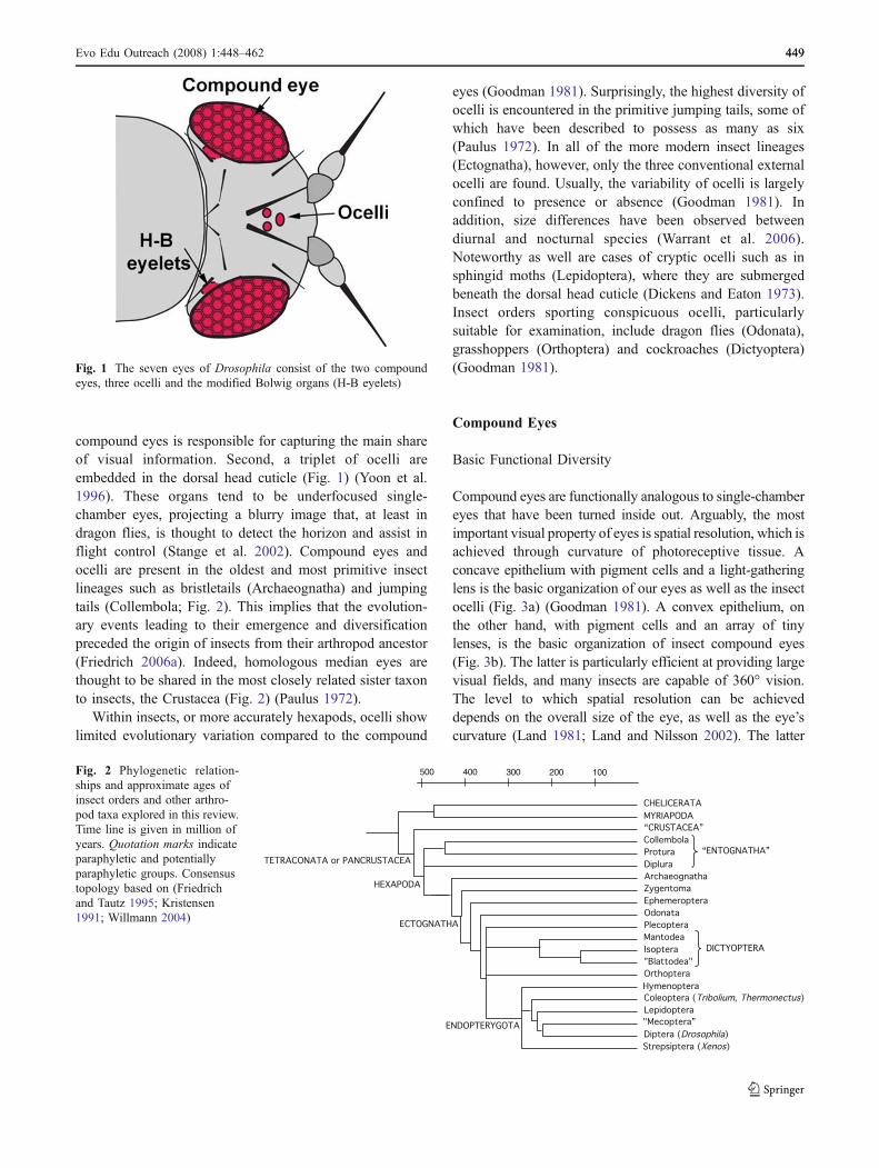
compound eyes is responsible for capturing the main shareof visual information. Second, a triplet of ocelli areembedded in the dorsal head cuticle (Fig. 1) (Yoon et al.1996). These organs tend to be underfocused single-chamber eyes, projecting a blurry image that, at least indragon flies, is thought to detect the horizon and assist inflight control (Stange et al. 2002). Compound eyes andocelli are present in the oldest and most primitive insectlineages such as bristletails (Archaeognatha) and jumpingtails (Collembola; Fig. 2). This implies that the evolution-ary events leading to their emergence and diversificationpreceded the origin of insects from their arthropod ancestor(Friedrich 2006a). Indeed, homologous median eyes arethought to be shared in the most closely related sister taxonto insects, the Crustacea (Fig. 2) (Paulus 1972).
Within insects, or more accurately hexapods, ocelli showlimited evolutionary variation compared to the compound
eyes (Goodman 1981). Surprisingly, the highest diversity ofocelli is encountered in the primitive jumping tails, some ofwhich have been described to possess as many as six(Paulus 1972). In all of the more modern insect lineages(Ectognatha), however, only the three conventional externalocelli are found. Usually, the variability of ocelli is largelyconfined to presence or absence (Goodman 1981). Inaddition, size differences have been observed betweendiurnal and nocturnal species (Warrant et al. 2006).Noteworthy as well are cases of cryptic ocelli such as insphingid moths (Lepidoptera), where they are submergedbeneath the dorsal head cuticle (Dickens and Eaton 1973).Insect orders sporting conspicuous ocelli, particularlysuitable for examination, include dragon flies (Odonata),grasshoppers (Orthoptera) and cockroaches (Dictyoptera)(Goodman 1981).
Compound Eyes
Basic Functional Diversity
Compound eyes are functionally analogous to single-chambereyes that have been turned inside out. Arguably, the mostimportant visual property of eyes is spatial resolution, which isachieved through curvature of photoreceptive tissue. Aconcave epithelium with pigment cells and a light-gatheringlens is the basic organization of our eyes as well as the insectocelli (Fig. 3a) (Goodman 1981). A convex epithelium, onthe other hand, with pigment cells and an array of tinylenses, is the basic organization of insect compound eyes(Fig. 3b). The latter is particularly efficient at providing largevisual fields, and many insects are capable of 360° vision.The level to which spatial resolution can be achieveddepends on the overall size of the eye, as well as the eye’scurvature (Land 1981; Land and Nilsson 2002). The latter
“CRUSTACEA”
Diplura
Protura
Collembola
Archaeognatha
Zygentoma
Ephemeroptera
Odonata
Plecoptera
Mantodea
Isoptera
"Blattodea"
Orthoptera
Coleoptera (Tribolium, Thermonectus)
Lepidoptera
Diptera (Drosophila)
"Mecoptera”
Hymenoptera
Strepsiptera (Xenos)
“ENTOGNATHA”
DICTYOPTERA
MYRIAPODA
CHELICERATA
ECTOGNATHA
TETRACONATA or PANCRUSTACEA
100200300400500
HEXAPODA
ENDOPTERYGOTA
Fig. 2 Phylogenetic relation-ships and approximate ages ofinsect orders and other arthro-pod taxa explored in this review.Time line is given in million ofyears. Quotation marks indicateparaphyletic and potentiallyparaphyletic groups. Consensustopology based on (Friedrichand Tautz 1995; Kristensen1991; Willmann 2004)
Fig. 1 The seven eyes of Drosophila consist of the two compoundeyes, three ocelli and the modified Bolwig organs (H-B eyelets)
Evo Edu Outreach (2008) 1:448–462 449449

frequently varies across different regions of individualcompound eyes, including regions with heightened acuity.Insects have evolved several sub-types of compound eyes,including apposition, superposition, and neural superpositioneyes. The review of these eye types is beyond the scope ofthis publication (but see Nilsson 1989 and Land and Nilsson2002 for comprehensive reviews on this topic), but we willgive a few examples of insect eyes that have evolved toexcel in specific ways.
Among the most basal insects with particularly well-developed compound eyes are the dragon- and damselflies(Odonata), which are characterized by up to 30,000ommatidia (Sherk 1978). It is no coincidence that the mostelaborate compound eyes are found among the best-flyinginsects. Insect flight requires high spatial resolution as wellas photoreceptors that are able to rapidly adjust to changesin visual scene. In addition, dragonflies are particularlygood at seeing small objects. These visually guidedpredators are able to intercept their prey by adjusting headpositions to maintain their victim in an area of the eye withparticularly high acuity (Olberg et al. 2007). Among themore basal insects, another group of interest is the prayingmantis (Mantodea). While they do not perform aerial pursuits,their high-acuity vision allows them to accurately gage thedistance to their prey (Kral 1998). In addition to othermethods of distance estimation, they are able to detect minordifferences between the image of the right and left eye tocorrectly estimate the distance of their prey. This has beenelegantly demonstrated through the placement of prisms infront of their eyes, leading to enhanced or reduced disparitiesof the two images: Accordingly, the praying mantisesundershoot or overshoot their prey (Rossel 1983).
While high acuity is important, other visual modalitieshave been highly adaptive throughout insect evolution.Among them is the ability to detect the direction of theelectric field component (e-vector) of linearly polarized light(see Wehner and Labhart 2006 and Horváth and Varjú 2004for recent reviews). This ability can be used for orientation
(Wehner 1992; von Frisch 1967), the detection of watersurfaces (Horvath and Varju 1997; Schwind 1991), and incommunication (Cronin et al. 2003; Marshall et al. 1999). Aspecialized region for polarized vision exists in the dorsalrim area of most compound eyes. The dorsal rim area hencerepresents an ancient component of the insect retina, havingbeen described in species ranging from dragonflies toDiptera (see Labhart and Meyer 1999).
Color vision is another specialty enabled by restrictingincoming light (see Kelber 2006). Multiple photoreceptorcells, each sampling a different portion of the spectrum, arecompared to facilitate the discrimination of color. The abilityof honeybees (Hymenoptera) to differentiate between colorswas already observed by Karl von Frisch early in thetwentieth century (von Frisch 1914). Indeed many insects arethought to possess color vision (Briscoe and Chittka 2001),an ability that requires bright light because each receptorsamples a relatively narrow visual spectrum. However, it hasrecently been demonstrated that the nocturnal hawkmothDeilephila elpenor (Lepidoptera) is able to discriminatecolors at light levels as low as dim starlight (Kelber et al.2002). While this ability may be exceptional, the eyes ofmany other insects are able to detect contrast at low lightlevels. Generally, this requires substantial pooling andtherefore tends to result in somewhat reduced spatialresolution (see Warrant 2006 for review). A particularlyinteresting example are the nocturnal halictid bees, which usevisual landmarks to find their nest (Warrant et al. 2004). Inthese species, light capture is thought to be facilitated byrelatively large rhabdoms (the site of photodetection) andthrough spatial pooling at the neurological level (Greiner etal. 2004). Drastically enlarged rhabdoms also are found innocturnal ants (Greiner et al. 2007).
Ancient Heritage
Regardless of adaptive fine-tuning, insect compound eyesare formed from ommatidia, each of which represents a
Fig. 3 Schematic of different eye types. a The single-chamber cameraeye is the basic type of the human eye but also exists within insects.Spatial resolution is achieved through the lens and through a concavecurvature of the retina. b Schematic of an insect compound eye.
Spatial resolution is achieved through a series of small lenses and aconvexly shaped retina. Striped areas indicate regions of photorecep-tion, which in real eyes tend to be separated by screening pigment
450 Evo Edu Outreach (2008) 1:448–462
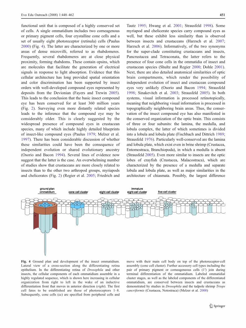
functional unit that is composed of a highly conserved setof cells. A single ommatidium includes two corneagenousor primary pigment cells, four crystalline cone cells and aset of usually eight photoreceptor (retinula) cells (Paulus2000) (Fig. 4). The latter are characterized by one or moreareas of dense microvilli, referred to as rhabdomeres.Frequently, several rhabdomeres are at close physicalproximity, forming rhabdoms. These contain opsins, whichare molecules that facilitate the generation of electricalsignals in response to light absorption. Evidence that thiscellular architecture has long provided spatial orientationand color discrimination has been supported by insectorders with well-developed compound eyes represented bydeposits from the Devionian (Fayers and Trewin 2005).This leads to the conclusion that the basic insect compoundeye has been conserved for at least 360 million years(Fig. 2). Surveying even more distantly related speciesleads to the inference that the compound eye may beconsiderably older. This is clearly suggested by thewidespread presence of compound eyes in crustaceanspecies, many of which include highly detailed blueprintsof insect-like compound eyes (Paulus 1979; Melzer et al.1997). There has been considerable discussion of whetherthese similarities could have been the consequence ofindependent evolution or shared evolutionary ancestry(Osorio and Bacon 1994). Several lines of evidence nowsuggest that the latter is the case. An overwhelming numberof studies show that crustaceans are more closely related toinsects than to the other two arthropod groups, myriapodsand chelicerates (Fig. 2) (Regier et al. 2005; Friedrich and
Tautz 1995; Hwang et al. 2001; Strausfeld 1998). Somemyriapod and chelicerate species carry compound eyes aswell, but these exhibit less similarity than is observedbetween insects and crustaceans (Harzsch et al. 2007;Harzsch et al. 2006). Informatively, of the two synonymsfor the super-clade constituting crustaceans and insects,Pancrustacea and Tetraconata, the latter refers to thepresence of four cone cells in the ommatidia of insect andcrustacean species (Shultz and Regier 2000; Dohle 2001).Next, there are also detailed anatomical similarities of opticbrain compartments, which render the possibility ofindependent evolution of insect and crustacean compoundeyes very unlikely (Osorio and Bacon 1994; Strausfeld1998; Sinakevitch et al. 2003; Strausfeld 2005). In bothsystems, visual information is processed retinotopically,meaning that neighboring visual information is processed intopographically neighboring brain areas. Thus, the conser-vation of the insect compound eye has also manifested inthe conserved organization of the optic brain. This consistsof three or four subunits: the lamina, the medulla, andlobula complex, the latter of which sometimes is dividedinto a lobula and lobula plate (Fischbach and Dittrich 1989;Strausfeld 1976). Particularly well-conserved are the laminaand lobula plate, which exist even in brine shrimp (Crustacea,Entomostraca, Branchiopoda), in which a medulla is absent(Strausfeld 2005). Even more similar to insects are the opticlobes of crayfish (Crustacea, Malacostraca), which arecharacterized by the presence of a medulla and separatelobula and lobula plate, as well as major similarities in thearchitecture of chiasmata. Possibly, the largest difference
Fig. 4 Ground plan and development of the insect ommatidium.Lateral view of a cross-section along the differentiating retinaepithelium. In the differentiating retina of Drosophila and otherinsects, the cellular components of each ommatidium assemble in ahighly regulated sequence, which is shown here increasing in cellularorganization from right to left in the wake of an inductivedifferentiation front that moves in anterior direction (right). The firstcell fates to be established are those of photoreceptors 1–8.Subsequently, cone cells (cc) are specified from peripheral cells and
move with their main cell body on top of the photoreceptor-cellassembly (cone cell cluster). Further accessory cell types including thepair of primary pigment or corneageneous cells (1′) join duringterminal differentiation of the ommatidium. Labeled ommatidialcluster stages, as well as the labeled components of the differentiatedommatidium, are conserved between insects and crustaceans asdemonstrated by studies in Drosophila and the tadpole shrimp Triopscancriformis (Crustacea, Notostraca) (Melzer et al. 2000)
Evo Edu Outreach (2008) 1:448–462 451451
between insects and malocostracean optic lobes is thepresence of a separated inner medulla in insects. Otherdifferences between the neuropils of crustacean and insectsinclude details in the layering of the medulla. Regardless, theoverwhelming number of similarities between insect andcrustacean optic brains is suggestive of ancient origin ofcentral nervous system components of the insect visual system(Strausfeld 1998; Sinakevitch et al. 2003; Harzsch 2002).
Last but not least, there are striking parities between theearliest steps of retinal development in crustaceans andinsects. As first described in Drosophila, the differentiationof the compound eye proceeds through a carefully choreo-graphed sequence of ommatidial patterning steps (forreview see Wolff and Ready 1993; Morante et al. 2007).This process starts with the specification of eight photore-ceptor cells, followed by cone cells, primary pigments cellsand further accessory cells (Fig. 4). Remarkably, thedifferentiating retina of crayfish (Crustacea, Malacostraca)and tadpole shrimps (Crustacea, Anacostraca) share manyof the ommatidial patterning stages (Melzer et al. 2000;Hafner and Tokarski 1998). This implies that the retinalpatterning of the ancestor to insects and crustaceans hasremained conserved for more than 500 million years(Walossek and Muller 1990). This conclusion is furthersupported by the similar retinal patterning in grasshoppers(Orthoptera), moths (Lepidoptera), and beetles (Coleoptera)(Egelhaaf 1988; Friedrich et al. 1996; Champlin and Truman1998; Friedrich and Benzer 2000). Seemingly, the differen-tiation of the insect compound eye is one of the mostconserved cellular patterning processes known.
Reduction and Deconstruction
Considering the enormous time range through which thestructure and design of the compound eye remainedconserved in the Pancrustacea (a.k.a. the Tetraconata;Fig. 2), one may wonder which, if any, factors may haveimposed modification. Nonetheless, the body of compara-tive research on compound-eye structure is considerable(Nilsson 1989; Oakley 2003; Meinertzhagen 1991; Bitschand Bitsch 2005; Land 1991; Land et al. 1999). After all,with at least 750,000 species, insects were given manyopportunities for “evolutionary tinkering” (Jacob 1977).Most variations in the insect retina represent slightreductions, or expansions, in number of photoreceptorcells per ommatidium, while cone cells appear to bevirtually numerically invariant (for review see Oakley2003). An interesting example is the addition of a ninthphotoreceptor, which occurred independently in Hymenopteraand Lepidoptera (Fig. 2) (Friedrich et al. 2006). In theLepidoptera especially, there are species with higher andmore variable numbers of photoreceptors per ommatidium(Egelhaaf 1988).
Another type of variation relates to eye size as measured indimensions or number of ommatidia. The largest known insecteyes belong to the previously mentioned dragon flies(Odonata) (30,000; Sherk 1978). Interestingly, compound-eye reduction has not yet been systematically explored,although reduction may well relate to the considerableenergetic costs therein. In Drosophila, it has been estimatedthat the ATP used to maintain electric currents of illuminatedphotoreceptor cells accounts for 8% of the total energy that isconsumed at rest (Laughlin et al. 1998). Note that thesecalculations do not account for any costs arising from theconstant renewal of the photoactive membrane, the mainte-nance of support cells, or the neural activity needed toprocess the visual information (Laughlin 2001). However thisrepresents an arbitrary sample. A comprehensive treatment ofthis topic may reveal ecological correlations to eye reduction.Considering paleontological and phylogenetic data, insectscontain examples of both very ancient as well as compara-tively recent eye reduction. In the former case, jumping tails(Collembola) are known for their reduced lateral eyes, whichconsist of few but complete ommatidia (Kristensen 1991).Other ancient hexapods with similarly or even moreextremely reduced eyes are the Protura and Diplura (Condeand Pages 1991; Imadate 1991). Importantly, becauseelaborate compound eyes are conserved in the Crustacea, onecan conclude that these basal insect orders underwentreduction of an initially more complete compound eye.Moreover, not all primitively wingless insects experiencedcompound-eye reduction, as the large lateral eyes of bristletails (Archaeognatha) indicate (Watson and Smith 1991).
It is possible to infer recent reduction if eye-reducedspecies are closely related to those with eyes of average size.Such examples are found among contributors to the faunas oflight-deprived environments like crevices, karsts, and caves.Facultative cave inhabitants (troglophilic) with reduced eyesand obligatory cave species (troglobitic) that are completelyeyeless are found in Orthoptera, Coleoptera, and Diptera(Harvey et al. 2000). Some species with highly reduced eyesare much closer to our homes, e.g., the harmless silverfish(Zygentoma) (Smith and Watson 1991). More harmful, butincreasingly more popular as a model system for develop-ment and insect genomics, is the starch-consuming darklingbeetle species Tribolium castaneum (red flour beetle)(Klingler 2004). In this case, the compound eye is only~90 ommatidia large, in contrast to closely related darklingbeetles, which possess hundreds of ommatidia per compoundeye.
Remodeling of Compound Eyes in Twisted Wing Insects(Strepsiptera)
One possible example of reduction with consecutiveelaboration is the eye of twisted wing insects (Strepsiptera).
452 Evo Edu Outreach (2008) 1:448–462

This group has evolved dramatically remodeled eyes thatsubstantially differ in function from those of other insectorders. While compound eyes generally tend to resolve oneimage point per lens, the eyes of Strepsiptera are amongvery few exceptions (Land and Nilsson 2002). This insectorder represents a particularly enigmatic group, oftenknown to systematists for their unclear position (Fig. 2)(see Beutel and Pohl 2006 for recent review), and whoseunusual eye organization has long been noted (MacCarthy1991; Rösch 1913). In fact, the eye organization is sopeculiar that it has led Mike Land to comment: “One istempted to conclude that these eyes, so unlike anything inany other insect, either came from outer space or were puthere by God to confuse scientists” (Land 1984).
Functionally, the strepsipteran eye represents a hybridof image-forming eyes and canonical compound eyes(Buschbeck et al. 1999; Buschbeck et al. 2003). Whiletypical compound eyes gain spatial resolution exclusivelythrough their convex surface (Fig. 3b), the strepsipteran eyeis composed of small, concave-image-forming units, calledeyelets (Fig. 5). Externally, Strepsiptera differ by presentingfar fewer but much larger lenses (Kinzelbach 1971). In theStrepsiptera, Xenos peckii, for example, there are onlyabout 50 lenses, much fewer than the over 700 facets of theslightly smaller fruit fly D. melanogaster. Moreover, atypical lens in X. peckii is about 65 μm in diameter andcovers about the same area as do 15 fruit fly lenses(Buschbeck et al. 1999). Typical insect compound eyespossess only eight to ten photoreceptor cells per facet. Incontrast, beneath each Strepsiptera lens lies a small, cup-shaped retina, which, in X. peckii, is comprised of over 100photoreceptor cells. Each lens and retina together, therefore,forms a small single-chamber eye that resolves only a partialimage, namely that of the small portion of the visual fieldthat lies above it. The collective resolution of each of theseeyelets is limited. Based on anatomical findings (Buschbecket al. 2003) and on behavioral studies (Maksimovic et al.2007), it is only around 13 points. However, in function, the
increase in total resolution is substantial, from about 50 to650. This resolution seems to assist in the one task that amale Strepsiptera needs to accomplish in the few hours of hisvery short life: finding a female to mate.
While strepsipteran eyes neither “came from outer space,”nor “were put here by God to confuse scientists,” theirevolutionary origin still remains somewhat unclear. This islargely due to major uncertainties in the systematic placementof Strepsiptera, as well as limited availability of especially themost basal strepsipteran groups. Nevertheless, there isevidence for the evolution of this eye from a more typicalinsect compound eye. Most of the systematic debate centersaround where within holometabolous insects Strepsiptera fitbest (Fig. 2) (Kristensen 1999; Hwang et al. 1998; Bonnetonet al. 2006), implying that their ancestors undoubtedly hadcompound eyes. Furthermore, at least at the level of grossmorphological development, many parallels with typicalinsect compound eyes have been observed. These includethe presence of separate larval eyes that degenerate and shiftduring development (Buschbeck 2005).
One ontogenic possibility is that as a first step, theancestral compound eye was largely reduced, possibly dueto adopting a nocturnal lifestyle. While little is knownabout the eyes of the most ancestral strepsipteran groups,the most ancestral living group of Strepsiptera, theMengenillidae, is indeed nocturnal (Pohl 2002). A noctur-nal history is also supported by the fact that strepsipteraneyelets resemble the ommatidia of some nocturnal insects,such as certain mosquitoes (Land et al. 1999). Severaleyelet characteristics are consistent with vision at low lightlevels. These include large lenses, short rhabdoms, slowreceptor-cell physiology, and possibly the absence of colorvision (Buschbeck et al. 2003). If strepsipteran eyes firstevolved to accommodate low-light vision, then additionalphotoreceptors in each unit could have evolved to enhancethe light capture of individual sample points. Concomitant-ly, the total number of lenses could have been reduced toenhance light capture at the cost of spatial resolution. If asecondary shift in lifestyle resulted in X. peckii’s ancestorsbecoming diurnal again (which indeed they are), then theabundance of receptor cells within each unit could have ledto a secondary increase of visual resolution within eacheyelet.
At this point, it remains unclear if this truly is howstrepsipteran eyes evolved, and many questions remain. Forexample, how did additional photoreceptors appear in eachunit, considering that facets are so rigidly conserved inother insect taxa? Several possibilities await furtherinvestigation. These include the fusion of ancestrallyseparate ommatidia, as has been suggested for diplopodsand chilopods (Harzsch et al. 2007), or the independentevolution of a large amount of additional receptor cellssimilar to some crepuscular Lepidoptera (Egelhaaf 1988).
Fig. 5 The strepsipteran eye represents a combination of a compoundeye and image-forming lens eyes. It consists of eyelets, each of whichresolves a small portion of the visual field of the insect
Evo Edu Outreach (2008) 1:448–462 453453
The Larval Eyes
Returning to the seven eyes of Drosophila, two eyes stillawait to be introduced. These are the so-called Hofbauer–Buchner (H-B) eyelets, small photoreceptor bundles deep inthe optic brain of the fly (Fig. 1) (Hofbauer and Buchner1989; Yasuyama and Meinertzhagen 1999). The DrosophilaH-B eyelets do not qualify as eyes in the strict sense ofimage-forming devices. However, they are without doubtvisual organs based on connectivity and the expression ofphotoreceptor specific proteins such as opsins (Malpel et al.2002). This leads to questions such as where the H-B-eyeletsmay have come from and what their function is. The answersare hidden deep inside the larvae of Drosophila, which usespecialized larval eyes, called stemmata.
Scorpion Flies and the Origin of Insect Larval Eyes
Stemmata are larval eyes found specifically in indirectlydeveloping insects (Endopterygota or Holometabola) likebutterflies, honeybees, and Drosophila, and which undergodramatic changes during postembryonic development(Kristensen 1999; Friedrich 2003). In endopterygoteinsects, the egg releases a juvenile instar, the larva, whosephysique has little in common with the adult (see Fig. 6 forcorrelated diversity of larval head morphologies). Onecorollary of this is the lack of compound eyes and the useof specialized larval eyes. From what did these larval eyesevolve? In many species, the larval eyes lack any similarityto the adult compound eye. However, in representatives ofthe order scorpion flies (Mecoptera), which are quite closelyrelated to the true flies (Diptera), the larvae are equipped withsmall compound eyes composed of ommatidia (Fig. 6b)(Steiner 1930). One may therefore ask what then definesthese as specialized larval eyes? It is the fact that, as inother endopterygote insects, they are replaced by the adultcompound eyes during pupation (Paulus 1989). Hence, themecopteran larval eyes are specialized larval eyes based ontheir life-history stage restriction. This is a definitivedifference to directly developing insects, where the firstjuvenile instar is born with true compound eyes that arecarried over into the adult stage (Fig. 6a). There is a second,less obvious difference as well. Juvenile compound eyescontinue to expand during postembryonic development bythe addition of new ommatidia in directly developingspecies (for review see Friedrich 2006b), in contrast to thecompound eyes of mecopteran larvae which do not furtherincrease in size (Paulus 1989).
The conservation of compound-eye-like larval eyes inscorpion flies was critical for relating the postembryonicvisual development of directly developing and endopter-ygote species. As the comparative entomologist HannesPaulus pointed out, the scorpion fly larval eyes represent
living proof that insect larval eyes evolved from acompound-eye-like precursor organ (Paulus 1986). Com-parative developmental biology has since shed additionallight on the evolutionary origin of the larval eye (Friedrich2006b). Through the use of molecular markers, forinstance, it was confirmed that the embryonic localizationof the differentiating larval eyes of endopterygote insectscorresponds to the developing embryonic compound eye indirectly developing species (Fig. 7) (Liu and Friedrich2004). From these findings, one can conclude the larvaleyes of endopterygote insects are homologous to thecompound eyes of the first instars of directly developingspecies (nymphs). Second, the adult eyes of endopterygotespecies correspond specifically to the portion of the adulteye that is added on during postembryonic development indirectly developing insects (Fig. 7). Hence, insect larvaleyes originated by the separation of the embryonic andpostembryonic partitions of the compound eye in the lastdirectly developing ancestor of endopterygote insects.
Evolutionary History of the Drosophila Larval Eyes
Insect larval eyes experienced numerous modifications in thehighly diversified lineages of endopterygote insect orders(Gilbert 1994). One of the most dramatic changes occurredin the higher Diptera, specifically in the lineage leading toDrosophila (Melzer and Paulus 1989). The Drosophila larvaperforms light-oriented behavior using a pair of photorecep-tor bundles that attach to each side of the cephalopharyngealhead skeleton of the maggot and constitute the Bolwig’sorgans (BO; Fig. 6d) (Busto et al. 1999). Originallydiscovered by Niels Bolwig in the closely related house fly(Bolwig 1946), the BOs had at first not been related to thecompound eye. However, comparative analysis in a widerange of Diptera established that even these enigmatic visualorgans are related to ommatidia-like precursor structures(Melzer and Paulus 1989). Consistent with this, it has beenfound that the genetic control of photoreceptor-specific opsinexpression in the BO is almost identical to the ommatidia ofthe adult eye (Friedrich in press). In addition, there are manydetailed parallels in the participation of genes during theinduction and specification of different cell fates in the twovisual organs (Friedrich 2006b). At this point, over tenshared regulators of early patterning have been identified(Friedrich in press, 2006a).
Is it possible to reconstruct the steps that lead from theneat arrangement of ommatidia in the ancestral larval eye tothe bare-bone BO of the Drosophila larva? An obviousdifference is the complete absence of accessory cells in theBO. This implies a reduction in pigment and cone cells.The actual evolutionary process leading to this “strip-down” becomes easy to conjecture if one recalls thesequence of cell fate determination steps during normal
454 Evo Edu Outreach (2008) 1:448–462

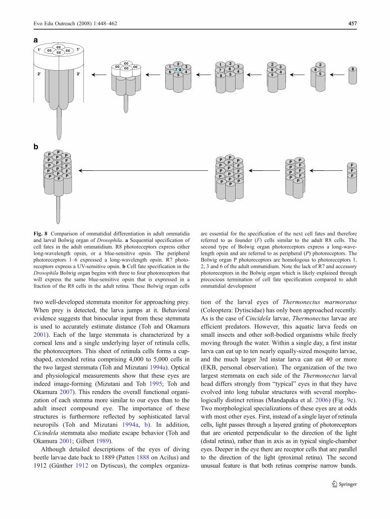
ommatidial development (Fig. 8). This is because thepigment and cone cell fates are established subsequent tothe photoreceptor-cell fates (Ready et al. 1976; Wolff andReady 1993). The evolutionary loss of accessory cells inthe BO is therefore simply explained by precociousabrogation of ommatidial differentiation (Fig. 8).
The deconstruction of the ommatidial ground plan goeseven one step further. Five photoreceptor sub-types exist inthe Drosophila adult eye, which are distinguished by thedifferential expression of opsin genes (for review see Cookand Desplan 2001). The central R8 photoreceptor is the firstcell to differentiate (for review see Morante et al. 2007,Freeman 1997). Since the specification of all subsequentphotoreceptor-cell fates is dependent on its presence, the R8photoreceptor is therefore also referred to as the foundercell (Dokucu et al. 1996). A second central photoreceptor,the R7 cell, is the last photoreceptor cell to differentiate. InDrosophila, the R7 photoreceptors are also unique inexpressing UV-sensitive opsin genes (Montell et al. 1987).The remaining six peripheral photoreceptors express long-wavelength-sensitive opsins. In the BO, there are only twophotoreceptor sub-types (Figs. 6d and 8) (Sprecher et al.2007). Three to four photoreceptors express a blue-sensitiveopsin and differentiate first. These photoreceptors representhomologs of R8 founder cells in the adult eye (Friedrich inpress). The second group is comprised of ten to 16 long-
wavelength opsin-expressing cells that correspond to theperipheral photoreceptors of the adult eye ommatidia. TheBO therefore lacks a UV-sensitive photoreceptor. Thissuggests that the R7 photoreceptor was lost during theevolution of the Drosophila larval eyes (Friedrich 2008).Strikingly, similar to the case of the accessory cells, the R7photoreceptor is the last photoreceptor cell to differentiateduring ommatidial development (Fig. 8). It thus is the firstphotoreceptor cell poised for disappearance by early termi-nation of the ommatidial cell fate specification sequence.
Recapitulated Fusion of Ancestral Ommatidal Unitsin the Larval Eye of Flour Beetle
The presence of more than four founder cell in the BOrepresents another interesting difference between it and theadult ommatidia (Fig. 8). It suggests that the BOs originatedby the ancestral fusion of three to four ommatidia.Intriguingly, the integration of ancestral ommatidia to largerlarval eyes was hypothesized earlier (Paulus 1986). Thisproposal was based on the observation that the larval eyesof many insects consist of a loose assemblage of a fewisolated but largely completely organized ommatidia.Developmental data from the red flour beetle T. castaneumprovide further support for this scenario. The Triboliumlarva is equipped with a normal head capsule that sports
Fig. 6 A snapshot of juvenileeye diversity in insects. aNymphal compound eye in thefirst instar nymph of a grass-hopper illustrating a typicalcompound eye. b Head ofmecopteran larva, which has acompound-eye-like eye. Modi-fied after (Steiner 1930). cLarval stemmata of the red flourbeetle T. castaneum. d Bolwigorgans of the Drosophila larva.Modified after (Melzer andPaulus 1989)
Evo Edu Outreach (2008) 1:448–462 455455

lateral stemmata as larval eyes (Fig 6c) (Liu and Friedrich2004). If examined closely, two discrete photoreceptorgroups can be seen on each side of the head, which arereferred to as dorsal and ventral stemmata (Fig. 6c).Investigation of their differentiation in the embryo, howev-er, revealed the initial formation of five discrete photore-ceptor clusters (Liu and Friedrich 2004). During lateembryonic development, two of the initial clusters fuse toform the dorsal stemma. In parallel, three ventral clustersmerge to form the ventral stemma. From an evolutionaryperspective, the morphogenesis of the Tribolium larval eyesappears to recapitulate the postulated fusion of ancestralommatidia.
The Tribolium larval eyes further document that cellularreduction is a pervasive theme in the evolution of insectlarval eyes. Like the Drosophila BOs, the Tribolium larvaleyes lack cone and pigment cells, implying a reduction ofall accessory cells. The direction of light flux is influencedby the heavy pigmentation of the photoreceptors them-selves, which force light to enter from a limited angle.Since species with primitive ommatidium-type larval eyesare known in both Coleoptera and Diptera (Gilbert 1994),one has to conclude that the reduction of accessory cellsoccurred independently in the lineages leading toDrosophilaand Tribolium. It is noteworthy that in both of these groups,the ecology of larvae appears to render vision lessimportant (Busto et al. 1999; Park 1934). The reductionmay therefore be the result of evolution against costlyvisual organs.
Remodeling of Larval Eyes
It would be misleading to conclude from Tribolium andDrosophila that the larval eyes of endopterygote insectsfaced only two possible evolutionary fates: conservation ordeconstruction. The evolution of the insect visual systemhas been far more versatile. Despite only inheriting a tinyfraction of the adult compound eye, not all larvae ofholometabolous insects have poor vision. In fact, there areseveral examples of larvae with quite good vision. Amongthem are certain Nematocera (the more basal flies) such asmosquitoes, which achieve improved vision simply byexpressing the adult compound eye early (Gilbert 1994)(Fig. 9a). This accelerated development of the adult eyeserves both the larvae and the pupa, which is extraordi-narily mobile and efficient at predator avoidance.
Several holometabolous insect larvae were able to inhabitunique habitats, presumably in part by evolving good eyes.Among them are predatory species that, relieved of the need foradult-specific visual functions (such as mate detection),evolved eyes specialized for prey capture. The most sophisti-cated stemmata have evolved in different groups of Coleoptera:tiger beetles (Cicindelidae) and diving beetles (Dytiscidae).
The best-studied larval visual system is that of the tigerbeetle, Cicindela chinensis. This consists of six stemmata oneach side, two of which are particularly sophisticated image-forming lens eyes with extended cup-shaped retinas (Fig. 9b)(Toh and Mizutani 1987). Cincindela larvae catch their preyby hiding near the entrance of a tunnel in the ground, while
Fig. 7 Homology of eyes in directly developing and endopterygoteinsects (after Friedrich 2006b). a Indirect development shown in D.melanogaster. 1 Differentiation of larval eyes (Bolwig organs).2 Specification and separation of eye–antennal imaginal disc cells. 3Functional Bolwig organs. 4 Nascent eye–antennal imaginal disc. 5Cell proliferation in eye–antennal imaginal disc during larvaldevelopment. 6 Degradation of larval epithelium. 7 Dedifferentiationof the Bolwig organs to H-B eyelets by relocalization into the brain.8 Onset of retinal differentiation in the eye–antennal imaginal disc. 9
Eversion of eye–antennal imaginal disc. 10 Functional Drosophilacompound eye generated by fully everted and differentiated eye–antennal imaginal disc. 11 Drosophila H-B eyelets. b Directdevelopment shown in the American desert locust Schistocercaamericana. 12 Onset of retinal differentiation in the embryo. 13Postembryonic expansion of the nymphal compound eye. 14 Adultgrasshopper compound eye. The embryonic phase of eye developmentis indicated by blue background. The postembryonic phase of eyedevelopment is indicated by red background
456 Evo Edu Outreach (2008) 1:448–462
two well-developed stemmata monitor for approaching prey.When prey is detected, the larva jumps at it. Behavioralevidence suggests that binocular input from these stemmatais used to accurately estimate distance (Toh and Okamura2001). Each of the large stemmata is characterized by acorneal lens and a single underlying layer of retinula cells,the photoreceptors. This sheet of retinula cells forms a cup-shaped, extended retina comprising 4,000 to 5,000 cells inthe two largest stemmata (Toh and Mizutani 1994a). Opticaland physiological measurements show that these eyes areindeed image-forming (Mizutani and Toh 1995; Toh andOkamura 2007). This renders the overall functional organi-zation of each stemma more similar to our eyes than to theadult insect compound eye. The importance of thesestructures is furthermore reflected by sophisticated larvalneuropils (Toh and Mizutani 1994a, b). In addition,Cicindela stemmata also mediate escape behavior (Toh andOkamura 2001; Gilbert 1989).
Although detailed descriptions of the eyes of divingbeetle larvae date back to 1889 (Patten 1888 on Acilus) and1912 (Günther 1912 on Dytiscus), the complex organiza-
tion of the larval eyes of Thermonectus marmoratus(Coleoptera: Dytiscidae) has only been approached recently.As is the case of Cincidela larvae, Thermonectus larvae areefficient predators. However, this aquatic larva feeds onsmall insects and other soft-bodied organisms while freelymoving through the water. Within a single day, a first instarlarva can eat up to ten nearly equally-sized mosquito larvae,and the much larger 3rd instar larva can eat 40 or more(EKB, personal observation). The organization of the twolargest stemmata on each side of the Thermonectus larvalhead differs strongly from “typical” eyes in that they haveevolved into long tubular structures with several morpho-logically distinct retinas (Mandapaka et al. 2006) (Fig. 9c).Two morphological specializations of these eyes are at oddswith most other eyes. First, instead of a single layer of retinulacells, light passes through a layered grating of photoreceptorsthat are oriented perpendicular to the direction of the light(distal retina), rather than in axis as in typical single-chambereyes. Deeper in the eye there are receptor cells that are parallelto the direction of the light (proximal retina). The secondunusual feature is that both retinas comprise narrow bands.
82
85
cccccc cc
234
16
5
1' 1'
2' 2'
cccc cccc
231
7 84
56
23
58 4
FFF
PFFF
PPPP
P
PP PP F
FF
PP
P
PPP P
P
P
PP PP F
FF
PP
P
PPP P
P
P
87 87
a
b
Fig. 8 Comparison of ommatidial differentiation in adult ommatidiaand larval Bolwig organ of Drosophila. a Sequential specification ofcell fates in the adult ommatidium. R8 photoreceptors express eitherlong-wavelength opsin, or a blue-sensitive opsin. The peripheralphotoreceptors 1–6 expressed a long-wavelength opsin. R7 photo-receptors express a UV-sensitive opsin. b Cell fate specification in theDrosophila Bolwig organ begins with three to four photoreceptors thatwill express the same blue-sensitive opsin that is expressed in afraction of the R8 cells in the adult retina. These Bolwig organ cells
are essential for the specification of the next cell fates and thereforereferred to as founder (F) cells similar to the adult R8 cells. Thesecond type of Bolwig organ photoreceptors express a long-wave-length opsin and are referred to as peripheral (P) photoreceptors. TheBolwig organ P photoreceptors are homologous to photoreceptors 1,2, 3 and 6 of the adult ommatidium. Note the lack of R7 and accessoryphotoreceptors in the Bolwig organ which is likely explained throughprecocious termination of cell fate specification compared to adultommatidial development
Evo Edu Outreach (2008) 1:448–462 457457
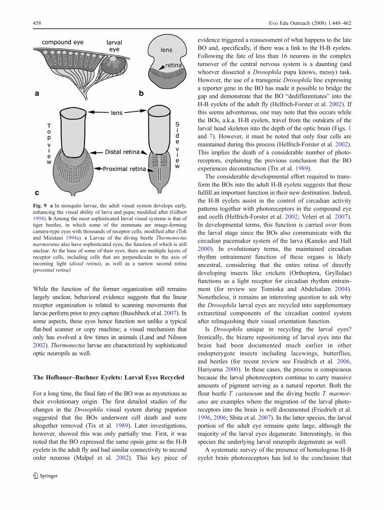
While the function of the former organization still remainslargely unclear, behavioral evidence suggests that the linearreceptor organization is related to scanning movements thatlarvae perform prior to prey capture (Buschbeck et al. 2007). Insome aspects, these eyes hence function not unlike a typicalflat-bed scanner or copy machine; a visual mechanism thatonly has evolved a few times in animals (Land and Nilsson2002). Thermonectus larvae are characterized by sophisticatedoptic neuropils as well.
The Hofbauer–Buchner Eyelets: Larval Eyes Recycled
For a long time, the final fate of the BO was as mysterious astheir evolutionary origin. The first detailed studies of thechanges in the Drosophila visual system during pupationsuggested that the BOs underwent cell death and werealtogether removed (Tix et al. 1989). Later investigations,however, showed this was only partially true. First, it wasnoted that the BO expressed the same opsin gene as the H-Beyelets in the adult fly and had similar connectivity to secondorder neurons (Malpel et al. 2002). This key piece of
evidence triggered a reassessment of what happens to the lateBO and, specifically, if there was a link to the H-B eyelets.Following the fate of less than 16 neurons in the complexturnover of the central nervous system is a daunting (andwhoever dissected a Drosophila pupa knows, messy) task.However, the use of a transgenic Drosophila line expressinga reporter gene in the BO has made it possible to bridge thegap and demonstrate that the BO “dedifferentiates” into theH-B eyelets of the adult fly (Helfrich-Forster et al. 2002). Ifthis seems adventurous, one may note that this occurs whilethe BOs, a.k.a. H-B eyelets, travel from the outskirts of thelarval head skeleton into the depth of the optic brain (Figs. 1and 7). However, it must be noted that only four cells aremaintained during this process (Helfrich-Forster et al. 2002).This implies the death of a considerable number of photo-receptors, explaining the previous conclusion that the BOexperiences deconstruction (Tix et al. 1989).
The considerable developmental effort required to trans-form the BOs into the adult H-B eyelets suggests that thesefulfill an important function in their new destination. Indeed,the H-B eyelets assist in the control of circadian activitypatterns together with photoreceptors in the compound eyeand ocelli (Helfrich-Forster et al. 2002; Veleri et al. 2007).In developmental terms, this function is carried over fromthe larval stage since the BOs also communicate with thecircadian pacemaker system of the larva (Kaneko and Hall2000). In evolutionary terms, the maintained circadianrhythm entrainment function of these organs is likelyancestral, considering that the entire retina of directlydeveloping insects like crickets (Orthoptera, Gryllidae)functions as a light receptor for circadian rhythm entrain-ment (for review see Tomioka and Abdelsalam 2004).Nonetheless, it remains an interesting question to ask whythe Drosophila larval eyes are recycled into supplementaryextraretinal components of the circadian control systemafter relinquishing their visual orientation function.
Is Drosophila unique in recycling the larval eyes?Ironically, the bizarre repositioning of larval eyes into thebrain had been documented much earlier in otherendopterygote insects including lacewings, butterflies,and beetles (for recent review see Friedrich et al. 2006,Hariyama 2000). In these cases, the process is conspicuousbecause the larval photoreceptors continue to carry massiveamounts of pigment serving as a natural reporter. Both theflour beetle T. castaneum and the diving beetle T. marmor-atus are examples where the migration of the larval photo-receptors into the brain is well documented (Friedrich et al.1996, 2006; Sbita et al. 2007). In the latter species, the larvalportion of the adult eye remains quite large, although themajority of the larval eyes degenerate. Interestingly, in thisspecies the underlying larval neuropils degenerate as well.
A systematic survey of the presence of homologous H-Beyelet brain photoreceptors has led to the conclusion that
Fig. 9 a In mosquito larvae, the adult visual system develops early,enhancing the visual ability of larva and pupa; modified after (Gilbert1994). b Among the most sophisticated larval visual systems is that oftiger beetles, in which some of the stemmata are image-formingcamera-type eyes with thousands of receptor cells; modified after (Tohand Mizutani 1994a). c Larvae of the diving beetle Thermonectusmarmoratus also have sophisticated eyes, the function of which is stillunclear. At the base of some of their eyes, there are multiple layers ofreceptor cells, including cells that are perpendicular to the axis ofincoming light (distal retina), as well as a narrow second retina(proximal retina)
458 Evo Edu Outreach (2008) 1:448–462
the presence of these specialized deep-brain visual organs canbe used to discriminate endopterygote species from otherinsects (Friedrich et al. 2006). In conclusion, the differenti-ation of the larval photoreceptors to H-B eyelet-like organsduring metamorphosis is a conserved trait of endopterygoteinsects. One may therefore assume that an important anduniversal evolutionary constraint precipitated for the preser-vation of circadian input functions in the adult.
The Manifold Evolutionary Trajectories of InsectEyes—Summary and Perspectives
In this review, we have discussed some of the examples ofhow evolution has shaped a diversity of different insecteyes, despite the fact that the basic ommatidium is ofancient origin and includes a well-conserved set of cells.The evolution of specific visual capabilities has allowedmany insects to successfully conquer specific niches. Someinsects are particularly good at spatial resolution, whereasothers are adapted for vision at low light levels. For otherinsects, vision lost some of its significance, resulting in thereduction of specific visual components. Many moreexamples are worth mentioning, such as dorsoventrallysplit eyes, sexually dimorphic eyes, or stalked eyes, toname a few (Hornstein et al. 2000; Hurley et al. 2002;Reifegerste and Moses 1999). Clearly, the total of insecteye diversity easily exceeds the scope of a single reviewand should be addressed in future work.
Recent advances in the field of comparative development(“evodevo”) have added a new level of understanding tomacro-evolutionary trajectories. For example, it is these kindsof data that allow us to understand the tight developmentalconnection between the stemmata of holometabolous larvaland adult eyes. The recycling of larval photoreceptors intodeep-brain circadian rhythm measurement devices furthershows that different functions can be linked at the geneticlevel, with evolution acting upon any or all of these functionsat the same time. The end result will be determined by overallfitness benefits, and any advantages from better eye perfor-mance must exceed any costs that arise from the new design.These include costs associated with matching sophisticatedbrain structures with sophisticated visual systems, pari passu.Considering again that only a few tips of many evolutionaryicebergs in insect eye diversity could be addressed here, itbecomes clear the insect eye holds promise for manydiscoveries of evolutionary versatility.
Acknowledgements We are grateful to T. Ryan Gregory for invitingus to contribute this review. Research in the Buschbeck lab has beenfunded by the National Science Foundation (IBN-0423963 and IOB-0545978). Emily J. Woods, Niles Eldredge and the anonymousreviewer helped with comments on the manuscripts.
References
Beutel RG, Pohl H. Endopterygote systematics—where do we standand what is the goal (Hexapoda, Arthropoda)? Syst Entomol2006;31:202–19. doi:10.1111/j.1365-3113.2006.00341.x.
Bitsch C, Bitsch J. Evolution of eye structure and arthropodphylogeny. Crustac Issues 2005;16:185–214.
Bolwig N. Senses and sense organs of the anterior end of the house flylarvae. Vidensk Med Dansk Naturh Foren 1946;109:81–217.
Bonneton F, Brunet FG, Kathirithamby J, Laudet V. The rapiddivergence of the ecdysone receptor is a synapomorphy forMecopterida that clarifies the Strepsiptera problem. Insect MolBiol 2006;15:351–62. doi:10.1111/j.1365-2583.2006.00654.x.
BriscoeAD, Chittka L. The evolution of color vision in insects. Annu RevEntomol 2001;46:471–510. doi:10.1146/annurev.ento.46.1.471.
Buschbeck EK. The compound lens eye of Strepsiptera: morpholog-ical development of larvae and pupae. Arthropod Struct Dev2005;34:315–26. doi:10.1016/j.asd.2005.04.002.
Buschbeck E, Ehmer B, Hoy R. Chunk versus point sampling: visualimaging in a small insect. Science 1999;286:1178–80.doi:10.1126/science.286.5442.1178.
Buschbeck EK, Ehmer B, Hoy RR. The unusual visual system of theStrepsiptera: external eye and neuropils. J Comp Physiol [A]2003;189:617–30. doi:10.1007/s00359-003-0443-x.
Buschbeck EK, Sbita SJ, Morgan RC. Scanning behavior by larvae ofthe predacious diving beetle, Thermonectus marmoratus (Cole-optera: Dytiscidae) enlarges visual field prior to prey capture. JComp Physiol a—Neuroethol Sens Neural Behav Physiol2007;193:973–82.
Busto M, Iyengar B, Campos AR. Genetic dissection of behavior:modulation of locomotion by light in the Drosophila mela-nogaster larva requires genetically distinct visual system func-tions. J Neurosci 1999;19:3337–44.
Carroll SB. Endless forms most beautiful: the new science of evo devoand the making of the animal kingdom. Norton; 2005.
Champlin DT, Truman JW. Ecdysteroids govern two phases of eyedevelopment during metamorphosis of the moth Manduca sexta.Development 1998;125:2009–18.
Conde B, Pages J. Diplura. In: Naumann ID, Carne PB, Lawrence JF,Nielsen ES, Spradberry JP, et al, editors. The insects of Australia:a textbook for students and research workers. Melbourne:CSIRO, Melbourne University Press; 1991. p. 269–71.
Cook T, Desplan C. Photoreceptor subtype specification: from flies tohumans. Semin Cell Dev Biol 2001;12:509–18. doi:10.1006/scdb.2001.0275.
Cronin TW, Shashar N, Caldwell RL, Marshall J, Cheroske AG, et al.Polarization vision and its role in biological signaling. IntegrComp Biol 2003;43:549–58. doi:10.1093/icb/43.4.549.
Dickens JC, Eaton JL. External ocelli in Lepidoptera previouslyconsidered to be anocellate. Nature 1973;242:205–6. doi:10.1038/242205a0.
Dohle W. Are the insects terrestrial crustaceans? A discussion of somefacts and arguments and the proposal of the proper name‘tetraconata’ for the monophyletic unit Crustacea+Hexapoda.Ann Soc Entomol Fr NS 2001;37:85–104.
Dokucu ME, Zipursky SL, Cagan RL. Atonal, rough and theresolution of proneural clusters in the developing Drosophilaretina. Development 1996;122:4139–47.
Egelhaaf A. Evidence for the priming role of the central retinula cell inommatidium differentiation of Ephestia kuehniella. Rouxs ArchDev Biol 1988;197:184–9. doi:10.1007/BF00427922.
Exner S. Die Physiologie der Facettierten Augen von Krebsen undInsekten. Wien: Franz Deuticke; 1891.
Fayers SR, Trewin NH. A hexapod from the early DevonianWindyfieldChert, Rhynie, Scotland. Palaeontology 2005;48:1117–30.
Evo Edu Outreach (2008) 1:448–462 459459

Fischbach KF, Dittrich APM. The optic lobe of Drosophila mela-nogaster. 1. A Golgi analysis of wild-type structure. Cell TissueRes 1989;258:441–75. doi:10.1007/BF00218858.
Freeman M. Cell determination strategies in the Drosophila eye.Development 1997;124:261–70.
Friedrich M. Evolution of insect eye development: first insights fromfruit fly, grasshopper and flour beetle. Integr Comp Biol2003;43:508–21. doi:10.1093/icb/43.4.508.
Friedrich M. Ancient mechanisms of visual sense organ developmentbased on comparison of the gene networks controlling larval eye,ocellus, and compound eye specification in Drosophila. ArthropodStruct Dev 2006a;35:357–78. doi:10.1016/j.asd.2006.08.010.
Friedrich M. Continuity versus split and reconstitution: exploringthe molecular developmental corollaries of insect eye primor-dium evolution. Dev Biol 2006b;299:310–29. doi:10.1016/j.ydbio.2006.08.027.
Friedrich M. Opsins and their regulation in the enigmatic DrosophilaBolwig organ: tricky lessons in homology inference. Bioessays2008;30:980–93.
Friedrich M, Benzer S. Divergent decapentaplegic expression patternsin compound eye development and the evolution of insectmetamorphosis. J Exp Zool 2000;288:39–55. doi:10.1002/(SICI)1097-010X(20000415)288:1<39::AID-JEZ5>3.0.CO;2-T.
Friedrich M, Tautz D. Ribosomal DNA phylogeny of the major extantarthropod classes and the evolution of myriapods. Nature1995;376:165–7. doi:10.1038/376165a0.
Friedrich M, Rambold I, Melzer RR. The early stages of ommatidialdevelopment in the flour beetle Tribolium castaneum (Coleop-tera, Tenebrionidae). Dev Genes Evol 1996;206:136–46.doi:10.1007/s004270050039.
Friedrich M, Dong Y, Jackowska M. Insect interordinal relationships:insights from the visual system. Arthropod Syst Phylogeny2006;64:133–48.
Gilbert C. Form and function of stemmata in larvae of holometabolousinsects. Annu Rev Entomol 1994;39:323–49. doi:10.1146/annurev.en.39.010194.001543.
Gilbert C. Visual determinants of escape in tiger beetle larvae(Cicindelidae). J Insect Behav 1989;2:557–74. doi:10.1007/BF01053354.
Goodman LJ. Organisation and physiology of the insect dorsal ocellarsystem. In: AutrumH, editor. Comparative physiology and evolutionof vision in invertebrates. Heidelberg: Springer; 1981. p. 201–86.
Greiner B, Ribi WA, Warrant EJ. Retinal and optical adaptations fornocturnal vision in the halictid bee Megalopta genalis. CellTissue Res 2004;316:377–90. doi:10.1007/s00441-004-0883-9.
Greiner B, Narendra A, Reid SF, DackeM, Ribi WA, et al. Eye structurecorrelates with distinct foraging-bout timing in primitive ants. CurrBiol 2007;17:R879–80. doi:10.1016/j.cub.2007.08.015.
Grenacher H. Untersuchungen ueber das Sehorgan der Arthropoden,insbesondere der Spinnen, Insekten und Crustaceen. Gottingen:Vandenhoeck & Ruprecht; 1879.
Günther K. Die Sehorgane der Larve und Imago von Dytiscus marginalis.Z Wiss Zool 1912;100:60–115.
Hafner GS, Tokarski TR. Morphogenesis and pattern formation in theretina of the crayfish Procambarus clarkii. Cell Tissue Res1998;293:535–50. doi:10.1007/s004410051146.
Hariyama T. The brain as a photoreceptor: intracerebral ocelli in thefirefly. Naturwissenschaften 2000;87:327–30. doi:10.1007/s001140050732.
Harvey M, Shear W, Hoch H. Cavernicolous onychophora, arachnids,myriapods and insects. In: Wilkens H, Culver DC, HumphreysWF, editors. Ecosystems of the world. Amsterdam: Elsevier;2000. p. 79–94.
Harzsch S. The phylogenetic significance of crustacean optic neuro-pils and chiasmata: a re-examination. J Comp Neurol2002;453:10–21. doi:10.1002/cne.10375.
Harzsch S, Vilpoux K, Blackburn DC, Platchetzki D, Brown NL, et al.Evolution of arthropod visual systems: development of the eyesand central visual pathways in the horseshoe crab Limuluspolyphemus Linnaeus, 1758 (Chelicerata, Xiphosura). Dev Dyn2006;235:2641–55. doi:10.1002/dvdy.20866.
Harzsch S, Melzer RR, Mueller CHG. Mechanisms of eye develop-ment and evolution of the arthropod visual system: the lateraleyes of myriapoda are not modified insect ommatidia. Org DiversEvol 2007;7:20–32. doi:10.1016/j.ode.2006.02.004.
Helfrich-Forster C, Edwards T, Yasuyama K, Wisotzki B, SchneuwlyS, et al. The extraretinal eyelet of Drosophila: development,ultrastructure, and putative circadian function. J Neurosci2002;22:9255–66.
Hofbauer A, Buchner E. Does Drosophila have seven eyes?Naturwissenschaften 1989;76:335–6. doi:10.1007/BF00368438.
Hornstein EP, O'Carroll DC, Anderson JC, Laughlin SB. Sexualdimorphism matches photoreceptor performance to behaviouralrequirements. Proc R Soc Lond B Biol Sci 2000;267:2111–7.doi:10.1098/rspb.2000.12572007.
Horvath G, Varju D. Polarization pattern of freshwater habitatsrecorded by video polarimetry in red, green and blue spectralranges and its relevance for water detection by aquatic insects. JExp Biol 1997;200:1155–63.
Horváth G, Varjú D. Polarized light in animal vision: polarizationpatterns in nature. Berlin: Springer; 2004.
Hurley I, Pomiankowski A, Fowler K, Smith H. Fate map of the eye–antennal imaginal disc in the stalk-eyed fly Cyrtodiopsisdalmanni. Dev Genes Evol 2002;212:38–42. doi:10.1007/s00427-001-0206-z.
Hwang UW, Kim W, Tautz D, Friedrich M. Molecular phylogeneticsat the Felsenstein-zone: new approaches to the Strepsipteraproblem using 5.8S and 28S rDNA sequences. Mol Phyl Evol1998;9:470–80. doi:10.1006/mpev.1998.0518.
Hwang UW, Friedrich M, Tautz D, Park CJ, Kim W. Mitochondrialprotein phylogeny joins myriapods with chelicerates. Nature2001;413:154–7. doi:10.1038/35093090.
Imadate G. Protura. In: Naumann ID, Carne PB, Lawrence JF, NielsenES, Spradberry JP, et al, editors. The insects of Australia: atextbook for students and research workers. Melbourne: CSIRO,Melbourne University Press; 1991. p. 265–8.
Jacob F. Evolution and tinkering. Science 1977;196:1161–6.doi:10.1126/science.860134.
Kaneko M, Hall JC. Neuroanatomy of cells expressing clock genes inDrosophila: transgenic manipulation of the period and timelessgenes to mark the perikarya of circadian pacemaker neurons andtheir projections. J Comp Neurol 2000;422:66–94. doi:10.1002/(SICI)1096-9861(20000619)422:1<66::AID-CNE5>3.0.CO;2-2.
Kelber A. Invertrbrate color vision. In: Warrant E, Nilsson DE, editors.Invertebrate vision. Cambridge University Press; 2006. pp. 250–90.
Kelber A, Balkenius A, Warrant EJ. Scotopic colour vision innocturnal hawkmoths. Nature 2002;419:922–5. doi:10.1038/nature01065.
Kinzelbach RK. Morphologische Befunde an Fächerflüglern und ihrephylogenetische Bedeutung (Insecta: Strepsiptera). Zoologica1971;41(119: 1. und 2. Hälfte):1–256.
Klingler M. Tribolium. Curr Biol 2004;14:R639–40. doi:10.1016/j.cub.2004.08.004.
Kral K. Spatial vision in the course of an insect’s life. Brain BehavEvol 1998;52:1–6. doi:10.1159/000006547.
Kristensen NP. Phylogeny of extant hexapods. In: Naumann ID, CarnePB, Lawrence JF, Nielsen ES, Spradberry JP, et al, editors. Theinsects of Australia: a textbook for students and researchworkers. 2nd ed. Melbourne: CSIRO, Melbourne UniversityPress; 1991. p. 125–40.
Kristensen N. Phylogeny of endopterygote insects, the most successfullineage of living organisms. Eur J Entomol 1999;96:237–53.
460 Evo Edu Outreach (2008) 1:448–462
Labhart T, Meyer EP. Detectors for polarized skylight in insects: a surveyof ommatidial specializations in the dorsal rim area of the compoundeye. Microsc Res Tech 1999;47:368–79. doi:10.1002/(SICI)1097-0029(19991215)47:6<368::AID-JEMT2>3.0.CO;2-Q.
Land MF. Optics and vision in invertebrates. In: Autrum H, editor.Comparative physiology and evolution of vision in invertebrates,handbook of sensory physiology. Berlin: Springer; 1981. p. 471–592.
Land MF. The eye: optics. In: Kerkut A, Gilbert LI, editors.Comprehensive insect physiology, biochemistry and pharmacol-ogy. Oxford: Pergamon; 1984. p. 225–75.
Land MF. Optics of the eyes of the animal kingdom. In: Cronly-DillonJR, Gregory RL, editors. Vision and visual dysfunction.Macmillan; 1991.
Land MF, Nilsson DE. In: Willmer P, Norman D, editors. Animaleyes. Oxford: Oxford University Press; 2002. 1–221 p.
Land MF, Gibson G, Horwood J, Zeil J. Fundamental differences in theoptical structure of the eyes of nocturnal and diurnal mosquitoes. JComp Physiol [A] 1999;185:91–103. doi:10.1007/s003590050369.
Laughlin SB. Energy as a constraint on the coding and processing ofsensory information. Curr Opin Neurobiol 2001;11:475–80.doi:10.1016/S0959-4388(00)00237-3.
Laughlin SB, van Steveninck RRD, Anderson JC. The metabolic cost ofneural information. Nat Neurosci 1998;1:36–41. doi:10.1038/236.
Liu Z, Friedrich M. The Tribolium homologue of glass and theevolution of insect larval eyes. Dev Biol 2004;269:36–54.doi:10.1016/j.ydbio.2004.01.012.
MacCarthy HR. Compund eye of male Stylops pacifica (Strepsiptera;Stylopidae). J Entomol Soc BC 1991;88:27–31.
Maksimovic S, Layne JE, Buschbeck EK. Behavioral evidence forwithin-eyelet resolution in twisted-winged insects (Strepsiptera).J Exp Biol 2007;210:2819–28. doi:10.1242/jeb.004697.
Malpel S, Klarsfeld A, Rouyer F. Larval optic nerve and adult extra-retinalphotoreceptors sequentially associate with clock neurons duringDrosophila brain development. Development 2002;129:1443–53.
Mandapaka K, Morgan RC, Buschbeck EK. Twenty eight retinas butonly twelve eyes; an anatomical analysis of the larval visualsystem of the diving beetle Thermonectus marmoratus (Dytisci-dae; Coleoptera). J Comp Neurol 2006;497:166–81. doi:10.1002/cne.20974.
Marshall J, Cronin TW, Shashar N, Land M. Behavioural evidence forpolarisation vision in stomatopods reveals a potential channel forcommunication. Curr Biol 1999;9:755–8. doi:10.1016/S0960-9822(99)80336-4.
Meinertzhagen IA. Evolution of the cellular organization of thearthropod compound eye and optic lobe. In: Cronly-Dillon JR,Gregory RL, editors. Vision and visual dysfunction. Boston:Macmillan; 1991. p. 341–62.
Melzer RR, Paulus HF. Evolutionswege zum Larvalauge der Insekten—Die Stemmata der höheren Dipteren und ihre Abwandlung zumBolwig-Organ. Z Zool Syst Evolutionsforsch 1989;27:200–45.
Melzer RR, Diersch R, Nicastro D, Smola U. Compound eye evolution:highly conserved retinula and cone cell patterns indicate a commonorigin of the insect and crustacean ommatidium. Naturwissenschaf-ten 1997;84:542–4. doi:10.1007/s001140050442.
Melzer RR, Michalke C, Smola U. Walking on insect paths? Earlyommatidial development in the compound eye of the ancestralcrustacean, Triops cancriformis . Naturwissenschaften2000;87:308–11. doi:10.1007/s001140050727.
Mizutani A, Toh Y. Optical and physiological-properties of the larvalvisual-system of the tiger beetle, Cicindela chinensis. J CompPhysiol [A] 1995;177:591–9. doi:10.1007/BF00207188.
Montell C, Jones K, Zuker C, Rubin G. A second opsin geneexpressed in the ultraviolet-sensitive R7 photoreceptor cells ofDrosophila melanogaster. J Neurosci 1987;7:1558–66.
Morante J, Desplan C, Celik A. Generating patterned arrays of photo-receptors. Curr Opin Genet Dev 2007;17:314–9. doi:10.1016/j.gde.2007.05.003.
Moses K. Drosophila visual system development. Berlin Heidelberg:Springer; 2002.
Nilsson DE. Optics and evolution of compound eyes. In: StavengaDG, Hardie RC, editors. Facets of vision. Berlin: Springer; 1989.p. 30–73.
Oakley TH. On homology of arthropod compound eyes. Integr CompBiol 2003;43:522–30. doi:10.1093/icb/43.4.522.
Olberg RM, Seaman RC, Coats MI, Henry AF. Eye movements andtarget fixation during dragonfly prey-interception flights. J CompPhysiol a—Neuroethol Sens Neural Behav Physiol 2007;193:685–93.
Osorio D, Bacon JP. A good eye for arthropod evolution. Bioessays1994;16:419–24. doi:10.1002/bies.950160610.
Park T. Observations on the general biology of the flour beetle,Tribolium confusum. Q Rev Biol 1934;9:36–64. doi:10.1086/394454.
Patten W. Studies on the eyes of arthropods. II Eyes of Acilius. JMorphol 1888;2:97–190. doi:10.1002/jmor.1050020106.
Paulus HF. Die Feinstruktur der Stirnaugen einiger Collembolen(Insecta, Entognatha) und ihre Bedeuting fuer die Stammesge-schichte der Insekten. Z Zool Syst Evolut-forsch 1972;10:81–122.
Paulus HF. Eye structure and the monophyly of the Arthropoda. In:Gupta AP, editor. Arthropod phylogeny. New York: Reinhold;1979. p. 299–371.
Paulus HF. Evolutionswege zum Larvalauge der Insekten—ein Modellfür die Entstehung und die Ableitung der ozellären Lateralaugen derMyriapoda von Fazettenaugen. Zool Jb Syst. 1986;113:353–71.
Paulus HF. Das Homologisieren in der Feinstrukturforschung: DasBolwig-Organ der hoeheren Dipteren und seine Homologisierungmit Stemmata und Ommatidien eines urspruenglichen Facettenaugesder Mandibulata. Zoologische Beitrage N F 1989;32:437–78.
Paulus HF. Phylogeny of the Myriapoda–Crustacea–Insecta: a newattempt using photoreceptor structure. J Zoological Syst Evol Res2000;38:189–208. doi:10.1046/j.1439-0469.2000.383152.x.
Pohl H. Phylogeny of the Strepsiptera based on morphological data ofthe first instar larvae. Zool Scr 2002;31:123–34. doi:10.1046/j.0300-3256.2001.00078.x.
Ready DF, Hanson TE, Benzer S. Development of the Drosophilaretina, a neurocrystalline lattice. Dev Biol 1976;53:217–40.doi:10.1016/0012-1606(76)90225-6.
Regier JC, Shultz JW, Kambic RE. Pancrustacean phylogeny:hexapods are terrestrial crustaceans and maxillopods are notmonophyletic. Proc Biol Sci 2005;272:395–401. doi:10.1098/rspb.2004.2917.
Reifegerste R, Moses K. Genetics of epithelial polarity and pattern inthe Drosophila retina. Bioessays 1999;21:275–85. doi:10.1002/(SICI)1521-1878(199904)21:4<275::AID-BIES3>3.0.CO;2-5.
Rösch P. Beiträge zur Kenntnis der Entwicklungsgeschichte derStrepsipteren. ZZeitschrift Naturwiss 1913;50:97–146.
Rossel S. Binocular stereopsis in an insect. Nature 1983;302:821–2.doi:10.1038/302821a0.
Sbita SJ, Morgan RC, Buschbeck EK. Eye and optic lobe metamor-phosis in the sunburst diving beetle, Thermonectus marmoratus(Coleoptera: Dytiscidae). Arthropod Struct Dev 2007;37:449–62.doi:10.1016/j.asd.2007.08.003.
Schwind R. Polarization vision in water insects and insects living on amoist substrate. J Comp Physiol a-Sens Neural Behav Physiol1991;169:531–40.
Sherk TE. Development of the compound eyes of dragonflies(Odonata) III. Adult compound eyes. J Exp Zool 1978;203:61–80. doi:10.1002/jez.1402030107.
Evo Edu Outreach (2008) 1:448–462 461461
Shultz JW, Regier JC. Phylogenetic analysis of arthropods using twonuclear protein-encoding genes supports a crustacean+hexapodclade. Proc R Soc Lond B Biol Sci 2000;267:1011–9. doi:10.1098/rspb.2000.1104.
Sinakevitch I, Douglass JK, Scholtz G, Loesel R, Strausfeld NJ.Conserved and convergent organization in the optic lobes ofinsects and isopods, with reference to other crustacean taxa. JComp Neurol 2003;467:150–72. doi:10.1002/cne.10925.
Smith GB, Watson JAL. Thysanura. In: Naumann ID, Carne PB,Lawrence JF, Nielsen ES, Spradberry JP, et al, editors. Theinsects of Australia: a textbook for students and researchworkers. Melbourne: CSIRO, Melbourne University Press;1991. p. 275–8.
Sprecher SG, Pichaud F, Desplan C. Adult and larval photoreceptorsuse different mechanisms to specify the same Rhodopsin fates.Genes Dev 2007;21:2182–95. doi:10.1101/gad.1565407.
Stange G, Stowe S, Chahl JS, Massaro A. Anisotropic imaging in thedragonfly median ocellus: a matched filter for horizon detection.J Comp Physiol [A] 2002;188:455–67. doi:10.1007/s00359-002-0317-7.
Steiner P. Studien and Panorpa communis. II. Zur Morphologie undpostembryonalen Entwicklung des Kopfskeletts von Panorpacommunis L. Z Morphol Oekol Tiere 1930;17:26–67.doi:10.1007/BF00406253.
Strausfeld NJ. Atlas of an insect brain. New York: Springer; 1976.Strausfeld NJ. Crustacean—insect relationships: the use of brain
characters to derive phylogeny amongst segmented invertebrates.Brain Behav Evol 1998;52:186–206. doi:10.1159/000006563.
Strausfeld NJ. The evolution of crustacean and insect optic lobes andthe origins of chiasmata. Arthropod Struct Dev 2005;34:235–56.doi:10.1016/j.asd.2005.04.001.
Tix S, Minden JS, Technau GM. Pre-existing neuronal pathways in thedeveloping optic lobes of Drosophila . Development1989;105:739–46.
Toh Y, Mizutani A. Visual-system of the tiger beetle (Cicindelachinensis) larva. 1. Structure. Zoolog Sci 1987;4:974.
Toh Y, Mizutani A. Structure of the visual system of the larva of the tigerbeetle (Cicindela chinensis). Cell Tissue Res 1994a;278:125–34.
Toh Y, Mizutani A. Neural organization of the lamina neuropil of thelarva of the tiger beetle (Cicindela chinensis). Cell Tissue Res1994b;278:135–44. doi:10.1007/BF00305785.
Toh Y, Okamura J. Behavioural responses of the tiger beetle larva tomoving objects: role of binocular and monocular vision. J ExpBiol 2001;204:615–25.
Toh Y, Okamura JY. Morphological and optical properties of thecorneal lens and retinal structure in the posterior large stemma ofthe tiger beetle larva. Vision Res 2007;47:1756–68. doi:10.1016/j.visres.2007.02.023.
Tomioka K, Abdelsalam S. Circadian organization in hemimetabolousinsects. Zoolog Sci 2004;21:1153–62. doi:10.2108/zsj.21.1153.
Trujillo-Cenoz O. The eye: development, structure and neuralconnections. In: Kerkut GA, Gilbert LI, editors. Comprehensiveinsect physiology, biochemistry and pharmacology. Oxford:Pergamon; 1985. p. 171–223.
Veleri S, Rieger D, Helfrich-Forster C, Stanewsky R. Hofbauer–Buchner eyelet affects circadian photosensitivity and coordinatesTIM and PER expression in Drosophila clock neurons. J BiolRhythms 2007;22:29–42. doi:10.1177/0748730406295754.
von Frisch K. Der Farbensinn und Formsinn der Biene. Zool Jahrb,Abt Allg Zool Physiol Tiere 1914;35:1–188.
von Frisch K. The dance language and orientation of bees. Cambridge,Mass.: Harvard University Press; 1967.
Walossek D, Muller KJ. Upper Cambrian stem-lineage crustaceansand their bearing upon the monophyletic origin of Crustacea andthe position of Agnostus. Lethaia 1990;23:409–27. doi:10.1111/j.1502-3931.1990.tb01373.x.
Warrant E. Invertebrate vision in dim light. In: Warrant E, Nilsson DE,editors. Invertebrate vision. UK: Cambridge University Press;2006. p. 83–126.
Warrant EJ, Kelber A, Gislen A, Greiner B, Ribi W, et al. Nocturnalvision and landmark orientation in a tropical halictid bee. CurrBiol 2004;14:1309–18. doi:10.1016/j.cub.2004.07.057.
Warrant EJ, Kelber A, Wallen R, Wcislo WT. Ocellar optics innocturnal and diurnal bees and wasps. Arthropod Struct Dev2006;35:293–305. doi:10.1016/j.asd.2006.08.012.
Watson JAL, Smith GB. Archaeognatha. In: Naumann ID, Carne PB,Lawrence JF, Nielsen ES, Spradberry JP, et al, editors. Theinsects of Australia: a textbook for students and researchworkers. Melbourne: CSIRO, Melbourne University Press;1991. p. 272–4.
Wehner R. Arthropods. In: Papi F, editor. Animal homing. London:Chapman and Hall; 1992. p. 45–144.
Wehner R, Labhart T. Polarisation vision. In: Warrant E, Nilsson DE,editors. Invertebrate vision. Cambridge University Press; 2006.pp. 291–348.
Willmann R. Phylogenetic relationships and evolution of insects. In:Cracraft J, Donoghue MJ, editors. Assembling the tree of life.New York: Oxford University Press; 2004. p. 330–44.
Wolff T, Ready D. Pattern formation in the Drosophila retina. In:Lawrence P, Martinez AM, editors. The development ofDrosophila melanogaster. New York: Cold Spring HarbourLaboratory Press; 1993. p. 1277–326.
Yasuyama K, Meinertzhagen IA. Extraretinal photoreceptors at thecompound eye's posterior margin in Drosophila melanogaster. JComp Neurol 1999;412:193–202. doi:10.1002/(SICI)1096-9861(19990920)412:2<193::AID-CNE1>3.0.CO;2-0.
Yoon CS, Hirosawa K, Suzuki E. Studies on the structure of ocellarphotoreceptor cells of Drosophila melanogaster with specialreference to subrhabdomeric cisternae. Cell Tissue Res1996;284:77–85. doi:10.1007/s004410050568.
462 Evo Edu Outreach (2008) 1:448–462