
Evolution of Dim-Light and Color Vision PigmentsEvolution of Dim-Light and Color Vision Pigments...
26
Evolution of Dim-Light and Color Vision Pigments Shozo Yokoyama Department of Biology, Emory University, Atlanta, Georgia 30322; email: [email protected] Annu. Rev. Genomics Hum. Genet. 2008. 9:259–82 First published online as a Review in Advance on June 10, 2008 The Annual Review of Genomics and Human Genetics is online at genom.annualreviews.org This article’s doi: 10.1146/annurev.genom.9.081307.164228 Copyright c 2008 by Annual Reviews. All rights reserved 1527-8204/08/0922-0259$20.00 Key Words spectral tuning, adaptive evolution, ancestral pigments, mutagenesis, protein engineering Abstract A striking level of diversity of visual systems in different species reflects their adaptive responses to various light environments. To study the adaptive evolution of visual systems, we need to understand how visual pigments, the light-sensitive molecules, have tuned their wavelengths of light absorption. The molecular basis of spectral tuning in visual pigments, a central unsolved problem in phototransduction, can be un- derstood only by studying how different species have adapted to various light environments. Certain amino acid replacements at 30 residues ex- plain some dim-light and color vision in vertebrates. To better under- stand the molecular and functional adaptations of visual pigments, we must identify all critical amino acid replacements that are involved in the spectral tuning and elucidate the effects of their interactions on the spectral shifts. 259 Click here for quick links to Annual Reviews content online, including: • Other articles in this volume • Top cited articles • Top downloaded articles • Our comprehensive search Further ANNUAL REVIEWS Annu. Rev. Genom. Human Genet. 2008.9:259-282. Downloaded from www.annualreviews.org by EMORY UNIVERSITY on 02/08/12. For personal use only.
Transcript of Evolution of Dim-Light and Color Vision PigmentsEvolution of Dim-Light and Color Vision Pigments...

ANRV353-GG09-14 ARI 25 July 2008 17:11
Evolution of Dim-Lightand Color Vision PigmentsShozo YokoyamaDepartment of Biology, Emory University, Atlanta, Georgia 30322;email: [email protected]
Annu. Rev. Genomics Hum. Genet. 2008. 9:259–82
First published online as a Review in Advance onJune 10, 2008
The Annual Review of Genomics and Human Geneticsis online at genom.annualreviews.org
This article’s doi:10.1146/annurev.genom.9.081307.164228
Copyright c© 2008 by Annual Reviews.All rights reserved
1527-8204/08/0922-0259$20.00
Key Words
spectral tuning, adaptive evolution, ancestral pigments, mutagenesis,protein engineering
AbstractA striking level of diversity of visual systems in different species reflectstheir adaptive responses to various light environments. To study theadaptive evolution of visual systems, we need to understand how visualpigments, the light-sensitive molecules, have tuned their wavelengthsof light absorption. The molecular basis of spectral tuning in visualpigments, a central unsolved problem in phototransduction, can be un-derstood only by studying how different species have adapted to variouslight environments. Certain amino acid replacements at 30 residues ex-plain some dim-light and color vision in vertebrates. To better under-stand the molecular and functional adaptations of visual pigments, wemust identify all critical amino acid replacements that are involved inthe spectral tuning and elucidate the effects of their interactions on thespectral shifts.
259
Click here for quick links to
Annual Reviews content online,
including:
• Other articles in this volume
• Top cited articles
• Top downloaded articles
• Our comprehensive search
FurtherANNUALREVIEWS
Ann
u. R
ev. G
enom
. Hum
an G
enet
. 200
8.9:
259-
282.
Dow
nloa
ded
from
ww
w.a
nnua
lrev
iew
s.or
gby
EM
OR
Y U
NIV
ER
SIT
Y o
n 02
/08/
12. F
or p
erso
nal u
se o
nly.

ANRV353-GG09-14 ARI 25 July 2008 17:11
Molecularadaptation: theprocess by whichmolecules arepositively selected bynatural selection
TM: transmembrane
λmax: the wavelengthof maximal absorptionof a visual pigment
Spectral tuning: thephenomenon in whicha chromophore attainsdifferent absorptionspectra when attachedto different opsins
Visual pigments:light-sensitivemolecules that consistof a chromophore(either 11-cis-retinal or11-cis-3,4-dehydroretinal) and anopsin
In vitro assay: a cellculture–based methodof measuring theabsorption spectra ofvisual pigments
Functionaladaptation: theprocess by which aprotein’s function ispositively selected bynatural selection
INTRODUCTION
Many vertebrates use vision as a principal meansto interpret their environments and have con-sequently evolved diverse visual systems (8, 40,71). The extensive data collected by vision sci-entists suggest strongly that this diversity is a re-sult of organisms’ adaptations to various photicenvironments and to their new lifestyles (8, 40,71, 81, 83, 94). Did animals really modify theirvisual systems to adapt to different environ-ments? If so, how did they do it? These ques-tions touch on a remarkably difficult problemof molecular adaptation in evolutionary biol-ogy as well as on a central unsolved problem ofphototransduction in vision science.
In most vertebrates, rod photoreceptors areresponsible for highly sensitive dim-light vi-sion, whereas cone photoreceptors mediatecolor discrimination and high visual acuity athigher light intensities (6, 60, 75). The noc-turnal Tokay gecko (Gekko gekko) and the di-urnal American chameleon (Anolis carolinensis)provide interesting oddities in that they havepure-rod retinas and pure-cone retinas, respec-tively (81).
The light-sensitive molecules, visual pig-ments, in these photoreceptor cells consist of anintegral transmembrane (TM) protein, opsin,and a chromophore, either 11-cis-retinal or11-cis-3,4-dehydroretinal. The chromophore iscovalently bound to an opsin via a Schiff baselinkage to a conserved lysine at residue 296(K296) (70). The 11-cis-retinal in solution ab-sorbs light maximally (λmax) at 440 nm (36);however, by interacting with various opsins, itdetects a wide range of λmaxs between 360 and560 nm, which is known as spectral tuning invisual pigments (37).
Animals live in diverse light environmentsthat range from the darkness at the bottom ofthe ocean to bright light on land. A strong as-sociation exists between the types of visual pig-ments animals possess and the environmentsthey live in. For example, humans have a totalof four types of visual pigments and their λmaxsrange from 414 nm to 560 nm (51); zebrafish,living in shallow water, have a total of nine types
of visual pigments and their λmaxs range from360 to 560 nm (14), which corresponds to thewide range of light available to them. Comparedwith these species, coelacanths, living at a depthof 200 m, have only two types of visual pigmentswhose λmaxs are very close to the maximal wave-length of downwelling sunlight at 480 nm (94)(Figure 1).
Molecular genetic analyses of spectral tun-ing in visual pigments became feasible whenthe bovine rhodopsin gene was cloned (49) andwhen an in vitro assay (Figure 2) became avail-able (46, 47, 54). Thanks to these developments,virtually any opsins in vertebrates can now bemanipulated, expressed in cultured cells, recon-stituted with 11-cis-retinal, and the λmax of theresulting visual pigments can be measured (82).The recent crystal structural analyses of bovinerhodopsin (Figure 3) also lay a solid founda-tion for studying the chemical basis of spectraltuning (53, 56). Despite these developments,the molecular basis of spectral tuning in visualpigments is still not well understood. Analysesof molecular adaptations in vertebrates are alsofraught with major problems because it is re-markably difficult not only to detect minute se-lective advantages caused by molecular changesin nature (38), but also to find genetic systemswhere evolutionary hypotheses can be tested(79).
To study the possible functional adaptationof visual pigments, we must understand howthe spectral tuning in visual pigments works.To understand the molecular basis of spectraltuning, we must identify amino acid changesthat shift the absorption spectra of visual pig-ments. To identify such amino acid changes, wemust know how animals modified their visualpigments in the past (79). Hence, the evolu-tionary biology and vision science approachesare closely intertwined and share an importantcommon goal of elucidating why and how or-ganisms modified their visual pigments to livein their new environments (79, 81, 83).
With the possibility of many adaptive events,the availability of the in vitro assay, and thecrystal structure of the bovine rhodopsin, dim-light and color vision provide an opportunity
260 Yokoyama
Ann
u. R
ev. G
enom
. Hum
an G
enet
. 200
8.9:
259-
282.
Dow
nloa
ded
from
ww
w.a
nnua
lrev
iew
s.or
gby
EM
OR
Y U
NIV
ER
SIT
Y o
n 02
/08/
12. F
or p
erso
nal u
se o
nly.

ANRV353-GG09-14 ARI 25 July 2008 17:11
400 450 500
Wavelength (nm)550
450 500 550Wavelength (nm)
400200
100
0
500 600 700
Wavelength (nm)
Wat
er d
epth
(m)
Land
400350 450 500Wavelength (nm)
550
Figure 1Photic environments and the visual pigments of human (51), zebrafish (14), and coelacanth (94). Thepictures of zebrafish and coelacanth were taken by M. Noren and JJ Photo, respectively. Seehttp://www.fshbase.org, version 09/2006.
to explore not only why and how molecularand functional adaptations of visual pigmentsoccurred, but also how various visual pigmentsmodulate their light sensitivities. Mutagenesisanalyses of visual pigments have improved ourunderstanding of the molecular bases of spec-tral tuning in visual pigments (25, 81). Conse-quently, visual pigments became one of a smallnumber of model systems in the exploration ofmolecular adaptations in vertebrates (19, 29, 79,81, 83, 93). In fact, the molecular analyses of theorigin and evolution of color vision producedarguably “the deepest body of knowledge link-ing differences in specific genes to differencesin ecology and to the evolution of species” (10).
Here I review recent developments in thefunctional differentiations of visual pigments
that have generated red-green color vision, UV-violet vision, and dim-light vision in ancestralas well as contemporary species. The key in-gredient in these analyses is mutagenesis exper-iments. A survey of mutagenesis results of visualpigments reveals that the direction and magni-tude of the spectral shift caused by a certainamino acid change can be affected strongly bythe amino acid composition of a specific visualpigment under study. Consequently, the spe-cific mutagenesis result may not apply even tothe identical mutation in other pigments, mak-ing it difficult not only to derive a general prin-ciple of the spectral tuning of visual pigments,but also to elucidate the molecular mechanismof functional adaptation of visual pigments. Theseemingly conflicting mutagenesis results can
www.annualreviews.org • Dim-Light and Color Vision Pigments 261
Ann
u. R
ev. G
enom
. Hum
an G
enet
. 200
8.9:
259-
282.
Dow
nloa
ded
from
ww
w.a
nnua
lrev
iew
s.or
gby
EM
OR
Y U
NIV
ER
SIT
Y o
n 02
/08/
12. F
or p
erso
nal u
se o
nly.

ANRV353-GG09-14 ARI 25 July 2008 17:11
a Opsin
cDNA
Opsin 11-cis-retinal
Wavelength (nm)A
bsor
banc
e
500
Ancestor
A308S
3000
0.2
0.4
400 600 700
COS 1+
+
b
Figure 2In vitro assay of the absorption spectra of the ancestral mammalian middle and long wavelength-sensitive(M/LWS) pigment and its mutant, containing A308S. (a) The opsin cDNAs are expressed in an expressionvector, pMT5, in COS1 cells after transient transfection. The opsins are then reconstituted with11-cis-retinal. The resulting visual pigments are purified by immunoaffinity chromatography with themonoclonal antibody 1D4 Sepharose 4B. (b) The absorption spectra (dark spectra) of the visual pigments arerecorded in the dark using a spectrophotometer. The amino acid change A308S was made by site-directedmutagenesis. The absorption spectra in the inset show the difference spectra by subtracting a spectrummeasured after photobleaching from a spectrum evaluated before light exposure.
Rhodopsins (RH1):visual pigments thatare expressed typicallyin rods
RH1-like (RH2)pigments: visualpigments whose aminoacid sequences aremost closely related tothose of RH1pigments
“make sense only in the light of evolution” (21)and the way we conduct mutagenesis experi-ments must be re-evaluated.
EVOLUTION OFVISUAL PIGMENTS
Since the first molecular clonings of the bovineand human opsin genes (49–51), more than 470opsin genes from ∼180 vertebrate species havebeen characterized. For 126 of these genes, notonly have the complete nucleotide sequences
of the coding regions been determined butalso the λmaxs of the corresponding visual pig-ments have been measured with in vitro assays.The phylogenetic analyses of these pigmentsshow that they are divided into five groups:rhodopsins (RH1 pigments), RH1-like (RH2)pigments, short wavelength–sensitive type 1(or SWS1) pigments, SWS type 2 (or SWS2)pigments, and middle and long wavelength–sensitive (M/LWS) pigments (79, 81, 83).These pigment groups have a tree topology of(M/LWS, (SWS1, (SWS2, (RH2, RH1)))), and
262 Yokoyama
Ann
u. R
ev. G
enom
. Hum
an G
enet
. 200
8.9:
259-
282.
Dow
nloa
ded
from
ww
w.a
nnua
lrev
iew
s.or
gby
EM
OR
Y U
NIV
ER
SIT
Y o
n 02
/08/
12. F
or p
erso
nal u
se o
nly.

ANRV353-GG09-14 ARI 25 July 2008 17:11
I
VII
VI
VIV
III
II
VIII
N
C
Figure 3The seven α helices and chromophore of bovinerhodopsin (Protein Data Bank ID 1U19).
reveal two major characteristics: First, the RH1group includes pigments from a wide rangeof vertebrate species, from fish to mammals,showing that the early vertebrate ancestor al-ready possessed all five types of visual pigments.Second, humans lack RH2 and SWS2 pig-ments. In fact, no placental mammals have RH2and SWS2 pigments, but platypus has SWS2pigment (18). Hence, the RH2 gene seemsto have become nonfunctional in the mam-malian ancestor, whereas the SWS2 gene inthe placental mammals became nonfunctionalafter the divergence between placental mam-mals and marsupials. The λmaxs of RH1 pig-ments (∼480–510 nm), RH2 pigments (∼450–530 nm), SWS1 pigments (∼360–440 nm),SWS2 pigments (∼400–450 nm), and M/LWSpigments (∼510–560 nm) can overlap amongdifferent groups. These λmaxs have been mea-sured in two different ways: dark spectra, which
SWS1 pigments:short wavelength–sensitive type 1pigments
SWS2 pigments:short wavelength–sensitive type 2pigments
M/LWS pigments:middle and longwavelength–sensitivepigments
were measured in the dark, and/or differencespectra, which were measured by subtracting aspectrum measured after photobleaching froma spectrum evaluated before light exposure(dark spectrum) (Figure 2).
Considering representative visual pigmentsof the five pigment groups, I now discuss whattypes of amino acid replacements generatedthe variable λmaxs in contemporary pigments.In these analyses, the amino acid sequencesof ancestral visual pigments were inferred us-ing a computer program, PAML (74). Unlessstated otherwise, the amino acid residues ofRH1, RH2, SWS1, and SWS2 pigments arestandardized by the corresponding amino acidpositions of the bovine RH1 pigment and theresidues of the M/LWS pigments are standard-ized by the corresponding amino acid positionsof the human M/LWS pigments.
Several ancestral RH1 pigments were engi-neered by introducing all necessary amino acidchanges into contemporary pigments or intoengineered ancestral pigments (S. Yokoyama,T. Tada, N. Takenaka, H. Zhang & L. Britt,unpublished data). The in vitro assays of thesepigments show that the RH1 pigments of earlyancestors had λmaxs of ∼500 nm (Figure 4a).The ancestral RH1 pigment in the archosaur,the major branch of the diapsid reptiles, wasalso engineered and its λmax was estimated tobe 508 nm (13). Studies with various mutationsintroduced into the engineered ancestralpigments show that a total of nine aminoacid replacements explain the λmaxs of mostancestral and contemporary RH1 pigments inFigure 4a (S. Yokoyama, T. Tada, N. Takenaka,H. Zhang & L. Britt, unpublished data). In-terestingly, D83N, E122Q, F261Y, and A292Soccurred seven, two, two, and seventimes, respectively (Table 1); in particu-lar, D83N/A292S occurred on five separateoccasions and caused similar functional changes(Figure 4a).
A limited number of ancestral RH2 pig-ments has been engineered and the evolution-ary mechanisms of λmax shifts of many RH2pigments are still largely unknown. However,molecular bases of spectral tuning in the four
www.annualreviews.org • Dim-Light and Color Vision Pigments 263
Ann
u. R
ev. G
enom
. Hum
an G
enet
. 200
8.9:
259-
282.
Dow
nloa
ded
from
ww
w.a
nnua
lrev
iew
s.or
gby
EM
OR
Y U
NIV
ER
SIT
Y o
n 02
/08/
12. F
or p
erso
nal u
se o
nly.

ANRV353-GG09-14 ARI 25 July 2008 17:11
zebrafish pigments (15), coelacanth 2 (P478)(94), chameleon 2 (P478), gecko 2 (P467), andmedaka 2-A (P452) (68) have been examined bymutagenesis experiments (Figure 4b). E122Qoccurred on four separate occasions and seemsto be the most common amino acid replace-ment that caused the major λmax shifts of RH2pigments. As in the case of the coelacanth RH1pigment, E122Q/A292S explains the λmax shiftin medaka 2-A (P452) (68). However, the aminoacid replacements at residues 49, 52, 83, 86, 97,and 164 explain only 65% of the λmax differ-
D83NA292S
E122Q/A292S
D83N
501
P194R/N195A/A292S
F261Y
E122I/F261Y
E122Q D83N/A292S
D83N/A292S
Y96V/Y102F
D83N/A292S
Clawed frog 1 (P502)
Coelacanth 1 (P482)*
Pigeon 1 (P502)
Chameleon 1 (P491)
Elephant 1 (P496)
Dolphin 1 (P488)*
Bovine 1 (P500)
Conger 1A (P486)
Eel 1B (P479)
Conger 1B (P485)
Eel 1A (P500)
Scabbard 1B (P481)
Zebrafish 1 (P501)
Scabbard 1A (P507)
Cavefish 1 (P503)
Lampfish 1 (P492)*
Thornyhead 1 (P483)
Medaka 1 (P502)
Viperfish 1 (P489)*
Zebra finch 1 (P501)
a
507
502
503
500
501
500
502
502
508
482
481
483
485
479
486
501
496
488
491
489
492
482
D83N
D83N/A292S
501
502
Zebrafish 2-1 (P467)
Goldfish 2-A (P511)*
Zebrafish 2-4 (P505)
Zebrafish 2-3 (P488)
Zebrafish 2-2 (P478)
Goldfish 2-B (P506)*
Chameleon 2 (P495)
Coelacanth 2 (P478)*
Gecko 2 (P467)
Pigeon 2 (P503)
Medaka 2-A (P452)
Medaka 2-C (P492)
Medaka 2-B (P516)
b
501
501
501
496
501
E122Q
E122Q/A292S
E122Q
E122Q
M207L
S49A/L52M/D83N/M86T/T97A
S49F/A164S
474
506506
505
506
503
516
511
467
495
478
492
452
488
478
467
ence between chameleon 2 (P495) and gecko 2(P467) (68).
The λmaxs of engineered SWS1 pigmentsat various stages of vertebrate evolution (62)show that those in early ancestors had λmaxs of∼360 nm and were UV sensitive (Figure 4c).Hence, most UV pigments in contemporaryspecies, including fish and mouse, have inher-ited UV vision directly from the early ancestors.An interesting exception is the avian lineage,where the common ancestor developed violetvision with a λmax of 393 nm, but a lineage ofdescendants, such as zebra finch and budgeri-gar, reinvented UV vision with a single aminoacid replacement, S90C (Figure 4c). This
←−−−−−−−−−−−−−−−−−−−−−−−−−−−−−−−−−−Figure 4Phylogenetic trees of visual pigments and amino acidreplacements that caused λmax shifts. The numbersafter P refer to dark and difference (∗) spectra. Thedecrease (blue line) and increase (red line) of λmaxs areshown. (a) Rhodopsin (RH1) pigments: conger, eel,lampfish, scabbard fish pigments (S. Yokoyama,T. Tada, N. Takenaka, H. Zhang & L. Britt,unpublished data); medaka pigment (41);thornyhead pigment (88); elephant pigment (91);and others (82). The ancestral pigment of thepigeon and zebra finch pigments and others arefrom 11 and S. Yokoyama, T. Tada, N. Takenaka,H. Zhang & L. Britt (unpublished data),respectively. The ovals denote surface (white),intermediate ( gray), and deep-sea (black) pigments.(b) RH1-like (RH2) pigments: zebrafish pigments(14), goldfish pigments (34), medaka pigments (41),and others (82). The ovals indicate the ancestralpigment (white) and its descendant pigment with ablue-shifted λmax (blue) (15). (c) Short wavelength–sensitive type 1 (SWS1) pigments: bluefin killifishpigment (92), bovine pigment (28), elephantpigment (91), wallaby pigment (20), and others (82).The ovals indicate UV pigments ( purple) and violetpigments (blue) (62). (d ) SWS type 2 (SWS2)pigments: bluefin killifish pigments (92), medakapigments (41), frog and newt pigments (66),platypus pigment (18), and others (82). The whiteoval indicates the ancestor of the goldfish andzebrafish pigments (16). (e) Middle and longwavelength–sensitive (M/LWS) pigments: zebrafishpigments (14), medaka pigments (41), wallabypigment (20), platypus pigment (18), and others(82). The ovals indicate LWS (red ) and MWSpigments ( green). Data for the ancestral pigmentsare taken from 83.
264 Yokoyama
Ann
u. R
ev. G
enom
. Hum
an G
enet
. 200
8.9:
259-
282.
Dow
nloa
ded
from
ww
w.a
nnua
lrev
iew
s.or
gby
EM
OR
Y U
NIV
ER
SIT
Y o
n 02
/08/
12. F
or p
erso
nal u
se o
nly.

ANRV353-GG09-14 ARI 25 July 2008 17:11
c
d
Chameleon S1 (P359)
Clawed frog S1 (P425)
Bfin killifish S1 (P354)
Zebrafish S1 (P355)
Goldfish S1 (P359)
Mouse S1 (P359)
Human S1 (P414)
Zebra finch S1 (P359)
Budgerigar S1 (P363)
Pigeon S1 (P393)
Elephant S1 (P419)
Bovine S1 (P438)
Wallaby S1 (P420)
F86M/V91I/T93P/V109A/E113D/L116V/S118T
F46T/F49L/T52F/F86L/T93P/A114G/S118T
F49V/F86S/L116V/S118A
S90C
F86S/T93L/L116V
F86Y
361
420
419
438
414
393
425
363
354
359
355
360
359
359
359
359
393
360360
Chameleon S2 (P437)
Bull frog S2 (P430)
Newt S2 (P474)
Bfin killifish S2-B (P397)
Goldfish S2 (P441)*
Zebrafish S2 (P416)
Bfin killifish S2-A (P448)
Zebra finch S2 (P440)
Pigeon S2 (P448)
Medaka S2-A (P439)
Medaka S2-B (P405)
Platypus S2 (P451)*
S91P/A94S/M122I/F261Y
A94C/S97C/A109G/T118A/W265Y
A118G
M44T/F46L/T116L/S292A
A94S/T116L
A292S
A269S
430
451
437
440
448
474
430
405
397
439
448
416
441
Medaka L-A (P561)
Zebrafish L-2 (P548)
Zebrafish L-1(P558)
Cavefish L (P558)
Cavefish M (P530)
Pigeon L (P559)
Chameleon L (P560)*
Clawed frog L (P557)
Human M (P530)
Human L (P560)
Sq. monkey M (P532)
Sq. monkey M (P545)
Sq. monkey L (P558)
Mouse M (P508)*
Squirrel M (P532)
Deer M (P531)
Dolphin M (P524)*
Wallaby M (P528)
Medaka L-B (P562)
Platypus L (P550)*
S180A
A308S
H197Y
530
Y277F
Y277F/T285A
S180A
S180A/Y277F T285A
S180A/Y277F/T285A
S180A/Y277F/T285A
A308S
Y277F/T285A
S180A
557
562
561
548
558
558
550
558
559
560
560
530
528
524
531
532
508
532
545
e
536
561
564
563
563
561
558
558
Figure 4(Continued )
www.annualreviews.org • Dim-Light and Color Vision Pigments 265
Ann
u. R
ev. G
enom
. Hum
an G
enet
. 200
8.9:
259-
282.
Dow
nloa
ded
from
ww
w.a
nnua
lrev
iew
s.or
gby
EM
OR
Y U
NIV
ER
SIT
Y o
n 02
/08/
12. F
or p
erso
nal u
se o
nly.
ANRV353-GG09-14 ARI 25 July 2008 17:11
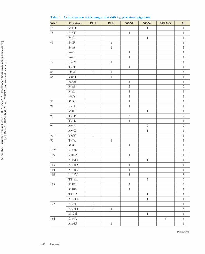
Table 1 Critical amino acid changes that shift λmaxs of visual pigments
Site1 Mutation RH1 RH2 SWS1 SWS2 M/LWS All44 M44T 1 1
46 F46T 1 1
F46L 1 1
49 S49F 1 1
S49A 1 1
F49V 1 1
F49L 1 1
52 L52M 1 1
T52F 1 1
83 D83N 7 1 8
86 M86T 1 1
F86M 1 1
F86S 2 2
F86L 1 1
F86Y 1 1
90 S90C 1 1
91 V91I 1 1
S91P 1 1
93 T93P 2 2
T93L 1 1
94 A94S 2 2
A94C 1 1
962 Y96V 1 1
97 T97A 1 1
S97C 1 1
1022 Y102F 1 1
109 V109A 1 1
A109G 1 1
113 E113D 1 1
114 A114G 1 1
116 L116V 3 3
T116L 2 2
118 S118T 2 2
S118A 1 1
T118A 1 1
A118G 1 1
122 E122I 1 1
E122Q 2 4 6
M122I 1 1
164 S164A 6 6
A164S 1 1
(Continued )
266 Yokoyama
Ann
u. R
ev. G
enom
. Hum
an G
enet
. 200
8.9:
259-
282.
Dow
nloa
ded
from
ww
w.a
nnua
lrev
iew
s.or
gby
EM
OR
Y U
NIV
ER
SIT
Y o
n 02
/08/
12. F
or p
erso
nal u
se o
nly.

ANRV353-GG09-14 ARI 25 July 2008 17:11
Table 1 (Continued )
Site1 Mutation RH1 RH2 SWS1 SWS2 M/LWS All181 H181Y 1 1
1942 P194R 1 1
1952 N195A 1 1
207 M207L 1 1
261 F261Y 2 1 3
Y261F 6 6
265 W265Y 1 1
269 A269S 1 1
T269A 5 5
292 A292S 7 2 1 2 12
S292A 1 1
1Sites 164, 181, 261, 269, and 292 correspond to 180, 197, 277, 285, and 308 of middle and long wavelength–sensitivepigments (M/LWS) pigments, respectively. H211C and A295S in bovine 1 (P500) also cause λmax shifts (32, 39, 46).2S. Yokoyama, T. Tada, N. Takenaka, H. Zhang & L. Britt (unpublished data).
interpretation is also supported by mutagenesisresults showing that the zebra finch andbudgerigar UV pigments become violet-sensitive by the mutation C90S (73, 86) andthe chicken and pigeon violet pigments becomeUV-sensitive by S90C (86).
Despite extensive mutagenesis analyses (27,61, 62, 67), the molecular basis of spectral tun-ing in the SWS1 pigments is still poorly un-derstood. This is simply because, with the ex-ception of some amino acid changes at residues86 and 90, extremely strong interactions ex-ist among different amino acid residues. Thefunctional differentiations of various violet pig-ments in some species from the ancestral UVpigment were caused by different sets of aminoacid replacements. The λmaxs of human S1(P414) (61) and clawed frog S1 (P425) (67) eachcan be explained by seven amino acid replace-ments, in which T93P and S118T were shared(Figure 4c). Intriguingly, all these 12 aminoacid changes cause no λmax shift individuallyand the λmaxs of two pigments have been modi-fied only through synergistic interactions of theseven amino acid replacements in each lineage(61, 67, Yokoyama & N. Takenaka, unpublishedresult). Conversely, F86Y in bovine S1 (P438)(27; see also 11, 17) and F86S in the ances-tral avian pigment and in elephant S1 (P419)(61, 91) increased the λmax significantly indi-
vidually. In the SWS1 pigments, F86S, T93P,L113V, and S118T occurred more than once(Table 1).
For the SWS2 pigments, only the commonancestor of goldfish S2 (P441) and zebrafish S2(P416) has been engineered (16). Because thisancestral pigment had a λmax of 430 nm andbluefin killifish S2-B (P397) decreased its λmax
to 397 nm (Figure 4d ), the ancestral SWS2pigment probably had a λmax of 400–430 nm.This relatively low λmax seems to have beencaused by A292S that occurred in the ancestralSWS2 pigment (Figure 4d ). Mutagenesis anal-yses suggest that goldfish S2 (P441) (16), bluefinkillifish S2-A (P448) (92), and newt S2 (P474)(66) increased their λmaxs, whereas bluefin killi-fish S2-B (P397) decreased it (Figure 4d ). Themutagenesis results of goldfish S2 (P441) (87)also suggest that A269S increased the λmax ofthe common ancestor of pigeon S2 (P448) andzebra finch S2 (P440). In this group, A94S andT116L occurred twice (Table 1).
The engineered ancestral M/LWS pigmentsreveal that early vertebrate ancestors used LWSpigments with λmaxs of ∼560 nm (85). Awide range of species, including fish, amphib-ians, reptiles, birds, and mammals have keptLWS pigments, whereas others have switchedfrom the ancestral LWS pigments to MWSpigments (Figure 4e). The ancestral LWS
www.annualreviews.org • Dim-Light and Color Vision Pigments 267
Ann
u. R
ev. G
enom
. Hum
an G
enet
. 200
8.9:
259-
282.
Dow
nloa
ded
from
ww
w.a
nnua
lrev
iew
s.or
gby
EM
OR
Y U
NIV
ER
SIT
Y o
n 02
/08/
12. F
or p
erso
nal u
se o
nly.

ANRV353-GG09-14 ARI 25 July 2008 17:11
pigments had a specific amino acid compositionof S180, H197, Y277, T285, and A308, fromwhich variable λmaxs of contemporary M/LWSpigments have been created by certain com-binations of S180A, H197Y, Y277F, T285A,and A308S. The ancestral rodent pigment isunique and reduced its λmax to 536 nm byH197Y (63). One striking aspect of these aminoacid replacements is that the identical changesS180A/Y277F/T285A occurred independentlyin cavefish M (P530), human M (P530), squir-rel monkey M (P532), deer M (P531), and wal-laby M (P528). In addition, the gecko MWSpigment with a λmax of 527 nm also incor-porated S180A/Y277F/T285A (N. S. Blow &S. Yokoyama, unpublished data). The probabil-ity that these six sets of S180A/Y277F/T285Aoccurred by chance alone is on the order of10−28. In addition, H198Y occurred once andS180A/A308S three times during vertebrateevolution, and the chance of the parallel re-placements under neutral evolution is on theorder of 10−9. These rare parallel changesstrongly suggest that the switches from the an-cestral LWS pigment to the six MWS pigmentswere caused by adaptive evolution (90).
When the five groups of visual pigments areconsidered together, a certain pattern of aminoacid replacements emerge. That is, amino acidreplacements, such as D83N and A292S, oc-curred repeatedly in different pigment groups.In particular, A292S occurred on at least 12separate occasions (Table 1). A292S offers animportant lesson in understanding the molecu-lar basis of spectral tuning in various pigments.A292S often decreases the λmax of visual pig-ments by ∼10 nm (Figure 4a,b,e). However,A292S that occurred in the ancestral pigmentof conger 1A (P486) does not decrease theλmax; conversely, the reverse mutation, S292A,in conger 1A (P486) increases the λmax by 12 nm(S. Yokoyama, T. Tada, N. Takenaka, H. Zhang& L. Britt, unpublished data). Furthermore, thereverse mutation, S292A, in human S1 (P414)does not increase the λmax at all (26). These mu-tagenesis results suggest that synergistic inter-actions can occur among different amino acidresidues. Such synergistic effects of different
amino acids have significant implications in theanalyses of spectral tuning, but they have beenpaid little attention.
As discussed below, critical amino acidchanges that cause significant λmax shifts are lo-calized to a total of 30 residues, most of whichare located near the N terminus of the TMsegments. Because the chromophore is also lo-cated near the N-terminal tail (luminal face)(Figure 3), these amino acid changes are ex-pected to interact with the chromophore andmodify the λmax of various visual pigments.However, residues 102, 194, and 195 in RH1pigments and 197 in M/LWS pigments, whichis equivalent to site 181 in the bovine RH1 pig-ment, are located in the luminal face, which isoutside of TM segments. In particular, residues194 and 195 are ∼20 A away from the chro-mophore (S. Yokoyama, T. Tada, N. Takenaka,H. Zhang & L. Britt, unpublished data). Atpresent, the molecular structural bases of suchamino acid interactions at long distance are notknown.
EVOLUTION OF DIM-LIGHTAND COLOR VISION
Dim-Light Vision
One of the critical times for the survival of an-imals in shallow water and on land is at twi-light, during which the most abundant lightwavelengths are 400–500 nm (45). In this en-vironment, a majority of animals use RH1 pig-ments (referred to as surface rhodopsins) withλmaxs of 500–507 nm. In contrast, in deep wa-ter, the distribution of downwelling sunlight isnarrower at ∼480 nm (33), and many deep-seafish use RH1 pigments (deep-sea rhodopsins)with λmaxs of ∼480–485 nm. The other RH1pigments with λmaxs of ∼490–495 nm can beclassified as intermediate rhodopsins. On thebasis of considerations of their light environ-ments, lifestyles, and the types of RH1 pig-ments they use, vertebrate dim-light visionsare classified mainly as surface, intermediate,and deep-sea vision (S. Yokoyama, T. Tada,N. Takenaka, H. Zhang & L. Britt, unpublished
268 Yokoyama
Ann
u. R
ev. G
enom
. Hum
an G
enet
. 200
8.9:
259-
282.
Dow
nloa
ded
from
ww
w.a
nnua
lrev
iew
s.or
gby
EM
OR
Y U
NIV
ER
SIT
Y o
n 02
/08/
12. F
or p
erso
nal u
se o
nly.

ANRV353-GG09-14 ARI 25 July 2008 17:11
data). The transitions among the three typesof dim-light visions have occurred on 12 sep-arate occasions (Figure 4a), strongly suggest-ing that dim-light vision has undergone adap-tive evolution. The evolution of dim-light vi-sion reveals two characteristics. First, naturalselection can be subtle and selective force candifferentiate even 5 nm of λmax differences ofRH1 pigments. Second, many transitions showthe ancestral surface vision → intermediate vi-sion (represented by lampfish and viperfish) orsurface vision → intermediate vision → deep-sea vision [represented by scabbard 1B (P481)].However, the lineage of scabbard 1A (P507)shows the transition of surface vision → inter-mediate vision → surface vision. Similarly, cer-tain lineages of squirrelfish have switched backto their ancestral surface and intermediate vi-sion from intermediate and deep-sea vision, re-spectively (89). In addition, as described below,a certain lineage of birds has experienced a UVvision → violet vision → UV vision transition.These observations show that evolution of dim-light vision and color vision can be reversible.To detect such reverse changes correctly, wemust engineer ancestral pigments at differentstages of vertebrate evolution.
Red-Green Color Vision
Vertebrates achieve red-green color vision us-ing not only M/LWS pigments but also RH2pigments. Some fish and primates, includinghumans, use LWS and MWS pigments withtypical λmaxs of ∼560 and ∼530 nm, respec-tively, for their red-green color vision. Toachieve red-green color vision, other specieshave modified their visual pigments and pho-toreceptor cells. That is, many fish species,birds, and reptiles do not have typical MWSpigments, but they can still achieve red-greencolor vision using 11-cis-3,4-dehydroretinal orcolored oil droplets. For example, goldfish 2-A(P511) with 11-cis-retinal has a λmax of 511 nm;however, when its 11-cis-retinal is replacedby 11-cis-3,4-dehydroretinal, the pigmentachieves a λmax of 537 nm (55). Conversely, thechicken RH2 pigment with 11-cis-retinal has
a λmax of 508 nm (52), but because it has agreen oil-droplet in its photoreceptor cell, thechicken cones with the RH2 pigments actuallyhave λmaxs of 533 nm (9).
Having neither 11-cis-3,4-dehydroretinalnor colored oil droplets, the red-green colorvision of mammals is mediated solely by theirM/LWS pigments. In higher primates, red-green color vision evolved in two separate ways.Hominoids and Old World monkeys use LWSand MWS opsins, encoded by two duplicatedX-linked loci (51). Most New World mon-keys have one M/LWS locus with three alleles(8, 31); for example, the squirrel monkey hasthree M/LWS alleles with λmaxs of 532, 545,and 558 nm (81). In such species, all males arered-green color blind, whereas females are ei-ther color blind or have red-green color vision,depending on their genotypes.
Because the molecular basis of spectral tun-ing in RH2 pigments is not well understood,I consider the subgroup of red-green color vi-sion that is based only on M/LWS pigmentswith 11-cis-retinal. As noted earlier, cavefish,gecko, human, squirrel monkey, deer, and wal-laby switched their LWS pigment into an MWSpigment independently. Furthermore, becauseof the extremely low chance of the occurrenceof S180A/Y277F/T285A in the six lineages, theswitches from the ancestral LWS pigment toMWS pigments in these species seem to haveundergone adaptive evolution (90). This con-clusion comprises one surprise; that is, the pos-itively selected MWS pigments are found inanimals with red-green color vision (cavefish,human, and squirrel monkey) and also in red-green color blind animals (gecko, deer, andwallaby). This finding contradicts a widely ac-cepted notion that animals with red-green colorvision have a selective advantage over those withcolor blindness (64), but it is compatible withthe observation that the majority of mammalianspecies and many other species are red-greencolor blind (31, 71).
Evidence is rather scant and is sometimescontroversial, but at least two observations sug-gest that animals with red-green color blind-ness can have a selective advantage over those
www.annualreviews.org • Dim-Light and Color Vision Pigments 269
Ann
u. R
ev. G
enom
. Hum
an G
enet
. 200
8.9:
259-
282.
Dow
nloa
ded
from
ww
w.a
nnua
lrev
iew
s.or
gby
EM
OR
Y U
NIV
ER
SIT
Y o
n 02
/08/
12. F
or p
erso
nal u
se o
nly.

ANRV353-GG09-14 ARI 25 July 2008 17:11
with red-green color vision. First, colorblindpeople can detect color-camouflaged objectsmuch better than those with red-green colorvision (44, 57). Second, color-blind individu-als of capuchin monkey, crab-eating monkeys,and chimpanzees are capable of discriminatingcolor-camouflaged stimuli, but those with red-green color vision failed to do so (58). However,in another survey no advantage was detectedbetween female tamarins with red-green colorvision and males without red-green color vision(23). Clearly, more analyses are needed to deter-mine whether the ability of decoding color cam-ouflage gives a selective advantage to color blindindividuals over those with red-green color vi-sion. The decoding of color-camouflage maybe only one facet of selective advantage of red-green color blindness over red-green color vi-sion. In the future, the other causes for the se-lective advantage of red-green color blindnessover red-green color vision may be discovered.
UV and Violet Vision
UV and violet (or blue) vision are mediatedby SWS1 and SWS2 pigments, which haveλmaxs of 360–440 and 400–450 nm, respectively.Hence, with the exception of UV pigments inthe SWS1 group, the λmaxs of the two groupsof visual pigments are indistinguishable, but themolecular mechanisms of functional differenti-ation of the two groups of pigments are verydifferent (Figure 4c,d ; Table 1). At present,the molecular basis of spectral tuning in theSWS1 pigments is better understood than thatin the SWS2 pigments. Therefore, I considerthe subgroup of UV-violet vision that is basedon SWS1 pigments.
The engineered ancestral pigments showthat early vertebrate ancestors had UV vision(Figure 4c). Because UV vision works underUV light, organisms are expected to switchfrom UV vision to violet vision or simply shutoff the function of the SWS1 gene in the ab-sence of UV light. However, given abundantUV light in their environments, many organ-isms also switched from UV vision to violet vi-sion. Two major causes for these changes can
be considered (30). First, UV light can dam-age retinal tissues, and the yellow pigments inthe lenses or corneas in many species, includinghumans, are devised to obviate most UV lightfrom reaching the retina. In such cases, UV pig-ments are of no use. Second, by achieving violetvision, organisms can improve visual resolutionand subtle contrast detection.
In the avian lineage, the ancestor lost UVvision, but some of its descendants restoredit (Figure 4c). The reinvention of UV visionseems to have been related to avian migration.For migratory birds, the pineal gland senseschanges in day length and releases hormonesthat initiate migration (1). UV vision is also es-sential in orientation based on the sun (7). Sur-prisingly, the mouse, a nocturnal animal, alsouses UV vision (Figure 4c). Voles mark theirrunway with urine and feces, which reflect UVlight and are used as a method of communi-cation (69). Furthermore, UV pigments are themajor visual pigments expressed in the third eye(or parietal eye) of chameleon (35). Clearly, UVdetection through this organ is important in ad-dition to UV vision.
Thus, the use of UV pigments and UV vi-sion by organisms is strongly associated withtheir light environments and behaviors. Com-pared with organisms with violet vision, thosewith UV vision have an advantage of recogniz-ing certain UV-reflecting objects much morequickly, but they lack precision in viewing theirsurroundings and are subjected to a higherchance of developing retinal damage caused byUV light. Whether or not organisms use UV vi-sion or violet vision must depend on the relativeimportance of these and other conflicting char-acteristics associated with UV vision to them(61). To appreciate the evolution of UV-violetvision in nature, we must study the roles of UVand violet pigments of many species in variouslight environments.
SPECTRAL TUNING
The Problem
Certain amino acid changes at a total of 26residues were known to have generated variable
270 Yokoyama
Ann
u. R
ev. G
enom
. Hum
an G
enet
. 200
8.9:
259-
282.
Dow
nloa
ded
from
ww
w.a
nnua
lrev
iew
s.or
gby
EM
OR
Y U
NIV
ER
SIT
Y o
n 02
/08/
12. F
or p
erso
nal u
se o
nly.

ANRV353-GG09-14 ARI 25 July 2008 17:11
Table 2 Forward and reverse mutations that shift λmaxs of visual pigments
Site RH1 RH2 SWS1 SWS2 M/LWS83 D83N (−6)a – – – –
N83D (2)b – – – –
86 – – F86Y (66)c – –
– – Y86F (−75)d – –
– – F86S (17)e – –
– – S86F (−52)b – –
90 G90S (−13)f – S90G (−7)g – –
S90C (−7)c
C90S (38)h
93 – – T93I (0)c – –
– – I93T (−6)b – –
113 E113D (7)i – E113D (−4)j – –
– – D113E (−12)k – –
116 – – L116V (0)j – –
– – V116L(−3)l – –
118 T118A (−16)f – A118T (3)m – –
122 E122Q (−20)i Q122E (10)n – – –
Q122E (10)n – – – –
164 A164S (2)o – – – S164A (−7)p
– – – – A164S (6)b
261 F261Y (10)o – – Y261F (−5)q Y261F (−10)p
Y261F (−8)r – – – F261Y (6)p
265 W265Y (−15)s – Y265W (10)g – –
269 A269T (14)o – – A269T (6)t A269T (10)p
T269A (−16)p
292 A292S (−10)f – S292A (0)g A292S (−8)q S292A (28)u
S292A (8)n
a46; b91; c28; d17; e62; f32; g26; h86; i96; j67; k5; l90; m73; n94; o12; p4; q66; r76; s39; t87; u27.
λmaxs of visual pigments in vertebrates (92).Amino acid replacements in Figure 4a–e coverchanges at 24 residues. Table 1 also lists aminoacid changes at four additional residues that areinvolved in the spectral tuning of RH1 pig-ments. Therefore, amino acid changes at a totalof 30 residues are now known to cause signifi-cant λmax shifts individually and synergistically.
Mutagenesis results reveal three character-istics of spectral tuning of visual pigments(Table 2). First, mutations in opposite direc-tions do not necessarily shift the λmax to oppo-site directions. For example, G90S in a RH1pigment decreases the λmax by 13 nm, but the
reverse change, S90G, in a SWS1 pigmentalso decreases the λmax by 7 nm. Similarly,E113D and D113E in two different SWS1 pig-ments both decrease the λmaxs. Second, iden-tical amino acid changes may cause differentmagnitudes of λmax shifts. For example, S292Ain a SWS1 pigment does not shift the λmax, butthe same mutation in a MWS pigment increasesthe λmax by 28 nm. Although it is not clear fromTable 2, the λmax shifts caused by S90C in dif-ferent SWS1 pigments range between −46 and0 nm (24, 27, 61, 62, 86). Third, even whenthe forward and reverse mutations shift the λmax
to opposite directions, the magnitudes of λmax
www.annualreviews.org • Dim-Light and Color Vision Pigments 271
Ann
u. R
ev. G
enom
. Hum
an G
enet
. 200
8.9:
259-
282.
Dow
nloa
ded
from
ww
w.a
nnua
lrev
iew
s.or
gby
EM
OR
Y U
NIV
ER
SIT
Y o
n 02
/08/
12. F
or p
erso
nal u
se o
nly.

ANRV353-GG09-14 ARI 25 July 2008 17:11
shifts can differ significantly. For example, pairsof F86Y and Y86F, F86S and S86F, S90C andC90S, T118A and A118T, E122Q and Q122E,A269T and T269A, and A292S and S292A shiftλmaxs to opposite directions, but the differencein the magnitudes of λmax shifts for each pairis more than 10 nm. As more mutagenesis re-sults accumulate, the list of these examples isexpected to grow.
Hence, λmax shifts caused by forward muta-tions that actually occurred in nature should notbe inferred from those of the identical aminoacid changes or corresponding reverse muta-tions in contemporary pigments. As the nexttwo examples illustrate, even if we are interestedin understanding the molecular basis of spec-tral tuning only, the actual evolutionary processcannot be ignored.
The Human M/LWS Pigments
An extensive mutagenesis analysis has beenconducted using human L (P560) and hu-man M (P530), whose difference spectraare given by 563 nm and 531 nm, respec-tively (4). S180A/Y277F/T285A in human L(P560) decrease the λmax by 33 nm and ex-plain fully the λmax difference between thetwo pigments. However, the reverse changesA180S/F277Y/A285T in human M (P530) in-crease the λmax only by 23 nm and do notexplain the λmax of human L (P560). In thiscase, not only A180S/F277Y/A285T but alsoY116S/T230I/S233A/F309Y are needed to ex-plain the λmax difference between the two pig-ments (4). Therefore, depending on which pig-ment we choose to mutate, we end up with twodifferent molecular mechanisms of spectral tun-ing! If we are not satisfied with two differentanswers, then how can we resolve the problem?One natural way is to try to understand themolecular mechanism of spectral tuning thatactually occurred in the past (78).
We have seen that the engineered ances-tral pigment of human L (P560) and humanM (P530) had a λmax of ∼560 nm (Figure 4e).The ancestral LWS pigment had the amino acidcomposition of S180/Y277/T285, and S180A,
Y277F, and T285A occurred in the past. Witha possible exception of S233A, it is highly un-likely that any of Y116S, A180S, T230I, F277Y,A285T, and F309Y occurred in the ancestralpigment (85). S233A decreases the λmax of hu-man L (P560) by 3 nm (4), but its actual ef-fect on the λmax shift in the ancestral pigmentis unknown. In fact, when 180A/Y277F/T285Awere introduced into the ancestral mammalianLWS pigment that was engineered previously(pigment d in 83), the mutant pigment hada λmax of 532 nm (S. Yokoyama & H. Yang,unpublished data). Hence, the three forwardmutations explain fully the spectral tuning inthe human M (P530) and the effect of S233Aon the λmax shift is negligible. Therefore, theevolutionary interpretation of the mutagene-sis results is simple: The λmax of human M(P530) was achieved by S180A/Y277F/T285A,whereas human L (P560) inherited its λmax di-rectly from the ancestral pigment without anycritical amino acid changes. Hence, the sevenreverse amino acid changes in human M (P530)describe a mostly hypothetical situation and areunrealistic.
The Clawed Frog SWS1 Pigmentand Its Ancestor
Two sets of chimeras of different SWS1 pig-ments (27, 62) suggested that the λmax dif-ferences between pairs of UV and violetpigments were generated by amino acid differ-ences at residues in TM I–III. Consequently,the search for amino acids that caused variableλmaxs among SWS1 pigments has been focusedin that region. To date, a total of 13 amino acidresidues in that region have been shown to beinvolved in the λmax shift of SWS1 pigments(Table 1). However, considering the chimericpigments between clawed frog S1 (P425) [orsimply, frog S1 (P425)] and its ancestral am-phibian pigment with a λmax of 359 nm [pig-ment (P359)], an entirely different picture ofthe molecular basis of spectral tuning of SWS1pigments has emerged (67).
The regions of interest in the two pig-ments were distinguished into four segments:
272 Yokoyama
Ann
u. R
ev. G
enom
. Hum
an G
enet
. 200
8.9:
259-
282.
Dow
nloa
ded
from
ww
w.a
nnua
lrev
iew
s.or
gby
EM
OR
Y U
NIV
ER
SIT
Y o
n 02
/08/
12. F
or p
erso
nal u
se o
nly.

ANRV353-GG09-14 ARI 25 July 2008 17:11
Table 3 The effects of transmembrane domain(TM) exchanges on the λmax shift in frog S1(P423) and its ancestor, pigment (P359)a
TM Forward ReverseI 0 −5II 24 −19III 51 −15IV–VII 1 −1I × II 6 5I × III 7 1I × IV–VII 1 4II × III −13 −28II × IV–VII 20 14III × IV–VII −7 −18I × II × III −12 −2I × II × IV–VII −3 −11I × III × IV–VII −8 −6II × III × IV–VII −17 3I × II × III × IV–VII 14 14Total 64 −64
aData from 65.
TM I (residues 31–66), TM II (residues 67–98), TM III (residues 99–151), and TM IV–VII(residues 152–311). The amino acids at the Nand C termini of the two pigments were re-placed by those of chameleon S1 (P359). Then,all single and multiple combinations of thesefour segments were constructed (67). Consid-ering the evolution of frog S1 (P425) from pig-ment (P359), the magnitudes of the λmax shiftcaused by replacing the TM I (θI), TM II (θII),TM III (θIII), and TM IV–VII (θIV−VII) of pig-ment (P359) by the corresponding segmentsof frog S1 (P425) and those of their syner-gistic effects θI × II, θI × III, θI × IV−VII, . . . , andθI × II × III × IV−VII on the λmax shift were evalu-ated (Table 3). The results show that TM IIand TM III have significant individual effects inthe spectral tuning of frog S1 (P425) and, at thesame time, TM IV–VII reveal significant inter-actions with the other TM segments. However,the overall effect of TM IV–VII on the λmax shift(θIV−VII +θI × IV−VII +θII × IV−VII +θIII × IV−VII +θI × II × IV−VII + θI × III × IV−VII + θII × III × IV−VII +θI × II × III × IV−VII) is only 1 nm. This negligi-
ble overall effect and negligible θIV−VII give animpression that the spectral tuning in frog S1(P425) is determined exclusively by amino acidchanges in TM I–III.
By considering the change from frog S1(P425) to pigment (P359), we can also evalu-ate the effects of amino acid changes in the op-posite direction (Table 3). In this case, TM IIand TM III also cause significant λmax shifts,but their impacts are much smaller than thoseof the forward changes; in particular, the de-crease in the λmax caused by TM III of pigment(P359) is 36 nm smaller than the expected valuefrom the λmax shift caused by that of frog S1(P425). In fact, the absolute values of the corre-sponding θIII, θII × III, θIII × IV−VII, θI × II × III, andθII × III × IV−VII values between the forward andreverse TM exchanges differ by 10 nm or more.For the reverse changes, the overall effect ofTM IV–VII on the λmax shift is −1 nm and isagain negligible.
The analyses of the chimeric pigments re-veal three main features of spectral tuning ofSWS1 pigments. First, amino acid changes notonly in TM I–III but also in TM IV–VII are in-volved in the spectral tuning in clawed frog S1(P425), where the critical amino acids in TMIV–VII remain to be discovered. Second, theeffects of forward and reverse TM changes andamino acid changes on the λmax shift can be verydifferent. Third, despite a significant amount ofinteraction between TM I–III and TM IV–VII,the overall effect of amino acid changes in TMIV–VII on the λmax shift is negligible. The causeand implications of the last observation are notimmediately clear.
A Solution
For RH1, RH2, SWS1, and SWS2 pigmentgroups, we do not have sufficient informationon the effects of forward amino acid changesand their interactions. However, we have a sig-nificant amount of data to study the molec-ular basis of spectral tuning in the M/LWSpigments. In 2001, applying multiple regres-sion analysis to all M/LWS pigments that wereknown at that time, various combinations of
www.annualreviews.org • Dim-Light and Color Vision Pigments 273
Ann
u. R
ev. G
enom
. Hum
an G
enet
. 200
8.9:
259-
282.
Dow
nloa
ded
from
ww
w.a
nnua
lrev
iew
s.or
gby
EM
OR
Y U
NIV
ER
SIT
Y o
n 02
/08/
12. F
or p
erso
nal u
se o
nly.

ANRV353-GG09-14 ARI 25 July 2008 17:11
0–2 2 4–4 6
5
10
15
D (47)
D-L (34)
–60
Δλmax (nm)
No.
of p
igm
ents
Figure 5The distribution of the differences (�λmax) between the expected λmax based onthe five-sites rule and actual λmax of middle and long wavelength–sensitivepigments (M/LWS) pigments; dark (D) (47 pigments) and difference (D-L)(34 pigments) spectra were evaluated separately.
λmax shifts caused by S180A (−7 nm), H197Y(−28 nm), Y277F (−8 nm), T285A (−15 nm),A308S (−27 nm), and S180A/H197Y (11 nm)were suggested to have generated the variableλmaxs of M/LWS pigments (85). In the analyses,the effects of forward amino acid replacementsS180A, H197Y, Y277F, and T285A on the λmax
shift were evaluated experimentally, but those ofA308S and S180A/H197Y were not. Recently,A308S, S180A/A308S, and H197Y/A308S wereintroduced into the ancestral mammalian LWSpigment. The results show that the respectivemutant pigments have λmaxs of 527, 525, and516 nm (S. Yokoyama & H. Yang, unpublisheddata).
At present, the dark spectra of a total of 9ancestral and 38 contemporary M/LWS pig-ments and the difference spectra of 9 ancestraland 25 contemporary pigments are available(4, 42, 43, 64, 83–85) (Figure 5). Applyingmultiple regression analysis to the λmaxs and theamino acid compositions at residues 180, 197,277, 285, and 308 of these pigments, the effectsof the individual and synergistic effects of thefive amino acid replacements on the λmax shiftwere evaluated. The results show that the λmaxsof M/LWS pigments are determined mainly byλmax shifts caused by S180A (−6 nm), H197Y
(−26 nm), Y277F (−10 nm), T285A (−16 nm),A308S (−33 nm), H197Y × A308S (15 nm),and S180A × H197Y × A308S (−8 nm). How-ever, the effects of S180A × H197Y (2 nm),S180A × Y277F (2 nm), S180A × T285A(1 nm), S180A × A308S (3 nm), H197Y ×T285A (−2 nm), Y277F × T285A (−1 nm),and S180A × Y277F × T285A (0 nm) on theλmax shift are much smaller and are negligible.Hence, the effect of S180A × H197Y is nownegligible; instead, the effects of interactionsH197Y × S308A and S180A × H197Y ×S308A become important. As suspected, theresults depend strongly on the data set used.
Only the rodent and dolphin pigments haveincorporated H197Y and A308S (Figure 4e).If we exclude them, the absorption spectra ofM/LWS pigments in a wide range of vertebratespecies are explained mostly by the additiveeffects of S180A, Y277F, and T285A, and aso-called three-sites rule holds (77, 80). If weexclude only the rodent pigments from con-sideration, then the λmaxs of M/LWS pigmentsare modulated mostly by the additive effects ofS180A, Y277F, T285A, and A308S.
The ancestral pigments with S180, H197,Y277, T285, and A308 have dark and differencespectra of 560 ± 2 and 561 ± 2 nm, respec-tively (S. Yokoyama & H. Yang, unpublisheddata). Theoretically, the λmaxs of all visual pig-ments can be evaluated by the λmaxs of the ances-tral pigment and θs. Hence, the expected λmaxsbased on the new five-sites rule can be com-pared with the corresponding observed valuesof M/LWS pigments. The differences betweenthe expected and observed λmaxs of M/LWS pig-ments were evaluated for the dark and differ-ence spectra separately (Figure 5). For the darkspectra, the λmax differences are within 4 nm.Because the standard deviation of λmaxs of an-cestral pigments is 2 nm, these λmax differencesare within the margin of experimental error.The majority of λmax differences for the dif-ference spectra are also within 4 nm, but thedifferences of two pairs of pigments are largerthan 4 nm. Because even when they are not re-liable, dark spectra are used in evaluating dif-ference spectra, the deviations might have been
274 Yokoyama
Ann
u. R
ev. G
enom
. Hum
an G
enet
. 200
8.9:
259-
282.
Dow
nloa
ded
from
ww
w.a
nnua
lrev
iew
s.or
gby
EM
OR
Y U
NIV
ER
SIT
Y o
n 02
/08/
12. F
or p
erso
nal u
se o
nly.

ANRV353-GG09-14 ARI 25 July 2008 17:11
caused by inaccurate estimates of the differencespectra. Overall, therefore, the variable λmaxsof the currently known M/LWS pigments canbe explained reasonably well by the new five-sites rule (S. Yokoyama & H. Yang, unpublishedresult).
ADAPTIVE EVOLUTION
By studying functional differentiations of an-cestral RH1 and SWS1 pigments and relatingthem to the associated environmental changesof organisms’ habitats and to new lifestyles,we have established that dim-light and UV-violet vision have undergone adaptive evolu-tion. For the adaptive evolution of red-greencolor vision, a more probabilistic argument ofparallel amino acid replacements in M/LWSpigments was used. Thus, surveying the aminoacid sequences and λmaxs of various visualpigments, followed by mutagenesis analyses,amino acid replacements that generated a widerange of λmaxs in nature have been uncovered(81).
Without an available functional assay,molecular adaptations have often been inferredby identifying amino acid changes using statisti-cal methods (65, 74, 95). The mutagenesis anal-yses of visual pigments establish five fundamen-tal features of molecular evolution that cannotbe learned from the standard statistical analysesof protein sequence data. First of all, mutage-nesis experiments can offer critical and deci-sive tests of whether or not amino acid changesthat are inferred as adaptive actually cause anyfunctional changes (19). Second, as exempli-fied by several sets of mutations (Table 2),the same amino acid replacements do not al-ways produce the same functional change butinstead the change can be affected by the back-ground amino acids of the opsin. Therefore, theprobability of parallel amino acid replacements,which may or may not result in any functionalchange, can overestimate the actual chance thatfunctional adaptive events occur. Third, simi-lar functional changes can be achieved by dif-ferent amino acid replacements. For exam-ple, D83N/A292S, P194R/N195A/A292S, and
E122Q all decrease the λmax by 14–20 nm(Figure 4a). Thus, by simply looking for par-allel replacements of specific amino acids, onecan miss other amino acid changes that gen-erate the same functional change, thereby un-derestimating the chance of finding functionaladaptations.
Fourth, as stressed already, not only canthe identical mutations in different pigmentscause different magnitudes of λmax shift, butalso the effects of forward and reverse aminoacid changes on the λmax shift can differ signifi-cantly. Hence, if we are interested in elucidatingthe evolutionary mechanisms of functional andphenotypic changes, we must study the effectsof forward mutations, not reverse mutations. Asnoted earlier, this evolutionary approach alsosimplifies our understanding of the molecularbasis of spectral tuning.
Fifth, even when the phylogenetic positionof a molecule is uncertain, its functional as-say can clarify the molecular evolution of func-tional adaptation. For example, the phyloge-netic position of lampfish 1 (P492) is uncertain(Figure 4a). However, because the E122Q mu-tation that generated its λmax is different fromthe other critical amino acid replacements inthe closely related thornyhead 1 (P483), scab-bard 1B (P481), and viperfish 1 (P489) proteins(Figure 4a), we can easily establish an indepen-dent origin of the functional change in lampfish1 (P492). Therefore, to explore the adaptiveevolution of certain traits, both functional andmolecular analyses of such traits are valuable(19). Analyses of functional adaption of visualpigments also demonstrate the importance ofrelating the functional changes to the environ-mental or behavioral changes that presumablycaused the functional and phenotypic changesin the first place.
To fully appreciate how adaptive evolutionof dim-light and color vision occurred, we muststudy the effects of critical forward amino acidreplacements on the λmax shift at the chemi-cal level as well. For example, quantum chem-ical analyses of the effects of forward aminoacid changes on the λmax shift will improve sig-nificantly our understanding of the molecular
www.annualreviews.org • Dim-Light and Color Vision Pigments 275
Ann
u. R
ev. G
enom
. Hum
an G
enet
. 200
8.9:
259-
282.
Dow
nloa
ded
from
ww
w.a
nnua
lrev
iew
s.or
gby
EM
OR
Y U
NIV
ER
SIT
Y o
n 02
/08/
12. F
or p
erso
nal u
se o
nly.

ANRV353-GG09-14 ARI 25 July 2008 17:11
basis of spectral tuning in visual pigments (2,3). The same analyses will, in turn, improvesignificantly our understanding of the chemicalbasis of the functional adaptations of dim-lightvision and color vision.
CONCLUSIONS
Studies on the structure and function of bovineRH1 pigment by Doi and coworkers (22) and aseries of subsequent papers by H. G. Khoranaand his colleagues as well as other vision re-searchers (46, 48, 59, 72, 96) have improved dra-matically our understanding of how key aminoacids in visual pigments work. Unfortunately,most mutations considered in these biochem-ical studies are not found in nature, so theirroles in the actual spectral tuning in various vi-sual pigments are not immediately clear (79,81). If we want to elucidate the mechanismsof spectral tuning that generated the λmaxs ofcontemporary visual pigments, then we mustconsider amino acid replacements that actuallyoccurred in nature. Such changes can be in-ferred only by comparing the amino acid se-quences of contemporary visual pigments, andthe actual functional changes caused by the pre-dicted amino acid changes can be evaluated us-ing in vitro assays. To date, using this approach,certain amino acid replacements at a total of30 residues have been shown to be involved inthe spectral tuning of different visual pigments(Table 1).
Phylogenetic analyses of contemporary vi-sual pigments show that early vertebrate an-cestors already had RH1, RH2, SWS1, SWS2,and M/LWS pigments (25, 81, 93). Many con-temporary species still use all five sets of visualpigments, and more recent gene duplications insome species generated additional variations inthe λmaxs of visual pigments, whereas RH2 andSWS2 pigments have become nonfunctional insome lineages, including placental mammals,and their color vision has become more special-ized (Figure 4a–e). The engineered visual pig-ments show that the RH1, SWS1, and M/LWSin early ancestors had λmaxs of ∼500, ∼360, and∼560 nm, respectively.
Depending on the organisms’ light environ-ments, lifestyles, and the λmaxs of their RH1pigments, dim-light vision of organisms can bedistinguished into deep-sea, intermediate, andsurface vision. The RH1 pigments of the re-spective groups have λmaxs of 480–485, 490–495, and 500–510 nm. Some species inher-ited the ancestral surface vision directly fromthe vertebrate ancestor, whereas others haveswitched to different types of dim-light vision.During vertebrate evolution, such transitionsoccurred on 12 separate occasions. As the λmaxsof the three types of dim-light vision indicate,natural selection can be subtle and selectiveforce may differentiate even 5 nm of λmax differ-ence. These adaptive events were accomplishedmostly by amino acid changes at nine residues,where D83N/A292S occurred seven timesindependently.
Many contemporary LWS pigments havemaintained the ancestral λmax of ∼560 nm,whereas others have decreased their λmaxsby using various combinations of S180A,H197Y, Y277F, T285A, and A308S. In par-ticular, identical amino acid replacements(S180A/Y277F/T285A) occurred on six sepa-rate occasions and shifted the λmaxs of M/LWSpigments in an additive fashion. In the lineageof rodent M/LWS pigments, H197Y occurredin their ancestral pigment, followed by A308Sin some MWS pigments, and they decreasedλmaxs individually and synergistically. Similarly,many contemporary SWS1 pigments inheritedtheir UV sensitivities from the common an-cestor, and others developed violet sensitivi-ties using different sets of amino acid replace-ments, many of which remain to be discovered.Most of the currently known critical amino acidreplacements modify the λmax mainly throughtheir synergistic effects, but some amino acidreplacements at residues 86 and 90, includingF86Y, F86S, and S90C, can cause significantλmax shifts individually as well as synergistically(91).
Despite these advances, our understandingof the molecular bases of adaptive evolution andspectral tuning of visual pigments is still frag-mental. This is because we still don’t have much
276 Yokoyama
Ann
u. R
ev. G
enom
. Hum
an G
enet
. 200
8.9:
259-
282.
Dow
nloa
ded
from
ww
w.a
nnua
lrev
iew
s.or
gby
EM
OR
Y U
NIV
ER
SIT
Y o
n 02
/08/
12. F
or p
erso
nal u
se o
nly.

ANRV353-GG09-14 ARI 25 July 2008 17:11
information on how the chromophore and dif-ferent amino acids interact with each other.In particular, amino acid changes in oppositedirections do not shift the λmax in the oppositedirection by the same magnitudes. Or, whenintroduced into different pigments, even theidentical amino acid replacements can cause dif-ferent magnitudes of λmax shifts. The most rea-sonable approach in resolving these seeminglycontradictory observations is to consider aminoacid changes that actually generated the variableλmaxs of contemporary visual pigments. Then,the functional adaptation and spectral tuning ofvisual pigments can be understood together bystudying the mechanisms of adaptive evolution
of visual pigments at the molecular and pheno-typic levels.
To solve the problem, we must engineer an-cestral pigments for the five groups of visualpigments at various stages of vertebrate evolu-tion and introduce mutations into them. Suchancestral pigments at various stages of verte-brate evolution have been engineered for RH1,SWS1, and M/LWS pigments, but those forRH2 and SWS2 pigments remain to be engi-neered. By dissecting these and contemporaryvisual pigments at the molecular level and relat-ing their λmaxs to organisms’ light environmentsand lifestyles, we can start to learn why and howorganisms adapted to their light environments.
SUMMARY POINTS
1. Visual pigments in vertebrates are classified into rhodopsins (RH1), RH1-like (RH2),short wavelength-sensitive type 1 (SWS1), SWS type 2 (SWS2), and middle and longwavelength–sensitive (M/LWS) groups with λmaxs of 480–510, 450–530, 360–440, 400–450, and 510–560 nm, respectively.
2. Dim-light vision is mediated by RH1 pigments and can be classified into three differenttypes; the evolutionary switches among them occurred on 12 separate occasions.
3. Red-green color vision and color blindness mediated by M/LWS pigments were gener-ated by certain combinations of amino acid changes (S180A, H197Y, Y277F, T285A, andA308S); S180A/Y277F/T285 occurred on six separate occasions.
4. The parallel replacements of S180A/Y277F/T285A in various vertebrate species suggestthat both red-green color vision and color blindness have undergone adaptive evolution,but the selective advantage of color blindness over red-green color vision is still not wellunderstood.
5. Many fish, reptile, and mammalian species inherited their UV vision from the vertebrateancestor, but the bird ancestor achieved violet vision by F4V/F86S/L116V/S118A, andsome of its descendants reinvented UV vision by S90C.
6. With the exception of some amino acid changes at residues 86 and 90, the molecularbasis of spectral tuning in SWS1 pigments is characterized by strong interactions amongamino acid residues.
7. Mutagenesis data show that mutations in opposite directions do not necessarily cause λmax
shifts to the opposite directions by the same magnitudes, implying that the molecularbasis of spectral tuning in visual pigments should be understood by considering forwardamino acid changes that actually generated the variable λmax shifts of contemporarypigments.
www.annualreviews.org • Dim-Light and Color Vision Pigments 277
Ann
u. R
ev. G
enom
. Hum
an G
enet
. 200
8.9:
259-
282.
Dow
nloa
ded
from
ww
w.a
nnua
lrev
iew
s.or
gby
EM
OR
Y U
NIV
ER
SIT
Y o
n 02
/08/
12. F
or p
erso
nal u
se o
nly.

ANRV353-GG09-14 ARI 25 July 2008 17:11
8. Among the five pigment groups, the molecular basis of spectral tuning is best understoodfor the M/LWS group and the most recent data show that the ancestral M/LWS pigmenthad a λmax of 560 nm. Significant λmax shifts have been caused mostly by S180A (−6 nm),H197Y (−26 nm), Y277F (−10 nm), T285A (−16 nm), A308S (−33 nm), H197Y/A308S(15 nm), and S180A/H197Y/A308S (−8 nm).
FUTURE ISSUES
1. All amino acid replacements that generated the variable λmaxs of the five groups of con-temporary visual pigments need to be identified.
2. Individual and synergistic effects of these forward amino acid changes on the λmax shiftsneed to be evaluated.
3. The molecular bases of spectral tuning in various visual pigments need to be understoodin terms of the individual and synergistic effects of the forward amino acid changes onthe λmax shifts.
4. The spectral tuning in visual pigments need to be understood at the chemical structurallevel, where quantum chemical computations of visual pigments at various stages ofvertebrate evolution should be performed.
5. The molecular bases of functional adaptation of visual pigments need to be understoodnot only by studying the molecular basis of differentiation of visual pigments, but alsoby relating them to organisms’ move to new photic environments or to new lifestyles.
DISCLOSURE STATEMENT
The author is not aware of any biases that might be perceived as affecting the objectivity of thisreview.
ACKNOWLEDGMENTS
The author’s research was supported by a grant from the National Institutes of Health.
LITERATURE CITED
1. Alcock J. 1997. Animal Behavior. Sunderland, MA: Sinauer2. Altun A, Yokoyama S, Morokuma K. 2008. Spectral tuning in visual pigments: An ONIOM (QM:MM)
study on bovine rhodopsin and its mutants. J. Phys. Chem. B 112:6814–273. Altun A, Yokoyama S, Morokuma K. 2008. Quantum mechanical/molecular mechanical studies on spectral
tuning mechanisms of visual pigments and other photoactive proteins. Photochem. Photobiol. In press4. Asenjo AB, Rim J, Oprian DD. 1994. Molecular determination of human red/green color discrimination.
Neuron 12:1131–385. Babu KR, Dukkipati A, Birge RR, Knox BE. 2001. Regulation of phototransduction in short-wavelength
cone visual pigments via the retinylidene Schiff base counterion. Biochemistry 40:13760–666. Baylor D. 1996. How photons start vision. Proc. Natl. Acad. Sci. USA 93:560–657. Bennett ATD, Cuthill IC. 1994. Ultraviolet vision in birds: What is its function? Vis. Res. 34:1471–78
278 Yokoyama
Ann
u. R
ev. G
enom
. Hum
an G
enet
. 200
8.9:
259-
282.
Dow
nloa
ded
from
ww
w.a
nnua
lrev
iew
s.or
gby
EM
OR
Y U
NIV
ER
SIT
Y o
n 02
/08/
12. F
or p
erso
nal u
se o
nly.

ANRV353-GG09-14 ARI 25 July 2008 17:11
8. Bowmaker JK. 1991. Evolution of photoreceptors and visual pigments. In Evolution of the Eye and VisualPigments, ed. JR Cronly-Dillon, RJ Gregory, pp. 63–81. Boca Raton, FL: CRC Press
9. Bowmaker JK, Knowles A. 1977. The visual pigments and oil droplets of the chicken retina. Vis. Res.17:755–64
10. Carroll SB. 2006. The Making of The Fittest. New York: Norton11. Carvalho LD, Cowing JA, Wilkie SE, Bowmaker JK, Hunt DM. 2006. Shortwave visual sensitivity in tree
and flying squirrels reflects changes in lifestyle. Curr. Biol. 16:R81–8312. Chan T, Lee M, Sakmar TP. 1992. Introduction of hydroxyl-bearing amino acids causes bathochromic
spectral shifts in rhodopsin: amino acid substitutions responsible for red-green color pigment spectraltuning. J. Biol. Chem. 267:9478–80
13. Chang BSW, Jonsson K, Kazmi MA, Donoghue MJ, Sakmar TP. 2002. Recreating a functional ancestralarchosaur visual pigment. Mol. Biol. Evol. 19:1483–89
14. Chinen A, Hamaoka T, Yamada Y, Kawamura S. 2003. Gene duplication and spectral diversification ofcone visual pigments of zebrafish. Genetics 163:663–75
15. Chinen A, Matsumoto Y, Kawamura S. 2005. Reconstitution of ancestral green visual pigments of zebrafishand molecular mechanism of their spectral differentiation. Mol. Biol. Evol. 22:1001–10
16. Chinen A, Matsumoto Y, Kawamura S. 2005. Spectral differentiation of blue opsins between phylogenet-ically close but ecologically distant goldfish and zebrafish. J. Biol. Chem. 280:9460–66
17. Cowing JA, Poopalasundaram S, Wilkie SE, Robinson PR, Bowmaker JK, et al. 2002. The molecularmechanism for the spectral shifts between vertebrate UV- and violet-sensitive cone visual pigments.Biochem. J. 367:129–35
18. Davies WL, Carvalho LS, Cowing JA, Beazley LD, Hunt DM, et al. 2007. Visual pigments of the platypus:A novel route to mammalian colour vision. Curr. Biol. 17:R161–63
19. Dean AM, Thornton JW. 2007. Mechanistic approaches to the study of evolution: the functional synthesis.Nat. Rev. Genet. 8:675–88
20. Deeb SS, Wakefield MJ, Tada T, Marotte L, Yokoyama S, Graves JAM. 2003. The cone visual pigmentsof an Australian marsupial, the tammar wallaby (Macropus eugenii): sequence, spectral tuning and evolution.Mol. Biol. Evol. 20:1642–49
21. Dobzhansky T. 1973. Nothing in biology makes sense except in the light of evolution. Am. Biol. Teach.35:125–29
22. Doi T, Molday RS, Khorana HG. 1990. Role of the intradiscal domain in rhodopsin assembly and function.Proc. Natl. Acad. Sci. USA 87:4991–95
23. Dominy NJ, Garber PA, Bicca-Marques JC, Azevedo-Lopes MADO. 2003. Do female tamarins use visualcues to detect fruit rewards more successfully than do males? Anim. Behav. 66:829–37
24. Dukkipati A, Vought BW, Singh D, Birge RR, Knox BE. 2001. Serine 85 in transmembrane helix 2of short-wavelength visual pigments interacts with the retinylidene Schiff base counterion. Biochemistry40:15098–108
25. Ebrey TG, Takahashi Y. 2002. Photobiology of retinal proteins. In Photobiology for the 21st Century, ed.TP Coohill, DP Valenzeno, pp. 101–33. Overland Park, KS: Valdenmar
26. Fasick JI, Lee N, Oprian DD. 1999. Spectral tuning in the human blue cone pigment. Biochemistry38:11593–96
27. Fasick JI, Applebury ML, Oprian DD. 2002. Spectral tuning in the mammalian short wavelength sensitivecone pigments. Biochemistry 41:6860–65
28. Fasick JI, Robinson PR. 1998. Mechanism of spectral tuning in the dolphin visual pigments. Biochemistry37:433–38
29. Golding GB, Dean AM. 1998. The structural basis of molecular adaptation. Mol. Biol. Evol. 15:355–6930. Jacobs GH. 1992. Ultraviolet vision in vertebrates. Am. Zool. 342:544–5431. Jacobs GH. 1993. The distribution and nature of color vision among the mammals. Biol. Rev. 68:413–7132. Janz JM, Farrens DL. 2001. Engineering a functional blue-wavelength-shifted rhodopsin mutant.
Biochemistry 40:7219–2733. Jerlov NG. 1976. Marine Optics. Amsterdam: Elsevier34. Johnson R, Grant KB, Zankel TC, Boehm MF, Merbs SL, et al. 1993. Cloning and expression of goldfish
opsin sequences. Biochemistry 32:208–14
www.annualreviews.org • Dim-Light and Color Vision Pigments 279
Ann
u. R
ev. G
enom
. Hum
an G
enet
. 200
8.9:
259-
282.
Dow
nloa
ded
from
ww
w.a
nnua
lrev
iew
s.or
gby
EM
OR
Y U
NIV
ER
SIT
Y o
n 02
/08/
12. F
or p
erso
nal u
se o
nly.

ANRV353-GG09-14 ARI 25 July 2008 17:11
35. Kawamura S, Yokoyama S. 1997. Expression of visual and nonvisual opsins in American chameleon.Vis. Res. 37:1867–71
36. Kito Y, Suzuki T, Azuma M, Sekoguchi Y. 1968. Absorption spectrum of rhodopsin denatured with acid.Nature 218:955–57
37. Kochendoerfer GG, Lin S, Sakmar TM, Mathies RA. 1999. How color visual pigments are tuned. TrendsBiochem. Sci. 24:300–5
38. Lewontin RC. 1979. Adaptation. Sci. Am. 239:156–6939. Lin SW, Kochendoerfer CKS, Wang D, Mathies RA, Sakmar TP. 1998. Mechanisms of spectral tuning
in blue cone visual pigments: visible and Raman spectroscopy of blue-shifted rhodopsin mutants. J. Biol.Chem. 273:24583–91
40. Lythgoe JN. 1979. The Ecology of Vision. Oxford: Clarendon Press41. Matsumoto Y, Fukamachi S, Mitani H, Kawamura S. 2006. Functional characterization of visual opsin
repertoire in Medaka (Oryzias latipes). Gene 371:268–7842. Merbs SL, Nathans J. 1992. Absorption spectrum of human cone pigments. Nature 356:433–3543. Merbs SL, Nathans J. 1993. Role of hydroxyl-bearing amino-acids in differentially tuning the absorption-
spectra of the human red and green cone pigments. Photochem. Photobiol. 58:706–1044. Morgan MJ, Adam A, Mollon JD. 1992. Dichromats detect colour-camouflaged objects that are not
detected by trichromats. Proc. R. Soc. London Ser. B 248:291–9545. Muntz FW, McFarland WN. 1977. Evolutionary adaptations of fishes to the photic environment. In
Handbook of Sensory Physiology, VII/5, ed. F Crescitelli, pp. 193–274. Berlin: Springer-Verlag46. Nathans J. 1990. Determinations of visual pigment absorbance: role of charged amino acids in the putative
transmembrane segments. Biochemistry 29:937–4247. Nathans J. 1990. Determinants of visual pigment absorbance: identification of the retinylidene Schiff ’s
base counterion in bovine rhodopsin. Biochemistry 29:9746–5248. Nathans J, Davenport CM, Maumenee IH, Lewis RA, Hejtmancik JF, et al. 1998. Molecular genetics of
human blue cone monochromacy. Science 245:831–38
49. Presents the firstcloning of a vertebrateopsin gene.
49. Nathans J, Hogness DS. 1983. Isolation, sequence analysis, and intron-exon arrangement of thegene encoding bovine rhodopsin. Cell 34:807–14
50. Nathans J, Hogness DS. 1984. Isolation and nucleotide sequence of the gene encoding human rhodopsin.Proc. Natl. Acad. Sci. USA 81:4851–55
51. Presents the firstcloning of cone opsingenes.
51. Nathans J, Thomas D, Hogness DS. 1986. Molecular genetics of human color vision: The genesencoding blue, green, and red pigments. Science 232:193–201
52. Okano T, Kojima D, Fukada Y, Shichida Y, Yoshizawa T. 1992. Primary structures of chicken cone visualpigments; Vertebrate rhodopsins have evolved out of cone visual pigments. Proc. Natl. Acad. Sci. USA89:5932–36
53. Okada T, Sugihara M, Bondar AN, Elstner M, Entel P, et al. 2004. The retinal conformation and itsenvironment in rhodopsin in light of a new 2.2 A crystal structure. J. Mol. Biol. 342:571–8354. Presents the first in
vitro assay of bovinerhodopsin.
54. Oprian DD, Molday RS, Kaufman RJ, Khorana HG. 1987. Expression of a synthetic bovinerhodopsin gene in monkey kidney cells. Proc. Natl. Acad. Sci. USA 84:8874–78
55. Palacios AG, Varela FJ, Srivastava R, Goldsmith TJ. 1998. Spectral sensitivity of cones in the goldfish,Carassius auratus. Vis. Res. 38:2135–4656. Presents the first
crystal structure of a Gprotein–coupledreceptor.
56. Palczewski K, Kumasaka T, Hori T, Behnke CA, Motoshima H, et al. 2000. Crystal structure ofrhodopsin: A G protein-coupled receptor. Science 289:739–45
57. Saitou A, Mikami A, Hosokawa T, Hasegawa T. 2006. Advantage of dichromats over trichromats indiscrimination of color-camouflaged stimuli in humans. Percept. Motor Skills 102:3–12
58. Saitou A, Mikami A, Kawamura S, Ueno Y, Hiramatsu C, et al. 2005. Advantage of dichromats overtrichromats in discrimination of colour-camouflaged stimuli in nonhuman primates. Am. J. Primatol.67:425–36
59. Sakmar TP, Franke RR, Khorana HG. 1989. Glutamic acid-113 serves as the retinylidene Schiff basecounterion in bovine rhodopsin. Proc. Natl. Acad. Sci. USA 86:8309–13
60. Schnapf JL, Baylor DA. 1987. How photoreceptor cells respond to light. Sci. Am. 256:32–3961. Shi Y, Yokoyama S. 2003. Molecular analysis of the evolutionary significance of UV vision in vertebrates.
Proc. Natl. Acad. Sci. USA 100:8308–13
280 Yokoyama
Ann
u. R
ev. G
enom
. Hum
an G
enet
. 200
8.9:
259-
282.
Dow
nloa
ded
from
ww
w.a
nnua
lrev
iew
s.or
gby
EM
OR
Y U
NIV
ER
SIT
Y o
n 02
/08/
12. F
or p
erso
nal u
se o
nly.

ANRV353-GG09-14 ARI 25 July 2008 17:11
62. Shi YS, Radlwimmer FB, Yokoyama S. 2001. Molecular genetics and the evolution of UV vision invertebrates. Proc. Natl. Acad. Sci. USA 98:11731–36
63. Sun H, Macke JP, Nathans J. 1997. Mechanisms of spectra tuning in the mouse green cone pigment. Proc.Natl. Acad. Sci. USA 94:8860–65
64. Surridge AK, Osorio D, Mundy NI. 2003. Evolution and selection of trichromatic vision in primates.Trends Ecol. Evol. 18:198–205
65. Suzuki Y, Gojobori T. 1999. A method for detecting positive selection at single amino acid sites.Mol. Biol. Evol. 16:1315–28
66. Takahashi Y, Ebrey TG. 2003. Molecular basis of spectral tuning in the newt short wavelength sensitivevisual pigment. Biochemistry 42:6025–34
67. Takahashi Y, Yokoyama S. 2005. Genetic basis of spectral tuning in the violet-sensitive visual pigments ofAfrican clawed frog, Xenopus laevis. Genetics 171:1153–60
68. Takenaka N, Yokoyama S. 2007. Mechanisms of spectral tuning in the RH2 pigments of Tokay gecko andAmerican chameleon. Gene 399:26–32
69. Viitala J, Korpimaki E, Palokangas P, Koivula M. 1995. Attraction of kestrels to vole scent marks visiblein UV light. Nature 373:425–27
70. Wald G. 1968. Molecular basis of visual excitation. Science 162:230–3971. Walls GL. 1942. The Vertebrate Eye and Its Adaptive Radiation. New York: Hafner72. Weitz CJ, Nathans J. 1993. Rhodopsin activation: effect of the metarhodopsin I-metarhodopsin II equilib-
rium of neutralization or introduction of charged amino acids within putative transmembrane segments.Biochemistry 32:14176–82
73. Wilkie SE, Robinson PR, Cronin TW, Poopalasundaram S, Bowmaker JK, et al. 2000. Spectral tuningof avian violet- and UV-sensitive visual pigments. Biochemistry 39:7895–901
74. Yang Z. 1997. PAML: a program package for phylogenetic analysis by maximum likelihood. Comput. Appl.Biosci. 13:555–56
75. Yau KW. 1994. Phototransduction mechanism in retinal rods and cones. Invest. Ophthalmol. Vis. Sci.35:9–32
76. Yokoyama R, Knox BE, Yokoyama S. 1995. Rhodopsin from the fish, Astyanax: Role of tyrosine 261 inthe red shift. Invest. Ophthalmol. Vis. Sci. 36:939–45
77. Presents the firstprediction that aminoacid differences at sites180, 277, and 285caused the λmax
difference betweenLWS (red) and MWS(green) pigments.
77. Yokoyama R, Yokoyama S. 1990. Convergent evolution of the red- and green-like visual pigmentgenes in fish, Astyanax fasciatus, and human. Proc. Natl. Acad. Sci. USA 87:9315–18
78. Yokoyama S. 1995. Amino acid replacements and wavelength absorption of visual pigments in vertebrates.Mol. Biol. Evol. 12:53–61
79. Yokoyama S. 1997. Molecular genetic basis of adaptive selection: Examples from color vision in vertebrates.Annu. Rev. Genet. 31:315–36
80. Yokoyama S. 1998. The “five-sites” rule and the evolution of red and green color vision in mammals. Mol.Biol. Evol. 15:560–67
81. Yokoyama S. 2000. Molecular evolution of vertebrate visual pigments. Prog. Retin. Eye Res. 19:385–41982. Yokoyama S. 2000. Phylogenetic analysis and experimental approaches to study color vision in vertebrates.
Methods Enzymol. 315:312–2583. Yokoyama S. 2002. Molecular evolution of color vision in vertebrates. Gene 300:69–7884. Yokoyama S, Radlwimmer FB. 1999. The molecular genetics of red and green color vision in mammals.
Genetics 153:919–32
85. Presents the firstgenetically engineeredancestral visualpigments.85. Yokoyama S, Radlwimmer FB. 2001. The molecular genetics of red and green color vision in
vertebrates. Genetics 158:1697–71086. Yokoyama S, Radlwimmer FB, Blow NS. 2000. Ultraviolet pigments in birds evolved from violet pigments
by a single amino acid change. Proc. Natl. Acad. Sci. USA 97:7366–7187. Yokoyama S, Tada T. 2003. The spectral tuning in the short wavelength-sensitive type 2 pigments. Gene
306:91–9888. Yokoyama S, Tada T, Yamato T. 2007. Modulation of the absorption maximum of rhodopsin by amino
acids in the C-terminus. Photochem. Photobiol. 83:236–4189. Yokoyama S, Takenaka N. 2004. The molecular basis of rhodopsin evolution of squirrelfish and soldierfish
(Holocentridae). Mol. Biol. Evol. 21:2071–78
www.annualreviews.org • Dim-Light and Color Vision Pigments 281
Ann
u. R
ev. G
enom
. Hum
an G
enet
. 200
8.9:
259-
282.
Dow
nloa
ded
from
ww
w.a
nnua
lrev
iew
s.or
gby
EM
OR
Y U
NIV
ER
SIT
Y o
n 02
/08/
12. F
or p
erso
nal u
se o
nly.
ANRV353-GG09-14 ARI 25 July 2008 17:11
90. Yokoyama S, Takenaka N. 2005. Statistical and molecular analyses of evolutionary significance of red-greencolor vision and color blindness in vertebrates. Mol. Biol. Evol. 22:968–75
91. Yokoyama S, Takenaka N, Agnew DW, Shoshani J. 2005. Elephants and human color-blind deuteranopeshave identical sets of visual pigments. Genetics 170:335–44
92. Yokoyama S, Takenaka N, Blow N. 2007. A novel spectral tuning in the short wavelength-sensitive (SWS1and SWS2) pigments of bluefin killifish (Lucania goodei). Gene 396:196–202
93. Yokoyama S, Yokoyama R. 1996. Adaptive evolution of photoreceptors and visual pigments in vertebrates.Annu. Rev. Ecol. Syst. 27:543–67
94. Yokoyama S, Zhang H, Radlwimmer FB, Blow NS. 1999. Adaptive evolution of color vision of theComoran coelacanth (Latimeria chalumnae). Proc. Natl. Acad. Sci. USA 96:6279–84
95. Zhang J. 2006. Parallel adaptive origins of digestive RNases in Asian and African leaf monkeys.Nat. Genet. 38:819–23
96. Zhukovsky EA, Oprian DD. 1989. Effect of carboxylic acid side chains on the absorption maximum ofvisual pigments. Science 246:928–30
282 Yokoyama
Ann
u. R
ev. G
enom
. Hum
an G
enet
. 200
8.9:
259-
282.
Dow
nloa
ded
from
ww
w.a
nnua
lrev
iew
s.or
gby
EM
OR
Y U
NIV
ER
SIT
Y o
n 02
/08/
12. F
or p
erso
nal u
se o
nly.
AR353-GG09-FM ARI 25 July 2008 5:19
Annual Review ofGenomics andHuman Genetics
Volume 9, 2008Contents
Human Telomere Structure and BiologyHarold Riethman � � � � � � � � � � � � � � � � � � � � � � � � � � � � � � � � � � � � � � � � � � � � � � � � � � � � � � � � � � � � � � � � � � � � � � � � � � � � � � � 1
Infectious Disease in the Genomic EraXiaonan Yang, Hongliang Yang, Gangqiao Zhou, and Guo-Ping Zhao � � � � � � � � � � � � � � � � � � �21
ENU Mutagenesis, a Way Forward to Understand Gene FunctionAbraham Acevedo-Arozena, Sara Wells, Paul Potter, Michelle Kelly,
Roger D. Cox, and Steve D.M. Brown � � � � � � � � � � � � � � � � � � � � � � � � � � � � � � � � � � � � � � � � � � � � � � � � � � � � � � � �49
Clinical Utility of Contemporary Molecular CytogeneticsBassem A. Bejjani and Lisa G. Shaffer � � � � � � � � � � � � � � � � � � � � � � � � � � � � � � � � � � � � � � � � � � � � � � � � � � � � � � �71
The Role of Aminoacyl-tRNA Synthetases in Genetic DiseasesAnthony Antonellis and Eric D. Green � � � � � � � � � � � � � � � � � � � � � � � � � � � � � � � � � � � � � � � � � � � � � � � � � � � � � � �87
A Bird’s-Eye View of Sex Chromosome Dosage CompensationArthur P. Arnold, Yuichiro Itoh, and Esther Melamed � � � � � � � � � � � � � � � � � � � � � � � � � � � � � � � � � � � � 109
Linkage Disequilibrium and Association MappingB. S. Weir � � � � � � � � � � � � � � � � � � � � � � � � � � � � � � � � � � � � � � � � � � � � � � � � � � � � � � � � � � � � � � � � � � � � � � � � � � � � � � � � � � � � 129
Positive Selection in the Human Genome: From Genome Scansto Biological SignificanceJoanna L. Kelley and Willie J. Swanson � � � � � � � � � � � � � � � � � � � � � � � � � � � � � � � � � � � � � � � � � � � � � � � � � � � 143
The Current Landscape for Direct-to-Consumer Genetic Testing:Legal, Ethical, and Policy IssuesStuart Hogarth, Gail Javitt, and David Melzer � � � � � � � � � � � � � � � � � � � � � � � � � � � � � � � � � � � � � � � � � � 161
Transcriptional Control of SkeletogenesisGerard Karsenty � � � � � � � � � � � � � � � � � � � � � � � � � � � � � � � � � � � � � � � � � � � � � � � � � � � � � � � � � � � � � � � � � � � � � � � � � � � � � 183
A Mechanistic View of Genomic ImprintingKy Sha � � � � � � � � � � � � � � � � � � � � � � � � � � � � � � � � � � � � � � � � � � � � � � � � � � � � � � � � � � � � � � � � � � � � � � � � � � � � � � � � � � � � � � � 197
Phylogenetic Inference Using Whole GenomesBruce Rannala and Ziheng Yang � � � � � � � � � � � � � � � � � � � � � � � � � � � � � � � � � � � � � � � � � � � � � � � � � � � � � � � � � � � 217
v
Ann
u. R
ev. G
enom
. Hum
an G
enet
. 200
8.9:
259-
282.
Dow
nloa
ded
from
ww
w.a
nnua
lrev
iew
s.or
gby
EM
OR
Y U
NIV
ER
SIT
Y o
n 02
/08/
12. F
or p
erso
nal u
se o
nly.

AR353-GG09-FM ARI 25 July 2008 5:19
Transgenerational Epigenetic EffectsNeil A. Youngson and Emma Whitelaw � � � � � � � � � � � � � � � � � � � � � � � � � � � � � � � � � � � � � � � � � � � � � � � � � � � 233
Evolution of Dim-Light and Color Vision PigmentsShozo Yokoyama � � � � � � � � � � � � � � � � � � � � � � � � � � � � � � � � � � � � � � � � � � � � � � � � � � � � � � � � � � � � � � � � � � � � � � � � � � � � � 259
Genetic Basis of Thoracic Aortic Aneurysms and Dissections: Focuson Smooth Muscle Cell Contractile DysfunctionDianna M. Milewicz, Dong-Chuan Guo, Van Tran-Fadulu, Andrea L. Lafont,
Christina L. Papke, Sakiko Inamoto, and Hariyadarshi Pannu � � � � � � � � � � � � � � � � � � � � � � � � � � � 283
Cohesin and Human DiseaseJinglan Liu and Ian D. Krantz � � � � � � � � � � � � � � � � � � � � � � � � � � � � � � � � � � � � � � � � � � � � � � � � � � � � � � � � � � � � 303
Genetic Predisposition to Breast Cancer: Past, Present, and FutureClare Turnbull and Nazneen Rahman � � � � � � � � � � � � � � � � � � � � � � � � � � � � � � � � � � � � � � � � � � � � � � � � � � � � � 321
From Linkage Maps to Quantitative Trait Loci: The Historyand Science of the Utah Genetic Reference ProjectStephen M. Prescott, Jean Marc Lalouel, and Mark Leppert � � � � � � � � � � � � � � � � � � � � � � � � � � � � � 347
Disorders of Lysosome-Related Organelle Biogenesis: Clinicaland Molecular GeneticsMarjan Huizing, Amanda Helip-Wooley, Wendy Westbroek,
Meral Gunay-Aygun, and William A. Gahl � � � � � � � � � � � � � � � � � � � � � � � � � � � � � � � � � � � � � � � � � � � � � � � 359
Next-Generation DNA Sequencing MethodsElaine R. Mardis � � � � � � � � � � � � � � � � � � � � � � � � � � � � � � � � � � � � � � � � � � � � � � � � � � � � � � � � � � � � � � � � � � � � � � � � � � � � 387
African Genetic Diversity: Implications for Human DemographicHistory, Modern Human Origins, and Complex Disease MappingMichael C. Campbell and Sarah A. Tishkoff � � � � � � � � � � � � � � � � � � � � � � � � � � � � � � � � � � � � � � � � � � � � � � 403
Indexes
Cumulative Index of Contributing Authors, Volumes 1–9 � � � � � � � � � � � � � � � � � � � � � � � � � � � � � � 435
Cumulative Index of Chapter Titles, Volumes 1–9 � � � � � � � � � � � � � � � � � � � � � � � � � � � � � � � � � � � � � � � 438
Errata
An online log of corrections to Annual Review of Genomics and Human Genetics articlesmay be found at http://genom.annualreviews.org/
vi Contents
Ann
u. R
ev. G
enom
. Hum
an G
enet
. 200
8.9:
259-
282.
Dow
nloa
ded
from
ww
w.a
nnua
lrev
iew
s.or
gby
EM
OR
Y U
NIV
ER
SIT
Y o
n 02
/08/
12. F
or p
erso
nal u
se o
nly.


![tmm/talks/bord04.2/bord2.6up.pdf · [ VisDB Windows group dimensions separate dimensions relevance factor dim. 3 dim. 1 dim. 4 dim. 2 dim. 5 dimensi ... wvvw.research.att.com/—rab/trellis/sunspot.html]](https://static.fdocuments.us/doc/165x107/5aabf4807f8b9a8d678c7324/tmmtalksbord042bord26uppdf-visdb-windows-group-dimensions-separate-dimensions.jpg)