Evoked Pain Analgesia in Chronic Pelvic Pain Patients ...
21
Evoked Pain Analgesia in Chronic Pelvic Pain Patients Using Respiratory-Gated Auricular Vagal Afferent Nerve Stimulation The Harvard community has made this article openly available. Please share how this access benefits you. Your story matters Citation Napadow, Vitaly, Robert R. Edwards, Christine M. Cahalan, George Mensing, Seth Greenbaum, Assia Valovska, Ang Li, et al. 2012. “Evoked Pain Analgesia in Chronic Pelvic Pain Patients Using Respiratory-Gated Auricular Vagal Afferent Nerve Stimulation.” Pain Medicine 13 (6) (June): 777–789. doi:10.1111/ j.1526-4637.2012.01385.x. Published Version doi:10.1111/j.1526-4637.2012.01385.x Citable link http://nrs.harvard.edu/urn-3:HUL.InstRepos:36303916 Terms of Use This article was downloaded from Harvard University’s DASH repository, and is made available under the terms and conditions applicable to Other Posted Material, as set forth at http:// nrs.harvard.edu/urn-3:HUL.InstRepos:dash.current.terms-of- use#LAA
Transcript of Evoked Pain Analgesia in Chronic Pelvic Pain Patients ...
Evoked Pain Analgesia in Chronic PelvicPain Patients Using Respiratory-Gated
Auricular Vagal Afferent Nerve StimulationThe Harvard community has made this
article openly available. Please share howthis access benefits you. Your story matters
Citation Napadow, Vitaly, Robert R. Edwards, Christine M. Cahalan,George Mensing, Seth Greenbaum, Assia Valovska, Ang Li,et al. 2012. “Evoked Pain Analgesia in Chronic Pelvic PainPatients Using Respiratory-Gated Auricular Vagal Afferent NerveStimulation.” Pain Medicine 13 (6) (June): 777–789. doi:10.1111/j.1526-4637.2012.01385.x.
Published Version doi:10.1111/j.1526-4637.2012.01385.x
Citable link http://nrs.harvard.edu/urn-3:HUL.InstRepos:36303916
Terms of Use This article was downloaded from Harvard University’s DASHrepository, and is made available under the terms and conditionsapplicable to Other Posted Material, as set forth at http://nrs.harvard.edu/urn-3:HUL.InstRepos:dash.current.terms-of-use#LAA
Evoked Pain Analgesia in Chronic Pelvic Pain Patients usingRespiratory-gated Auricular Vagal Afferent Nerve Stimulation
Vitaly Napadow1,2, Robert R Edwards2, Christine M Cahalan2, George Mensing2, SethGreenbaum2, Assia Valovska2, Ang Li1, Jieun Kim1, Yumi Maeda1, Kyungmo Park3, andAjay D. Wasan2,4
1Martinos Center for Biomedical Imaging, Department of Radiology, Massachusetts GeneralHospital, Charlestown, MA, USA2Department of Anesthesiology, Brigham and Women’s Hospital, Boston, Massachusetts3Department of Biomedical Engineering, Kyunghee University, Yongin, Republic of Korea4Department of Psychiatry, Brigham and Women’s Hospital, Boston, Massachusetts
AbstractObjective—Previous Vagus Nerve Stimulation (VNS) studies have demonstrated anti-nociceptive effects, and recent non-invasive approaches; termed transcutaneous-VNS, or t-VNS,have utilized stimulation of the auricular branch of the vagus nerve in the ear. The dorsalmedullary vagal system operates in tune with respiration, and we propose that supplying vagalafferent stimulation gated to the exhalation phase of respiration can optimize t-VNS.
Design—counterbalanced, crossover study.
Patients—patients with chronic pelvic pain (CPP) due to endometriosis in a specialty pain clinic.
Interventions/Outcomes—We evaluated evoked pain analgesia for Respiratory-gatedAuricular Vagal Afferent Nerve Stimulation (RAVANS) compared with Non-Vagal AuricularStimulation (NVAS). RAVANS and NVAS were evaluated in separate sessions spaced at leastone week apart. Outcome measures included deep tissue pain intensity, temporal summation ofpain, and anxiety ratings, which were assessed at baseline, during active stimulation, immediatelyfollowing stimulation, and 15 minutes after stimulus cessation.
Results—RAVANS demonstrated a trend for reduced evoked pain intensity and temporalsummation of mechanical pain, and significantly reduced anxiety in N=15 CPP patients, comparedto NVAS, with moderate to large effect sizes (eta2>0.2).
Conclusion—Chronic pain disorders such as CPP are in great need of effective, non-pharmacological options for treatment. RAVANS produced promising anti-nociceptive effects forQST outcomes reflective of the noted hyperalgesia and central sensitization in this patientpopulation. Future studies should evaluate longer-term application of RAVANS to examine itseffects on both QST outcomes and clinical pain.
IntroductionPrevious Vagus Nerve Stimulation (VNS) studies have demonstrated anti-nociceptiveeffects [1], particularly in patients with depression [2]. However, moderate morbidity hasbeen associated with the surgical procedure and maintenance of VNS [3]. Furthermore, it is
*Corresponding Address: Vitaly Napadow, PhD, Martinos Center for Biomedical Imaging, 149 Thirteenth St. #2301, Charlestown,MA 02129, 617-724-3402, [email protected].
NIH Public AccessAuthor ManuscriptPain Med. Author manuscript; available in PMC 2013 June 01.
Published in final edited form as:Pain Med. 2012 June ; 13(6): 777–789. doi:10.1111/j.1526-4637.2012.01385.x.
NIH
-PA Author Manuscript
NIH
-PA Author Manuscript
NIH
-PA Author Manuscript

still unclear whether VNS is an analgesic treatment in general or for a specific chronic painsyndrome. In this study, we propose a novel, non-invasive procedure based on theneurobiology of VNS treatment - Respiratory-gated Auricular Vagal Afferent NerveStimulation (RAVANS), which synchronizes stimulation to the respiratory cycle. Theauricular branch of the vagus nerve extends to the pinna of the ear and can be electricallydepolarized with minimal invasiveness, a procedure referred to as transcutaneous-VNS, or t-VNS [4, 5]. Respiration is known to cyclically modulate activity in both input and outputvagal brainstem regions. Hence, the brainstem vagal input-output system operates in tunewith respiration and t-VNS can be synchronized with respiratory events to better optimizestimulation, which may improve the analgesic benefits of VNS.
Multiple studies have suggested that VNS can produce anti-nociceptive effects. Studies in arat model have linked stimulation of vagal afferents with antinociception [6, 7]. Both animalstudies [8] and a recent study in humans [9], suggest that during active VNS, pro-nociception can occur when stimulus intensity is low, but anti-nociceptive effectspredominate when stimulus intensity is high (non-noxious, detectable stimulation, in mArange). Moreover, Kirchner et al. have found in humans that chronic VNS (at mean 0.7 to1.4mA) raises pain thresholds for both tonic pinch and heat pain, as well mitigating painwind-up phenomenon for mechanical stimuli [10, 11]. These results demonstrate promisinganalgesic effects of VNS, although it is unclear whether findings involving implanted vagalstimulators in patients with intractable seizure disorders will generalize to trials of t-VNS inpatients with chronic pain.
Classical VNS involves surgery, with the stimulator lead implanted within the carotidsheath, wrapped around the vagus nerve in the left neck [12]. This can induce morbiditystemming either from co-activation of efferent vagal fibers (e.g. bradycardia, asystole [13],larynx/pharynx disorders [14], dysphagia [15]), or from infection or hardware failure [15].Ultimately, as the mechanisms for VNS likely involve afferent, and not efferent vagal fibers[16], isolation of afferent fibers in vagal stimulation would eliminate potential negativeeffects due to efferent stimulation, while accessing these fibers without surgical interventionwould eliminate infection-associated morbidity. In sum, there are many advantages to aminimally invasive and less costly vagal nerve stimulation device, which would serve tobenefit a larger number of chronic pain patients.
The analgesic mechanisms of VNS have not been fully elucidated, but are likely mediatedby afferent (not efferent) input to supraspinal brain regions [16]. Vagal afference is relayedto the nucleus tractus solitarious (NTS) in the medullary brainstem. Importantly, the NTSalso receives somatosensory afference via the auricular branch of the vagus (ABV) nervefrom specific portions of the auricle [17]. ABV afference is transmitted to both the NTS [17]and the spinal trigeminal nucleus (SpV) [18], by neurons located in the superior (jugular)ganglion of the vagus nerve. Respiration can modulate NTS activity directly (the lungs areinnervated by the vagus nerve) and indirectly. In regard to the latter, inspiration increasesvenous return to the thorax, which increases arterial pressure, and hence vagal (andglossopharyngeal n.) afference to the NTS via aortic and carotid baroreceptors, respectively[19]. The NTS then inhibits efferent vagal outflow to the heart [20, 21], leading to atransient inspiratory tachycardia with every breath. This feedback loop is termed“respiratory sinus arrhythmia” [22]. Hence, the dorsal medullary vagal system operates intune with respiration, and we propose that supplying vagal afferent stimulation gated to theexhalation phase of respiration (i.e. when thoracic baroreceptor afference does not enter theNTS), will optimize t-VNS therapy (see Figure 1 for schematic). Furthermore, suchintermittent, irregular stimulation (i.e., varying with respiration) will also mitigate classicalneuronal adaptation/accomodation, which can occur with continuous stimulation of NTSneurons [23].
Napadow et al. Page 2
Pain Med. Author manuscript; available in PMC 2013 June 01.
NIH
-PA Author Manuscript
NIH
-PA Author Manuscript
NIH
-PA Author Manuscript
While VNS is a general analgesic mechanism at the level the brain, perhaps enhancing theactivity of descending inhibitory systems, the vagus nerve has widespread projectionsthroughout the abdominal and pelvic viscera. Thus, a likely target of VNS in initial clinicaluse could be abdominal and/or pelvic pain. Chronic pelvic pain (CPP) is a syndrome inurgent need of innovative and effective therapies [24]. CPP encompasses a number ofcommon and debilitating syndromes including interstitial cystitis, endometriosis-mediatedpain, and cancer pain [25]. Evidence from quantitative sensory testing (QST) studies hasindicated that hyperalgesic mechanisms and central sensitization play a role in the chronicityand severity of this pain syndrome [26–28], supporting the use of QST measures as primaryoutcomes in evaluating potential therapeutic interventions for pelvic pain. In this study, weevaluated the effects of RAVANS on evoked, experimental pain ratings in patients with CPPdue to endometriosis, using a counterbalanced crossover design. Patients completed twosessions utilizing QST evaluations before and after either RAVANS or an active controlprocedure, Non-Vagal Auricular Stimulation (NVAS). This was identical to RAVANS,except for the auricular location of stimulation. We hypothesized that RAVANS wouldproduce greater evoked pain analgesia compared to the NVAS control.
MethodsOur randomized, crossover, pilot study was conducted at the Pain Management Center in theDepartment of Anesthesiology at Brigham and Women’s Hospital in Boston, MA. Allpatients completed informed consent procedures according to the protocol approved by thePartners Human Research Committee (PHRC).
SubjectsIn an effort to select a more homogenous pelvic pain condition, patients with CPP due toendometriosis were recruited from the Pain Management Center of Brigham and Women’sHospital. However, we recognize that endometriosis-linked CPP is difficult to classify andcharacterize as well, and that the etiology of pain is often unclear. For this initial study ofRAVANS treatment, we targeted a sample size of approximately 15 patients. Crossoverstudies in patient groups which use QST as outcome measures often employ sample sizes of20 or lower (e.g., n=10 in Staahl et al., 2007).
Inclusion criteria consisted of the following: a) female volunteers between 21 and 64 yearsof age with chronic pelvic pain for more than six months thought to be due to endometriosisby self report (six months of chronic pain is the criteria most often used in CPP research[24]); b) confirmed by determination of a gynecologist or pain physician specializing inpelvic pain (AV); c) average pain intensity of ≥4 on a scale from 0 to 10; and d) at least an8th grade English-reading level. In addition, exclusion criteria consisted of the following: a)any interventional procedure for CPP two weeks prior to the study or during the two-weekstudy period, such as lumbar epidural steroids, nerve root blocks, etc.; b) any etiology forCPP due to a known other local somatic lesion for the pain (e.g. fibroids) documented by thepatient’s gynecologist, surgery and/or imaging; c) opioid usage, either oral or intrathecal; d)surgical therapy in the previous 12 weeks, the intent to undergo surgery during the studyperiod, or any clinically unstable systemic illness that is judged to interfere with the trial; e)non-ambulatory status; f) history of severe cardiac or nervous system disease; g) cancer orother malignant disease; and i) pregnancy.
We did not evaluate study subjects during a specific phase of menstrual cycle. While theeffects of menstrual cycle phase on pain sensitivity have been controversial [29], we chosenot to control for this factor as (1) we anticipated that multiple subjects would bemenopausal due to either post-hysterectomy, or other endometriosis treatment, (2) our study
Napadow et al. Page 3
Pain Med. Author manuscript; available in PMC 2013 June 01.
NIH
-PA Author Manuscript
NIH
-PA Author Manuscript
NIH
-PA Author Manuscript
outcomes focused on within-session change scores, and (3) multiple subjects would be onoral contraceptives which are known to blunt any potential cycle related variability in painsensitivity [30, 31].
Session ProtocolSubjects completed two experimental sessions, spaced at least one week apart, though giventhe duration of the treatment, we did not expect any carryover effects. The two sessionsincluded either RAVANS (patent pending by Massachusetts General Hospital, not by theauthors) or non-vagal auricular stimulation (NVAS), occurring in a counter-balanced order.
Subjects were seated in a reclined position for both sessions. During the RAVANSstimulation session, two 0.20 x 1.5mm modified press-tack electrodes (DBC, Korea andVinco, China) were inserted in the left ear. Auricular locations were (1) the cymba conchaand (2) the slope between the antihelix and cavum concha (Figure 2). These locations werechosen based on previous knowledge of vagus innervation of the human auricle. Whilevariability exists, anatomic dissection in 7 cadavers (14 ears) found that the cymba concha,anti-helix, and cavum concha were innervated by the afferent branch of the vagus nerve in100%, 75%, and 45% of ears, respectively [32]. During the control, NVAS, procedure, twoelectrodes were inserted into the ear lobe of the left auricle. Peuker et al. found that the earlobe was innervated by the great auricular nerve in 100% of ears studied [32]. The stimulusduration, intensity, pulse frequency, and all other stimulation parameters were the samebetween RAVANS and NVAS. As all aspects of the protocol including transcutaneouselectrical stimulation parameters, but not site of stimulation, were matched in the 2 treatmentconditions, NVAS should be considered an active control.
Electrical stimulation was provided by a Cefar Acus II (Cefar Medical, Lund, Sweden).Stimuli consisted of rectangular pulses with 450 μS pulse width, delivered at 30Hz.Stimulus duration was 0.5 seconds, and was gated to the exhalation phase of respiration (seebelow). Current intensity was set to achieve moderate to strong (but not painful) sensation,and pulse frequency/duration was set following pilot testing to achieve a subjectivelycomfortable stimulus sensation.
Respiratory gating for stimulation required real-time evaluation of the respiratory cycle. Apneumatic belt was placed around the subject’s lower thorax. Low-compliance tubingconnected this belt to a pressure transducer (PX138-0.3D5V, Omegadyne, Inc., Sunbury,Ohio), thereby producing voltage data that corresponded to changes in respiratory volume[33]. The voltage signal from the transducer was acquired by a laptop-controlled device(National Instruments USB DAQCard 6009, 14bit i/o, with Labview 7.0 data acquisitionsoftware). Computer code detected end-inspiration and end-expiration in real-time and aTTL signal was output to a miniature high-frequency relay (G6Z-1P-DC5, OmronElectronics Components, Schaumburg, IL). The TTL pulse was output to the relay 0.5second after end-inspiration (i.e. during expiration), which allowed stimuli to pass to the earelectrodes for 0.5 seconds. Real-time evaluation of respiratory cycle is non-trivial, and anadaptive threshold detection method was employed. Correct expiratory-cycle stimulationwas confirmed in real-time by the experimenter via running chart of the respiration signaland stimulus pulse. Post-hoc review of these tracings was also performed and demonstratedaccurate expiratory stimulation.
Physiological MonitoringIn addition to QST and clinical outcomes, we also collected physiological monitoring data,as our RAVANS intervention was mediated by the afferent vagus nerve, and efferent vagalfeedback may have also been affected at medullary and higher brain levels. We collected
Napadow et al. Page 4
Pain Med. Author manuscript; available in PMC 2013 June 01.
NIH
-PA Author Manuscript
NIH
-PA Author Manuscript
NIH
-PA Author Manuscript
both respiratory and electrocardiography (ECG) data, at 400Hz. Respiration was monitoredwith a pneumatic belt as part of the RAVANS procedure.
Respiration and ECG data were used to calculate respiratory rate, heart rate (HR), and heartrate variability (HRV). HRV analysis has been applied to indirectly estimate sympatheticand parasympathetic modulation to the heart [34–37]. While some controversy ininterpretation remains, the spectral peak in a low frequency band (LF, 0.01–0.15Hz) isthought to be influenced by both parasympathetic and sympathetic activity, while the peakin a high frequency band (HF, 0.15–0.50Hz) is influenced solely by parasympathetic(cardiovagal) activity [36]. The LF/HF ratio has been used to approximate the balancebetween sympathetic and parasympathetic modulation to the heart. All physiological metricswere evaluated for 5-minute windows at baseline, and at the end of stimulation (windowending at termination of stimulus). ECG data were processed with the WFDB (WaveFormDataBase) Software Package [38] and MATLAB 7.4.0 (The Mathworks, Inc. Natick, MA).Data were automatically annotated with careful manual correction for QRS peak detection inorder to form an R-R interval time series. Respiration rate and HRV were evaluated usingspectral methods over the window of interest. We used a conventional FFT-based analysisusing the Yule-Walker algorithm, a parametric spectral estimation method.
Within each window of interest, modulation of physiological metrics (HR, respiratory rate,LF-HRV, HF-HRV, LF/HF) was evaluated with a 2 x 2 ANOVA (PASW Statistics 18,SPSS Inc., Chicago, IL) was performed with factors STIM (RAVANS and NVAS) andTIME (baseline and end-stim) as independent variables. Post-hoc testing was performedwith Student’s t-tests, significant at alpha = 0.05.
Quantitative Sensory Testing (QST)Our primary outcome measures included psychophysical responses to several forms ofnoxious mechanical stimulation. QST measures serve as markers of sensitization andhyperalgesia, and have been studied as predictors of pain treatment outcomes. Prior researchin a variety of patient samples has indicated that QST measures predict responses to opioidmedications in both patients [39] and controls [40]. Other treatment studies have revealedthat changes in responses to standardized noxious stimuli are associated with changes inclinical pain [41–44].
Since numerous studies have demonstrated that CPP is associated with generalizedhyperalgesia at various body sites [26, 45, 46], we elected to study RAVANS’ impact onindices of hyperalgesia and central sensitization. Hence, we evaluated repeated mechanicalstimuli that produce windup (a phenomenon related to central sensitization) and tonic, deep-tissue mechanical pain.
During the session, subjects were seated comfortably in a reclining chair. Tonic, deep tissuemechanical pain was assessed using an inflatable cuff. Cuff pressure algometry (CPA) is arecently-characterized method that is now included in many quantitative sensory testingstudies. In brief, tonic, deep-tissue, mechanical stimulation is applied using a pneumatictourniquet cuff, which is inflated to and maintained at a particular pressure [47]. Oneadvantage to the application of cuff algometry is that unlike more superficial methods ofevaluating mechanical sensitivity, cuff pain responses are unaffected by sensitization ordesensitization of the skin, indicating that this procedure primarily assesses sensitivity inmuscle and other deep tissues [48, 49]. The present protocol utilized a Hokanson rapid cuffinflator, as in some of our previous cuff studies [50, 51]. A standard blood pressure cuff waswrapped comfortably around the lower leg, over the gastrocnemius muscle. A computer-controlled air compressor maintained the pressure at a level that was individually tailored,for each subject, to produce a pain intensity rating of 40/100. Cuff inflation was maintained
Napadow et al. Page 5
Pain Med. Author manuscript; available in PMC 2013 June 01.
NIH
-PA Author Manuscript
NIH
-PA Author Manuscript
NIH
-PA Author Manuscript
for 2 minutes, and subjects rated pain intensity and unpleasantness at 30-second intervals.Cuff pain intensity and unpleasantness were averaged across the 2-minute cuff stimulationperiod.
Mechanical probes were used to assess windup. First, as in previous work [52], participantsunderwent an assessment of mechanical temporal summation using a set of seven custom-made weighted pinprick stimulators developed by the German research Network onNeuropathic Pain [53, 54]. These punctuate mechanical probes have a flat contact area of 0.2mm in diameter, and exert forces between 8 and 512 mN. Punctate stimuli were delivered tothe skin on the dorsum of the middle finger of the right hand. In each session, we determinedthe lowest force stimulator that produced a sensation of mild to moderate pain (128 or 256mN for most subjects), and then applied a train of 10 stimuli at the rate of 1 per second.Participants rated the painfulness of the first, fifth, and tenth stimulus. All ratings were on a0–100 verbal pain intensity scale used in previous studies [55, 56]. We used these ratings toevaluate temporal summation of mechanical pain (i.e., the human analog to “wind-up”), afrequently used index of central pain facilitation. The assessment of temporal summationinvolves rapidly applying a series of identical noxious stimuli and determining the increasein pain across trials. Animal studies have suggested that temporal summation occurscentrally in second-order neurons in the spinal cord as a consequence of sustained C-fiberafferent input [57].
We also evaluated the two measures described above, concurrently, in order to study themodulatory effects of one stimulus on the other. Recent psychophysical pain research hasrecognized the role of endogenous inhibitory systems in shaping an individual’s perceptionof pain. In particular, diffuse noxious inhibitory controls (DNIC), refers to one noxiousstimulus inhibiting the pain produced by a second noxious stimulus [58]. DNIC depends onopioid-mediated supraspinal mechanisms [59], is a sensitive measure of deficits in painmodulation in fibromyalgia and related disorders [60] and predicts the development of long-term clinical pain [61]. In this study, we assessed the effects of RAVANS and NVASstimulation on the magnitude of DNIC by assessing changes in the painfulness of punctuatemechanical stimulation during cuff algometry. That is, at the conclusion of the 2-minute cuffstimulus, the sequence of 10 punctate mechanical probe stimuli was repeated whilemaintaining cuff inflation around the gastrocnemius.
Each set of pain responses (temporal summation, cuff algometry, DNIC) was assessed atbaseline, at the midpoint (15 minutes) of a half-hour-long period of RAVANS (or NVAS),immediately post-RAVANS (or post-NVAS), and 15 minutes after the conclusion ofRAVANS (or NVAS). A 2 X 3 repeated measures ANOVA was performed on changescores from baseline. The factor with 2 levels was STIM (RAVANS vs. NVAS), and thefactor with 3 levels was TIME (change from baseline at the 3 time points: duringstimulation, immediately after stimulation, and 15 min following the end of stimulation).Post-hoc testing was performed with Student’s t-tests, significant at alpha = 0.05.
Exploratory OutcomesExploratory, or secondary, outcome measures included clinical pain ratings (on a 0–10scale), which were obtained at numerous time points during the psychophysical testingsession. Sensations evoked by RAVANS and NVAS stimulation were assessed using apsychophysical instrument, the MASS scale, developed for acupuncture and acupuncture-like interventions [62]. The MASS scale can be summarized by the MASS Index, whichaggregates the breadth and depth of different sensations evoked by needle penetration andstimulation [62]. In addition, as in prior QST studies (Edwards, Smith et al. 2006;Kuzminskyte, Kupers et al. 2010), current verbal ratings of anxiety (on a 0–100 scale, with
Napadow et al. Page 6
Pain Med. Author manuscript; available in PMC 2013 June 01.
NIH
-PA Author Manuscript
NIH
-PA Author Manuscript
NIH
-PA Author Manuscript

“no anxiety” and “severe anxiety” as the respective anchors) were also obtained during thetesting session. Finally, we performed exploratory correlation analyses to evaluate ifchanges in perceived anxiety were correlated to changes in pain report for both cuff painratings and windup scores.
ResultsA total of eighteen (18) women were enrolled in the study. Fifteen (15) women completedthe study. Their mean age was 36.3 years old (SD = 10.6, range = 20–58 years), and themean pelvic pain duration was 12.3 years (SD = 9.2, range = 1–39 years). Subjectscompleted two experimental sessions, spaced at least one week apart (Mean: approximately2 weeks, Range: 1 week to 6 weeks). 3 more subjects completed a single session but did notreturn for the second session. No subjects dropped out due to stimulus discomfort.
All of the subjects tolerated the RAVANS and NVAS procedures. The average electricalcurrent intensity used for stimulation did not differ (p=0.31) between RAVANS (0.43 ± 0.25mA, μ±σ), and NVAS (0.34 ± 0.20 mA). Similarly, the intensity of sensations evoked bythe stimulation did not differ, as MASS Index (assessed in only 9 of the 15 patients due to apaperwork error) did not differ (p=0.18) across the testing sessions (RAVANS: 3.3 ± 2.3, μ±σ; NVAS: 2.5 ± 1.4).
Subjects rated the pain intensity and unpleasantness evoked by cuff pressure. One subject’scuff algometry data was dropped due to inadvertent within-session alterations in the cuffpressure. Hence, 14 participants are included in this analysis. Average cuff inflation pressureto reach a 40/100 pain rating did not differ (p=0.34) between RAVANS (133.8 ± 43.0mmHg, μ±σ) and NVAS (144.6 ± 45.4 mmHg) visits. In addition, baseline cuff painintensity and unpleasantness ratings did not differ across study visits (p’s > 0.5). A 2 x 3repeated measures ANOVA with factors STIM and TIME demonstrated that for cuff painintensity, a significant main effect of STIM was observed [F(1,13)= 4.7, p=0.049, eta2 =0.27], with no significant main effect of TIME [F(2, 12)= 1.8, p=0.21] or interaction[F(2,12)= 0.5, p=0.65]. Follow-up t-tests (see Figure 3) revealed that cuff pain intensityratings were reduced from baseline at each time point in both sessions (p’s< 0.05), but thatthe reduction tended to be larger in the RAVANS session for each time period: during thestimulation [t(13)=1.9, p= 0.08], immediately after the stimulation [t(13)= 2.0, p= 0.07], and15 minutes after the end of stimulation [t(13)= 1.9, p= 0.08]. For cuff pain unpleasantness,neither main effect nor the interaction was significant (p’s> .1).
Temporal summation of mechanical pain was calculated by subtracting the pain rating of thefirst stimulus from the maximum pain rating during the sequence of 10 punctate stimuli. Theamount of temporal summation at baseline did not differ significantly (p=0.16) between theRAVANS (30.7 ± 20.8, μ±σ) and NVAS (20.7 ± 18.9) sessions. As with the cuff algometrydata, a 2 (STIM) X 3 (TIME) repeated measures ANOVA was performed on temporalsummation change scores from baseline. While no significant main effects of STIM[F(1,14)= 1.1, p=0.33] or TIME [F(2, 13)= 0.8, p=0.45] were observed, the interaction wassignificant [F(2,13)= 3.6, p=0.04, eta2 = 0.20]. Follow-up t-tests (Figure 4) revealed that theonly significant change from baseline was observed during stimulation in the RAVANSsession (p=0.05), and there was a similar trend for windup to be reduced immediately post-stimulation in the RAVANS session (p=0.07). At 15 minutes after RAVANS stimulation,and at all 3 time points in the NVAS session, there was no significant change from baselinein windup (p’s > 0.1). Comparing change scores in the RAVANS and NVAS sessions, therewas a trend for reductions in windup to be greater during stimulation in the RAVANSrelative to the NVAS session [t(15)= 1.8, p=0.09), but no trend for any session differences atthe immediate post-stimulation and 15-minute post-stimulation time points (p’s>0.4).
Napadow et al. Page 7
Pain Med. Author manuscript; available in PMC 2013 June 01.
NIH
-PA Author Manuscript
NIH
-PA Author Manuscript
NIH
-PA Author Manuscript
DNIC was explored by evaluating temporal summation on the fingers during cuff algometryon the leg, at both RAVANS and NVAS sessions. At baseline, temporal summation ofmechanical pain was unchanged during cuff algometry (p’s> 0.1 for both RAVANS andNVAS), suggesting an absence of DNIC effects in these patients. A 2 X 3 repeated measuresANOVA on change scores from baseline revealed no significant main effects of TIME orSTIM and no interaction (p’s> 0.1).
Clinical Pain was also explored by having patients rate (0–100) the intensity of their pelvicpain prior to QST and at each of the study time points. Pain ratings at baseline differedsignificantly (p=0.02) between the RAVANS (32.8 ± 28.7, μ±σ) and NVAS (mean= 44.0 ±27.0) sessions. However, a 2 (STIM) X 3 (TIME) repeated measures ANOVA on changescores from baseline revealed no significant main effects of Time or Session and nointeraction (p’s> 0.3).
We also specifically assessed anxiety prior to QST at each of the study time points (Figure5). Anxiety ratings at baseline did not differ significantly (p=0.12) between the RAVANS(17.0 ± 16.9, μ±σ) and NVAS (10.5 ± 17.2) sessions. A 2x3 ANOVA on change scoresrevealed a significant main effect of STIM [F(1,14)= 9.1, p< 0.01, eta2 = 0.40], but no maineffect of TIME or interaction (p’s> 0.2). Follow-up t-tests revealed that anxiety scores werelower (compared to baseline) at each of the subsequent time points in the RAVANS session(p’s< 0.01), but there was no change from baseline at any time points in the NVAS session(p’s> 0.3). Direct comparison of change scores at each time point indicated that reductionsin anxiety were significantly larger in the RAVANS than the NVAS session at each timepoint (p’s< 0.05).
We examined associations between changes in anxiety and changes in pain responses usingcorrelation coefficients. Because correlations can be strongly affected by outlying values, weevaluated the distributions of change scores. Visual inspection of these distributions did notreveal any obvious outliers, and Grubb’s test indicated that no individual values weresignificant outliers (p’s> 0.05). After Bonferroni correction for multiple comparisons, noneof the correlations were significant (p’s> 0.05), suggesting that treatment-associated changesin anxiety and treatment-associated changes in pain responses were largely independent.
While RAVANS stimulation specifically targeted afferent, and not efferent, vagalstimulation, physiological outflow variables (HR, respiratory rate, and HRV metrics) wereevaluated to investigate potential feedback modulation of ANS outflow. These metrics wereevaluated at baseline, before QST, and at the very end of RAVANS and NVAS stimulation.Due to excessive noise in the ECG signal (stemming from concurrent stimulation and linenoise), the ECG data for some subjects were excluded from HR and HRV analyses. Due tovariable cross-interference between electrical stimulation and ECG signal acquisition, wewere only able to successfully annotate ECG data for 10 RAVANS and 12 NVAS sessions.A 2x2 ANOVA demonstrated no significant effect of STIM, TIME, or interaction (p’s>0.7)on HR. A similar result was also found for HRV indices HF-HRV, LF-HRV, and LF/HFratio (p’s>0.7), with 1 subject’s data dropped because their respiratory rate (at both baselineand end-stimulation) was below the HF frequency band cutoff). Respiratory rate wasassessed in 11 RAVANS sessions and 14 NVAS sessions. A 2x2 ANOVA demonstrated nosignificant effect of STIM, TIME, or interaction (p’s>0.8) on respiratory rate (RAVANS:baseline = 14.6 ± 1.3 breaths per minute, μ±σ; end-stim = 14.6 ± 1.2 bpm; NVAS: baseline= 15.1 ± 1.0 bpm; end-stim = 16.1 ± 1.0 bpm).
Napadow et al. Page 8
Pain Med. Author manuscript; available in PMC 2013 June 01.
NIH
-PA Author Manuscript
NIH
-PA Author Manuscript
NIH
-PA Author Manuscript
DiscussionOur pilot counterbalanced, crossover study found that RAVANS demonstrated a trendingreduction of both evoked deep pain intensity and temporal summation of mechanical pain(windup) in patients with chronic pelvic pain due to endometriosis. RAVANS was alsofound to reduce anxiety levels. These reductions in pain responses and anxiety showedmoderate to large effect sizes (eta2>0.2 [63]) and tended to be greater than those producedby control stimulation at auricular sites not innervated by the ABV nerve. Furthermore,analgesic responses were independent of reductions in anxiety, suggesting independentmechanisms. These results are promising and further longitudinal studies are warranted,utilizing QST and clinical outcomes as primary endpoints.
Analgesic effects of the auricular, non-invasive variant of VNS, t-VNS, have been evaluatedby several studies in the previous decade. In healthy adults, Johnson et al. found thatelectrical stimulation of auricular locations, including the cavum concha (a noted site ofABV receptors), increased experimental pain threshold by 30% to 50% in a subset ofsubjects [64]. While we also did not find modulation of autonomic variables such as HR orHRV, we did find significant evoked pain analgesia in our group of CPP patients. Notabledifferences between our study and that of Johnson et al. included the group of subjectsevaluated (i.e. CPP patients versus healthy adults), and the duration of stimulation, whichwas only 15 minutes in the study by Johnson et al, and was 30 minutes in our study.Interestingly, several previous longitudinal trials of electrical stimulation on three points onthe auricle (one of which was the anti-helix, noted to be innervated by vagal afferents [32])have demonstrated analgesia for chronic low back pain [65], cervical pain [66], and foracute pain during in-vitro fertilization [67]. Similarly, future studies should also evaluatepotential analgesia produced by RAVANS in a longitudinal trial in CPP and other chronicpain populations.
While RAVANS produced more significant analgesia compared to NVAS, some mildanalgesia was also noted following this active control stimulation. Auricular vagalstimulation accesses higher brain regions through both the NTS and SpV [17]. As ourcontrol stimulation provided input to great auricular nerve receptors localized on the earlobe, the SpV nucleus would also be processing NVAS stimulation. Thus, the mild evokedpain analgesia imparted by NVAS stimulation, suggests that input to the SpV might alsocontribute to anti-nociception, though less significant compared to vagal input relayed byboth NTS and SpV. In addition to the scientific rationale for being an active control, the lackof differences in stimulation parameters (e.g. current amplitude) or in subject ratings of thestimulation sensations between treatment conditions support the credibility of NVAS as anactive control.
Our lack of response in different cardiac autonomic variables (reflecting the safety ofRAVANS) may simply reflect the innervation of the auricle, which is innervated by afferent,and not efferent, fibers of the vagus nerve [32], the latter of which innervate thechronotropic sinoatrial node of the heart. In fact, this specificity of innervation is one of theadvantages of t-VNS stimulation over that of classical VNS stimulation, which affects bothafferent and efferent branches of the main vagus nerve, with multiple side-effects resultingfrom the stimulation of the latter. However, side-effects for t-VNS via afferent-efferentvagal reflexes may also exist and include Arnold’s cough reflex (incidence 2.3–4.2%) [18,68], ear-gag reflex (~1.8%), ear-lacrimation reflex (~2%), and ear-syncope reflex (~0.6%).Thus, feedback loops, similar to the more extensively studied autonomic baroreflex [69],also exist for ABV signaling, but are rare and we did not encounter any side effectsconsistent with such reflexes in our study. Interestingly, somatosensory afference tuned tothe respiratory rhythm has been found in previous studies to modulate autonomic outflow.
Napadow et al. Page 9
Pain Med. Author manuscript; available in PMC 2013 June 01.
NIH
-PA Author Manuscript
NIH
-PA Author Manuscript
NIH
-PA Author Manuscript
For instance, when stimulation was applied to the arm, gated to respiration, heart rate wasfound to decrease more substantially than for continuous stimulation at the same location[70]. Thus, future studies should continue to evaluate cardiac and other autonomic measuresin response to RAVANS, as subtle modulations noted in this study may demonstratesignificance with larger sample sizes, and may ultimately relate to clinically-relevantoutcomes.
There is a dearth of studies exploring t-VNS mechanisms of action. The afferent vagusnerve, including the ABV, synapses bilaterally on the nucleus tractus solitarius (NTS) in thedorsal medulla of the brainstem. The NTS sends information to efferent (premotor)parasympathetic nuclei, including the dorsal motor nucleus of the vagus (DMNX) and thenucleus ambiguus (NAmb), as well as higher brain regions known to modulate pain, such asthe rostral ventromedial medulla, periaqueductal gray, and anterior cingulate cortex [71–74].Thus the NTS connects with a diffuse system of brain regions modulating pain. Thissupraspinal network of brain regions has been hypothesized to be the mechanistic substrateof VNS therapeutic effects [16]. In humans, Fallgetter et al. report evoked brainstempotentials following t-VNS [75]. Additionally, fMRI has demonstrated that t-VNSmodulates limbic brain regions and induces positive effects on mood [4]. The latter findingis supported by our data, which showed reduced anxiety following RAVANS, and notNVAS. Reduced anxiety was not correlated with reductions in pain outcomes, suggesting anindependent mechanism specific to ABV stimulation. More study is needed on the neuralmechanisms of t-VNS and on the optimum location for stimulation, as neither of theseneuroimaging studies stimulated the cymba or cavum concha, instead focusing on the tragus,which was found by Peuker et al. to be innervated by the ABV in only 45% of ears studied[32].
Future studies will need to more thoroughly optimize various stimulus parameters forlongitudinal application of RAVANS. In clinical application, classical VNS uses stimulusparameters that vary depending on patient tolerance. However, typical usage includes a 30–90 second, 20–50hz (0.5mS pulse width) burst of stimulation with current amplitude 1–3mA, which is applied every 5–10 minutes throughout the day [12]. Furthermore, thespecific contribution of respiratory gating should be addressed by adding controlintervention groups with ABV stimulation only during inspiration, intermittentlyirrespective of respiratory cycle, and/or continuously throughout the stimulus period.Important design parameters would have to be addressed, including whether stimulation inthis control group is continuous at the same frequency (perhaps leading to greater energyinput, but also more chance for habituation or sensitization, compared to respiratory-gatedstimulation). Another option would be to have pulsed stimuli gated to exhalation (similar toRAVANS), but instead of a fixed delay, these control stimuli could occur after a randomdelay, i.e. during exhalation or inhalation for the next breath.
Several limitations should be noted. We did not find any reduction of clinical pain by eitherRAVANS or the control NVAS stimulation. This is not surprising given that chronic painwas assessed after a single treatment. Future studies may need to include longer-durationRAVANS stimulation over the course of multiple treatment sessions. Another issue is theeffect size of the analgesia observed. Clinically significant analgesia for clinical painoutcomes is at least a 30% improvement [76]. For evoked pain outcomes (i.e., QST), noconsensus has emerged to define the magnitude of clinically meaningful analgesia, andeffects vary as a function of numerous factors such as the modality of the noxious stimulus,the location of its application, etc. [77]. However, recent studies of oral opioids haverevealed that oxycodone reduces deep-tissue mechanical pain by approximately 15–25% inhealthy volunteers [78, 79] and by 40–50% in a study of chronic pain patients [80]. Wereport RAVANS-associated reductions of evoked pain ratings of approximately 30–50% in
Napadow et al. Page 10
Pain Med. Author manuscript; available in PMC 2013 June 01.
NIH
-PA Author Manuscript
NIH
-PA Author Manuscript
NIH
-PA Author Manuscript
models of deep tissue mechanical pain and mechanical temporal summation. This suggeststhat RAVANS stimulation may have effects on deep-tissue evoked pain that are comparablein magnitude to those of potent opioids such as oxycodone, though direct comparisonstudies would be necessary to confirm this. An additional limitation stems from thepossibility that CPP patients may have disrupted central pain modulation circuitry [26–28].While we did not find any significant DNIC effects during RAVANS stimulation, healthysubjects, who would have intact DNIC circuitry, should also be evaluated in future studies,as a comparison group. While we have included our rationale for not controlling for phase ofmenstrual cycle in our patient cohort, this lack of control should nevertheless be noted asanother limitation. Finally, due to technical difficulties we were not able to use the ECGsignal in all subjects. Thus, the negative findings of RAVANS effects on autonomic outflowto the heart, while consistent with similar investigations in healthy adults [64], should beconfirmed in future studies.
In conclusion, RAVANS demonstrated a trend for reduced evoked pain intensity andtemporal summation of mechanical pain, and significantly reduced anxiety in CPP patients.Chronic pain disorders such as CPP are in great need of effective, non-pharmacologicaloptions for treatment. RAVANS produced promising anti-nociceptive effects for QSToutcomes reflective of the noted hyperalgesia and central sensitization in this patientpopulation. Future studies should evaluate RAVANS for longitudinal reduction of both QSToutcomes and clinical pain.
AcknowledgmentsWe would like to thank NIH for funding support (VN: R01-AT004714, P01-AT002048; RRE: R01 AG034982,R21 AR057920; KP: F05-AT003770; ADW: K23-DA020681), Dr. Park was also supported by the Institute ofInformation Technology Advancement, Korea IITA-2008-(C1090-0801-0002). The content is solely theresponsibility of the authors and does not necessarily represent the official views of our sponsors.
References1. Multon S, Schoenen J. Pain control by vagus nerve stimulation: from animal to man...and back.
Acta Neurol Belg. 2005; 105(2):62–7. [PubMed: 16076058]
2. Borckardt JJ, Kozel FA, Anderson B, Walker A, George MS. Vagus nerve stimulation affects painperception in depressed adults. Pain Res Manag. 2005; 10(1):9–14. [PubMed: 15782242]
3. Fahy BG. Intraoperative and perioperative complications with a vagus nerve stimulation device.Journal of clinical anesthesia. 2010; 22(3):213–22. [PubMed: 20400010]
4. Kraus T, Hosl K, Kiess O, Schanze A, Kornhuber J, Forster C. BOLD fMRI deactivation of limbicand temporal brain structures and mood enhancing effect by transcutaneous vagus nervestimulation. J Neural Transm. 2007; 114(11):1485–93. [PubMed: 17564758]
5. Ventureyra EC. Transcutaneous vagus nerve stimulation for partial onset seizure therapy. A newconcept. Childs Nerv Syst. 2000; 16(2):101–2. [PubMed: 10663816]
6. Ren K, Randich A, Gebhart GF. Vagal afferent modulation of spinal nociceptive transmission in therat. J Neurophysiol. 1989; 62(2):401–15. [PubMed: 2549208]
7. Ren K, Zhuo M, Randich A, Gebhart GF. Vagal afferent stimulation-produced effects onnociception in capsaicin-treated rats. J Neurophysiol. 1993; 69(5):1530–40. [PubMed: 8389827]
8. Randich A, Gebhart GF. Vagal afferent modulation of nociception. Brain Res Brain Res Rev. 1992;17(2):77–99. [PubMed: 1327371]
9. Ness TJ, Fillingim RB, Randich A, Backensto EM, Faught E. Low intensity vagal nerve stimulationlowers human thermal pain thresholds. Pain. 2000; 86(1–2):81–5. [PubMed: 10779664]
10. Kirchner A, Birklein F, Stefan H, Handwerker HO. Left vagus nerve stimulation suppressesexperimentally induced pain. Neurology. 2000; 55(8):1167–71. [PubMed: 11071495]
Napadow et al. Page 11
Pain Med. Author manuscript; available in PMC 2013 June 01.
NIH
-PA Author Manuscript
NIH
-PA Author Manuscript
NIH
-PA Author Manuscript
11. Kirchner A, Stefan H, Bastian K, Birklein F. Vagus nerve stimulation suppresses pain but haslimited effects on neurogenic inflammation in humans. Eur J Pain. 2006; 10(5):449–55. [PubMed:16125425]
12. Hatton KW, McLarney JT, Pittman T, Fahy BG. Vagal nerve stimulation: overview andimplications for anesthesiologists. Anesth Analg. 2006; 103(5):1241–9. [PubMed: 17056962]
13. Asconape JJ, Moore DD, Zipes DP, Hartman LM, Duffell WH Jr. Bradycardia and asystole withthe use of vagus nerve stimulation for the treatment of epilepsy: a rare complication ofintraoperative device testing. Epilepsia. 1999; 40(10):1452–4. [PubMed: 10528943]
14. Vassilyadi M, Strawsburg RH. Delayed onset of vocal cord paralysis after explantation of a vagusnerve stimulator in a child. Childs Nerv Syst. 2003; 19(4):261–3. [PubMed: 12715196]
15. Smyth MD, Tubbs RS, Bebin EM, Grabb PA, Blount JP. Complications of chronic vagus nervestimulation for epilepsy in children. J Neurosurg. 2003; 99(3):500–3. [PubMed: 12959437]
16. Henry TR. Therapeutic mechanisms of vagus nerve stimulation. Neurology. 2002; 59(6 Suppl4):S3–14. [PubMed: 12270962]
17. Nomura S, Mizuno N. Central distribution of primary afferent fibers in the Arnold’s nerve (theauricular branch of the vagus nerve): a transganglionic HRP study in the cat. Brain Res. 1984;292(2):199–205. [PubMed: 6692153]
18. Gupta D, Verma S, Vishwakarma SK. Anatomic basis of Arnold’s ear-cough reflex. Surg RadiolAnat. 1986; 8(4):217–20. [PubMed: 3107144]
19. Piepoli M, Sleight P, Leuzzi S, Valle F, Spadacini G, Passino C, Johnston J, Bernardi L. Origin ofrespiratory sinus arrhythmia in conscious humans. An important role for arterial carotidbaroreceptors. Circulation. 1997; 95(7):1813–21. [PubMed: 9107168]
20. Wang J, Irnaten M, Mendelowitz D. Characteristics of spontaneous and evoked GABAergicsynaptic currents in cardiac vagal neurons in rats. Brain Res. 2001; 889(1–2):78–83. [PubMed:11166689]
21. Wang J, Irnaten M, Neff RA, Venkatesan P, Evans C, Loewy AD, Mettenleiter TC, MendelowitzD. Synaptic and neurotransmitter activation of cardiac vagal neurons in the nucleus ambiguus.Ann N Y Acad Sci. 2001; 940:237–46. [PubMed: 11458681]
22. Eckberg DL. The human respiratory gate. J Physiol. 2003; 548(Pt 2):339–52. [PubMed: 12626671]
23. Zhou Z, Champagnat J, Poon CS. Phasic and long-term depression in brainstem nucleus tractussolitarius neurons: differing roles of AMPA receptor desensitization. The Journal ofneuroscience :the official journal of the Society for Neuroscience. 1997; 17(14):5349–56.[PubMed: 9204919]
24. Williams RE, Hartmann KE, Steege JF. Documenting the current definitions of chronic pelvicpain: implications for research. Obstet Gynecol. 2004; 103(4):686–91. [PubMed: 15051560]
25. Stratton P, Berkley KJ. Chronic pelvic pain and endometriosis: translational evidence of therelationship and implications. Human reproduction update. 2011; 17(3):327–46. [PubMed:21106492]
26. Bajaj P, Bajaj P, Madsen H, Arendt-Nielsen L. Endometriosis is associated with centralsensitization: a psychophysical controlled study. J Pain. 2003; 4(7):372–80. [PubMed: 14622679]
27. Neziri AY, Haesler S, Petersen-Felix S, Muller M, Arendt-Nielsen L, Manresa JB, Andersen OK,Curatolo M. Generalized expansion of nociceptive reflex receptive fields in chronic pain patients.Pain. 2010; 151(3):798–805. [PubMed: 20926191]
28. Pukall CF, Baron M, Amsel R, Khalife S, Binik YM. Tender point examination in women withvulvar vestibulitis syndrome. Clin J Pain. 2006; 22(7):601–9. [PubMed: 16926575]
29. de Tommaso M. Pain Perception during Menstrual Cycle. Current pain and headache reports. 2011
30. Hooper AE, Bryan AD, Eaton M. Menstrual cycle effects on perceived exertion and pain duringexercise among sedentary women. Journal of women’s health. 2011; 20(3):439–46.
31. Kowalczyk WJ, Evans SM, Bisaga AM, Sullivan MA, Comer SD. Sex differences and hormonalinfluences on response to cold pressor pain in humans. The journal of pain : official journal of theAmerican Pain Society. 2006; 7(3):151–60. [PubMed: 16516820]
32. Peuker ET, Filler TJ. The nerve supply of the human auricle. Clin Anat. 2002; 15(1):35–7.[PubMed: 11835542]
Napadow et al. Page 12
Pain Med. Author manuscript; available in PMC 2013 June 01.
NIH
-PA Author Manuscript
NIH
-PA Author Manuscript
NIH
-PA Author Manuscript
33. Binks AP, Banzett RB, Duvivier C. An inexpensive, MRI compatible device to measure tidalvolume from chest-wall circumference. Physiol Meas. 2007; 28(2):149–59. [PubMed: 17237587]
34. Akselrod S, Gordon D, Ubel FA, Shannon DC, Berger AC, Cohen RJ. Power spectrum analysis ofheart rate fluctuation: a quantitative probe of beat-to-beat cardiovascular control. Science. 1981;213(4504):220–2. [PubMed: 6166045]
35. Luczak H, Laurig W. An analysis of heart rate variability. Ergonomics. 1973; 16(1):85–97.[PubMed: 4702067]
36. Malik M, Bigger JT Jr, Camm AJ, Kleiger RE, Malliani A, Moss AJ, Schwartz PJ. Heart ratevariability. Standards of measurement, physiological interpretation, and clinical use. Task Force ofthe European Society of Cardiology and the North American Society of Pacing andElectrophysiology. Eur Heart J. 1996; 17(3):354–81. [PubMed: 8737210]
37. Sayers BM. Analysis of heart rate variability. Ergonomics. 1973; 16(1):17–32. [PubMed: 4702060]
38. Goldberger AL, Amaral LA, Glass L, Hausdorff JM, Ivanov PC, Mark RG, Mietus JE, Moody GB,Peng CK, Stanley HE. PhysioBank, PhysioToolkit, and PhysioNet: components of a new researchresource for complex physiologic signals. Circulation. 2000; 101(23):E215–20. [PubMed:10851218]
39. Edwards RR, Haythornthwaite JA, Tella P, Max MB, Raja S. Basal heat pain thresholds predictopioid analgesia in patients with postherpetic neuralgia. Anesthesiology. 2006; 104(6):1243–8.[PubMed: 16732096]
40. Eisenberg E, Midbari A, Haddad M, Pud D. Predicting the analgesic effect to oxycodone by‘static’ and ‘dynamic’ quantitative sensory testing in healthy subjects. Pain. 2010; 151(1):104–9.[PubMed: 20621419]
41. Bialosky JE, Bishop MD, Robinson ME, Price DD, George SZ. Heightened pain sensitivity inindividuals with signs and symptoms of carpal tunnel syndrome and the relationship to clinicaloutcomes following a manual therapy intervention. Manual therapy. 2011
42. Poitras P, Riberdy Poitras M, Plourde V, Boivin M, Verrier P. Evolution of visceral sensitivity inpatients with irritable bowel syndrome. Digestive diseases and sciences. 2002; 47(4):914–20.[PubMed: 11991628]
43. Kleinbohl D, Gortelmeyer R, Bender HJ, Holzl R. Amantadine sulfate reduces experimentalsensitization and pain in chronic back pain patients. Anesthesia and analgesia. 2006; 102(3):840–7. [PubMed: 16492838]
44. Fenton BW, Palmieri PA, Durner C, Fanning J. Quantification of abdominal wall pain using painpressure threshold algometry in patients with chronic pelvic pain. The Clinical journal of pain.2009; 25(6):500–5. [PubMed: 19542798]
45. Granot M, Friedman M, Yarnitsky D, Zimmer EZ. Enhancement of the perception of systemic painin women with vulvar vestibulitis. BJOG. 2002; 109(8):863–866. [PubMed: 12197364]
46. Pukall CF, Binik YM, Khalife S, Amsel R, Abbott FV. Vestibular tactile and pain thresholds inwomen with vulvar vestibulitis syndrome. Pain. 2002; 96(1–2):163–175. [PubMed: 11932072]
47. Graven-Nielsen T, Arendt-Nielsen L. Assessment of mechanisms in localized and widespreadmusculoskeletal pain. Nat Rev Rheumatol. 2010
48. Polianskis R, Graven-Nielsen T, Arendt-Nielsen L. Modality-specific facilitation and adaptation topainful tonic stimulation in humans. Eur J Pain. 2002; 6(6):475–84. [PubMed: 12413436]
49. Polianskis R, Graven-Nielsen T, Arendt-Nielsen L. Pressure-pain function in desensitized andhypersensitized muscle and skin assessed by cuff algometry. J Pain. 2002; 3(1):28–37. [PubMed:14622851]
50. Edwards RR, Fillingim RB. Age-associated differences in responses to noxious stimuli. J GerontolA Biol Sci Med Sci. 2001; 56(3):M180–M185. [PubMed: 11253160]
51. Edwards RR, Haythornthwaite JA, Sullivan MJ, Fillingim RB. Catastrophizing as a mediator ofsex differences in pain: differential effects for daily pain versus laboratory-induced pain. Pain.2004; 111(3):335–341. [PubMed: 15363877]
52. Edwards RR, Wasan AD, Michna E, Greenbaum S, Ross E, Jamison RN. Elevated Pain Sensitivityin Chronic Pain Patients at Risk for Opioid Misuse. The journal of pain : official journal of theAmerican Pain Society. 2011
Napadow et al. Page 13
Pain Med. Author manuscript; available in PMC 2013 June 01.
NIH
-PA Author Manuscript
NIH
-PA Author Manuscript
NIH
-PA Author Manuscript
53. Rolke R, Baron R, Maier C, Tolle TR, Treede RD, Beyer A, Binder A, Birbaumer N, Birklein F,Botefur IC, Braune S, Flor H, Huge V, Klug R, Landwehrmeyer GB, Magerl W, Maihofner C,Rolko C, Schaub C, Scherens A, Sprenger T, Valet M, Wasserka B. Quantitative sensory testing inthe German Research Network on Neuropathic Pain (DFNS): standardized protocol and referencevalues. Pain. 2006; 123(3):231–243. [PubMed: 16697110]
54. Rolke R, Magerl W, Campbell KA, Schalber C, Caspari S, Birklein F, Treede RD. Quantitativesensory testing: a comprehensive protocol for clinical trials. Eur J Pain. 2006; 10(1):77–88.[PubMed: 16291301]
55. Edwards RR, Grace E, Peterson S, Klick B, Haythornthwaite JA, Smith MT. Sleep continuity andarchitecture: Associations with pain-inhibitory processes in patients with temporomandibular jointdisorder. Eur J Pain. 2009
56. Edwards RR, Wasan AD, Bingham CO III, Bathon J, Haythornthwaite JA, Smith MT, Page GG.Enhanced reactivity to pain in patients with rheumatoid arthritis. Arthritis Res Ther. 2009;11(3):R61. [PubMed: 19413909]
57. Stein C, Clark JD, Oh U, Vasko MR, Wilcox GL, Overland AC, Vanderah TW, Spencer RH.Peripheral mechanisms of pain and analgesia. Brain research reviews. 2009; 60(1):90–113.[PubMed: 19150465]
58. Pud D, Granovsky Y, Yarnitsky D. The methodology of experimentally induced diffuse noxiousinhibitory control (DNIC)-like effect in humans. Pain. 2009; 144(1–2):16–19. [PubMed:19359095]
59. Sprenger C, Bingel U, Buchel C. Treating pain with pain: supraspinal mechanisms of endogenousanalgesia elicited by heterotopic noxious conditioning stimulation. Pain. 2011; 152(2):428–439.[PubMed: 21196078]
60. van Wijk G, Veldhuijzen DS. Perspective on diffuse noxious inhibitory controls as a model ofendogenous pain modulation in clinical pain syndromes. J Pain. 2010; 11(5):408–419. [PubMed:20075013]
61. Yarnitsky D, Crispel Y, Eisenberg E, Granovsky Y, Ben Nun A, Sprecher E, Best LA, Granot M.Prediction of chronic post-operative pain: pre-operative DNIC testing identifies patients at risk.Pain. 2008; 138(1):22–28. [PubMed: 18079062]
62. Kong J, Gollub R, Huang T, Polich G, Napadow V, Hui K, Vangel M, Rosen B, Kaptchuk TJ.Acupuncture de qi, from qualitative history to quantitative measurement. J Altern ComplementMed. 2007; 13(10):1059–70. [PubMed: 18166116]
63. Cohen J. A power primer. Psychol Bull. 1992; 112(1):155–9. [PubMed: 19565683]
64. Johnson MI V, Hajela K, Ashton CH, Thompson JW. The effects of auricular transcutaneouselectrical nerve stimulation (TENS) on experimental pain threshold and autonomic function inhealthy subjects. Pain. 1991; 46(3):337–42. [PubMed: 1758713]
65. Sator-Katzenschlager SM, Scharbert G, Kozek-Langenecker SA, Szeles JC, Finster G, SchiesserAW, Heinze G, Kress HG. The short-and long-term benefit in chronic low back pain throughadjuvant electrical versus manual auricular acupuncture. Anesth Analg. 2004; 98(5):1359–64.table of contents. [PubMed: 15105215]
66. Sator-Katzenschlager SM, Szeles JC, Scharbert G, Michalek-Sauberer A, Kober A, Heinze G,Kozek-Langenecker SA. Electrical stimulation of auricular acupuncture points is more effectivethan conventional manual auricular acupuncture in chronic cervical pain: a pilot study. AnesthAnalg. 2003; 97(5):1469–73. [PubMed: 14570667]
67. Sator-Katzenschlager SM, Wolfler MM, Kozek-Langenecker SA, Sator K, Sator PG, Li B, HeinzeG, Sator MO. Auricular electro-acupuncture as an additional perioperative analgesic methodduring oocyte aspiration in IVF treatment. Hum Reprod. 2006; 21(8):2114–20. [PubMed:16679325]
68. Tekdemir I, Aslan A, Elhan A. A clinico-anatomic study of the auricular branch of the vagus nerveand Arnold’s ear-cough reflex. Surg Radiol Anat. 1998; 20(4):253–7. [PubMed: 9787391]
69. Karemaker JM, Wesseling KH. Variability in cardiovascular control: the baroreflex reconsidered.Cardiovasc Eng. 2008; 8(1):23–9. [PubMed: 18041583]
Napadow et al. Page 14
Pain Med. Author manuscript; available in PMC 2013 June 01.
NIH
-PA Author Manuscript
NIH
-PA Author Manuscript
NIH
-PA Author Manuscript
70. Tanaka TH, Leisman G, Nishijo K. The physiological responses induced by superficialacupuncture: a comparative study of acupuncture stimulation during exhalation phase andcontinuous stimulation. Int J Neurosci. 1997; 90(1–2):45–58. [PubMed: 9285287]
71. Benarroch EE. The central autonomic network: functional organization, dysfunction, andperspective. Mayo Clin Proc. 1993; 68(10):988–1001. [PubMed: 8412366]
72. Janig, W. The Integrative Action of the Autonomic Nervous System. Cambridge: CambridgeUniversity Press; 2006.
73. Loewy, A.; Spyer, K. Central Regulation of Autonomic Functions. New York: Oxford UniversityPress; 1990. p. 390
74. Saper CB. The central autonomic nervous system: conscious visceral perception and autonomicpattern generation. Annu Rev Neurosci. 2002; 25:433–69. [PubMed: 12052916]
75. Fallgatter AJ, Neuhauser B, Herrmann MJ, Ehlis AC, Wagener A, Scheuerpflug P, Reiners K,Riederer P. Far field potentials from the brain stem after transcutaneous vagus nerve stimulation. JNeural Transm. 2003; 110(12):1437–43. [PubMed: 14666414]
76. Farrar JT, Young JP Jr, LaMoreaux L, Werth JL, Poole RM. Clinical importance of changes inchronic pain intensity measured on an 11-point numerical pain rating scale. Pain. 2001; 94(2):149–58. [PubMed: 11690728]
77. Staahl C, Olesen AE, Andresen T, Arendt-Nielsen L, Drewes AM. Assessing efficacy of non-opioid analgesics in experimental pain models in healthy volunteers: an updated review. Britishjournal of clinical pharmacology. 2009; 68(3):322–41. [PubMed: 19740390]
78. Olesen AE, Upton R, Foster DJ, Staahl C, Christrup LL, Arendt-Nielsen L, Drewes AM. Apharmacokinetic and pharmacodynamic study of oral oxycodone in a human experimental painmodel of hyperalgesia. Clinical pharmacokinetics. 2010; 49(12):817–27. [PubMed: 20873879]
79. Staahl C, Christrup LL, Andersen SD, Arendt-Nielsen L, Drewes AM. A comparative study ofoxycodone and morphine in a multi-modal, tissue-differentiated experimental pain model. Pain.2006; 123(1–2):28–36. [PubMed: 16600508]
80. Staahl C, Dimcevski G, Andersen SD, Thorsgaard N, Christrup LL, Arendt-Nielsen L, DrewesAM. Differential effect of opioids in patients with chronic pancreatitis: an experimental painstudy. Scandinavian journal of gastroenterology. 2007; 42(3):383–90. [PubMed: 17354119]
Napadow et al. Page 15
Pain Med. Author manuscript; available in PMC 2013 June 01.
NIH
-PA Author Manuscript
NIH
-PA Author Manuscript
NIH
-PA Author Manuscript
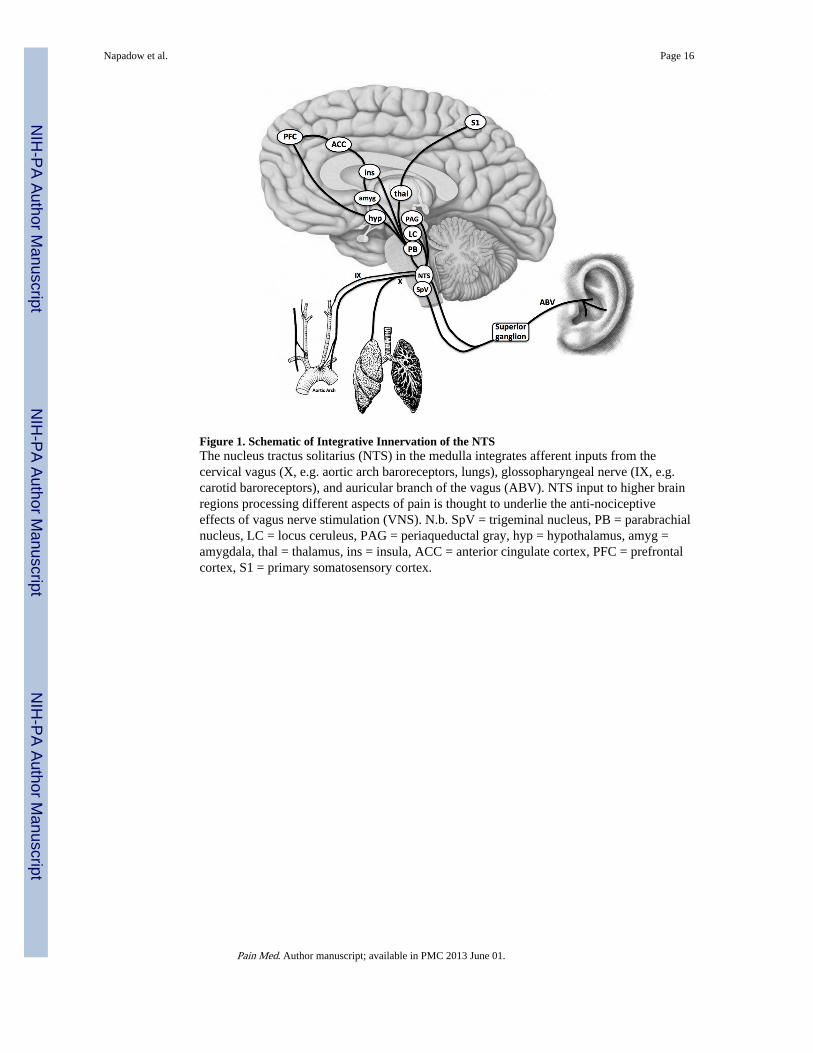
Figure 1. Schematic of Integrative Innervation of the NTSThe nucleus tractus solitarius (NTS) in the medulla integrates afferent inputs from thecervical vagus (X, e.g. aortic arch baroreceptors, lungs), glossopharyngeal nerve (IX, e.g.carotid baroreceptors), and auricular branch of the vagus (ABV). NTS input to higher brainregions processing different aspects of pain is thought to underlie the anti-nociceptiveeffects of vagus nerve stimulation (VNS). N.b. SpV = trigeminal nucleus, PB = parabrachialnucleus, LC = locus ceruleus, PAG = periaqueductal gray, hyp = hypothalamus, amyg =amygdala, thal = thalamus, ins = insula, ACC = anterior cingulate cortex, PFC = prefrontalcortex, S1 = primary somatosensory cortex.
Napadow et al. Page 16
Pain Med. Author manuscript; available in PMC 2013 June 01.
NIH
-PA Author Manuscript
NIH
-PA Author Manuscript
NIH
-PA Author Manuscript
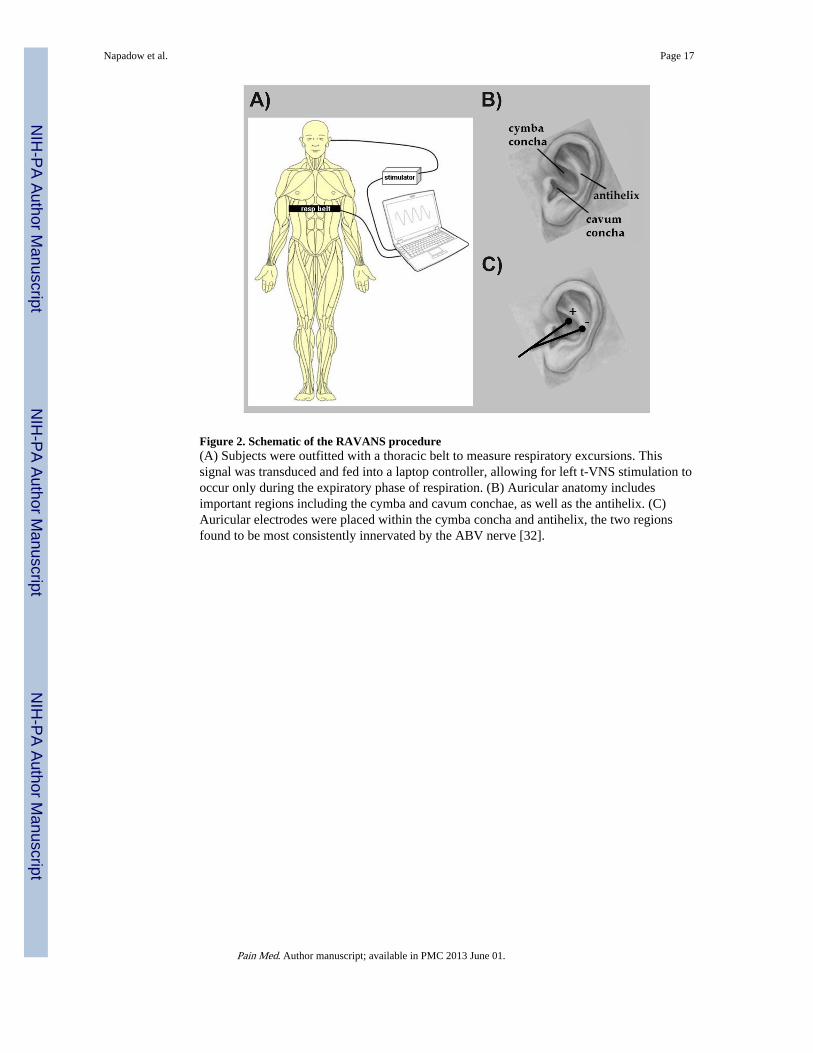
Figure 2. Schematic of the RAVANS procedure(A) Subjects were outfitted with a thoracic belt to measure respiratory excursions. Thissignal was transduced and fed into a laptop controller, allowing for left t-VNS stimulation tooccur only during the expiratory phase of respiration. (B) Auricular anatomy includesimportant regions including the cymba and cavum conchae, as well as the antihelix. (C)Auricular electrodes were placed within the cymba concha and antihelix, the two regionsfound to be most consistently innervated by the ABV nerve [32].
Napadow et al. Page 17
Pain Med. Author manuscript; available in PMC 2013 June 01.
NIH
-PA Author Manuscript
NIH
-PA Author Manuscript
NIH
-PA Author Manuscript
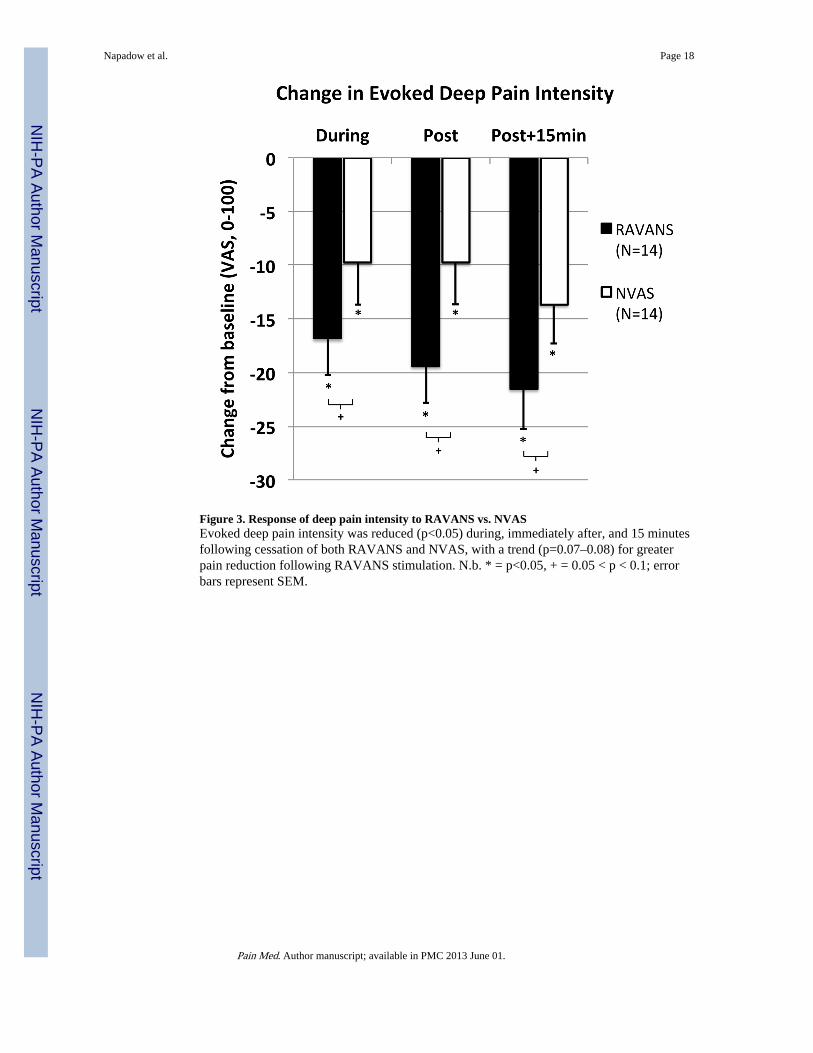
Figure 3. Response of deep pain intensity to RAVANS vs. NVASEvoked deep pain intensity was reduced (p<0.05) during, immediately after, and 15 minutesfollowing cessation of both RAVANS and NVAS, with a trend (p=0.07–0.08) for greaterpain reduction following RAVANS stimulation. N.b. * = p<0.05, + = 0.05 < p < 0.1; errorbars represent SEM.
Napadow et al. Page 18
Pain Med. Author manuscript; available in PMC 2013 June 01.
NIH
-PA Author Manuscript
NIH
-PA Author Manuscript
NIH
-PA Author Manuscript
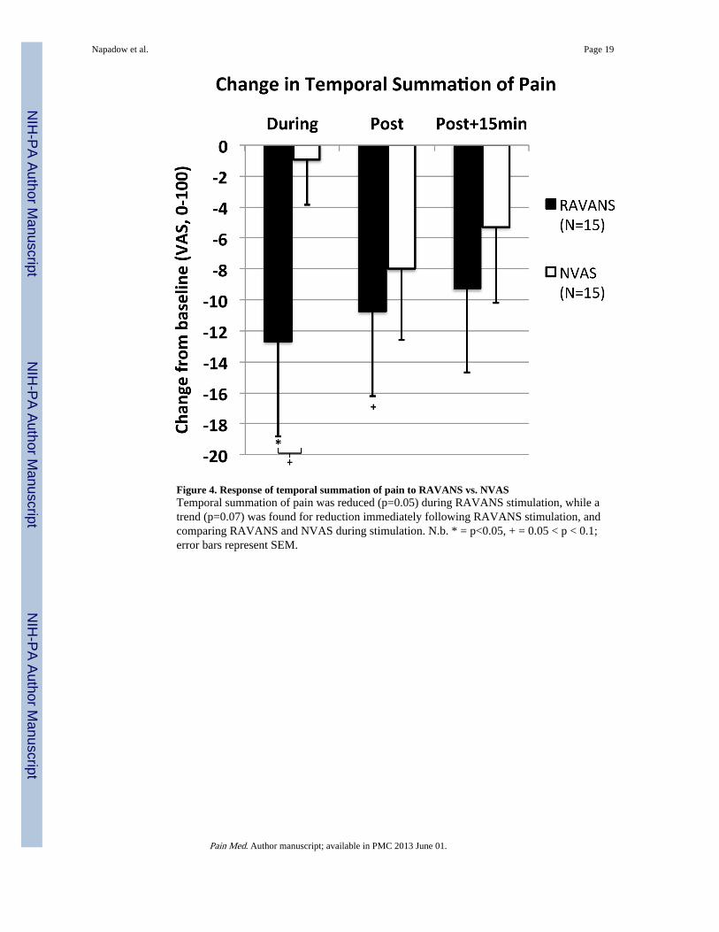
Figure 4. Response of temporal summation of pain to RAVANS vs. NVASTemporal summation of pain was reduced (p=0.05) during RAVANS stimulation, while atrend (p=0.07) was found for reduction immediately following RAVANS stimulation, andcomparing RAVANS and NVAS during stimulation. N.b. * = p<0.05, + = 0.05 < p < 0.1;error bars represent SEM.
Napadow et al. Page 19
Pain Med. Author manuscript; available in PMC 2013 June 01.
NIH
-PA Author Manuscript
NIH
-PA Author Manuscript
NIH
-PA Author Manuscript
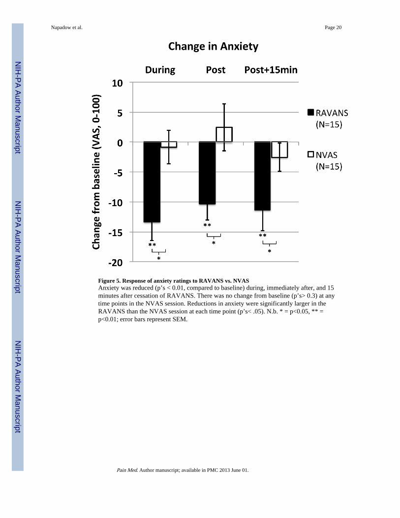
Figure 5. Response of anxiety ratings to RAVANS vs. NVASAnxiety was reduced (p’s < 0.01, compared to baseline) during, immediately after, and 15minutes after cessation of RAVANS. There was no change from baseline (p’s> 0.3) at anytime points in the NVAS session. Reductions in anxiety were significantly larger in theRAVANS than the NVAS session at each time point (p’s< .05). N.b. * = p<0.05, ** =p<0.01; error bars represent SEM.
Napadow et al. Page 20
Pain Med. Author manuscript; available in PMC 2013 June 01.
NIH
-PA Author Manuscript
NIH
-PA Author Manuscript
NIH
-PA Author Manuscript