Evidence for Decreased Coronary Flow Reserve in...
11
1201 Evidence for Decreased Coronary Flow Reserve in Viable Postischemic Myocardium Johan Vanhaecke, Willem Flameng, Marcel Borgers, Ik-Kyung Jang, Frans Van de Werf, and Hilaire De Geest To try to unravel the complexity and heterogeneity of the "no-reflow" phenomenon and its underlying mechanisms, we studied tissue perfusion in reperfused heart muscle by using tracer microspheres in an anesthetized dog model of 90-minute coronary occlusion followed by reperfusion for 2½2 hours, 24 hours, or 1 week. Regional myocardial blood flow was determined both in basal flow conditions and during reactive hyperemia. The effect of intracoronary adenosine administration was examined, and the ultrastructure of postischemic myocardium was analyzed. In viable reperfused tissue (as delineated by triphenyltetrazolium chloride staining), reflow in basal conditions is unimpaired. Coronary flow reserve (as approximated by peak reactive hyperemic flow) is intact at the start of reperfusion, decreases by more than half after 2Y2 hours, and recovers completely within 1 week. This impairment of coronary reserve can be relieved by intracoronary adenosine administration. On ultrastructural examination, the capillaries are patent. On the other hand, in irreversibly damaged myocardium, both the basal reflow impairment and the decrease in coronary flow reserve are severe and permanent. Coronary flow reserve is already decreased at the start of reperfusion, and the pharmacological intervention has no beneficial effect. Ultrastructurally, extracellular and intracellular edema invariably are present, whereas the vascular endothelium is damaged and the capillaries are packed with red blood cells. We conclude that the no-reflow phenomenon (i.e., mechanical obstruction to blood flow) is limited to infarcted tissue. In viable myocardium, however, coronary flow reserve is transiently diminished, probably because of washout and subsequent insufficient availability of the chemical mediator adenosine after breakdown and slow recovery of the precursor ATP pool. (Circulation Research 1990;67:1201-1210) W hen arterial flow is restored after a pro- longed period of interrupted blood sup- ply, tissue perfusion does not regain the preischemic level. This impairment of tissue reperfu- sion has been called the "no-reflow" phenomenon, although in most instances reflow is not absent but merely is reduced to a variable degree. This phenom- enon of reduced reflow has been demonstrated in kidney,' brain,2 and skin.3 It was described in canine myocardium by Kloner et al,4 and there is some evidence for its occurrence in humans.5 Several mechanisms for the no-reflow phenome- non have been proposed, including capillary damage, cell swelling, red blood cell packing, neutrophil plug- ging, and compression by cardiac contraction.46 All of these assume a mechanical obstruction to blood From the Departments of Cardiology and Cardiovascular Surgery, University Hospital Gasthuisberg, Leuven, Belgium, and the Jans- sen Research Foundation, Beerse, Belgium. Address for correspondence: J. Vanhaecke, Department of Cardiology, University Hospital Gasthuisberg, Herestraat 49, B-3000 Leuven, Belgium. Received November 17, 1987; accepted July 2, 1990. flow. Leaf hypothesized a vicious circle of ischemia causing cell swelling, resulting in further impairment of tissue perfusion and obstruction to reflow, ulti- mately leading to cell death. Support for the idea that tissue edema is responsible for reduced reflow came from the work of Willerson et al,8 who improved myocardial reflow by administration of hypertonic mannitol in dogs on right ventricular heart bypass. Tranum-Jensen et a19 obtained similar results using selective hypertonic reperfusion in isolated porcine hearts. These earlier studies did not make a distinc- tion between reflow in irreversibly damaged myocar- dium and reflow in tissue that remained viable after a period of ischemia. Also, the term no-reflow was restricted to blood flow in basal conditions. Data on coronary flow reserve in reperfused heart muscle are scarce and conflicting. One hour after release of a 2-hour coronary ligation, Bloor and White10 in five unanesthetized dogs found an un- changed peak reactive hyperemia but a decreased flow debt repayment, as measured with an electro- magnetic flow probe. Blumenthal et all" reported no change in reactive hyperemia during reperfusion in by guest on May 28, 2018 http://circres.ahajournals.org/ Downloaded from
Transcript of Evidence for Decreased Coronary Flow Reserve in...

1201
Evidence for Decreased Coronary FlowReserve in Viable Postischemic Myocardium
Johan Vanhaecke, Willem Flameng, Marcel Borgers, Ik-Kyung Jang,
Frans Van de Werf, and Hilaire De Geest
To try to unravel the complexity and heterogeneity of the "no-reflow" phenomenon and itsunderlying mechanisms, we studied tissue perfusion in reperfused heart muscle by using tracermicrospheres in an anesthetized dog model of 90-minute coronary occlusion followed byreperfusion for 2½2 hours, 24 hours, or 1 week. Regional myocardial blood flow was determinedboth in basal flow conditions and during reactive hyperemia. The effect of intracoronaryadenosine administration was examined, and the ultrastructure of postischemic myocardiumwas analyzed. In viable reperfused tissue (as delineated by triphenyltetrazolium chloridestaining), reflow in basal conditions is unimpaired. Coronary flow reserve (as approximated bypeak reactive hyperemic flow) is intact at the start of reperfusion, decreases by more than halfafter 2Y2 hours, and recovers completely within 1 week. This impairment of coronary reserve
can be relieved by intracoronary adenosine administration. On ultrastructural examination,the capillaries are patent. On the other hand, in irreversibly damaged myocardium, both thebasal reflow impairment and the decrease in coronary flow reserve are severe and permanent.Coronary flow reserve is already decreased at the start of reperfusion, and the pharmacologicalintervention has no beneficial effect. Ultrastructurally, extracellular and intracellular edemainvariably are present, whereas the vascular endothelium is damaged and the capillaries arepacked with red blood cells. We conclude that the no-reflow phenomenon (i.e., mechanicalobstruction to blood flow) is limited to infarcted tissue. In viable myocardium, however,coronary flow reserve is transiently diminished, probably because of washout and subsequentinsufficient availability of the chemical mediator adenosine after breakdown and slow recoveryof the precursor ATP pool. (Circulation Research 1990;67:1201-1210)
W hen arterial flow is restored after a pro-longed period of interrupted blood sup-ply, tissue perfusion does not regain the
preischemic level. This impairment of tissue reperfu-sion has been called the "no-reflow" phenomenon,although in most instances reflow is not absent butmerely is reduced to a variable degree. This phenom-enon of reduced reflow has been demonstrated inkidney,' brain,2 and skin.3 It was described in caninemyocardium by Kloner et al,4 and there is someevidence for its occurrence in humans.5
Several mechanisms for the no-reflow phenome-non have been proposed, including capillary damage,cell swelling, red blood cell packing, neutrophil plug-ging, and compression by cardiac contraction.46 Allof these assume a mechanical obstruction to blood
From the Departments of Cardiology and Cardiovascular Surgery,University Hospital Gasthuisberg, Leuven, Belgium, and the Jans-sen Research Foundation, Beerse, Belgium.Address for correspondence: J. Vanhaecke, Department of
Cardiology, University Hospital Gasthuisberg, Herestraat 49,B-3000 Leuven, Belgium.
Received November 17, 1987; accepted July 2, 1990.
flow. Leaf hypothesized a vicious circle of ischemiacausing cell swelling, resulting in further impairmentof tissue perfusion and obstruction to reflow, ulti-mately leading to cell death. Support for the idea thattissue edema is responsible for reduced reflow camefrom the work of Willerson et al,8 who improvedmyocardial reflow by administration of hypertonicmannitol in dogs on right ventricular heart bypass.Tranum-Jensen et a19 obtained similar results usingselective hypertonic reperfusion in isolated porcinehearts. These earlier studies did not make a distinc-tion between reflow in irreversibly damaged myocar-dium and reflow in tissue that remained viable aftera period of ischemia. Also, the term no-reflow wasrestricted to blood flow in basal conditions.Data on coronary flow reserve in reperfused heart
muscle are scarce and conflicting. One hour afterrelease of a 2-hour coronary ligation, Bloor andWhite10 in five unanesthetized dogs found an un-changed peak reactive hyperemia but a decreasedflow debt repayment, as measured with an electro-magnetic flow probe. Blumenthal et all" reported nochange in reactive hyperemia during reperfusion in
by guest on May 28, 2018
http://circres.ahajournals.org/D
ownloaded from

1202 Circulation Research Vol 67, No 5, November 1990
43 dogs studied
20 dogs 23 dogs
open chest closed chest
90'LAD ligation 90'LAD thrombosisrelease snare thrombolysis
GROUP I (n = 20) GROUP II (n = 8) GROUP IIl (n = 15)open chest study after open chest study after open chest study after
2.5 hrs reperfusion 24 hrs reperfusion 1 week reperfusion
FIGURE 1. Experimental protocol.
two dogs without a rise in serum enzyme levels after1-11/2 hours of coronary occlusion; reactive hyper-emia was reduced in nine dogs with enzymatic evi-dence of infarction. Cobb et a112 related flows inreperfused canine myocardium to the extent of his-tological necrosis. Basal flow was decreased in sam-ples containing more than 50% infarction. Transientischemic stimulation and intravenous adenosine in-fusion effected increases in myocardial blood flowthat were inversely proportional to the extent ofinfarction. These reactive hyperemic and adenosineflows, however, were not compared with measure-ments in control conditions. Finally, a recent ab-stract13 reports decreases in coronary flow reserveafter a transient (15-minute) period of ischemia inopen-chest dogs.We studied myocardial blood flow in a canine
model of coronary occlusion and reperfusion. Viablepostischemic myocardium was analyzed separatelyfrom irreversibly damaged tissue, and the temporalevolution of reflow was determined not only in basalconditions but also during reactive hyperemia, re-flecting the evolution of coronary flow reserve in theocclusion-reperfusion setting. The mechanisms re-sponsible for reflow impairment were explored bypharmacological manipulation of reflow and by ultra-structural tissue analysis.
Materials and MethodsExperimental Model and Protocol
Basically, in our canine experimental preparation,a coronary artery was occluded for 90 minutes andthereafter reperfused for 21/2 hours (group I), 24hours (group II), or 1 week (group III) (see Figure1). Forty-three mongrel dogs of either sex, weighing18-25 kg, were premedicated with Hypnorm (10 mgfluanisone/0.2 mg fentanyl per ml) (Duphar, Amster-dam, The Netherlands) (0.25 ml/kg i.m.) and wereanesthetized with sodium pentobarbital (15 mg/kgi.v.). After endotracheal intubation, the lungs wereventilated with a 50/50 mixture of oxygen and room
air using a Bird Mark 7 respirator (Bird ElectronicCorp., Cleveland). Arterial blood gases and pH weredetermined repeatedly throughout the experiment(pH/blood gas 166 microanalyzer, Corning GlassWorks, Corning, N.Y.), and ventilation was adjustedif necessary to keep these values within the normalrange. Additional doses of pentobarbital were ad-ministered as required to maintain anesthesia. Cor-onary occlusion was produced either by ligation orthrombosis.
Ligation protocol. In the ligation protocol (group I,n = 20), the chest was opened through the left fifthintercostal space, and the heart was suspended in apericardial cradle. The left anterior descending cor-onary artery (LAD) was isolated from the surround-ing tissue and encircled by a snare. Coronary occlu-sion and reperfusion were produced by tighteningand release of the snare.
In this group of dogs studied up to 21/2 hours ofreperfusion, LAD occlusion was produced by ligationto ensure the accuracy of some of the regionalmyocardial blood flow (RMBF) measurements thatwere planned (testing of coronary flow reservethrough reactive hyperemia and intracoronary aden-osine infusion, see below). In six of the 20 experi-ments of group I, a 24-gauge needle connected to asmall tubing was inserted in the LAD at the site offuture ligation. At the desired times during theexperiment, adenosine was infused through this nee-dle into the coronary artery in a dose of 0.5 mg/min;the infused volume was 0.5-1.0 ml/min. This dosepreviously has been shown to produce maximal vaso-dilation,14 and the intracoronary route of administra-tion was chosen to minimize alterations in peripheralhemodynamics.
Thrombosis protocol. In the thrombosis protocol(groups II and III, n=23), a copper coil attached to aguide wire was advanced under fluoroscopic controlvia the carotid artery into the LAD. The formation ofan occlusive thrombus took 5-10 minutes and wasconfirmed by coronary angiography using a SF Leh-
by guest on May 28, 2018
http://circres.ahajournals.org/D
ownloaded from
Vanhaecke et al Mechanisms of No-Reflow 1203
man catheter (USCI, Billerica, Mass.). Reperfusionwas achieved by coronary thrombolysis, using an in-travenous infusion of a thrombolytic agent. As throm-bolytic substances, urokinase (10 ,g.kg-l.min-1), re-combinant tissue-type plasminogen activator (10,ug.kg-'.min-1), or recombinant single-chain uroki-nase-type plasminogen activator (20 ,ggkg- .min-')were used. The thrombolytic infusion was started 1hour after thrombus formation. Reperfusion occurredwithin 15-30 minutes and was documented by coro-nary angiography. Then the carotid artery was ligated,skin wounds were sutured, and the dog was allowed torecover. Twenty-four hours (group II, n=8) or 1 week(group III, n= 15) later, a final open-chest experimentwas performed, at the end of which the animal waskilled and the heart removed for further processing.
In groups II and III (24 hours and 1 week ofreperfusion, respectively), LAD occlusion was pro-duced by coronary thrombosis to avoid two consecu-tive open-chest experiments on the same animal.Coronary flow reserve in these animals was studiedonly during the final open-chest experiment.
Regional Myocardial Blood Flow MeasurementsMethodology. RMBF was measured with the tracer
microsphere technique.15 We used 15-,um micro-spheres labeled with 141Ce, 113Sn, 103Ru, or 95Nb (NENChemicals GmbH, Dreieich, FRG). Microspherehandling, dilution, and mixing were performed asdescribed previously.16At different times during an experiment, micro-
spheres were injected in basal flow conditions as wellas during intracoronary adenosine infusion and dur-ing peak reactive hyperemia after release of a 60-second LAD occlusion. The hyperemic response wasmonitored by means of an electromagnetic flowmeterto ensure good timing of the microsphere injection.At the end of an experiment, left ventricular slices
were cut into multiple tissue samples after epicardialfat and vessels were removed. The left ventricularwall was divided into three layers of equal thickness(epimyocardium, midmyocardium, and endomyocar-dium). The area at risk (LAD area) and the controlarea (circumflex area) were separated, and LADsamples were labeled by their macroscopic appear-ance after triphenyltetrazolium chloride (ITC) stain-ing as either viable, patchy infarcted, or homogene-ously infarcted (see below). This procedure typicallyresulted in 60-80 tissue samples of ± 1 g (range,0.5-2.0 g) per experiment. Tissue samples were putinto test tubes and immersed in 3% glutaraldehydesolution. Radioactivity in reference blood samplesand tissue samples was counted (5 min/sample) usingan automatic gamma counter and sample changersystem (analyzer model 45, M0lsgaard Medical, H0r-sholm, Denmark) connected to an ND 680 program-mable analyzer/computer system (Nuclear DataGmbH, Frankfurt/Main, FRG).
Values for blood flow in every myocardial samplewere computed using a slight adaptation17 of the MICii program of Schosser et al.18RMBF in a specific region of the left ventricle
(e.g., epimyocardium in the circumflex area andviable epimyocardium in the LAD area) was calcu-lated by averaging the values of all tissue samplesbelonging to that region.
Timing. Under preischemic control conditions,basal myocardial blood flow measurements were ob-tained in 32 animals from groups I, II, and III; alldata were pooled. Coronary flow reserve was deter-mined by RMBF measurement at peak reactivehyperemia in all 20 animals of group I. In six of thesedogs, coronary reserve also was tested by RMBFmeasurement during infusion of adenosine.Under coronary occlusion conditions, RMBF mea-
surement was performed in 40 animals from groups I,II, and III at the end of 90 minutes ofLAD occlusion.Under reperfusion conditions, RMBF was mea-
sured in six dogs of group I at 1 minute of reperfu-sion, during the reactive hyperemia immediately fol-lowing the release of the snare after 90 minutes ofcoronary occlusion. At 21/2 hours of reperfusion,RMBF was measured in 15 dogs of group I. Coronaryflow reserve was determined by RMBF measurementat peak reactive hyperemia in 18 dogs of group I. Insix of these dogs, coronary reserve also was tested byRMBF measurement during intracoronary adenosineinfusion. At 24 hours of reperfusion (group II),RMBF was measured in all eight dogs. Coronary flowreserve was determined by RMBF measurement atpeak hyperemic response in five dogs. At 1 week ofreperfusion (group III), RMBF was measured in all15 dogs in basal flow conditions as well as duringpeak reactive hyperemia.
Hemodynamic MeasurementsSystolic and diastolic aortic pressures and left
ventricular end-diastolic pressure or left atrial pres-sure were monitored through fluid-filled cathetersconnected to a Siemens pressure transducer 746(Siemens Elema, Solna, Sweden). These catheterswere inserted via carotid and/or femoral arteries, orfor the left atrial pressure (open-chest experiments),directly into the left atrium via an incision in the leftatrial appendage. The pressure signals togetherwith electrocardiographic lead II were displayed onan oscilloscope and recorded on a multichannel inkjet recorder (Siemens Corp., Iselin, N.J.) through-out the experiment.Delineation of Viable and Infarcted Tissue in the Areaat Risk
For this purpose, we used a dye perfusion andfixation technique. At the end of each experiment,the heart was removed and both right and leftcoronary ostia were cannulated as well as the LADdistal to the site of previous occlusion. The LAD wasperfused with Ringer's solution, and the ostia wereperfused at the same pressure with a mixture ofRinger's solution and Evans blue. Two minutes later,the LAD perfusion was switched to a TTC solution'9at 370 C for 10 minutes. Finally, the heart was fixed by
by guest on May 28, 2018
http://circres.ahajournals.org/D
ownloaded from

1204 Circulation Research Vol 67, No 5, November 1990
TABLE 1. Temporal Evolution of Regional Myocardial Blood Flow in Viable Subepicardium
Basal conditions (ml/min/100 g) PRH in LAD area (mlmin/100 g)
Group n Cx LAD n Cx LAD
Control 32 49±+17 49±19 20 57+22 338+137Occlusion 40 55±19 25± 16* ... ... ...
Reperfusion1 min ... 6 66+27 292+7321/2hr 15 60±19 53+15t 18 67+44 149+75t24 hr 8 53+20 52+22 5 51+20 160+72§1 wk 15 48+25 54±32 15 69±22 345±145
Dogs underwent a 90-min occlusion of the left anterior descending artery followed by reperfusion.Values are mean±+SD. PRH, peak reactive hyperemia; LAD, left anterior descending; Cx, circumflex.*p<0.001 vs. Cx flow at the same time.tp<0.02 vs. Cx flow at the same time.tp<0.001 vs. control PRH in LAD area.§p<0.02 vs. control PRH in LAD area.
perfusing the LAD area for another 5 minutes with2% glutaraldehyde and both coronary ostia with a
mixture of 2% glutaraldehyde and Evans blue.After the right ventricle, the atria, and the valvar
structures were removed, the isolated left ventriclewas cut in 1-cm-thick slices perpendicularly to thelong axis.As a result of the above-described dye perfusion
technique, myocardium supplied by nonoccludedcoronary arteries (referred to as the circumflex area)was colored blue, whereas in the occluded bed (theLAD area), viable tissue stained brick red, andinfarcted tissue was white.
Ultrastructural AnalysesAfter perfusion staining and fixation, small samples
of myocardial tissue were immersed in a fixativecontaining 3% glutaraldehyde and 90 mmol/potassiumoxalate, adjusted to pH 7.4 with sodium potassiumhydroxide. These samples then were washed thor-oughly in the same buffer supplemented with 0.22%sucrose. Postfixation was done in 1% osmic acid,buffered to pH 7.4 with 0.05 M veronal acetatecontaining 0.093 M sucrose, for 1 hour at 40 C. After arinse in the buffer, samples were dehydrated in agraded series of ethanol and routinely embedded inepoxy resin. Ultrathin sections were examined with a
Philips EM300 electron microscope (Philips Neder-land, Eindhoven, The Netherlands) after staining withuranyl acetate and lead citrate.
Statistical Methods
Statistical analyses were done using the SAS sta-tistical package.20 Student's t test (paired or unpaired)was used for comparisons between two groups. Whenmore than two groups were compared, one-way anal-ysis of variance was used to assess overall significanceof group differences. If a statistically significant Fvalue was observed, an appropriate multiple compar-ison method (Tukey's modification of the honestlysignificant difference test) was applied to evaluate thedifference between any two groups. Values are givenas mean+SD unless stated otherwise.
ResultsTime Dependency of the No-Reflow PhenomenonThe data on RMBF measurements at different
times during reperfusion are presented separately forviable subepicardium in Table 1 and for homogene-ously infarcted subendocardium in Table 2.
Viable subepicardium. In viable subepicardium ofthe LAD area, the preischemic control flow is similarto the flow in the subepicardial circumflex area.
TABLE 2. Temporal Evolution of Regional Myocardial Blood Flow in Infarcted Subendocardium
Basal conditions (ml/min/100 g) PRH in LAD area (ml/min/100 g)
Group n Cx LAD n Cx LAD
Control 24 68+21 57±21 20 71+25 483+140Occlusion 25 74±27 3+3* ...
Reperfusion1 min ... ... ... 4 86+30 120±73t21/2hr 12 66±19 22+16* 12 64+27 56+27t24 hr 6 66+14 18+8* 5 68±22 50±21t1 wk 9 62±23 21±8* 9 76±13 107±82t
Dogs underwent a 90-min occlusion of the left anterior descending artery followed by reperfusion.Values are mean±SD. PRH, peak reactive hyperemia; LAD, left anterior descending; Cx, circumflex.*p<O0.01 vs. Cx flow at the same time.tp<O.00l vs. control PRH in LAD area.
by guest on May 28, 2018
http://circres.ahajournals.org/D
ownloaded from

Vanhaecke et al Mechanisms of No-Reflow 1205
During occlusion, collateral flow in the LAD area isabout 45% of the preischemic flow. Reflow in thesubepicardial LAD area after reperfusion for 21/2hours, 24 hours, and 1 week is slightly but notsignificantly higher than the preischemic value. Ascompared with flow in the circumflex area, there isinitially a small but significant flow deficit (p<0.02)because of a small increase in circumflex flow. Thisflow deficit disappears by 24 hours and 1 week ofreperfusion.
The coronary flow reserve as assessed by the peakreactive hyperemic (PRH) flow is intact at the start ofreperfusion. PRH flow during the first minute ofreflow after 90 minutes of occlusion is similar to thepreischemic control PRH flow elicited by transientischemic stimulation: 292+73 and 338±137 ml/min/100 g, respectively (p=NS). Thus, a 90-minute cor-
onary occlusion per se does not adversely affectcoronary flow reserve in viable tissue. The decreasein PRH flow (to about 45% of control values) ob-served after 21/2 hours of reflow occurs during theinitial phase of reperfusion. After 24 hours of reper-fusion, the PRH flow still is depressed (47% ofcontrol values), but at the end of 1 week of reperfu-sion, there is a complete recovery of the coronaryflow reserve in viable subepicardium: PRH flowreturned to a value of 345-+±145 ml/min/100 g.
Infarcted subendocardium. In homogeneously in-farcted subendocardium, the preischemic controlflow is 15% lower than in the subendocardial circum-flex area because of real and/or apparent micro-sphere loss.21
Collateral flow during occlusion is very low (4% ofthe preischemic LAD flow). Reflow after 21/2 hours ofreperfusion is severely impaired and amounts to only33% of preischemic LAD flow. In the subsequentdays of reperfusion, there is no recovery of this flowdeficit: at 1 week, reflow still is 34% of preischemicLAD flow. Thus, in infarcted tissue, the basal flowdeficit is permanent, whereas in viable myocardium,basal flow normalizes after reperfusion.The coronary flow reserve in homogeneously in-
farcted subendocardium also behaves differently inseveral aspects. First, the vasodilatory capacity isalready severely compromised during the 90-minuteperiod of coronary occlusion: PRH flow at the start ofreperfusion has decreased to 25% of preischemiccontrol values. So, in contrast to the viable subepi-cardium, here the adverse effects have occurredprimarily during the period of ischemia. During thesubsequent hours of reperfusion, there is a trendtoward a further decrease of PRH flow (p=NS). At21/2 and 24 hours of reperfusion, a transient ischemicstimulation elicits very little hyperemic response(flows increase slightly over the basal values), and thePRH flow does not even reach the level of basal flowin the corresponding circumflex area. Finally, there isno significant recovery of PRH flow in infarctedsubendocardium after 1 week of reperfusion (22% ofcontrol values).
TABLE 3. Hemodynamics During Peak Reactive Hyperemia
Mean aorticDuration of Heart rate pressure LVEDPreperfusion n (beats/min) (mm Hg) (mm Hg)
21/2hr 18 117+29 93±15 6.3±2.724 hr 5 161±33 81±19 4.8±1.01 wk 15 148±32 102±16 2.2±1.3
Values are mean±SD. LVEDP, left ventricular end-diastolicpressure.
Hemodynamics during reperfusion. It is appropriateto consider briefly the most important hemodynamicparameters at these different times during reperfu-sion (Table 3). After 24 hours of reperfusion, theanimals are not in a very stable condition: they oftenhave severe arrhythmias, accounting for the highheart rate, and their blood pressure is usually ratherlow. Together with the relatively small number ofobservations at this time, these considerations call forsome caution in the interpretation of the data onPRH flow at this time of reperfusion. The importantpoint, however, is that hemodynamics after 21/2 hoursand after 1 week of reperfusion are comparable.Mean aortic pressure is on average 9 mm Hg higherafter 1 week, and heart rate is 31 beats/min faster.The combination of these factors permits a safeexclusion of a major role of hemodynamic variablesin the observed changes in PRH flow.
Intracoronary Adenosine Infusion. In six open-chestdogs of group I, instrumented as described in "Ma-terials and Methods," RMBF was measured duringpeak reactive hyperemia and during intracoronaryadenosine infusion, both in preischemic control con-ditions and after 21/2 hours of reperfusion. Theresults are shown in Figure 2. During intracoronaryinfusion of adenosine, mean aortic blood pressurefell slightly but not significantly. To correct for thesedifferences in perfusion pressure during reactivehyperemia and intracoronary adenosine infusion, wealso calculated coronary resistance (diastolic bloodpressure divided by coronary flow).
In viable subepicardium, blood flows during intra-coronary adenosine infusion in preischemic controlconditions tended to be higher than PRH flows(456±153 and 414±184 ml/min/100 g, respectively;p=NS), and coronary resistance was lower duringintracoronary adenosine infusion than during peakreactive hyperemia (0.16±0.08 versus 0.21+0.07;p<0.05). This confirms previous evidence that coro-nary vasodilation during reactive hyperemia is notmaximal.14
After 21/2 hours of reperfusion, PRH flow droppedexpectedly to 200±+71 ml/min/100 g, or 48% of con-trol values (p<0.02 versus control). The adenosineflow at this time, however, remained unchanged(463+±150 ml/min/100 g). Comparison of coronaryresistance after 21/2 hours of reperfusion with pre-ischemic control values gave similar results: coronaryresistance during reactive hyperemia doubled(0.21±0.07 to 0.42±0.17; p<0.02), whereas it re-
by guest on May 28, 2018
http://circres.ahajournals.org/D
ownloaded from

1206 Circulation Research Vol 67, No 5, November 1990
RMBF (mlImin/10Ogr) CORONARY RESISTANCEImmHg. min 10Ogr / m)
VIABLE SUBEPICARDIUM (n=6)ADENOSINE I.C REACTIVE
HYPEREMIA
INFARCTED SUBENDOCARDIUM (n=3)ADENOSINE I.C. REACTIVE ADENOSINE C.
HYPEREMIA
1.0
.8
.6-
.4-
FIGURE 2. Regional myocardial blood flow (RMBF)and coronary resistance in the left anterior descendingarea duringpeak reactive hyperemia and intracoronary(I.C.) adenosine infusion (0.5 mg/min) in preischemiccontrol conditions and after 2½/2 hours of reperfusionfollowing a 90-minute coronary occlusion. *p<0.05;**p <0.02 vs. preischemic control; tp<0.05 vs. reactivehyperemia.
= controlM 2112 hrs of reperfusion
mained unchanged during intracoronary adenosineinfusion (0.16+0.08 versus 0.17±0.08).Only three of the six dogs had homogeneously
infarcted subendocardial tissue blocks, which makesstatistical analysis of these data not very meaningful.It is evident from Figure 2, however, that in thesethree animals, the very distinct decrease in PRH flowafter 21/2 hours of reperfusion is matched by a similardecrease in flows during intracoronary adenosineinfusion (430±+174 to 76±+10 and 502±260 to 102±35ml/min/100 g, respectively). The rises in coronaryresistance in infarcted subendocardium during bothreactive hyperemia and intracoronary adenosine in-fusion are roughly comparable (0.19+±0.03 to 1.01+±0.17 and 0.15+±0.05 to 0.80±0.24, respectively) andeven statistically significant (p<0.02 andp>0.05) de-spite the small number of observations. Thus, al-though the hyperemic response to a transient ischemicstimulus is markedly blunted in viable reperfusedmyocardium, the coronary vasodilatory capacity assuch is preserved, because exogenous adenosine ad-ministration restores maximal flows. On the otherhand, in irreversibly damaged tissue, adenosine infu-sion cannot relieve the severe reflow impairment.
Ultrastructural Analysis of Reperfused MyocardiumTissue samples from viable and infarcted portions
of the LAD area (as indicated by TTC staining) andsamples from the control circumflex area were pro-cessed as described above. At least three ultrathinsections from different sites were analyzed per exper-iment. These sections were scanned systematicallyfor mitochondrial, nuclear, and myofibrillar changes;for the integrity of the cell membrane; and for thepresence of intracellular and/or extracellular edema.
Vascular structures (i.e., almost exclusively capillar-ies) were analyzed separately in a number of exper-iments. A normal appearance of the control myocar-dium from the circumflex bed should testify to theappropriate processing of the tissue samples. In fourdogs (21/2 hours, n=1; 24 hours, n=2; and 1 week,n=1, of reperfusion) in which control myocardiumwas analyzed, the ultrastructure looked normal. Intwo of these animals, confluent mitochondrial cristaewere present, suggesting minimal signs of tissueinjury. The capillary lumen was always patent, andendothelial cells were intact.The characteristic signs of irreversible myocardial
damage include the presence of amorphous densebodies in the mitochondrial matrix, pyknosis of thecell nucleus, the development of contraction bands inthe myofilaments, and rupture of the cell membrane.Hallmarks of irreversible injury were absent in TTC-stained tissue of 16 dogs (21/2 hours, n=6; 24 hours,n =2; and 1 week, n=8, of reperfusion), but they wereinvariably present in YITC-unstained tissue of fivedogs (21/2 hours, n=4; and 1 week, n=l, or reperfu-sion).As for the relation of ultrastructural changes to
reflow impairment, the absence of edema in viabletissue samples is remarkable. Except for a smallpericapillary rim, which is a known artifact aftersequential perfusion staining and fixation, extracellu-lar and intracellular edema were completely absentin 15 of 16 and 13 of 16 animals, respectively. On theother hand, edema was an almost universal feature ofinfarcted tissue samples. Representative electron mi-crographs are shown in Figures 3 and 4. In thiscontext it should be pointed out that the perfusionmedia used in the postmortem staining and fixation
by guest on May 28, 2018
http://circres.ahajournals.org/D
ownloaded from
Vanhaecke et al Mechanisms of No-Reflow 1207
-,,4.
if
9'~~~~~~~~~~~~~~~~~J
80<Ij 4+_5% jt , :
AS' A >D'"
:'
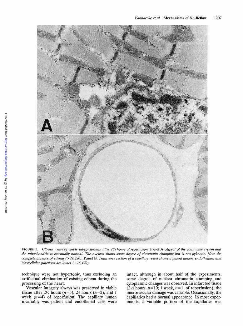
FIGURE 3. Ultrastracture of viable subepicardium after 2'12 hours of reperfusion. Panel A: Aspect of the contractile system andthe mitochondria is essentially normaL The nucleus shows some degree of chromatin clumping but is not pyknotic. Note thecomplete absence of edema (x24,820). Panel B: Transverse section of a capillary vessel shows a patent lumen; endothelium andintercellular junctions are intact (x15,470).
technique were not hypertonic, thus excluding anartifactual elimination of existing edema during theprocessing of the heart.
Vascular integrity always was preserved in viabletissue after 21/2 hours (n=5), 24 hours (n=2), and 1week (n=4) of reperfusion. The capillary lumeninvariably was patent and endothelial cells were
intact, although in about half of the experiments,some degree of nuclear chromatin clumping andcytoplasmic changes was observed. In infarcted tissue(21/2 hours, n=10; 1 week, n=1, of reperfusion), themicrovascular damage was variable. Occasionally, thecapillaries had a normal appearance. In most exper-iments, a variable portion of the capillaries was
i';j 1-
,m-1~,-
A
.711e--.
k. 1l! .ok-q--i.
r'
by guest on May 28, 2018
http://circres.ahajournals.org/D
ownloaded from

1208 Circulation Research Vol 67, No 5, November 1990
i. 4;
./ .i
* ..gi*r. .
.. ",
¾ a. NSt
r, Ct,
'?Z
B j6
FIGURE 4. Ultrastructure of infarcted subendocardium after 2'12 hours of reperfusion. Panel A: Myofilaments are relaxed andpartially distorted; the mitochondrial matrix is cleared and there is fragmentation of the cristae; numerous intramitochondrialamorphous dense bodies are present; the cell membrane is ruptured; edema is evident (x24,820). Panel B: A capillary vesseltypically is packed with red blood cells. The capillary lumen also contains some cellular debnrs (x 15,470).
packed with red blood cells, whereas the endotheliumof these vessels was usually intact. In other animals,the capillary lumina were obstructed, and endothelialcells were completely necrotic and ruptured.
DiscussionThe perfusion bed of an acutely occluded coronary
artery is a very inhomogeneous entity. For a variable
period of time, it contains all degrees of tissue injury,varying from completely normal myocardial cells tonecrosis; the capillary network may be intact or
obstructed to a variable degree. This inhomogeneityresults from inhomogeneities in collateral blood sup-ply and oxygen demand. A priori, one could there-fore expect the reflow pattern in this tissue to beinhomogeneous too. Previous studies documenting
¾-v*
4.I1
'
f
'^
.0. 'g, /
-J
t0
;4
c:, ;
by guest on May 28, 2018
http://circres.ahajournals.org/D
ownloaded from

Vanhaecke et al Mechanisms of No-Reflow 1209
the no-reflow phenomenon have failed to highlightits complexity and inhomogeneity. Several mecha-nisms have been invoked to explain the no-reflowphenomenon; their common feature is that they allassume some form of mechanical obstruction toblood flow, a "shut down" of capillaries, caused bydamage to the vascular endothelium, edema, plug-ging by packed red blood cells or neutrophils, andcompression by cardiac contraction.46 Finally, theterm no-reflow largely has been restricted to bloodflow in basal conditions, without regard to the coro-nary flow reserve of the myocardium under study.The data presented here provide evidence that aftercoronary reperfusion, different mechanisms are re-sponsible for the observed flow patterns in viablepostischemic heart muscle as opposed to irreversiblydamaged myocardium.
In viable tissue, reflow in basal conditions is similarto the preischemic flow at all times after reperfusion(21/2 hours, 24 hours, and 1 week). Because changesin hemodynamics between the preischemic controlcondition and the reperfusion phase may influencemyocardial blood flow, it may be more appropriate tocompare flow in the postischemic LAD area withflow in the circumflex bed at the same time point.Thus, after 21/2 hours of reperfusion, we found aslightly lower basal reflow in viable postischemictissue. The presence of a zone of "low reflow" in theviable subepicardium overlying an infarct also hasbeen shown by Kloner and Alker.22 In that study, theauthors found that no-reflow was associated withirreversibly injured myocardium (TTC-negative),whereas low reflow (mild reperfusion abnormality)occurred in viable TTC-positive tissue. Becausereperfused myocardium is stunned and thereby haslower metabolic requirements, the reduced reflow inbasal conditions suggests that autoregulation is in-tact, that is, that the resistance vessels are not"stunned." An alternative explanation would be thatthe difference between circumflex and LAD basalflows at 21/2 hours of reperfusion indicates increasedcircumflex flow, caused by hyperkinesis of the cir-cumflex territory, to overcome the hypokinesis of thepostischemic zone. However, after 24 hours, whenstunning of the postischemic zone probably is stillpresent, the basal circumflex and LAD flows aresimilar.When PRH flow is used as a measure of coronary
flow reserve, two points need to be addressed. First,from previous evidence14 as well as from our owndata, it appears that PRH flow to some extentunderestimates coronary flow reserve as measuredafter intracoronary adenosine administration. Thus,PRH flow is only a useful approximation of true flowreserve. Second, we assume that a brief coronaryocclusion causes the same ischemic insult in viablepostischemic myocardium versus control myocar-dium. Although we are not aware of data to thecontrary, this point is hard to verify. In three dogs notincluded in this study, extension of the coronary
occlusion up to 180 seconds did not further enhancethe hyperemic response in postischemic tissue.With these caveats in mind, our results indicate
that coronary flow reserve in viable postischemictissue is preserved at the onset of reperfusion. Itdecreases during the initial phase of reperfusion,remains depressed for at least 24 hours, and hascompletely recovered after 1 week of reperfusion.The ultrastructure of this viable postischemic myo-cardium is intact at the different times of reperfusion.Thus, the evolution of coronary flow reserve duringreperfusion must be explained either by a reducedavailability of vasodilating mediator substances or bythe inability of the vasculature to vasodilate maxi-mally. The results with intracoronary adenosine in-fusion rule out the latter and indicate that thevasculature in this tissue remains not only structur-ally but also functionally intact after reperfusion. Thetransient decrease in coronary flow reserve occursprobably because vasoactive metabolites responsiblefor the autoregulation of coronary flow (primarilyadenosine), which are formed by the rapid break-down of ATP during ischemia, are washed out fromthe postischemic bed on reperfusion and thereafterare insufficiently available for a period of time be-cause of depletion of their precursor pool of nucle-otides, which has a slow rate of recovery.23,24 In arecent study, Reimer et a125 demonstrated that evenshort periods of ischemia followed by reperfusion areassociated with a considerable loss of adenine nucle-otides.
Undoubtedly, this hypothetical scheme oversimpli-fies a complex interplay of factors that regulatesblood flow in viable postischemic myocardium. Forexample, the amount of adenosine needed for coro-nary vasodilation is probably small,26 and althoughtissue levels of ATP after 21/2 hours of reperfusionare low, they are by no means negligible (data notshown). Therefore, one has to call on the concept ofcompartmentalization of ATP stores2728 to explainthe observed flow patterns on the basis of insufficientavailability of adenosine. The question of whetherthis compartmentalization is intracellular, within thecardiac myocyte, or intercellular, between differentcell types (myocytes, pericytes, vascular endotheli-um), further complicates the matter. An alternativeexplanation for the observed flow patterns would bea decrease in sensitivity and/or number of vascularadenosine receptors in postischemic viable tissue.More experimental work is needed to corroborate orrefute these conjectures.
In irreversibly damaged myocardium, the severityand permanence of the basal reflow impairment perse do not permit any conclusion about mechanisms ofno-reflow. Indeed, dead myocytes do not requireoxygen, so there is no reason to suspect that basalreflow should ever return to control levels in in-farcted tissue. However, the ultrastructural findings(edema, capillary plugging, and vascular damage)and the fact that intracoronary adenosine effects onlya very minor relief of the reflow impairment are
by guest on May 28, 2018
http://circres.ahajournals.org/D
ownloaded from

1210 Circulation Research Vol 67, No 5, November 1990
consistent with the current idea that no-reflow largelyis a mechanical phenomenon.
In conclusion, it would be preferable if the termno-reflow were restricted to its original definition, thatis, basal reflow impairment due to physical restrictionof capillary flow. This phenomenon is limited toirreversibly damaged myocardium. In viable postische-mic heart muscle, the vasculature remains functionallyand structurally intact; the transient decrease of flowreserve in this tissue possibly is due to a reducedproduction of the chemical mediator adenosine.
References1. Summers WK, Jamison RL: The no-reflow phenomenon in
renal ischemia. Lab Invest 1971;25:635-6432. Majno G, Ames A III, Chaing J, Wright RL: No reflow after
cerebral ischemia. Lancet 1967;2:569-5703. Willms-Kretschmer K, Majno G: Ischemia of the skin: Elec-
tron microscopic study of vascular injury. Am J Pathol 1969;54:327-343
4. Kloner RA, Ganote CE, Jennings RB: The "no-reflow" phe-nomenon after temporary coronary occlusion in the dog. J ClinInvest 1974;54:1496-1508
5. Schofer J, Spielmann R, Montz R: The "no-reflow" phenom-enon in man (abstract). Circulation 1983;68(suppl III):III-393
6. Lucchesi BR: Leukocytes and ischemia-induced myocardialinjury. Annu Rev Phramacol Toxicol 1986;26:201-224
7. Leaf A: Cell swelling: A factor in ischemic tissue injury.Circulation 1973;48:455-458
8. Willerson JT, Watson JT, Hutton I, Templeton GH, FixlerDE: Reduced myocardial reflow and increased coronary vas-cular resistance following prolonged myocardial ischemia inthe dog. Circ Res 1975;36:771-781
9. Tranum-Jensen J, Janse MJ, Fiolet JWT, Krieger WJG,Naumann d'Alnoncourt C, Durrer D: Tissue osmolality, cellswelling and reperfusion in acute regional myocardial isch-emia in the isolated porcine heart. Circ Res 1981;49:364-381
10. Bloor CM, White FC: Coronary artery reperfusion: Effects ofocclusion duration on reactive hyperemia responses. Basic ResCardiol 1975;70:148-158
11. Blumenthal MR, Wang H, Liu LMP: Experimental coronaryarterial occlusion and release: Effects on enzymes, electrocar-diograms, myocardial contractility and reactive hyperemia.AmJ Cardiol 1975;36:225-233
12. Cobb FR, Bache RJ, Greenfield JC Jr: Regional myocardialblood flow in awake dogs. J Clin Invest 1974;53:1618-1625
13. Nicklas JM, Gips S, Van Heyningen A: Decreased flow reserve
following transient myocardial ischemia (abstract). JAm CollCardiol 1984;3:546
14. Warltier DC, Gross GJ, Brooks HL: Pharmacologic- vs. isch-emia-induced coronary artery vasodilation.Am JPhysiol 1981;240:H767-H774
15. Winkler B: The tracer microsphere method, in Schaper W(ed): The Pathophysiology ofMyocardial Perfusion. Amsterdam,Elsevier/North Holland Biomedical Press, 1979, pp 13-42
16. Flameng W, Winkler B, Wuesten B, Schaper W: Minimumrequirements for the measurement of regional myocardialblood flow using tracer microspheres. BiblAnat 1977;15:24-29
17. Prinzen WF: Gradients in myocardial blood flow, metabolismand mechanics across the ischemic left ventricular wall (the-sis). Rijksuniversiteit Limburg, Maastricht, The Netherlands,1982
18. Schosser R, Arfors K-E, Messmer K: MIC II: A program forthe determination of cardiac output, arterio-venous shunt andregional blood flow using the radioactive microsphere method.Comput Progr Biomed 1979;9:19-39
19. Fishbein MC, Meerbaum S, Rit J, Lando U, Kanmatsuse K,Mercier JC, Corday E, Ganz W: Early phase acute myocardialinfarct size quantification: Validation of the triphenyl tetra-zolium chloride tissue enzyme staining technique. Am Heart J1981;101:593-600
20. SAS Institute Inc. SAS User's Guide: Statistics, Version 5Edition. Cary, NC, SAS Institute Inc, 1985
21. Reimer KA, Jennings RB: The changing anatomic referencebase of evolving myocardial infarction: Underestimation ofmyocardial collateral blood flow and overestimation of exper-imental anatomic infarct size due to tissue edema, hemorrhageand acute inflammation. Circulation 1979;60:866-876
22. Kloner RA, Alker KJ: The effect of streptokinase on intra-myocardial hemorrhage, infarct size, and the no-reflow phe-nomenon during coronary reperfusion. Circulation 1984;70:513-521
23. De Boer LWV, Ingwall JS, Kloner RA, Braunwald E: Pro-longed derangements of canine myocardial purine metabolismafter a brief coronary artery occlusion not associated withanatomic evidence of necrosis. Proc Nati Acad Sci USA 1980;77:5471-5475
24. Ellis SG, Henschke CI, Sandor T, Wynne J, Braunwald E,Kloner RA: Time course of functional and biochemical recov-ery of myocardium salvaged by reperfusion. JAm Coll Cardiol1983;1:1047-1055
25. Reimer KA, Murry CE, Yamasawa I, Hill ML, Jennings RB:Four brief periods of myocardial ischemia cause no cumulativeATP loss or necrosis. Am J Physiol 1986;251:H1306-H1315
26. Olsson RA, Khouri EM, Bedynek JL Jr, McLean J: Coronaryvasoactivity of adenosine in the conscious dog. Circ Res1979;45:468-478
27. Saks VA, Kupriyanov VV, Elizarova GV, Jacobus WE: Studiesof energy transport in heart cells: The importance of creatinekinase localization for the coupling of mitochondrial phospho-ryl creatine production to oxidative phosphorylation. J BiolChem 1980;255:755-763
28. Opie LH: High energy phosphate compounds, in Drake-Holland AJ, Noble MIM (eds): Cardiac Metabolism. NewYork, John Wiley & Sons Ltd, 1983, pp 279-307
KEY WORDS myocardial reperfusion * coronary flow reserve* no-reflow * adenosine
by guest on May 28, 2018
http://circres.ahajournals.org/D
ownloaded from

J Vanhaecke, W Flameng, M Borgers, I K Jang, F Van de Werf and H De GeestEvidence for decreased coronary flow reserve in viable postischemic myocardium.
Print ISSN: 0009-7330. Online ISSN: 1524-4571 Copyright © 1990 American Heart Association, Inc. All rights reserved.is published by the American Heart Association, 7272 Greenville Avenue, Dallas, TX 75231Circulation Research
doi: 10.1161/01.RES.67.5.12011990;67:1201-1210Circ Res.
http://circres.ahajournals.org/content/67/5/1201World Wide Web at:
The online version of this article, along with updated information and services, is located on the
http://circres.ahajournals.org//subscriptions/
is online at: Circulation Research Information about subscribing to Subscriptions:
http://www.lww.com/reprints Information about reprints can be found online at: Reprints:
document. Permissions and Rights Question and Answer about this process is available in the
located, click Request Permissions in the middle column of the Web page under Services. Further informationEditorial Office. Once the online version of the published article for which permission is being requested is
can be obtained via RightsLink, a service of the Copyright Clearance Center, not theCirculation Researchin Requests for permissions to reproduce figures, tables, or portions of articles originally publishedPermissions:
by guest on May 28, 2018
http://circres.ahajournals.org/D
ownloaded from