Evaluation of Thyroid Nodules
12
“Evaluation of Thyroid and Adrenal Nodules” Stephen Kowalyk, MD POMA District VIII 31 st Annual Educational Winter Seminar January 25‐28, 2018 1 Evaluation of Thyroid Nodules Stephan Kowalyk, MD
Transcript of Evaluation of Thyroid Nodules
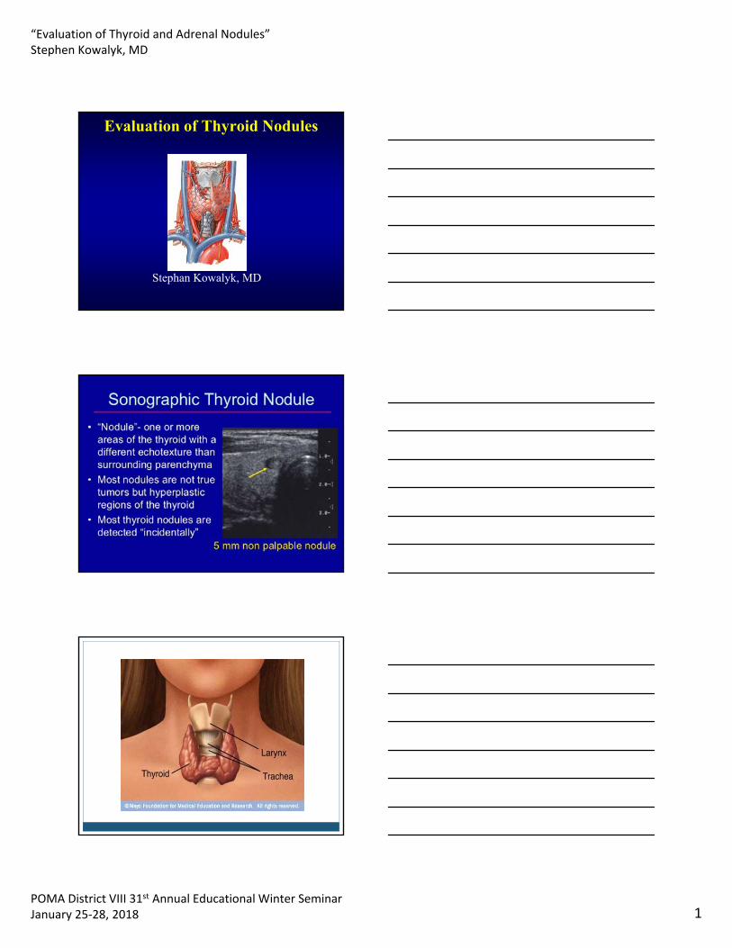
“Evaluation of Thyroid and Adrenal Nodules”Stephen Kowalyk, MD
POMA District VIII 31st Annual Educational Winter SeminarJanuary 25‐28, 2018 1
Evaluation of Thyroid Nodules
Stephan Kowalyk, MD

“Evaluation of Thyroid and Adrenal Nodules”Stephen Kowalyk, MD
POMA District VIII 31st Annual Educational Winter SeminarJanuary 25‐28, 2018 2
Primary goal
Exclude malignancy
Incidental thyroid nodules
• If found on CT, MRI, PET scan, carotid Doppler
• ULTRASOUND!!

“Evaluation of Thyroid and Adrenal Nodules”Stephen Kowalyk, MD
POMA District VIII 31st Annual Educational Winter SeminarJanuary 25‐28, 2018 3
Sonographic monitoring without biopsy may be an acceptable alternative, but stability without biopsy does not entirely exclude malignancy
Risk Factors
– young patients (<20 years of age)
– older (>60 years of age) -higher risk, especially for more aggressive thyroid tumors
– history of head or neck radiation
-first degree relatives with thyroid cancer
-uptake on PET scanning
-calcitonin >100
-MEN II, Gardner’s Syndrome, Cowden’s disease.
Gender and Thyroid Nodules
• Gender – male -higher risk if nodule present
– females• have many more nodules
• less likely to be malignant.
• still have majority of thyroid cancers

“Evaluation of Thyroid and Adrenal Nodules”Stephen Kowalyk, MD
POMA District VIII 31st Annual Educational Winter SeminarJanuary 25‐28, 2018 4
Concerning Personal History
• Recent growth
• Soft tissue swelling
• Vocal changes(recurrent nerve involvement)
• Dysphagia
Thyroid Scans
• Purpose – Determine function of the gland and/or a
nodule within the gland
• Hot nodules - usually independently functioning nodules – Rarely malignant
• Cold nodules - either adenoma or maligancy– 15% chance of malignancy in adults.

“Evaluation of Thyroid and Adrenal Nodules”Stephen Kowalyk, MD
POMA District VIII 31st Annual Educational Winter SeminarJanuary 25‐28, 2018 5
Fine-Needle Aspiration• Best tool for determining pathology other
than surgical excision
• Can be as high as 80 % sensitive and 95% specific
HIGH RISK FEATURES
• microcalcifications
• hypoechoic
• increased vascularity
• infiltrative margins(‘irregular”)
• taller than wide on transverse view

“Evaluation of Thyroid and Adrenal Nodules”Stephen Kowalyk, MD
POMA District VIII 31st Annual Educational Winter SeminarJanuary 25‐28, 2018 6
Interpreting the Biopsy Report• What you get:
– benign (low probability)
– follicular lesion unknown significance
– suspicious (high probability)
– inadequate specimen
• What it means:– benign - 90-95% likelihood it is benign
– FLUS-need molecular markers
– suspicious- it’s malignant.
– inadequate specimen - do it again (and again)
Molecular markers
• Typically give % risk malignancy
• Useful to guide need for resection
What about those benign nodules?
• No specific treatment is needed.
• Thyroid suppression may shrink size of adenomasnot recommeded
• Not proven to be effective or necessary
• May hide malignancies - ? Periodic biopsies
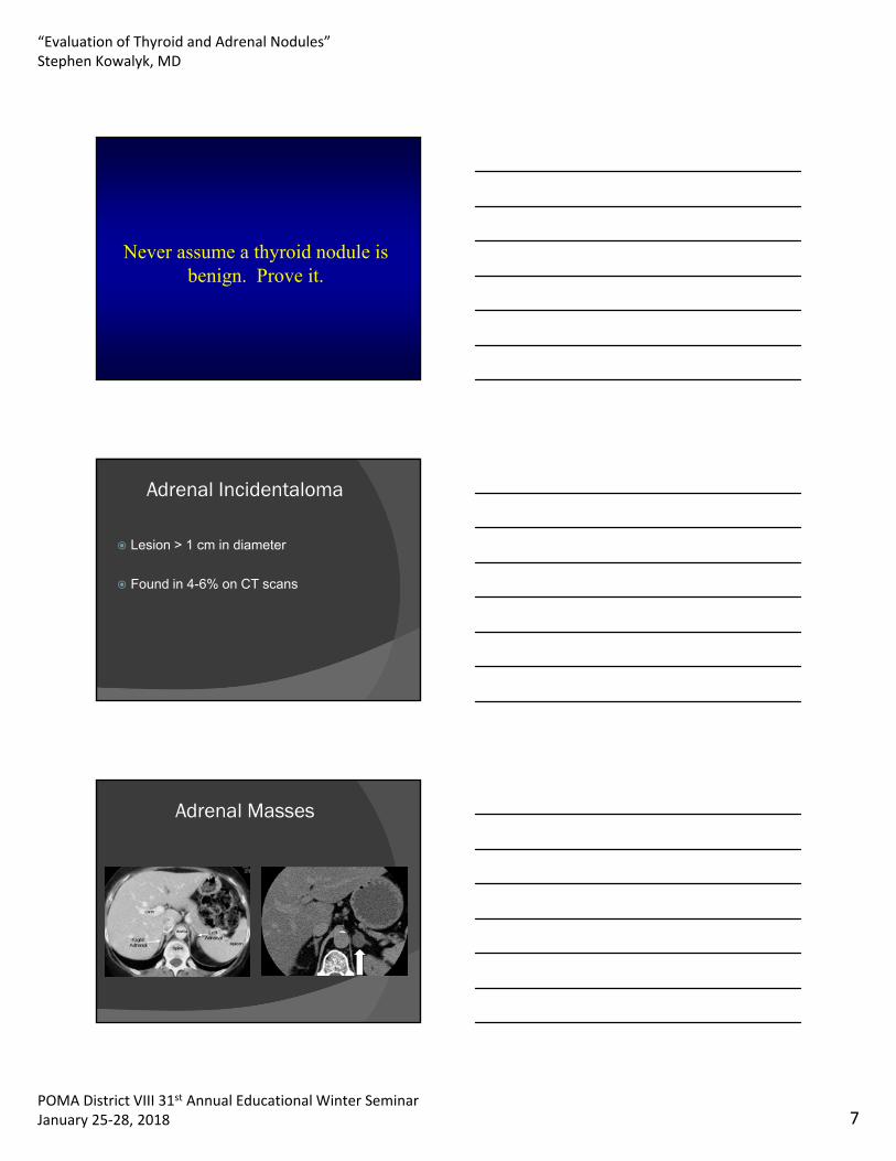
“Evaluation of Thyroid and Adrenal Nodules”Stephen Kowalyk, MD
POMA District VIII 31st Annual Educational Winter SeminarJanuary 25‐28, 2018 7
Never assume a thyroid nodule is benign. Prove it.
Adrenal Incidentaloma
Lesion > 1 cm in diameter
Found in 4-6% on CT scans
Adrenal Masses

“Evaluation of Thyroid and Adrenal Nodules”Stephen Kowalyk, MD
POMA District VIII 31st Annual Educational Winter SeminarJanuary 25‐28, 2018 8
Differential diagnosis
Functioning
Nonfunctioning
Malignancy
Benign features
Imaging –fat results in low attenuation on CT=benign
<10 Housefield units and rapid washout >50% in 10 minutes
Homogenous
Smooth borders
<4 cm
Malignancy
Adrenocortical carcinoma
Pheochromocytoma
Metastasis

“Evaluation of Thyroid and Adrenal Nodules”Stephen Kowalyk, MD
POMA District VIII 31st Annual Educational Winter SeminarJanuary 25‐28, 2018 9
Malignancy features
>4cm
Irregular shape
Inhomogeneous High attenuation > 20 Housefield units Delayed washout <50% in 10 minutes
Functioning tumors
Cortisol secreting adenoma
Aldosterone secreting adenoma
Adrenocortical carcinoma
Pheochromocytoma
Cushing’s syndrome
Overnight dexamethasone suppression test
11pm 1mg dexamethasone 8am blood cortisol normal <5

“Evaluation of Thyroid and Adrenal Nodules”Stephen Kowalyk, MD
POMA District VIII 31st Annual Educational Winter SeminarJanuary 25‐28, 2018 10
Pheochromocytoma
24 hour urine catecholamines and metanephrines
Plasma catecholamines/free metanephrines
Hyperaldosteronism
If patient hypertensive/hypokalemic
Plasma renin aldosterone ratio
Adrenocortical carcinoma
DHEAS not DHEA

“Evaluation of Thyroid and Adrenal Nodules”Stephen Kowalyk, MD
POMA District VIII 31st Annual Educational Winter SeminarJanuary 25‐28, 2018 11
Evaluation based on imaging
Adrenal Mass on CT Scan <1 cm in greatest diameter (especially if fatty or cystic consistency) Functional(laboratory evaluation)
Adrenal mass >4 cm
Laboratory evaluation
Biopsy unless clearly benign(cyst, myelolipoma)
Surgical consultation
Lipid rich adrenal mass 1-4cm
Laboratory evaluation
Repeat CT 12 months

“Evaluation of Thyroid and Adrenal Nodules”Stephen Kowalyk, MD
POMA District VIII 31st Annual Educational Winter SeminarJanuary 25‐28, 2018 12
Lipid poor adrenal mass1-4cm
Laboratory evaluation
Consider MRI
PET scan
Surgical consultation
Follow-Up
If functional studies are normal and no high risk imaging characteristics Repeat imaging at 6-12 months
Surgery if grows >1cm
Repeat adrenal screening annually for 4 years
If concern for malignant potential based on imaging the biopsy or excision