Evaluation of the Inpatient With Anemia Fina - Handout.ppt of the Inpatient...Anemia of Inflammation...
27
1 Camila Masias, M.D. Assistant Professor Division of Hematology The Ohio State University Wexner Medical Center Evaluation of the Inpatient with Anemia Case 1 Case 1 • 22 yo woman presents to the ED with fatigue and sob. • Afebrile, HR 100 BP 100/60 • 10 week pregnant • WBC 14, Hb 7, Plt count 372
Transcript of Evaluation of the Inpatient With Anemia Fina - Handout.ppt of the Inpatient...Anemia of Inflammation...
1
Camila Masias, M.D.Assistant Professor
Division of HematologyThe Ohio State University Wexner Medical Center
Evaluation of the Inpatient with Anemia
Case 1 Case 1
• 22 yo woman presents to the ED with fatigue and sob.
• Afebrile, HR 100 BP 100/60
• 10 week pregnant
• WBC 14, Hb 7, Plt count 372

2
More details… More details…
• Hb 7 g/dL
• HCT 26.8
• MCV 64.1
• MCH 17.2
• MCHC 26.8
• RDW 19.2
• Retic count 4.89
Hemoglobin Oxygen carrying molecule >13.5 g/dL in men, > 12 g/dL in women
Hematocrit Packed cell volume >41% in men, >36% in women
Mean CorpuscularVolume (MCV)
Average size of the patient’s RBC
80-95 fL
Mean Corpuscular Hb(MCH)
Average Hb content per RBC
25-32 pg
Mean Corpuscular HbConcentration (MCHC)
Average [Hb] per RBC 32- 35 g/dL
Red cell distribution width (RDW)
Measure of RBC size variation (anisocytosis)
11-14 %
Reticulocyte count % of RBC 0.8-1.5%
Absolute retic count Relative reticulocyte count x RBC count
Normal 50,000–75,000/µl
3
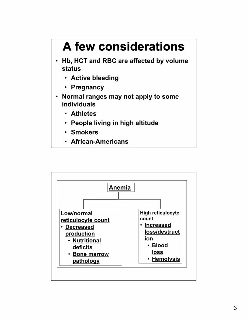
A few considerationsA few considerations• Hb, HCT and RBC are affected by volume
status
• Active bleeding
• Pregnancy
• Normal ranges may not apply to some individuals
• Athletes
• People living in high altitude
• Smokers
• African-Americans
Anemia
Low/normal reticulocyte count• Decreased
production• Nutritional
deficits• Bone marrow
pathology
High reticulocyte count• Increased
loss/destruction• Blood
loss • Hemolysis
4
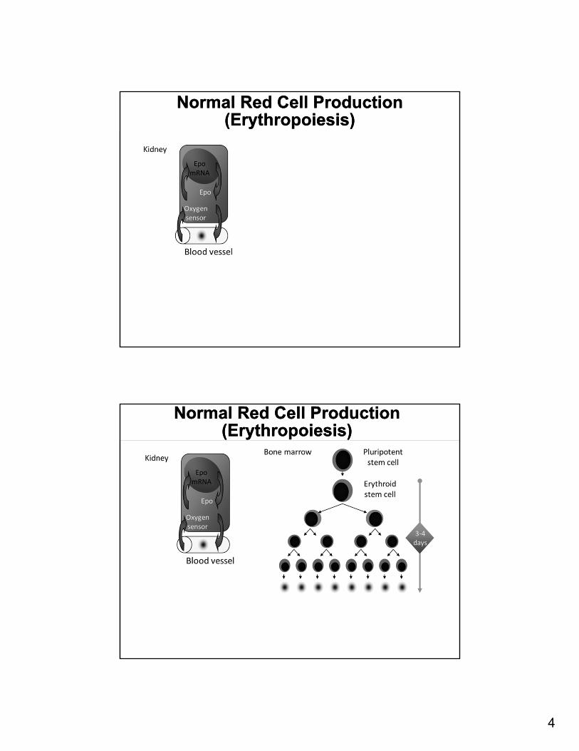
Normal Red Cell Production (Erythropoiesis)
Normal Red Cell Production (Erythropoiesis)
Normal Red Cell Production (Erythropoiesis)
Normal Red Cell Production (Erythropoiesis)
5
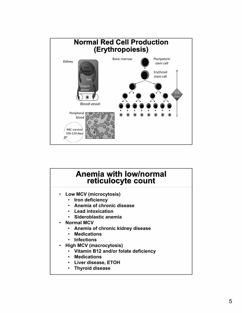
Normal Red Cell Production (Erythropoiesis)
Normal Red Cell Production (Erythropoiesis)
Anemia with low/normal reticulocyte count
Anemia with low/normal reticulocyte count
• Low MCV (microcytosis)• Iron deficiency• Anemia of chronic disease• Lead intoxication• Sideroblastic anemia
• Normal MCV• Anemia of chronic kidney disease• Medications• Infections
• High MCV (macrocytosis)• Vitamin B12 and/or folate deficiency• Medications• Liver disease, ETOH• Thyroid disease
6
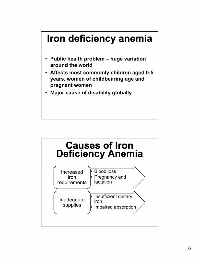
Iron deficiency anemia Iron deficiency anemia
• Public health problem – huge variation around the world
• Affects most commonly children aged 0-5 years, women of childbearing age and pregnant women
• Major cause of disability globally
Causes of Iron Deficiency Anemia
Causes of Iron Deficiency Anemia
• Blood loss• Pregnancy and
lactation
Increased iron
requirements
• Insufficient dietary iron
• Impaired absorption
Inadequate supplies

7
Iron IntakeIron Intake• Recommended dietary iron:
• Ages 9-13: 8 mg• Ages 14 -18: 11 mg for boys and 15 mg for
girls • > 19: 8 mg for men, 18 mg for women
(until age 50)• Pregnant women: 30 mg
• Main source of iron intake is meat (especially red meat)
Iron metabolismIron metabolism
• Duodenum absorption: ~1-2 mg a day• Iron loss (sloughed mucosal cells, menstruation,
other blood loss): ~1-2 mg a day • Total body iron storage: 3000-4000 mg
• Hb: 2g• Iron containing proteins: 400 mg • Iron in plasma bound to transferrin: 3 -7 mg• Storage iron (ferritin or hemosiderin): 0.5 g in
women and 1 g in men

8
Diagnosis: Symptoms Diagnosis: Symptoms • Pica (25%): Compulsive ingestion of a
non-food substance such as starch, clay, ground, ice
• Beeturia (49-80%): urine turns red after ingestion of beets
• Restless legs syndrome (10%): Urge to move the legs usually accompanied by uncomfortable sensations that begins or worsens during periods of rest and relieved by movement. Worse in the evening/night
Diagnosis: Laboratory Diagnosis: Laboratory Test IDA ACD
Iron Measures circulating iron bound to transferrin. NOT a marker of iron status (will change with even just one meal)
↓ ↓
Transferrin (TIBC)
Circulating transport protein for iron ↑ ↓
Transferrin saturation
Serum iron ÷ TIBC x 100 ↓↓ ↓
Ferritin Circulating iron storage protein, acute phase reactant. Best value for iron deficiency
↓ ↑
9
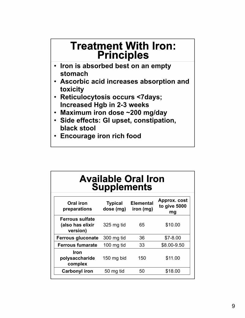
Treatment With Iron: Principles
Treatment With Iron: Principles
• Iron is absorbed best on an empty stomach
• Ascorbic acid increases absorption and toxicity
• Reticulocytosis occurs <7days; Increased Hgb in 2-3 weeks
• Maximum iron dose ~200 mg/day• Side effects: GI upset, constipation,
black stool• Encourage iron rich food
Available Oral Iron Supplements
Available Oral Iron Supplements
Oral iron preparations
Typical dose (mg)
Elementaliron (mg)
Approx. cost to give 5000
mg
Ferrous sulfate (also has elixir
version)325 mg tid 65 $10.00
Ferrous gluconate 300 mg tid 36 $7-8.00
Ferrous fumarate 100 mg tid 33 $8.00-9.50
Iron polysaccharide
complex150 mg bid 150 $11.00
Carbonyl iron 50 mg tid 50 $18.00
10
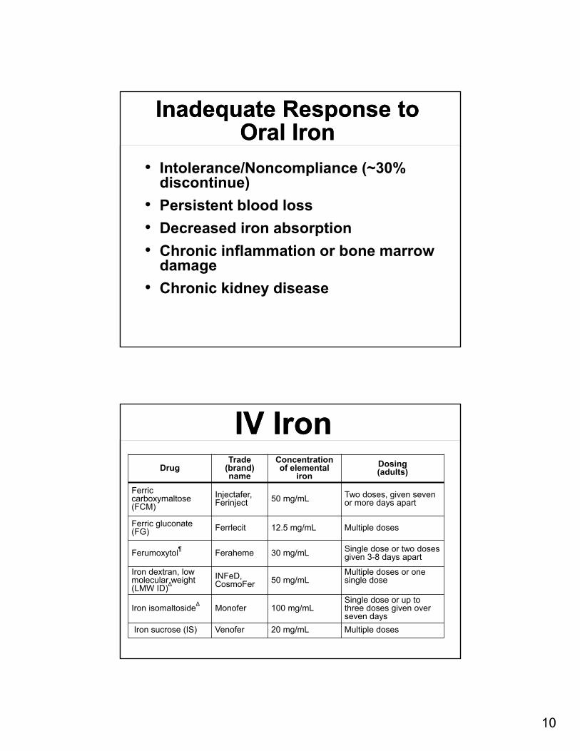
Inadequate Response to Oral Iron
Inadequate Response to Oral Iron
• Intolerance/Noncompliance (~30% discontinue)
• Persistent blood loss
• Decreased iron absorption
• Chronic inflammation or bone marrow damage
• Chronic kidney disease
IV Iron IV Iron Drug
Trade (brand) name
Concentration of elemental
iron
Dosing(adults)
Ferric carboxymaltose(FCM)
Injectafer, Ferinject 50 mg/mL Two doses, given seven
or more days apart
Ferric gluconate (FG) Ferrlecit 12.5 mg/mL Multiple doses
Ferumoxytol
Feraheme 30 mg/mL Single dose or two doses given 3-8 days apart
Iron dextran, low molecular weight (LMW ID)
∆INFeD, CosmoFer 50 mg/mL
Multiple doses or one single dose
Iron isomaltoside∆
Monofer 100 mg/mLSingle dose or up to three doses given over seven days
Iron sucrose (IS) Venofer 20 mg/mL Multiple doses

11
Anemia of Inflammation
Anemia of Inflammation
• Decreased RBC production + decreased RBC survival
• Reduced iron absorption in the GI tract and trapping of iron in macrophages
• Relative decrease in EPO production
• Decrease bone marrow response to EPO mediated by inflammatory cytokines
Treatment Options for Anemia of Chronic Disease
Treatment Options for Anemia of Chronic Disease
• Treat the underlying diseases• RBC Transfusions• For anemia of chronic kidney
disease: • Erythroid-stimulating agents
(ESA) and potentially iron supplementation (ferritin <100 and/or iron sat <20%)
12
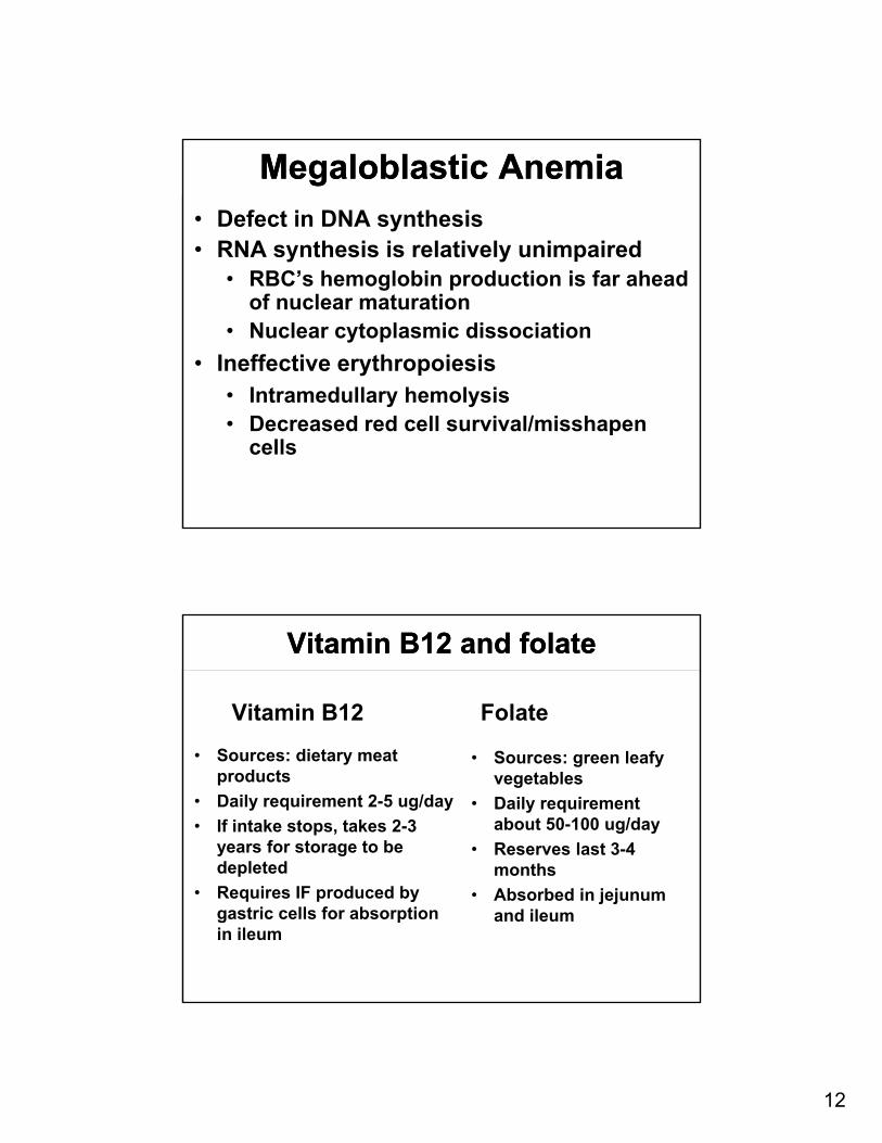
Megaloblastic AnemiaMegaloblastic Anemia• Defect in DNA synthesis• RNA synthesis is relatively unimpaired
• RBC’s hemoglobin production is far ahead of nuclear maturation
• Nuclear cytoplasmic dissociation
• Ineffective erythropoiesis• Intramedullary hemolysis• Decreased red cell survival/misshapen
cells
Vitamin B12 and folateVitamin B12 and folate
Vitamin B12
• Sources: dietary meat products
• Daily requirement 2-5 ug/day
• If intake stops, takes 2-3 years for storage to be depleted
• Requires IF produced by gastric cells for absorption in ileum
Folate
• Sources: green leafy vegetables
• Daily requirement about 50-100 ug/day
• Reserves last 3-4 months
• Absorbed in jejunum and ileum

13
Signs/Symptoms of B12 Deficiency
Signs/Symptoms of B12 Deficiency
• Anemia, hypersegmented neutrophils• “Beefy Red” tongue, smooth surface of the
tongue• Neurologic
• demyelination of the posterior and lateral columns of the spinal cord
• paresthesia, loss of position/vibratory sense
• in advanced disease, neuropathy, muscle weakness, and even CNS symptoms (irritability, somnolence, psychosis)
Diagnosis and Treatment Diagnosis and Treatment • Check MMA with borderline levels of Vit
B12 and treat if elevated
• Folate deficiency: 1-5mg daily of oral folic acid
• Vit B12 deficiency:
• IV: 1000 mcg once per week until the deficiency is corrected and then once per month
• Oral: 1000 to 2000 mcg daily

14
Miscellaneous: Bone marrow process
Miscellaneous: Bone marrow process
• Broad DDx, including• Acute or chronic leukemia• Myelodysplastic syndrome (MDS)• Myeloproliferative diseases (MPD)• Involvement of malignancies in the bone marrow• Disseminated infections in the bone marrow
• Patients usually have more symptoms such as unexplained weight loss, petechiae, fever, hepatosplenomegaly, etc
• More than one cell line is abnormal and could be severe
• Referral to hematology and bone marrow biopsy is needed for definitive diagnosis
http://www.pathpedia.com/education/eatlas/histopathology/bone_marrow/acute_lymphoblastic_leukemia,_b-cell_(b-all).aspx
Case 2 Case 2 • 43 yo man with no remarkable PMhx that
presents to the ED with fatigue
• PE: hepato/splenomegaly and jaundice
• Hb 7, WBC 5, plt count 200K
• MCV 100
• Retic count 5%
15
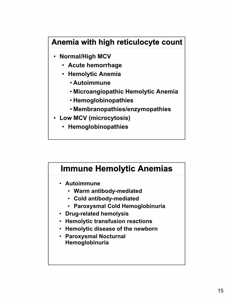
Anemia with high reticulocyte countAnemia with high reticulocyte count
• Normal/High MCV
• Acute hemorrhage
• Hemolytic Anemia
• Autoimmune
• Microangiopathic Hemolytic Anemia
• Hemoglobinopathies
• Membranopathies/enzymopathies
• Low MCV (microcytosis)
• Hemoglobinopathies
Immune Hemolytic AnemiasImmune Hemolytic Anemias
• Autoimmune• Warm antibody-mediated• Cold antibody-mediated• Paroxysmal Cold Hemoglobinuria
• Drug-related hemolysis • Hemolytic transfusion reactions• Hemolytic disease of the newborn• Paroxysmal Nocturnal
Hemoglobinuria

16
Auto-Immune Hemolytic AnemiasAuto-Immune Hemolytic Anemias
• Antibodies causing hemolysis can be broken down into 2 general categories: warm and cold
• Warm antibodies react with RBCs best at 37°and typically do not agglutinate red cells
• Cold antibodies typically react best at <32° and do cause RBC agglutination

17
Warm-Antibody Hemolytic AnemiasEtiology
Warm-Antibody Hemolytic AnemiasEtiology
• Primary or Secondary
• Drugs
• Solid or hematologic malignancy
• Infection
• Collagen Disease
• Pregnancy
• Can be associated with immune
platelet destruction = Evan’s syndrome
Warm-Antibody Hemolytic AnemiasClinical Features
Warm-Antibody Hemolytic AnemiasClinical Features
• Splenomegaly, jaundice is usually present
• Depending on degree of anemia and rate of fall in hemoglobin, patients can have VERY symptomatic anemia
• Lab Dx -
• reticulocytes, bili, LDH, ↓haptoglobin
• Positive Coomb’s test - both direct and indirect
• Spherocytes are seen on the peripheral smear
18
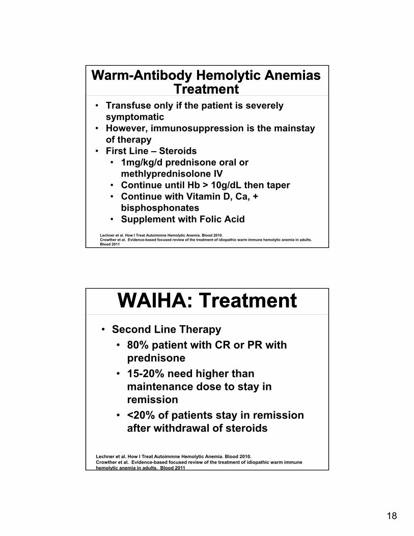
Warm-Antibody Hemolytic AnemiasTreatment
Warm-Antibody Hemolytic AnemiasTreatment
• Transfuse only if the patient is severely symptomatic
• However, immunosuppression is the mainstay of therapy
• First Line – Steroids• 1mg/kg/d prednisone oral or
methlyprednisolone IV• Continue until Hb > 10g/dL then taper• Continue with Vitamin D, Ca, +
bisphosphonates• Supplement with Folic Acid
Lechner et al. How I Treat Autoimmne Hemolytic Anemia. Blood 2010.Crowther et al. Evidence-based focused review of the treatment of idiopathic warm immune hemolytic anemia in adults. Blood 2011
WAIHA: TreatmentWAIHA: Treatment• Second Line Therapy
• 80% patient with CR or PR with prednisone
• 15-20% need higher than maintenance dose to stay in remission
• <20% of patients stay in remission after withdrawal of steroids
Lechner et al. How I Treat Autoimmne Hemolytic Anemia. Blood 2010.Crowther et al. Evidence-based focused review of the treatment of idiopathic warm immune hemolytic anemia in adults. Blood 2011

19
WAIHA: TreatmentWAIHA: Treatment• 2nd Line Therapy
• Splenectomy
• Rituxan
• Other Therapies
• Danazol
• Cyclophosphamide
• Mycophenolate Mofetil
• Cyclosporine
• Vincristine
• Alemtuzumab
• Ofatumumab
• Ineffective therapies
• Azathioprine
• BMT
• IVIG
• Plasma Exchange
Lechner et al. How I Treat Autoimmne Hemolytic Anemia. Blood 2010.Crowther et al. Evidence-based focused review of the treatment of idiopathic warm immune hemolytic anemia in adults. Blood 2011
Drug-Induced Immune HemolysisThree general mechanisms
Drug-Induced Immune HemolysisThree general mechanisms
• Innocent bystander
• Quinine, Quinidine, Isoniazide
• Hapten
• Penicillins, Cephalosporins
• True autoimmune
• Alpha-methyldopa, L-DOPA, Procainamide
20
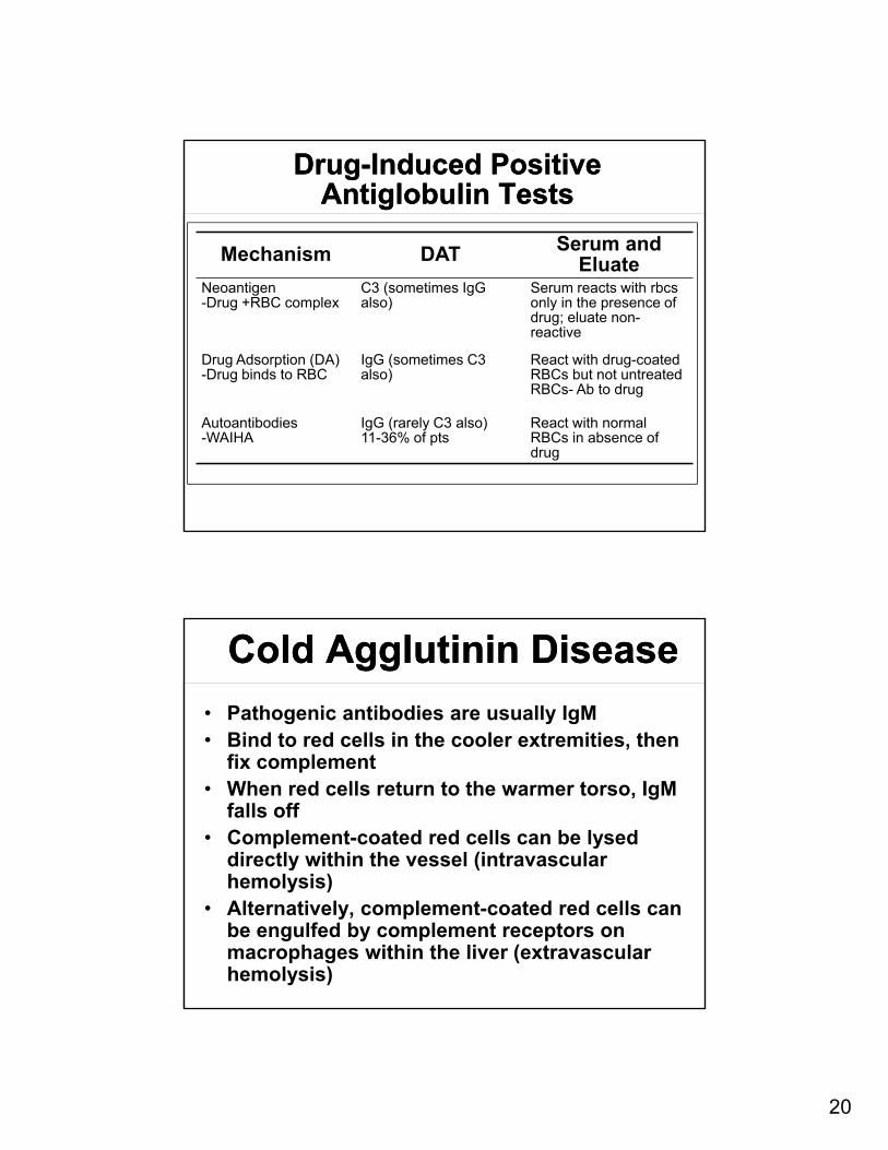
Drug-Induced Positive Antiglobulin Tests
Drug-Induced Positive Antiglobulin Tests
Mechanism DAT Serum and Eluate
Neoantigen-Drug +RBC complex
C3 (sometimes IgGalso)
Serum reacts with rbcsonly in the presence of drug; eluate non-reactive
Drug Adsorption (DA)-Drug binds to RBC
IgG (sometimes C3 also)
React with drug-coated RBCs but not untreated RBCs- Ab to drug
Autoantibodies-WAIHA
IgG (rarely C3 also)11-36% of pts
React with normal RBCs in absence of drug
Cold Agglutinin DiseaseCold Agglutinin Disease
• Pathogenic antibodies are usually IgM• Bind to red cells in the cooler extremities, then
fix complement• When red cells return to the warmer torso, IgM
falls off• Complement-coated red cells can be lysed
directly within the vessel (intravascular hemolysis)
• Alternatively, complement-coated red cells can be engulfed by complement receptors on macrophages within the liver (extravascular hemolysis)
21
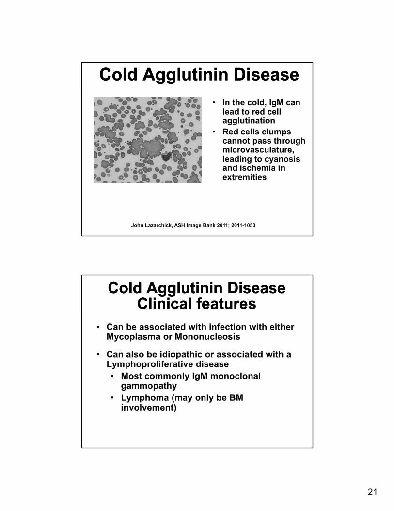
Cold Agglutinin DiseaseCold Agglutinin Disease
• In the cold, IgM can lead to red cell agglutination
• Red cells clumps cannot pass through microvasculature, leading to cyanosis and ischemia in extremities
John Lazarchick, ASH Image Bank 2011; 2011-1053
Cold Agglutinin DiseaseClinical features
Cold Agglutinin DiseaseClinical features
• Can be associated with infection with either Mycoplasma or Mononucleosis
• Can also be idiopathic or associated with a Lymphoproliferative disease• Most commonly IgM monoclonal
gammopathy• Lymphoma (may only be BM
involvement)
22
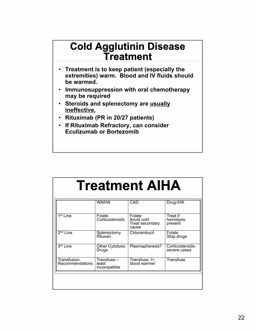
Cold Agglutinin DiseaseTreatment
Cold Agglutinin DiseaseTreatment
• Treatment is to keep patient (especially the extremities) warm. Blood and IV fluids should be warmed.
• Immunosuppression with oral chemotherapy may be required
• Steroids and splenectomy are usually ineffective.
• Rituximab (PR in 20/27 patients)• If Rituximab Refractory, can consider
Eculizumab or Bortezomib
Treatment AIHATreatment AIHAWAIHA CAD Drug-IHA
1st Line FolateCorticosteroids
FolateAvoid coldTreat secondary cause
Treat if hemolysispresent
2nd Line SplenectomyRituxan
Chlorambucil FolateStop drugs
3rd Line Other CytotoxicDrugs
Plasmapheresis? Corticosteroids-severe cases
TransfusionRecommendations
Transfuse –least incompatible
Transfuse- I+, blood warmer
Transfuse

23
Microangiopathic Hemolytic Anemia
Microangiopathic Hemolytic Anemia
• Non-immune hemolytic anemia
• reticulocytes, bili, LDH, ↓haptoglobin
• NEGATIVE Coomb’s
• Prosthetic Valves, Heart valve induced, Pregnancy Associated Syndrome, HTN, Infections, Immune D/os, DIC
• Thrombotic Microangiopathy
• TTP, aHUS, HUS, Drug-Induced TMA
Structural abnormalities of Hb
Structural abnormalities of Hb
• Thalassemia
• Sickle Cell disease
• G6PD deficiency
• Hereditary Spherocytosis
24
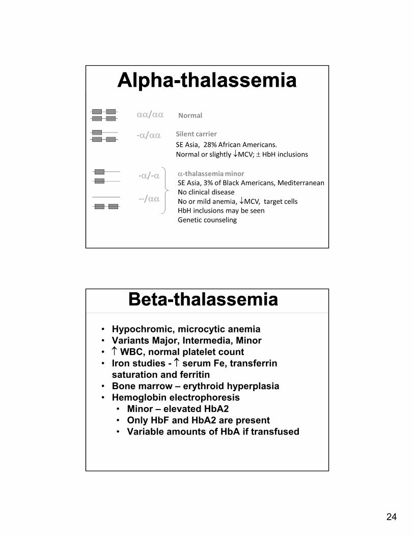
Alpha-thalassemiaAlpha-thalassemia
Beta-thalassemiaBeta-thalassemia
• Hypochromic, microcytic anemia• Variants Major, Intermedia, Minor• WBC, normal platelet count • Iron studies - serum Fe, transferrin
saturation and ferritin• Bone marrow – erythroid hyperplasia• Hemoglobin electrophoresis
• Minor – elevated HbA2• Only HbF and HbA2 are present• Variable amounts of HbA if transfused

25
Name Genotype Percent
Homozygous SS(Sickle Cell Anemia) S-S 65
Heterozygous SC S- C 24
Heterozygous S-+ thal S- + thal 7
Heterozygous S-° thal S- ° thal 3
The Common Variants of Sickle Cell Disease
The Common Variants of Sickle Cell Disease
Sickle Cell Anemia Pathophysiology
Sickle Cell Anemia Pathophysiology
• Manifestations of SCD are driven by:
• Vaso-occlusion with ischemia-reperfusion injury
• Hemolytic anemia
• Endothelial Activation
Owusu-Ansah 2015

26
Complications of SCDComplications of SCD
Konotey-Ahulu FID. The Sickle Cell Disease. Clinical Manifestations Including the “Sickle Crisis”. Arch Intern Med. 1974;133(4):611-619.
Sepsis
Multiorgan Failure
Myocardial Infarction
Priapism
Transfusion Reaction
Evaluation of inpatient with anemia
Evaluation of inpatient with anemia
Patient history: acuity, prior
medical history, family history and
associated symptoms
Physical exam: hepato/splenome
galy, LNP, associated infection
Lab: MCV, MCH, retic count,
peripheral smear
27
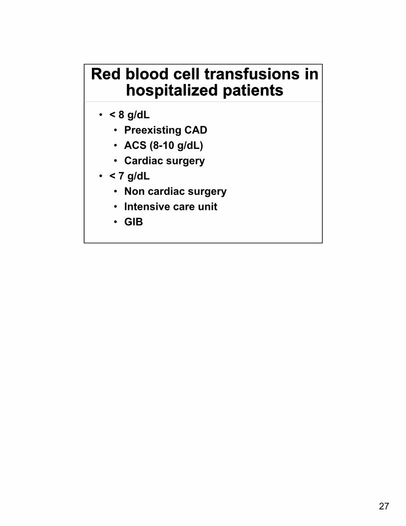
Red blood cell transfusions in hospitalized patients
Red blood cell transfusions in hospitalized patients
• < 8 g/dL
• Preexisting CAD
• ACS (8-10 g/dL)
• Cardiac surgery
• < 7 g/dL
• Non cardiac surgery
• Intensive care unit
• GIB


![Lecture – 3 Dr. Zahoor Ali Shaikh 1. What is Anemia? Anemia means - Decreased hemoglobin - Decreased RBC count - Decreased Hematocrit [PCV] Therefore,](https://static.fdocuments.us/doc/165x107/56649c9e5503460f9495e870/lecture-3-dr-zahoor-ali-shaikh-1-what-is-anemia-anemia-means-decreased.jpg)