Evaluation of Some Traditional Antidiabetic Plants on ...
9
Medicine Journal 2015; 2(5): 79-87 Published online October 9, 2015 (http://www.openscienceonline.com/journal/med) Evaluation of Some Traditional Antidiabetic Plants on Testis in the Alloxan-Induced Diabetic Male Albino Rats: Histological, Histochemical, Immunohistochemical and Morphometric Study Tamer M. M. Abu-Amara 1, * , Abd EL Razek A. Meselhy 2 , Howada I. Abdel Aziz 3 , Amal Said 4 1 Histology and Cytology Department, Faculty of Medicine, Al-Azhar University, Cairo, Egypt 2 Anatomy and Embryology Department, Faculty of Medicine, Al-Azhar University, Assiut, Egypt 3 Histology and Cytology Department, Faculty of Medicine, Suze-Canal University, Ismailia, Egypt 4 Zoology Department, Faculty of Science (Girls), Al-Azhar University, Cairo, Egypt Email address [email protected] (T. M. M. Abu-Amara) To cite this article Tamer M. M. Abu-Amara, Abd EL Razek A. Meselhy, Howada I. Abdel Aziz, Amal Said. Evaluation of Some Traditional Antidiabetic Plants on Testis in the Alloxan-Induced Diabetic Male Albino Rats: Histological, Histochemical, Immunohistochemical and Morphometric Study. Medicine Journal. Vol. 2, No. 5, 2015, pp. 79-87. Abstract Background: Diabetes mellitus is one of the common and widely distributed metabolic diseases all over the world. This disease is characterized by hyperglycemia that results from defects in insulin secretion, insulin action or both. Previous studies reported negative complications of diabetes on the male genital system with associated gonadal dysfunction. Different medicinal plant species were used as a traditional treatment for diabetes mellitus long times ago. Aim of the work: As diabetes had been reported to affect the male genital system, this work was aimed to investigate the antidiabetic effects of the aqueous extract of the following herbs: Ambrosia maritima, L. (Damsissa), Chrozophoratinctoria, L. (Sammo), Artemisia annua, L. (Kaysom) and Artemisia judaica, L. (Sheeh) on the testis’ histology in the alloxan-induced diabetic male albino rats. Material and Methods: This study was performed on sixty male albino rats with an average 100-110 g body weight. The animals were divided into six groups (10/cage); Group1 (Control untreated-group), Group 2 (Alloxan-induced diabetic group), Group 3 (diabetic group treated orally with “28.5 mg/kg body wt. twice/day” of the Damsissa extract), Group 4 (diabetic group treated orally with “28.5 mg/kg body wt. twice/day” of the Sammo extract), Group 5(diabetic group treated orally with “28.5 mg/kg body wt. twice/day” of the Kaysom extract)and Group 6 (diabetic group treated orally with “28.5 mg/kg body wt. twice/day” of the Sheeh extract). Results: Considerable improvements in the testicular tissue morphological changes that were observed in diabetic groups had been detected after treatment with Damsissa and Sammo in comparison to the control group. These improvements were less obvious after Kaysom and Sheeh treatment. Conclusion: It could be concluded that Damsissa and Sammo can guard against the negative effects of diabetes on the testis. Keywords Diabetes Mellitus, Alloxan, Hyperglycemia, Damsissa, Sammo, Kaysom, Sheeh 1. Introduction Diabetes mellitus is a serious metabolic disorder with numerous complications [1]. The increase of the blood glucose levels leads to many structural and functional changes in various tissues and organs [2] Experimentally, the induction of diabetes in male rats had been found to be associated with altered functions of the reproductive system [3]. Induction of diabetes in rats was used as an in vivo model for studying the effects of diabetes on the various organs [4, 5]. Induction of diabetes affects the testicular functions due to the lack of insulin and subsequently the impairment of insulin regulatory action on the Leydig and Sertoli cells [6] This gonadal dysfunction and the subsequent decrease in the testosterone production lead to insufficiency in spermatozoids production [7, 8, 9]. Moreover, several previous studies reported change in the reproductive system
Transcript of Evaluation of Some Traditional Antidiabetic Plants on ...
Medicine Journal 2015; 2(5): 79-87
Published online October 9, 2015 (http://www.openscienceonline.com/journal/med)
Evaluation of Some Traditional Antidiabetic Plants on Testis in the Alloxan-Induced Diabetic Male Albino Rats: Histological, Histochemical, Immunohistochemical and Morphometric Study
Tamer M. M. Abu-Amara1, *
, Abd EL Razek A. Meselhy2, Howada I. Abdel Aziz
3, Amal Said
4
1Histology and Cytology Department, Faculty of Medicine, Al-Azhar University, Cairo, Egypt 2Anatomy and Embryology Department, Faculty of Medicine, Al-Azhar University, Assiut, Egypt 3Histology and Cytology Department, Faculty of Medicine, Suze-Canal University, Ismailia, Egypt 4Zoology Department, Faculty of Science (Girls), Al-Azhar University, Cairo, Egypt
Email address
[email protected] (T. M. M. Abu-Amara)
To cite this article Tamer M. M. Abu-Amara, Abd EL Razek A. Meselhy, Howada I. Abdel Aziz, Amal Said. Evaluation of Some Traditional Antidiabetic
Plants on Testis in the Alloxan-Induced Diabetic Male Albino Rats: Histological, Histochemical, Immunohistochemical and Morphometric
Study. Medicine Journal. Vol. 2, No. 5, 2015, pp. 79-87.
Abstract
Background: Diabetes mellitus is one of the common and widely distributed metabolic diseases all over the world. This disease
is characterized by hyperglycemia that results from defects in insulin secretion, insulin action or both. Previous studies
reported negative complications of diabetes on the male genital system with associated gonadal dysfunction. Different
medicinal plant species were used as a traditional treatment for diabetes mellitus long times ago. Aim of the work: As diabetes
had been reported to affect the male genital system, this work was aimed to investigate the antidiabetic effects of the aqueous
extract of the following herbs: Ambrosia maritima, L. (Damsissa), Chrozophoratinctoria, L. (Sammo), Artemisia annua, L.
(Kaysom) and Artemisia judaica, L. (Sheeh) on the testis’ histology in the alloxan-induced diabetic male albino rats. Material
and Methods: This study was performed on sixty male albino rats with an average 100-110 g body weight. The animals were
divided into six groups (10/cage); Group1 (Control untreated-group), Group 2 (Alloxan-induced diabetic group), Group 3
(diabetic group treated orally with “28.5 mg/kg body wt. twice/day” of the Damsissa extract), Group 4 (diabetic group treated
orally with “28.5 mg/kg body wt. twice/day” of the Sammo extract), Group 5(diabetic group treated orally with “28.5 mg/kg
body wt. twice/day” of the Kaysom extract)and Group 6 (diabetic group treated orally with “28.5 mg/kg body wt. twice/day”
of the Sheeh extract). Results: Considerable improvements in the testicular tissue morphological changes that were observed in
diabetic groups had been detected after treatment with Damsissa and Sammo in comparison to the control group. These
improvements were less obvious after Kaysom and Sheeh treatment. Conclusion: It could be concluded that Damsissa and
Sammo can guard against the negative effects of diabetes on the testis.
Keywords
Diabetes Mellitus, Alloxan, Hyperglycemia, Damsissa, Sammo, Kaysom, Sheeh
1. Introduction
Diabetes mellitus is a serious metabolic disorder with
numerous complications [1]. The increase of the blood
glucose levels leads to many structural and functional
changes in various tissues and organs [2] Experimentally, the
induction of diabetes in male rats had been found to be
associated with altered functions of the reproductive system
[3]. Induction of diabetes in rats was used as an in vivo model
for studying the effects of diabetes on the various organs [4,
5]. Induction of diabetes affects the testicular functions due
to the lack of insulin and subsequently the impairment of
insulin regulatory action on the Leydig and Sertoli cells [6]
This gonadal dysfunction and the subsequent decrease in the
testosterone production lead to insufficiency in
spermatozoids production [7, 8, 9]. Moreover, several
previous studies reported change in the reproductive system

Medicine Journal 2015; 2(5): 79-87 80
structure in diabetic cases [2, 10, 11]. Also, the effect of
diabetes on the changes of the body and testicular weights
have been reported in several studies. For instance, in
diabetic rats, testicular weight was decreased around 20% in
comparison to the healthy rats [12]. A wide variety of
Egyptian folk medicinal plants are used in the treatment of
diabetes [13]. For instance, Damsissa was commonly used in
the treatment of rheumatic pains, asthma, bilharziasis,
diabetes, stomach, and renal troubles [14]. Moreover, several
effects such as anti-inflammatory, anti-oxidative effects, anti-
hyperlipidemia and antihypertensive effects have been
reported for Kaysom treatment [15, 16, 17]. In this study,
was aimed to investigate the antidiabetic effects of the
aqueous extract of the following herbs: Ambrosia maritima,
L. (Damsissa), Chrozophoratinctoria, L. (Sammo), Artemisia
annua, L. (Kaysom) and Artemisia judaica, L. (Sheeh) on the
testis’ histology in the alloxan-induced diabetic male albino
rats.
2. Material and Methods
2.1. Plant Material
The aerial parts of Ambrosia maritima, L. (Damsissa),
Chrozophoratinctoria, L. (Sammo), Artemisia annua, L.
(Kaysom) and Artemisia judaica, L. (Sheeh) were collected
from El-Arbaeen valley, Saint Catherine, Wadi Gebal, South
Sinai, Egypt. The plants were grinded and the aqueous
extracts of them were prepared by boiling 2 g of each of them
with 200ml of tap water for 15 min, left to cool at room
temperature then filtered through filter paper. Later, the
extracts were stored in glass containers in refrigerator. Fresh
extract preparations were done every two days.
2.2. Animals
Sixty male adult albino rats (8-10 weeks/ 100-110 g) were
used in this experiment. The rats were kept under observation
for about 2 weeks before the start of the experiment for
adaptation. Diabetes mellitus was induced in animals by
single dose of alloxan (120 mg/kg B. W. dissolved in saline)
injected intraperitoneally to induce diabetes mellitus in rats18
.
The rats were deprived of food for 16 hours before alloxan
injection. After three days of alloxan injection, the rats were
deprived of food overnight and they were then given glucose
(3g/kg B. W.) by gastric intubation. After 2 hours of oral
glucose administration, blood samples were taken from tail
vein and the fasting blood glucose (FBG) concentration was
determined by means of one touch ultra-glucometer (Johnson
& Johnson Company, USA) and compatible blood glucose
strips. After 2 h of oral glucose administration, the rats’
glucose concentrations “ranging from 180 to 300 mg/dl”
were considered as mild diabetic animals and included in the
experiment.
2.3. Experimental Design
Experimental animals were divided into six groups, ten
each, as follows:
• Group 1 (Control group): Non-diabetic rats.
• Group 2 (Diabetic group): Rats were injected
intraperitoneally with a single dose of alloxan (120
mg/kg dissolved in saline solution).
• Group 3 (Damsissa group): Diabetic rats treated orally
with Ambrosia maritime extract (28.5 mg/kg twice /day)
for 30 days.
• Group 4 (Sammo group): Diabetic rats treated orally
with Chrozophoratinctoria extract (28.5 mg/kg twice
/day) for 30 days.
• Group 5 (Kaysoom group): Diabetic rats treated orally
with Artemisia annua extract (28.5 mg/kg twice /day)
for 30 days.
• Group 6 (Sheeh group): Diabetic rats treated orally with
Artemisia judaica extract (28.5 mg/kg twice /day) for
30 days.
Histological and Histochemical studies: Therats from the
control and treated groups were sacrificed after one month
and small pieces of the testis were taken for the histological
and histochemical studies. The specimens were prepared via
fixation in 10% neutral buffered formalin solution and
Carnoy’s fluid. For histological study, paraffin sections were
stained with Harris’s haematoxylin and eosin (H&E)
19. For
detection of collagen fibers, paraffin sections were stained by
using Mallory’s trichrome stain19
. For histochemical study,
Paraffin sections of 5µm thickness were prepared and stained
with Feulgen stain19
. Later, the stained sections were
examined via light microscope, photographed and all the
detected variations between the three groups on the level of
the microscopic findings had been scientifically discussed.
Immunohistochemical study: Sections of testes were
deparaffinized with xylene, followed by antigen retrieval by
heating in citrate buffer (10 mM, 20 min). This was followed
by endogenous peroxidase blocking in 3% H2O2for 10 min
and incubation with anti-caspase–3 (1:100; Abcam, Ab4051).
After washing the slides with phosphate buffered saline, the
sections were incubated with the related secondary antibodies
at room temperature for 1 h, followed by detection with 3-
amino-9-ethylcarbazole, a chromogen. The slides were
mounted in paramount aqueous mounting medium.
Morphometric analysis: The image analyzer (ImageJ 1.46r)
was used to obtain the following morphometric data:
• The mean diameter of the seminepherous tubules for
the different groups using H&E - stained sections at
400x magnification.
• The mean thickness of the seminepherous tubular
diameters for the different groups using Mallory’s
trichrome-stained sections at 400x magnification.
• The area percentage of the collagen fibers in the
seminepherous tubules for the different groups using
Mallory’s trichrome-stained sections at 400x
magnification.
• The mean apoptotic changes of the seminiferous tubular
germinal cells nuclei for the different groups of the
study using Feulgen-stained sections at 400x
magnification.
• The mean number of the caspase-3+ve expressed cells

81 Tamer M. M. Abu-Amara et al.: Evaluation of Some Traditional Antidiabetic Plants on Testis in the Alloxan-Induced
Diabetic Male Albino Rats: Histological, Histochemical, Immunohistochemical and Morphometric Study
of the seminepherous tubular germinal cells for the
different groups of the study using caspase-3
immnuostained sections at 400x magnification.
Statistical analysis: All statistical analyses were performed
via PAleontological Statistics Version 3.0 (PAST 3.0)
statistical software (Hammer et al., 2001)20
. The obtained
data were expressed as mean ± standard deviation (SD) and
analyzed using analysis of variance (ANOVA)-Bonferroni
with p<0.05 considered statistically significant.
3. Results
3.1. Histological Results
Section of the control adult albino rat testis showed several
seminiferous tubules with interstitial tissue in between
(Fig.1). The seminiferous tubules were lined with
spermatogenic cells in different developmental stages and
mature sperms in the lumen. Seminiferous tubules (S. Ts)
had two cell types: Sertoli cells and spermatogenic cells
(Fig.1). Spermatogenic cells were arranged from the basal to
the adluminal compartments in the following order;
spermatogonia, spermatocytes, spermatids and spermatozoa.
While spermatogonia were basal in position and had small
rounded nuclei, primary spermatocytes were next to it and
had large rounded central nuclei. Next to it, primary
spermatocytes, spermatids are detected (Fig. 1). Spermatids
have smaller nuclei with pale chromatin and an acrosomal
cap. The elongated spermatids were identified via their
elongated deeply stained nuclei. Sertoli cells with ovoid
nuclei were detected in between spermatogonia resting on the
basement membrane (Fig. 1). Within the interstitial
connective tissue (I. T), the blood vessels were surrounded
by polygonal or rounded Leydig cells that had granular
cytoplasm, single or doubled nuclei and appeared singly or in
groups (Fig. 1). The histological investigations of testicular
tissue in the untreated diabetic rats demonstrated irregularity
of the S. Ts shapes with significant decrease in S. Ts
diameters in comparison to the control group (Figs. 2, 6 & T.
1). All the treated groups showed a considerable significant
recovery of the S. Ts diameters in comparison to the diabetic
group (Figs. 2, 6& T. 1). Also, the germinal epithelium
showed obvious disorganization of the germinal epithelium
with abnormal cellular attachment. Moreover, the
spermatogonia cells were the major cell type that was seen
(Figs. 2, 6& T. 1). Also, multinucleated cells with two or
three nucleus were detected in S. Ts (Fig. 2). Moreover, the
interstitial connective tissue had an amorphous material with
marked destruction of the connective tissues with subsequent
widening of the I.T spaces (Figs. 3, 7, 8 & T. 1). All the
treated groups showed a considerable recovery of the I.T to
normal levels with significant increase in the amount of the
collagen fibers in comparison to the diabetic group (Figs. 3, 7,
8 & T. 1).
3.2. Histochemical Results
Fig. 1. A) Light photomicrograph of a section in a rat testicular tissue from the control group. The seminiferous tubules (S.Ts) have ordinary shape, their
epithelium is structurally intact and shows normal association of germ cells. The interstitial connective tissue (I.T) shows normal architecture. H&E (200×).B)
Light photomicrograph of a section in a rat testicular tissue from the control group shows the normal association of the germ cells and normal architecture of
the interstitial tissue. G= Spermatogonium, P=Spermatocytes, D=Spermatid, SP=Sperm, S=Sertoli cells, Ly=Lydig cell, B.V= Blood vessel. H&E (400×).
Medicine Journal 2015; 2(5): 79-87 82
Feulgen-stained testicular tissue sections of the control
group revealed ordinary S. Ts shapes with intact epithelium
and normal association of the germinal cells (Fig. 4). Within
the untreated diabetic group, marked significant reduction,
apoptotic changes (pyknosis and karyolysis), disorganization
and depletion of the S. Ts germinal epithelium were observed
in comparison to the control group (Figs. 4, 9 & T. 1). In
both Damsissa- and Sammo-treated groups, marked
significant recovery of the germinal epithelium apoptotic
changes were observed in comparison to the diabetic group
(Figs. 4, 9 & T. 1). While in both Kaysom - and Sheeh-
treated groups, mild non-significant recovery of the germinal
epithelium apoptotic changes was observed in comparison to
the diabetic group (Figs. 4, 9 & T. 1).
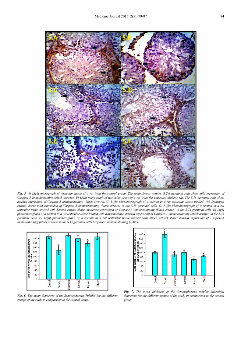
3.3. Immunohistochemical Results
The immunohistochemical investigations of the testicular
tissue for the control group showed mild expression of
Caspase-3 immunostaining with few caspase-3 +ve cells in
the S. Ts (Figs. 5, 10& T. 1). In diabetic group, a marked
significant increase in the number of caspase-3+ve cells was
observed in the S. Ts in comparison to the control ones (Figs.
5, 10& T. 1). In both Damsissa- and Sammo-treated groups,
mild expression of caspase-3+ve cells were detected with
significant decrease compared with the diabetic group (Figs.
5, 10& T. 1). However, in both Kaysom - and Sheeh-treated
groups, a marked significant increase in the number of
caspase-3+ve cells was observed in the S. Ts in comparison
to the control ones (Figs. 5, 10& T. 1).
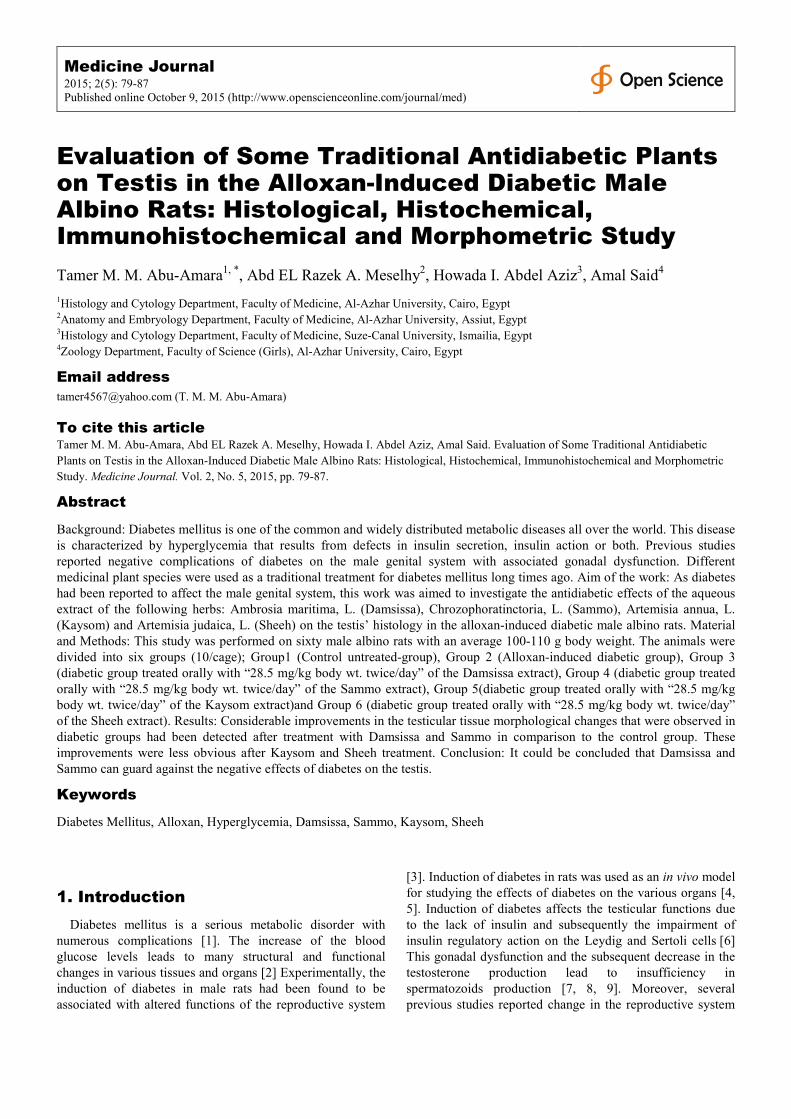
Fig. 2. A) Light photomicrograph of a section in a rat testicular tissue from the control group. The seminiferous tubules (S.Ts) have ordinary shape. S.Ts
epithelium is structurally intact and shows normal association of germ cells. B) Light photomicrograph of a section in a rat testicular tissue form untreated
diabetic rat. The S. Ts have irregular shape and the germinal epithelium is disorganized. Depletion of germ cells, pyknotic germ cells (black arrows) and
karyolysis (green arrow) are seen. The giant cell formation with two or three nucleus (red arrow) is seen in the lumen of irregular shaped seminiferous tubule
(ST). C)Light photomicrograph of a section in a rat testicular tissue treated with Damsissa extract. The S.Ts have partial recovery to normal structure. D)
Light photomicrograph of a section in a rat testicular tissue treated with Sammo extract. The S.Ts have partial recovery to normal structure. E) Light
photomicrograph of a section in a rat testicular tissue treated with Kaysom extract. The S.Ts have partial recovery to normal structure. F) Light
photomicrograph of a section in a rat testicular tissue treated with Sheeh extract. The S.Ts have partial recovery to normal structure. H&E (400×).

83 Tamer M. M. Abu-Amara et al.: Evaluation of Some Traditional Antidiabetic Plants on Testis in the Alloxan-Induced
Diabetic Male Albino Rats: Histological, Histochemical, Immunohistochemical and Morphometric Study
Fig. 3. A) Light photomicrograph of a section in a rat testicular tissue from the control group shows normal distribution of collagen fibers in the interstitial
tissue (I.T) around the seminiferous tubules. B) Light photomicrograph of a section in a rat testicular tissue form untreated diabetic rat shows marked
reduction of collagen fibers in the I.T around the seminiferous tubules, increase diameter of the interstitial spaces. C) Light photomicrograph of a section in a
rat testicular tissue treated with Damsissa extract shows more or less similar distribution of collagen fibers in the I.T around the seminiferous tubules in
comparison to the control group. D) Light photomicrograph of a section in a rat testicular tissue treated with Sammo extract shows more or less similar
distribution of collagen fibers in the I.T around the seminiferous tubules in comparison to the control group. E) Light photomicrograph of a section in a rat
testicular tissue treated with Kaysom shows mild distribution of collagen fibers in the I.T around the seminiferous tubules in comparison to the control group.
F) Light photomicrograph of a section in a rat testicular tissue treated with Sheeh extract shows more or less similar distribution of collagen fibers in the I.T
around the seminiferous tubules in comparison to the control group. Mallory’s trichrome (200×).
Fig. 4. A) Light photomicrograph of a section in a rat testicular tissue from the control group. The seminiferous tubules (S.Ts) have ordinary shape, their
epithelium is structurally intact and shows normal association of germ cells. B) Light photomicrograph of a section in a rat testicular tissue form untreated
diabetic rat shows marked reduction, pyknosis, disorganization and depletion of the germinal epithelium in the S.Ts. C) Light photomicrograph of a section in
a rat testicular tissue treated with Damsissa extract shows marked recovery of the germinal epithelium to the normal architecture. D) Light photomicrograph
of a section in a rat testicular tissue treated with Sammo extract shows marked recovery of the germinal epithelium to the normal architecture. E) Light
photomicrograph of a section in a rat testicular tissue treated with Kaysom shows mild recovery of the germinal epithelium to the normal architecture. F)
Light photomicrograph of a section in a rat testicular tissue treated with Sheeh extract shows mild recovery of the germinal epithelium to the normal
architecture. Feulgen stain (400×).
Medicine Journal 2015; 2(5): 79-87 84
Fig. 5. A) Light micrograph of testicular tissue of a rat from the control group. The seminiferous tubules (S.Ts) germinal cells show mild expression of
Caspase-3 immunostaining (black arrows). B) Light micrograph of testicular tissue of a rat from the untreated diabetic rat. The S.Ts germinal cells show
marked expression of Caspase-3 immunostaining (black arrows). C) Light photomicrograph of a section in a rat testicular tissue treated with Damsissa
extract shows mild expression of Caspase-3 immunostaining (black arrows) in the S.Ts germinal cells. D) Light photomicrograph of a section in a rat
testicular tissue treated with Sammo extract shows moderate expression of Caspase-3 immunostaining (black arrows) in the S.Ts germinal cells. E) Light
photomicrograph of a section in a rat testicular tissue treated with Kaysom shows marked expression of Caspase-3 immunostaining (black arrows) in the S.Ts
germinal cells. F) Light photomicrograph of a section in a rat testicular tissue treated with Sheeh extract shows marked expression of Caspase-3
immunostaining (black arrows) in the S.Ts germinal cells.Caspase-3 immunostaining (400×).
Fig. 6. The mean diameters of the Seminepherous Tubules for the different
groups of the study in comparison to the control group.
Fig. 7. The mean thickness of the Seminepherous tubular interstitial
diameters for the different groups of the study in comparison to the control
group.

85 Tamer M. M. Abu-Amara et al.: Evaluation of Some Traditional Antidiabetic Plants on Testis in the Alloxan-Induced
Diabetic Male Albino Rats: Histological, Histochemical, Immunohistochemical and Morphometric Study
Fig. 8. The mean area percentage of the collagen fibres of the
Seminepherous tubular interstitial diameters for the different groups of the
study in comparison to the control group.
Fig. 9. The mean apoptotic changes of the Seminepherous Tubular germinal
cells nuclei for the different groups of the study in comparison to the control
group.
Fig. 10. The mean number of the caspase-3+ve cells of the seminepherous tubular germinal cells for the different groups of the study in comparison to the
control group.
Table. 1. Diameters of the seminepherous tubules, thickness of the seminepherous tubular interstitial diameters, percentage of the collagen fibers of the
seminepherous tubular interstitial diameters, apoptotic changes of the seminepherous tubular germinal cells nuclei and number of the caspase-3+ve cells of
the seminepherous tubular germinal cells for the different groups of the study expressed as mean ± SD.
Paramaters Mean diameters
of the
seminepherous
tubules
Mean thickness of
the seminepherous
tubular interstitial
diameters
Mean area percentage of
the collagen fibres of the
seminepherous tubular
interstitial diameters
Mean apoptotic changes
of the seminepherous
tubular germinal cells
nuclei
Mean number of the
caspase-3+ve cells of the
seminepherous tubular
germinal cells Study Groups
Group 1
(Control)
188.11±
13.92
99.89±
7.57
140.03±
23.25
30.073±
1.48
12.00±
1.94
Group 2
(Diabetic)
128.61±
32.17 **a
180.70±
23.50**b
65.97±
16.55**a
14.08±
4.02**a
37.81±
2.78**b
Group 3
(Demcica)
192.78±
10.67**c
90.38±
15.29**d
148.96±
16.79**c
25.75±
6.28**c
15.00±
3.31**d
Group 4
(Samwaa)
179.50±
21.97**c
100.10±
16.51**d
141.48±
20.43**c
22.63±
655.96**c
13.18±
2.92**d
Group5
(Kaysom)
158.13±
19.95**c
69.64±
11.06**d
57.8911±
11.51**a
16.88±
3.48**a
33.90±
2.54**b
Group 6
(Sheeh)
187.43±
20.38**c
84.05±
4.74**d
95.37±
21.31**a
17.55±
2.73**a
33.72±
2.41**b
*Significantly different from the control group (P < 0.05).
**Significantly different from the control group (P< 0.001). aSignificant decrease in the parameters levels of the treated group in comparison to the control group. bSignificant increase in the parameters levels in the treated group in comparison to the control group. cSignificant increase in the parameters levels of the treated group in comparison to the diabetic group. dSignificant decrease in the parameters levels in the treated group in comparison to the diabetic group.
Medicine Journal 2015; 2(5): 79-87 86
4. Discussion
Diabetes is a common health problem that decrease
sexual functions such as; infertility, adverse effect on
pregnancy outcomes, sexual disinclination, reduction of
clitoral sensitivity and penile erection loss [21, 22]. The
male reproductive dysfunction is a common complication
in the diabetic patients [23, 24]. These dysfunction may be
persistent or temporary according to the degree and the
duration of the disease [23, 24]. Diabetic associated-tissue
injury and its subsequent complications are most probably
induced by free radicals [25] Prior studies on diabetic
subjects showed decreased testosterone levels and
vacuolization in the germinal epithelium (spermatogonia
and spermatocytes) with subsequent decrease in the
testicular weight, sperm number and motility [26, 27].
Other studies pointed to increasing S. Ts thickness and
germ cell depletionin both the diabetic human and rats [28,
29]. Our study results showed similar results as
irregularity of the S.Ts shapes with significant decrease in
S.Ts diameters. Furthermore, similar to several previous
studies results [10, 11, 30], this study’ results showed
degeneration and necrosis of the S. Ts, giant cell
formation and interstitialchanges in the diabetic rats.
These S. Ts changes were significantly improved after
Damsissa- and Sammo-treatment, while no significant
improvement was detected after Kaysom- and Sheeh-
treatment. Moreover, marked significant reduction,
disorganization, apoptotic changes (e.g. pyknosis and
karyolysis) and depletion of the S. Ts germinal epithelium
was detected in our results. Control of apoptosis is
essential factor for the healthy spermatogenesis within the
adult testes [31]. During non-physiological stresses, such
as diabetes, significant induction of apoptotic cell death
may happen [32]. Many herbal extracts or derivatives with
high antioxidant activity are useful for diabetes treatment
and other metabolic syndrome [33]. Thus, antioxidant
therapy is one of the major strategies for diabetes
treatment [33]. Our results showed that Damsissa- and
Sammo have significant protective effects against the
apoptotic changes. This protective effect may be due to
their antioxidant properties that were proved in previous
studies [14, 33]. Abnormal high levels of free radicals and
the simultaneous decline of antioxidant defense
mechanisms may damages the cellular organelles and
enzymes, increases lipid peroxidation and leads to insulin
resistance [34]. Currently, it is proved that, the essential
oils components of Damsissa have antioxidant, anti-
inflammatory and anti-hypoinsulinemic properties [35].
In conclusion, Considerable improvements in the testicular
tissue morphological changes that were observed in diabetic
groups had been detected after Damsissa- and Sammo-
treatment in comparison to the control group. These
improvements were less obvious after Kaysom- and Sheeh-
treatment.
References
[1] Yanardag, R., Ozsoy-Sacan O., Bolkent, S. (2005): Protective effects of metform in treatment on the liver injury of streptozotocin-diabetic rats. Hum ExpToxicol 2005, 24: 129-135.
[2] Cai L., Chen S., Evans, T. (2000): Apoptotic germ-cell death and testicular damage in experimental diabetes: prevention by endothelin antagonism. Urol Res., 28: 342-347.
[3] Orth, M., Murray, T., Bardin, W. (1979): Ultrastructural Changes in Leydig Cells of Streptozotocin-induced Diabetic Rats. Anat Rec., 195: 415-430.
[4] Kuhn-Velten, N., Waldenburger D., Staib, W. (1982): Evaluation of Steroid Biosynthetic Lesions in Isolated Leydig Cells from the Testes of Streptozotocin-Diabetic Rats. Diabetol, 23: 529-533.
[5] Morimoto, S., Mendoza-Rodriguez, A., Hiriat, M. (2005): Protective effect of testosterone on early apoptotic damage induced by streptozotocin in rat pancreas. J Endocrinol, 187: 217-224.
[6] Ballester, J., Carmen, M., Dominguez, J. (2004): Insulin-Dependent Diabetes Affect Testicular Function by FSH- and LH-linked Mechanisms. J Androl, 25: 706-719.
[7] Cameron, F., Orth, J., Murray T. (1982): Morphological alteration in the testes from diabetic man and rat. Diabetes., 31: 11A.
[8] Steger, W. and Rabe, M. (1997): The effect of diabetes mellitus on endocrine and reproductive function. ProcSocExpBiol Med., 214: 1-11.
[9] Ozdemir, O., Akalin, P., Baspinar, N. (2009): Pathological changes in the acute phase of streptozotocin-induced diabetic rats. Bull Vet InstPulawy, 53: 783-790.
[10] Sanguinetti, E., Ogawa K., Kurohmaru, M. (1995): Ultrastructural changes in mouse Leydigcells after streptozotocin administration. Exp Anim., 44: 71-73.
[11] Ozturk, F., Gul, M., Agkadir, M. (2002): Histological alterations of rat testes in experimental diabetes. T Kin J Med Sci., 22: 173-178.
[12] Navarro-Cassado, L., Juncos-Tobarra, MA., Chafer-Rudilla, M. (2010): Effect of experimental diabetes and STZ on male fertility capacity. Study in rats. J Androl., 108: 007260.
[13] Abdel Wahab, M., Wassel, M., Ammar, M. and Hanna, T. (1987): HerbaHungarica Tom., 26 (1): 27-31.
[14] Ghazanfer, S. (1994): CRC Hand book of Arabian Medicinal Plants. CRC Press, Boca Raton., Pp. 265.
[15] Bora, KS., and Sharma, A. (2011): The genus Artemisia: A comprehensive review. Pharm. Biol., 49: 101–109.
[16] Al-Mustafa, H., and Al-Thunibat, OY. (2008): Antioxidant capacity of some Jordanian medicinal plants used traditionally for the treatment of diabetes. Pak J Biol Sci., 1: 351–358. http://www.ncbi.nlm.nih.gov/pubmed/1881715
[17] Bhakuni, S., Jain, C., Sharma, P. and Kumar, S. (2001): Secondary metabolites of Artemisia annua and their biological activity. Current Science, 80: 35–48.
87 Tamer M. M. Abu-Amara et al.: Evaluation of Some Traditional Antidiabetic Plants on Testis in the Alloxan-Induced
Diabetic Male Albino Rats: Histological, Histochemical, Immunohistochemical and Morphometric Study
[18] Malaisse, J. (1982): Alloxan toxicity to the pancreatic B-cell. Anew hypothesis. Biochem. Pharmacol, 31: 3527-3537.
[19] Bancroft, D., and Gamble, M. (2008): Bancroft's Theory and Practice of Histological Techniques. 6th Ed. Elsevier, Churchill Livingst one, Edinburgh, Scotland, Pp 178-186, 221-224.
[20] Hammer, Ø., Harper, D., Ryan, D. (2001): PAST: Paleontological Statistics Software Package for Education and Data Analysis. Palaeontologia Electronica., 4(1): 9-15.
[21] Kim, N., Stankovic, M., Cushman, T., Goldstein, I., Munarriz, R. and Traish, M. (2006): Streptozotocin-induced diabetes in the rat is associated with changes in vaginal hemodynamics, morphology and biochemical markers. BMC Physiol., 6:1–9.
[22] Amaral, S., Mota, C., Lacerda, B., Alves, M., Pereira, Mde L., Oliveira, J. and Ramalho-Santos, J. (2009): Testicular mitochondrial alterations in untreated streptozotocin-induced diabetic rats. Mitochondrion, 9: 41–50.
[23] Armagan, A., Uz, E., Yilmaz, R., Soyupek, S., Oksay, T. and Ozcelik, N. (2006): Effects of melatonin on lipid peroxidation and antioxidant enzymes in streptozotocin-induced diabetic rat testis. Asian J Androl., 8: 595–600.
[24] Amaral, S., Oliveira, J. and Ramalho-Santos, J. (2008): Diabetes and the impairment of reproductive function: Possible role of mitochondria and reactive oxygen species. Curr Diabetes Rev., 4: 46–54.
[25] Annunziata, L., Domenico, F. and Pietro, T. (2005): Glyco-oxidation in diabetes and related diseases. Clin Chim Acta, 2: 236–250.
[26] Bhasin, S., Enzlin, P., Coviello, A., and Basson, R. (2007): Sexual dysfunction in men and women with endocrine disorders. The Lancet, 369(9561): 597-611.
[27] Seethalakshmi, L., Menon, M. and Diamond, D. (1987): The effect of streptozotocin-induced diabetes on the neuroendocrine-male reproductive tract axis of the adult rat. J Urol., 138: 190–194.
[28] Cameron, F., Murray, T. and Drylie, D. (1985): Interstitial compartment pathology and spermatogenic disruption in testes from impotent diabetic men. Anat Rec., 213: 53–62.
[29] Sadik, H., El-Seweidy, M. and Shaker, G. (2011): The antiapoptotic effects of sulphurous mineral water and sodium hydrosulphide on diabetic rat testes. Cell PhysiolBiochem, 28: 887–898.
[30] Anderson, E. and Thliveris, A. (1986): Testicular histology in streptozotocin-induced diabetes. Anat Rec., 214: 378-382.
[31] Koh, O (2007): Streptozotocin-induced diabetes increases apoptosis through JNK phosphorylation and Bax activation in rat testes. J Vet Med Sci., 69: 969–971.
[32] Cai, L., Hales, F. and Robaire, B. (1997): Induction of apoptosis in the germ cells of adult male rats after exposure to cyclophosphamide. BiolReprod, 56: 1490–1497.
[33] Samad, A., Shams, S., Ullah, Z., Wais, M., Nazish, I., Sultana, Y. and Aqil, M. (2009): Status of herbal medicines in the treatment of diabetes. Curr. Diabetes Rev., 5: 102-111
[34] Yang, H., Jin, X., Kei Lam, W. and Yan, K. (2011): Oxidative stress and diabetes mellitus. Clinical Chemistry & Laboratory Medicine, 49(11): 1773-1782.
[35] Bakkali, F., Averbeck, S., Averbeck, D. And Idaomar, M (2008): Biological effects of essential oils. Food Chem. toxicol, 46: 446-475.