Evaluation of Routine Sonography for Early … of Ultrasonography The Osak, a Medical Center for...
6
Evaluation of Routine Sonography for Early Detection of Pancreatic Cancer Sachiko Tanaka 1 , Tsugio Kitamra 1 , Kiyomi Yamamoto 1 , Sachiko Fujikawa, 1 Tomoko Imaoka 1 , Shigeri Nishikawa 1 , Akihiko Nakaizumi 2 , Hiroyuki Uehara 2 , Osamu Ishikawa 3 and Hiroaki Ohigashi 3 laboratory of Ultrasonography, Departments of Gastrointestinal Oncology and 'Surgery, The Osaka Medical Center for Cancer and Cardiovascular Diseases, Osaka The diagnostic accuracy of routine abdominal sonography for the detection of pancreatic cancer was examined. During the one-year period of 1994, sonographic examination of the upper ab- dominal region was performed 12,761 times on a total of 9410 patients for the screening of abdominal disorders. In 655 cases (7%) part of the pancreas could not be observed. Based on the "Diagnostic criteria for pancreatic cancer" published by the Japanese Society of Ultrasound in Medicine, sonographic finding was evaluated to be positive for pancreatic tumor in a total of 411 cases. At the end of 1995, 51 patients were proven to have pancreatic cancer, and 45 of these cases were ductal adenocarcinoma. In 26 cases the tumor was surgically resected. Fifty cases were true sonographic positives and one was a false negative. The sensitivity, specifici- ty, overall accuracy, and positive and negative predictive values of sonography for pancreatic cancer were 98.0%, 95.9%, 95.9%, 12.2% and 100.0%, respectively. Among the 50 true posi- tive cases, the tumor diameter was less than 1 cm in four (8%). In conclusion, the diagnostic accuracy of sonography for the detection of pancreatic cancer is sufficiently high. Therefore, a detailed study aimed at mass screening for pancreatic cancer using sonography as the main modality seems warranted as a countermeasure for the rapid increase of pancreatic cancer in Japan. (Jpn J Clin Oncol 26: 422-427, 1996) Key words:' Pancreatic cancer—Ultrasonography—Diagnostic accuracy Introduction The prognosis of pancreatic cancer has been reported to be very poor. 1 " 3 ' However, recent reports have described a better prognosis if the cancers are small and detected early. Crist et al. l) reported that the 5-year actuarial survival rate af- ter surgical treatment for pancreatic cancer with- out lymph node metastasis was quite high, 48%. Ishikawa et al. 4) summarized 32 reported cases of small pancreatic cancer measuring 1 cm or less, and described a good 5-year survival of 67% after sur- gery. Moreover Nakaizumi et a/. 5 ' reported that cytologically-diagnosed in situ pancreatic cancer or Received: February 29, 1996 Accepted: June 3, 1996 For reprints and all correspondence: Sachiko Tanaka, Laboratory of Ultrasonography, The Osaka Medical Center for Cancer and Cardiovascular Diseases, 3-3, Nakamichi 1-chome, Higashinari, Osaka 537 cancer with minimal invasion could be expected to have a good long-term prognosis. Hence, early de- tection of pancreatic cancer with a highly sensitive modality is required to improve the prognosis of this disease. Following the introduction of real-time sonogra- phy, the diagnostic accuracy of ultrasonographic ex- amination (US) for hepatocellular carcinoma has improved markedly, 6 - v and a mass survey with US was reported to be effective for early diagnosis of hepatocellular carcinoma. 8 ' Although the pancreas has been considered difficult to visualize by US, 9) introduction of the convex-type probe has brought an obvious improvement in the visualization of this organ. Furthermore, many reports 2 " 4 ' have described that US has been the major aid for initial detection of resectable pancreatic cancer. In the present study, the diagnostic accuracy of routine abdominal US for pancreatic cancer was ex- amined retrospectively, and the possibility of mass 422 Jpn J Clin Oncol 26(6) 1996
Transcript of Evaluation of Routine Sonography for Early … of Ultrasonography The Osak, a Medical Center for...
Evaluation of Routine Sonography for Early Detection of Pancreatic Cancer
Sachiko Tanaka1, Tsugio Kitamra1, Kiyomi Yamamoto1, Sachiko Fujikawa,1 Tomoko Imaoka1,Shigeri Nishikawa1, Akihiko Nakaizumi2, Hiroyuki Uehara2, Osamu Ishikawa3 and HiroakiOhigashi3
laboratory of Ultrasonography, Departments of Gastrointestinal Oncology and 'Surgery, The Osaka MedicalCenter for Cancer and Cardiovascular Diseases, Osaka
The diagnostic accuracy of routine abdominal sonography for the detection of pancreatic cancerwas examined. During the one-year period of 1994, sonographic examination of the upper ab-dominal region was performed 12,761 times on a total of 9410 patients for the screening ofabdominal disorders. In 655 cases (7%) part of the pancreas could not be observed. Based onthe "Diagnostic criteria for pancreatic cancer" published by the Japanese Society of Ultrasoundin Medicine, sonographic finding was evaluated to be positive for pancreatic tumor in a totalof 411 cases. At the end of 1995, 51 patients were proven to have pancreatic cancer, and 45of these cases were ductal adenocarcinoma. In 26 cases the tumor was surgically resected. Fiftycases were true sonographic positives and one was a false negative. The sensitivity, specifici-ty, overall accuracy, and positive and negative predictive values of sonography for pancreaticcancer were 98.0%, 95.9%, 95.9%, 12.2% and 100.0%, respectively. Among the 50 true posi-tive cases, the tumor diameter was less than 1 cm in four (8%). In conclusion, the diagnosticaccuracy of sonography for the detection of pancreatic cancer is sufficiently high. Therefore,a detailed study aimed at mass screening for pancreatic cancer using sonography as the mainmodality seems warranted as a countermeasure for the rapid increase of pancreatic cancer inJapan.
(Jpn J Clin Oncol 26: 422-427, 1996)
Key words:' Pancreatic cancer—Ultrasonography—Diagnostic accuracy
Introduction
The prognosis of pancreatic cancer has beenreported to be very poor.1"3' However, recentreports have described a better prognosis if thecancers are small and detected early. Crist et al.l)
reported that the 5-year actuarial survival rate af-ter surgical treatment for pancreatic cancer with-out lymph node metastasis was quite high, 48%.Ishikawa et al.4) summarized 32 reported cases ofsmall pancreatic cancer measuring 1 cm or less, anddescribed a good 5-year survival of 67% after sur-gery. Moreover Nakaizumi et a/.5' reported thatcytologically-diagnosed in situ pancreatic cancer or
Received: February 29, 1996Accepted: June 3, 1996For reprints and all correspondence: Sachiko Tanaka,Laboratory of Ultrasonography, The Osaka Medical Centerfor Cancer and Cardiovascular Diseases, 3-3, Nakamichi1-chome, Higashinari, Osaka 537
cancer with minimal invasion could be expected tohave a good long-term prognosis. Hence, early de-tection of pancreatic cancer with a highly sensitivemodality is required to improve the prognosis ofthis disease.
Following the introduction of real-time sonogra-phy, the diagnostic accuracy of ultrasonographic ex-amination (US) for hepatocellular carcinoma hasimproved markedly,6-v and a mass survey with USwas reported to be effective for early diagnosis ofhepatocellular carcinoma.8' Although the pancreashas been considered difficult to visualize by US,9)
introduction of the convex-type probe has broughtan obvious improvement in the visualization of thisorgan.
Furthermore, many reports2"4' have describedthat US has been the major aid for initial detectionof resectable pancreatic cancer.
In the present study, the diagnostic accuracy ofroutine abdominal US for pancreatic cancer was ex-amined retrospectively, and the possibility of mass
422 Jpn J Clin Oncol 26(6) 1996

EVALUATION OF US FOR PANCREATIC CANCER
Table I. Diagnostic Criteria for Pancreatic Cancer
Existence of pancreatic tumor10'A. Definite
1: Markedly abnormal echoic area* (echo level,pattern, boundary)
2: Abnormal echoic area with dilatation of the distalpancreatic duct
3: Abnormal echoic area with stenosis or obstructionof the common bile duct in the pancreas and thepancreatic area
4: Abnormal echoic area with localized swelling of thepancreas
B. Suspected mass1: Abnormal echoic area in the pancreas2: Abnormal echoic area in the vicinity of the pancreas3: Localized swelling of the pancreas including the
pancreatic marginC. Further examination recommended
. 1: Dilatation of the pancreatic duct2: Dilatation of the common bile duct and/or swelling
of the gallbladder
Remarks: *, an abnormal echoic area is defined as one which is eitherlighter or darker, more or less dense, and with a different arrange-ment of echoic elements than the surrounding tissues. In addition,there is a clearly discernible border and a change from the normalechoic patterns.
screening for pancreatic cancer was assessed.
Patients and Methods
During the one-year period from January 1 toDecember 31, 1994, US of the upper abdominalregion was performed 12,761 times on a total of9410 patients (5309 males and 4101 females; agerange: 14-96 years; mean: 59 years) at the hospi-tal of the Osaka Medical Center for Cancer andCardiovascular Diseases. US was performed routine-ly by several well trained sonographers and doctorsspecializing in gastroenterology. The US took about15 min. for each patient in a supine position andsometimes in a sitting position. Requests for USwere received from various departments of ourhospital and sometimes from other hospitals, forthe screening of abdominal disorders.
The instruments used were a EUB-565, EUB-450and EUB-40 with 3.5- and 5-MHz convex probes(Hitachi, Tokyo), and a UM9-HDI with 3- and 4to 7-MHz convex probes (ATL, Seattle, WA).
Based on the "Diagnostic criteria for pancreaticcancer" (Table I), published by the Japanese Soci-ety of Ultrasound in Medicine,10' the existence ofpancreatic tumors was evaluated using three ranks:definite (A), suspected (B) and further examinationrecommended (C). For the final diagnosis of pan-
Table II. Results of US Findings According to the Criteriafor Pancreatic Cancer
US findings No. ofpositive cases
Pancreatic cancer(<1 cm)
DefiniteA-1 (solid)
(cystic)A-2*A-3A-4
6211840107
152(1)
19(2)73
SuspectedB-lB-2B-3
Further exam.C-l*C-2
Total
528
recommended12237
411
020
1 (1)1
50(4)
*A-2, C-l: 3 mm or more is regarded as dilatation of the pancreaticduct.
creatic cancer, all subjects were checked against theCancer Registry in our hospital at the end of 1995.For patients who ceased attending, their recentphysical condition was confirmed by telephoningthe patients or their families.
Results
US Findings
Of the 9410 cases, the pancreas was successfullyobserved and evaluated by US in 8755 (93.0%). Inthe remaining 655 cases, part of the pancreas couldnot be visualized clearly, although no abnormal USfindings were detected. The results of US areshown in Table II: 237 cases were ranked as A, 15as B and 159 as C.
For the total of 411 patients with positive USfindings, CT, ERCP, pancreatic juice cytology,repeated US and/or other examinations were per-formed.
Pancreatic Cancer Cases
Of the 9410 patients, 51 (0.54%) (31 males and20 females) were proven to have pancreatic cancerby the end of 1995. Of these cases, 36 (71%) werehistologically proven to be pancreatic cancer, andthe remaining cases were cytologically proven. Thecancers included 45 ductal adenocarcinomas, twocystadenocarcinomas, one adenosquamous cell car-cinoma and three islet cell carcinomas. The patientsranged in age from 30 to 54 years (mean: 42 years)for islet cell carcinoma and 41 to 81 years (mean:
423
TANAKA ET AL.
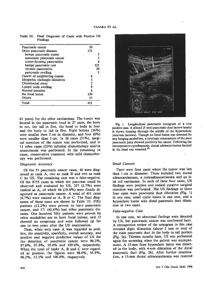
Table III. Final Diagnoses of CasesFindings
Pancreatic cancerOther pancreatic diseases
benign pancreatic tumormetastatic pancreatic cancertumor-forming pancreatitisbenign pancreatic cystchronic pancreatitispancreatic swelling
Cancer of neighboring organsIdiopathic cholangio dilatationCholedochal stoneLymph node swellingNormal intestineNo focal lesionOthers
Total
with Positive
50171
63288
15013
411
US
534
125295
61 years) for the other carcinomas. The tumor waslocated in the pancreatic head in 27 cases, the bodyin ten, the tail in five, the head to body in fourand the body to tail in five. Eight lesions (16%)were smaller than 2 cm in diameter, and four (8%)were smaller than 1 cm. In 26 cases (51%), surgi-cal resection of the tumor was performed, and in11 other cases (22%) infusion chemotherapy and/oranastomosis was performed. In the remaining 14cases, conservative treatment with'mild chemother-apy was performed.
Diagnostic Accuracy
Of the 51 pancreatic cancer cases, 46 were diag-nosed as rank A, two as rank B and two as rankC by US. The remaining case was a false-negative.Of the 8755 cases in which the pancreas could beobserved and evaluated by US, 237 (2.7%) wereranked as A, of which 46 (19.4%) were finally di-agnosed as pancreatic cancer. A total of 411 cases(4.7%) were ranked as A, B or C. The final diag-noses of these cases are shown in Table III. Fiftypatients (12.2%) were proven to have pancreaticcancer, and 171 (41.6%) had other pancreatic dis-eases. One hundred fifty patients were proven byother modalities not to have focal lesions, and 13showed no symptoms and were followed up forone to two years after the US examination.
Thus, when only rank A was regarded as posi-tive, the sensitivity, specificity, overall accuracy, andpositive and negative predictive values of US forthe detection of pancreatic cancer were 90.2%,97.8%, 97.9%, 19.4% and 100.0%, respectively.When the total of ranks A, B and C were regard-ed as positive, the figures were 98.0%, 95.9%,96.2%, 12.2% and 100.0%, respectively.
Fig. 1. Longitudinal pancreatic sonogram of a truepositive case. A dilated (5 mm) pancreatic duct (arrow heads)is shown running through the middle of the hyperechoicpancreas (arrows). Though no focal lesion was detected byany imaging modalities, a cytologic examination of the purepancreatic juice showed positivity for cancer. Following theintraoperative cytodiagnosis, ductal adenocarcinoma locatedin the head was resected.18'
Small Cancers
There were four cases where the tumor was-lessthan 1 cm in diameter. These included two ductaladenocarcinomas, a cystoadenocarcinoma and an is-let cell carcinoma, in each of these four cases, USfindings were positive and radical curative surgicalresection was performed. The US findings in thesefour cases were pancreatic duct dilatation (Fig. 1)in one case, small cystic lesion in one case, and ahypoechoic lesion with distal pancreatic duct dilata-tion in two cases.
False-negative Case
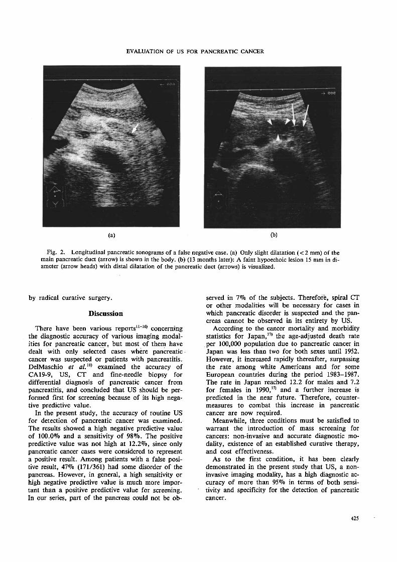
In one case, no abnormal findings were detectedby US, but pancreatic cancer was confirmed later.A retrospective review of the sonogram in this caserevealed slight dilatation (about 2 mm or less) ofthe main pancreatic duct in the body to tail portion(Fig. 2a). Thirteen months later, US was performedagain for screening when the patient was asympto-matic. A 15-mm faint hypoechoic lesion was detect-ed in the body, with 4-mm dilatation of the distalpancreatic duct (Fig. 2b). After further examina-tion, a 15-mm ductal adenocarcinoma was resected
424 Jpn J Clin Oncol 26(6) 1996
EVALUATION OF US FOR PANCREATIC CANCER
Fig. 2. Longitudinal pancreatic sonograms of a false negative case, (a) Only slight dilatation (<2 mm) of themain pancreatic duct (arrow) is shown in the body, (b) (13 months later): A faint hypoechoic lesion 15 mm in di-ameter (arrow heads) with distal dilatation of the pancreatic duct (arrows) is visualized.
by radical curative surgery.
Discussion
There have been various reports""16* concerningthe diagnostic accuracy of various imaging modal-ities for pancreatic cancer, but most of them havedealt with only selected cases where pancreaticcancer was suspected or patients with pancreatitis.DelMaschio et al.n) examined the accuracy ofCA19-9, US, CT and fine-needle biopsy fordifferential diagnosis of pancreatic cancer frompancreatitis, and concluded that US should be per-formed first for screening because of its high nega-tive predictive value.
In the present study, the accuracy of routine USfor detection of pancreatic cancer was examined.The results showed a high negative predictive valueof 100.0% and a sensitivity of 98%. The positivepredictive value was not high at 12.2%, since onlypancreatic cancer cases were considered to representa positive result. Among patients with a false posi-tive result, 47% (171/361) had some disorder of thepancreas. However, in general, a high sensitivity orhigh negative predictive value is much more impor-tant than a positive predictive value for screening.In our series, part of the pancreas could not be ob-
served in 7% of the subjects. Therefore, spiral CTor other modalities will be necessary for cases inwhich pancreatic disorder is suspected and the pan-creas cannot be observed in its entirety by US.
According to the cancer mortality and morbiditystatistics for Japan,17' the age-adjusted death rateper 100,000 population due to pancreatic cancer inJapan was less than two for both sexes until 1952.However, it increased rapidly thereafter, surpassingthe rate among white Americans and for someEuropean countries during the period 1983-1987.The rate in Japan reached 12.2 for males and 7.2for females in 1990,17) and a further increase ispredicted in the near future. Therefore, counter-measures to combat this increase in pancreaticcancer are now required.
Meanwhile, three conditions must be satisfied towarrant the introduction of mass screening forcancers: non-invasive and accurate diagnostic mo-dality, existence of an established curative therapy,and cost effectiveness.
As to the first condition, it has been clearlydemonstrated in the present study that US, a non-invasive imaging modality, has a high diagnostic ac-curacy of more than 95% in terms of both sensi-tivity and specificity for the detection of pancreaticcancer.
4IS
TANAKA ET AL.
As to the second condition, recent reports5'18)
have described that pancreatic cancer is curable ifit is diagnosed in an early stage.
Cost efficiency, the third condition, means thatthe cost of examination to detect one curablecancer must be reasonable. This is related to theexamination cost per person and the incidence ofcancer cases in the study population. Although, theincidence of pancreatic cancer has become higher,it is still low in comparison with that of hepatocel-lular carcinoma or gastric cancer.
Based on the age- and sex-specific pancreaticcancer mortality rates in Japan (1990), we proposethat only males aged 45 to 74 years and femalesaged 50 to 74 years should be included, since theserepresent the group in which the pancreatic cancermortality rate is more than five per 100,000 popu-lation and who can also tolerate radical surgery. Asa result, the expected incidence of pancreatic cancerwould become 0.021% (0.024% for men and0.017% for women). This value is considered to berather low for cost-effective mass screening.
With regard to periodic check-ups for hepatocel-lular carcinoma by US,8) started in 1987 and con-tinued successfully, the subjects were previouslylimited to those at high risk of the disease. Thusthe incidence of cancer cases among the subjectswas rather high at more than 3% in one year. Itis desirable to establish proper criteria for deciding"high-risk" for pancreatic cancer. Many retrospec-tive or prospective studies have "attempted to dothis using the presence of symptoms, elevation ofthe serum amylase level,2'3'19) or elevation of theserum CA 19-9 and elastase-1 levels.19"22' However,these trials resulted in detection of only advancedcases, and the establishment of "high-risk" criter-ia was reported to be very difficult.
On the other hand, Inamoto et al.19> reportedthe results of an abdominal mass survey using US,based on a questionnaire distributed to 14 institu-tions. According to their report, the incidence ofpancreatic cancer among the subjects was low at0.011%, but small (<2 cm) cancers were includedat a higher rate of 30% compared with the ratebased on the data from the national registration ofpancreatic cancer.3'
However, the use of US as a first choice for thescreening of pancreatic cancer may be consideredexpensive. According to Mihara et al.,23) morethan ten times the number of malignant cases otherthan pancreatic cancer are also detectable by massscreening with US. Considering the present situa-tion, therefore, the criteria used for defining pa-tients at high risk for pancreatic cancer must besettled by US.
In our series, there was one false-negative case inwhich the tumor was detected at the next US ex-
amination performed 13 months after the first,when the cancer was still curable. Here, slight dila-tation of the main pancreatic duct was evidentupon retrospective review of the first sonogram.Also, all of four cancers less than 1 cm in diametershowed positive US findings.
Thus, it seems reasonable to perform periodic ex-aminations once a year, and every three or sixmonths for cases showing slight (less than 3 mm)pancreatic duct dilatation. This contrasts with theexamination of all subjects every three or sixmonths for hepatocellular carcinoma.
In conclusion, to counteract the rapid increase inthe mortality rate due to pancreatic cancer, and forimproving the diagnostic accuracy and prognosis af-ter treatment, a detailed study aimed at massscreening for pancreatic cancer is warranted.
References
1) Crist DW, Sitzmann JV, Cameron JL: Improvedhospital morbidity, mortality and survival after theWhipple procedure. Ann Surg 206: 358-365, 1987
2) Miyazaki N, Saitoh Y: Clinical features of the pan-creatic carcinoma smaller than 2 cm in diameter.Fukubu Gazoushindan 12: 8-12, 1992 (in Japanese)
3) Pancreatic Cancer Registration Committee of JapanPancreas Society: Annual report of national registra-tion of pancreatic cancer patients in 1994. Suizo 10:535-564, 1995 (in Japanese)
4) Ishikawa O, Ohigashi H, Nakamori S, Imaoka S,Sasaki Y, Furukawa H, Iwanaga T, Nakaizumi A,Uehara H, Kitamura T: Diagnosis and long-termresult of minute and occult carcinoma of the pan-creas. Shokaki Gan 3: 87-91, 1993 (in Japanese)
5) Nakaizumi A, Tatsuta M, Uehara H, Takenaka A,Iishi H, Kitamra T, Ohigashi H, Ishikawa O, OkudaS, Wada A: Effectiveness of the cytologic examina-tion of pure pancreatic juice in the diagnosis of earlyneoplasia of the pancreas. Cancer 76: 750-757, 1995
6) Tanaka S, Kitamura T, Ohshima A, Umeda K,Okuda S, Ohtani T, Tatsuta M, Yamamoto K: Di-agnostic accuracy of ultrasonography for hepatocel-lular carcinoma. Cancer 58: 344-347, 1986
7) Tanaka S, Kitamura T, Nakanishi K, Okuda S,Kojima J, Fujimoto I: Recent advances in ultrasono-graphic diagnosis of hepatocellular carcinoma. Cancer63: 1313-1317, 1989
8) Tanaka S, Kitamura T, Nakanishi K, Okuda S,Yamazaki H, Hiyama T, Fujimoto I: Effectiveness ofperiodic checkup by ultrasonography for the early di-agnosis of hepatocellular carcinoma. Cancer 66:2210-2214, 1990
9) Kamin PD, Bernardino ME, Wallace S, Jing BC:Comparison of ultrasound and computed tomographyin the detection of pancreatic malignancy. Cancer 46:2410-2412, 1980
10) The Committee for the Diagnostic Criteria in theJapanese Society of Ultrasound in Medicine: Diagnos-tic criteria for pancreatic cancer. Chouonpa Igaku 19:
426 Jpn J Clin Oncol 26(6) 1996
EVALUATION OF US FOR PANCREATIC CANCER
553-557, 1992 (in Japanese)11) DelMaschio A, Vanzulli A, Sironi S, Castrucci M,
Mellone R, Staudacher C, Carlucci M, Zerbi A,Parolini D, Faravelli A, Cantaboni A, Garancini P,Carlo VD: Pancreatic cancer versus chronic pancrea-titis: diagnosis with CA19-9 assessment, US, CT andCT-guided fine-needle biopsy. Radiology 178: 95-99,1991
12) Vellet AD, Romano W, Bach DB, Passi RB, TavesDH, Munk PL: Adenocarcinoma of the pancreaticducts: comparative evaluation with CT and MR im-aging at 1.5 T. Radiology 183: 87-95, 1992
13) Muller MF, Meyenberger C, Bertschinger P, SchaerR, Marincek B: Pancreatic tumors: evaluation withendoscopic US, CT and MR imaging. Radiology 190:745-751, 1994
14) Bluemke DA, Cameron JL, Hruban RH, Pitt HA,Siegelman SS, Soyer P, Fishman EK: Potentiallyresectable pancreatic adenocarcinoma: spiral CT as-sessment with surgical and pathologic correlation.Radiology 197: 381-385, 1995
15) Stollfuss JC, Glatting G, Friess H, Kocher F, BegerHG, Reske SN: 2-(Fluorine-18) fluoro-2-deoxy-D-glucose PET in detection of pancreatic cancer: valueof quantitative image interpretation. Radiology 195:339-344, 1995
16) Megibow AJ, Zhou XH, Rotterdam H, Francis IR,Zerhouni EA, Balfe DM, Weinreb JC, Aisen A,Kuhlman J, Heiken JP, Gatsonis C, McNeil B: Pan-creatic adenocarcinoma: CT versus MR imaging inthe evaluation of resectability: report of the Radiol-ogy Diagnostic Oncology Group. Radiology 195:327-332, 1995
17) Tominaga S, Aoki K, Hanai A, Kurihara N: CancerMortality and Morbidity Statistics-1993. ShinoharaShuppann, Tokyo. 1993 (in Japanese)
18) Ishikawa O, Imaoka S, Ohigashi H, Nakaizumi A,Uehara H, Wada A, Nagumo S, Yamamoto R,
Sasaki Y, Iwanaga T: A new method of intra-operative cytodiagnosis for more precisely locating theoccult neoplasms of the pancreas. Surgery 111:294-.300, 1991
19) Inamoto Y, Kawamura S, Takemoto T: The actualcondition and feature of pancreatic carcinomas de-tected by mass examination using ultrasonography.Fukubu Gazoushindan 13: 471-478, 1993 (in
, Japanese)20) Hayashi S, Takebe T, Ohyama K, Odake Y,
Takiyama Y, Koike D, Miyakawa H, Moriai T,Yamadera K, Myoen R, Yokotama Y, Satoh M,Terasawa K, Ishii K, Nohda K, Miyashita F: A tri-al of the mass examination of pancreatic cancer us-ing measurement of serum CA 19-9 and elastase-1and ultrasonography. Nippon Shokakibyo GakkaiZasshi 83: 1360-1366, 1986 (in Japanese)
21) Frebourg T, Bercoff E, Manchon N, Senant J,Basuyau JP, Breton P, Janvresse A, Brunelle P,Bourreille J: The evaluation of CA19-9 antigen levelin the early detection of pancreatic cancer: a prospec-tive study of 866 patients. Cancer 62: 2287-2290,1988
22) Nakaizumi A, Tatsuta M, Uehara H, Iishi H,Yamamura H, Okuda S, Kitamra T: A prospectivetrial of early detection of pancreatic cancer byultrasonographic examination combined with measure-ment of serum elastasel. Cancer 69: 936-940, 1992
23) Mihara S, Yoshioka R, Sawatari M, Koba H,Narimatu R, Nagano K, Fujita S, Nishiono A, HiraoS, Tanaka S, Migita K, Miyata T, Machihara M,Okazaki T, Yamabe H, Ueno K, Suminaga Y,Fujimoto Y, Goto Y, Hondou K, Morimoto E,Tanaka K, Ishihara E, Koyama W: Actual status ofmalignant neoplasm cases detected by ultrasonic masssurvey. Shokaki Shudan Kenshin 33: 69-79, 1995 (inJapanese)
427