Evaluation of programs for the prediction of GPI-anchor in proteins.
57
1 Evaluation of programs for the prediction of GPI-anchor in proteins. Report of Practical Training performed at Swiss-Prot group, Swiss Institute of Bioinformatics CMU, 1 Michel-Servet CH-1211 Genève 4 - Suisse submitted for: The Master Degree in Proteomics and Bioinformatics by Natalia V. Sernova. Jury: Dr. Anne-Lise Veuthey (responsable de stage) Dr. David Perret Dr. Patricia Palagi Prof. Amos Bairoch (président de jury) To be defended at the University of Geneva, June 16, 2006 at 4:00 PM
Transcript of Evaluation of programs for the prediction of GPI-anchor in proteins.
1
EEvvaalluuaattiioonn ooff pprrooggrraammss ffoorr tthhee pprreeddiiccttiioonn ooff GGPPII--aanncchhoorr iinn pprrootteeiinnss..
Report of Practical Training performed at Swiss-Prot group, Swiss Institute of Bioinformatics CMU, 1 Michel-Servet CH-1211 Genève 4 - Suisse submitted for: The Master Degree in Proteomics and Bioinformatics by Natalia V. Sernova. Jury: Dr. Anne-Lise Veuthey (responsable de stage) Dr. David Perret Dr. Patricia Palagi Prof. Amos Bairoch (président de jury) To be defended at the University of Geneva, June 16, 2006 at 4:00 PM

2
Table of contents:
Summary………………………………………………………………………………………………………………………………………………………3
Work objectives…………………………………………………………………………………………………………………………………5
1.Introduction……………………………………………………………………………………………………………………………………6
1.1 The PTM and their importance
for protein function…………………………………………………………………………………………7
1.2 Challenges in PTM-prediction……………………………………………………………………8
1.3 PTM data resources and their
features. UniProtKB/Swiss-Prot…………………………………………………………………………9
1.3.1 UniProtKB/Swiss-Prot annotation……………………………………10
1.3.2 Non-experimental qualifiers………………………………………………10
1.4 GPI anchor PTM……………………………………………………………………………………………………11
1.4.1 The mechanism of GPI-anchor attachment…………………12
1.4.2 Experimental way to prove GPI-anchoring……………14
1.4.3 GGPPII--aanncchhoorriinngg pprreeddiiccttiioonn
pprrooggrraamm aallggoorriitthhmmss………………………………………………………………………………………………………………………………………………………………………………1166
2. Materials and methods
2.1 Annotation of GPI-anchor in
UniProtKB/Swiss-Prot database…………………………………………………………………………22
2.2 Building datasets………………………………………………………………………………………………23
2.2.1 Positive dataset creation……………………………………………………23
2.2.2 Dataset with experimentally verified
cleavage site……………………………………………………………………………………………………23
2.2.3 Negative dataset creation……………………………………………………23
2.2.4 Special case: transmembrane proteins………………………24
2.2.5 Automated dataset development and update……………24
2.3 Datasets used for development of the
GPI-prediction programs…………………………………………………………………………………………25
2.4 Access to GPI-anchor prediction programs…………………………………25
2.5 Program performance evaluation……………………………………………………………27
3. Results……………………………………………………………………………………………………………………………………………28
4. Discussion and concluding remarks………………………………………………………………………39
5. Acknowledgements……………………………………………………………………………………….…………………………41
6. References……………………………………………………………………………………………………………………………………42
7. Appendix
7.1 Perl-scripts …………………………………………………………………………………………………………47
7.2 Additional tables and images…………………………………………………………………52

3
SUMMARY.
Purposes:
• UniProtKB/Swiss-Prot is a curated database of proteins, which
integrates biological information retrieved from the biomedical
literature. In addition, UniProtKB/Swiss-Prot annotation relies on
sequence analysis tools and protein feature prediction methods, since
genome sequencing projects are providing large amounts of potential
protein coding regions without any experimental characterization.
Thus, promoting the development of reliable predictive methods, as
well as evaluating their performances is an essential task for improving
the database quality.
• Protein modifications play crucial structural and functional roles (Han
et al, 1992), so their identification is very important. Experimental data
are available for limited number of proteins, so prediction is the only
way to deal with huge amount of protein sequence data. Any PTM
prediction program for UniProtKB/Swiss-Prot annotation has to be
evaluated, and since new data information may help to improve
program performance, evaluation has to be done regularly.
Methods:
• Evaluation implies the estimation of prediction method performance on
experimentally proved negative and positive protein datasets, making
proper dataset construction a priority. Being constructed, besides
evaluation the datasets can be also used to develop new prediction
programs, as well as to improve existing methods.
• In the present work we evaluated the performance of three currently
existing programs, predicting GPI-anchor protein modification
(namely BigPI, DGPI and GPI-SOM). Protein data for evaluation
datasets were extracted from the UniProtKB/Swiss-Prot, the most
accurate and up-to-date protein knowledgebase currently available.
• This work was started with data from UniProtKB/Swiss-Prot release
48.1 (September 2005), and near finishing results were updated
according to the data from release 49.7 (May 16, 2006). The script for
later fully automated updates is provided by the author.

4
Results:
• The most reliable program in terms of its specificity is BigPI, which
was investigated on experimentally verified metazoan dataset. Lack of
verified data prevented us from proper investigation of other taxons, as
well as some program features like cleavage site prediction.
Conclusions:
• Better datasets are needed to create better prediction programs,
although currently existing programs, like BigPI, may already give
some hints on what proteins experimentalists should concentrate to
have verified GPI-anchored protein sequences.

5
Work objectives:
These studies were initiated to pursue the following goals:
• to evaluate currently existing GPI-anchor predictive methods
• to update experimentally verified datasets for the purpose to
develop new prediction programs, as well as to improve existing
tools
• to develop automatic update tools
To achieve these goals, the subject was sub-divided into the following tasks:
• to create positive and negative dataset of experimentally verified
proteins, being/not being GPI-anchored, respectively.
• to write Perl-scripts, which would access web-sites of three GPI-
anchor prediction programs and parse the output, what will allow
to calculate specificity/sensitivity of the three programs.
• to analyse and compare the results of all three programs.

6
1. Introduction.
Epigraph:
““IInnccrreeaasseedd ccoonnffiiddeennccee tthhaatt aa pprrootteeiinn iiss ppuuttaattiivveellyy GGPPII--aanncchhoorreedd sshhoouulldd
eennccoouurraaggee mmoorree rreesseeaarrcchheerrss ttoo eexxppeerriimmeennttaallllyy vveerriiffyy tthhiiss mmooddiiffiiccaattiioonn,, wwhhiicchh iinn ttuurrnn
wwiillll aallllooww tthhee ccoonnssttrruuccttiioonn ooff aa pprreeddiiccttoorr wwiitthh eevveenn hhiigghheerr aaccccuurraaccyy iinn tthhee ffuuttuurree..””
EEiisseennhhaabbeerr eett aall,, 11999988
Almost all proteins analyzed to date carry some post-translational
modifications (PTMs). The modified protein function is often strongly affected, or
even determined by these modifications (Blom et al, 2004). Increased knowledge
about the potential PTMs of a target protein may deepen our understanding of the
molecular processes it is involved in, and ultimately of its function. High-throughput
methods for the identification of PTMs are being developed, in particular within the
fields of proteomics and mass spectrometry.
However, most of these methods are still in their infancy, and to cut down on the
number of experimental steps by integrating computational approaches into the
validation procedures is indeed advantageous. Many advanced methods for the
prediction of PTMs exist and many are made publicly available.
The current work is dedicated to evaluation of performance for three existing GPI-
anchor prediction programs.
In this introduction we will talk about:
• the importance of post-translational modifications for protein function
with a description of GPI-anchor PTM as an example;
• UniProtKB/Swiss-Prot database as a reliable source of PTM-information, with
its daily data curation which is necessary to maintain data high quality;
• existing experimental GPI-detection techniques as well as computational
GPI-prediction methods

7
1.1 The PTMs and their importance for protein function
Many years ago, in the early days of molecular biology, the function of a
protein was typically known before the sequence of amino acids encoded by the gene
was determined. Nowadays the situation is reversed, and as long as sequences have
been accumulating in the databases, corresponding protein functional
analysis/prediction will remain the most important issue. Currently the number of
experimentally validated examples of post-translational modifications (PTMs) grows
tremendously, since PTMs make a particular protein molecule unique in terms of its
structure and function.
Three main classes of post-translational modifications (the name is misleading
because the modifications may also occur before and during protein synthesis) are the
following:
• cleavage
• linkage
• cross-linking
and can be combined – for instance, glycosylphosphotidylinositol (GPI) – anchor
attachment implies both cleavage and linkage. (Farriol-Mathis et al, 2004)
Proteins appear to be modified several times along their life-time (Eisenhaber
at al, chapter 5, 2003). Most proteins cannot perform their molecular function as
unmodified folded polypeptides. In most cases, proteins need to acquire permanent or
transient molecular features in order to become functional.
There is enormous amount of known DNA-sequences (as well as complete
genomes) nowadays, which raise a question of PTM-prediction from amino acid
sequence for genome annotation (with further goal of protein function prediction).
PTMs influence protein size, hydrophobicity and other physico-chemical properties;
can change, enhance or block a specific activity; can also target the protein to the
specific subcellular location. Because of that, PTM-prediction from protein sequence
data is very important.
The paradigm has been that protein sequence determines its structure, and
knowing the structure yields the functional information (Bork et al, 1998; Attwood,
2000). A somewhat complementary approach is currently being developed with
bioinformatics, namely the idea of making use of protein features and then assign
8
function using the features in an integrated fashion (Jensen et al, 2002; Jensen et al,
2003).
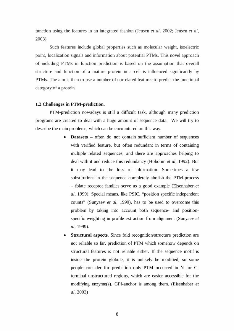
Such features include global properties such as molecular weight, isoelectric
point, localization signals and information about potential PTMs. This novel approach
of including PTMs in function prediction is based on the assumption that overall
structure and function of a mature protein in a cell is influenced significantly by
PTMs. The aim is then to use a number of correlated features to predict the functional
category of a protein.
1.2 Challenges in PTM-prediction.
PTM-prediction nowadays is still a difficult task, although many prediction
programs are created to deal with a huge amount of sequence data. We will try to
describe the main problems, which can be encountered on this way.
• Datasets – often do not contain sufficient number of sequences
with verified feature, but often redundant in terms of containing
multiple related sequences, and there are approaches helping to
deal with it and reduce this redundancy (Hobohm et al, 1992). But
it may lead to the loss of information. Sometimes a few
substitutions in the sequence completely abolish the PTM-process
– folate receptor families serve as a good example (Eisenhaber et
al, 1999). Special means, like PSIC, “position specific independent
counts” (Sunyaev et al, 1999), has to be used to overcome this
problem by taking into account both sequence- and position-
specific weighting in profile extraction from alignment (Sunyaev et
al, 1999).
• Structural aspects. Since fold recognition/structure prediction are
not reliable so far, prediction of PTM which somehow depends on
structural features is not reliable either. If the sequence motif is
inside the protein globule, it is unlikely be modified; so some
people consider for prediction only PTM occurred in N- or C-
terminal unstructured regions, which are easier accessible for the
modifying enzyme(s). GPI-anchor is among them. (Eisenhaber et
al, 2003)

9
• Modification process itself. The complexity of the process and
the number of enzymes involved determine the chance of
successful predictions. Often PTM implies a sequence of events,
which make a successful prediction even less probable. GPI-
anchoring include transport to the ER by leader peptide
mechanism, cleavage of C-terminal propeptide and attachment of
the anchor itself. (Udenfriend and Kodukula, 1995), so it is
complex enough to be a real challenge.
The first and the most important question for any PTM-prediction program is datasets,
which it was based on and with which it was evaluated. Now we will discuss the
databases, which can be a proper source for those datasets.
11..33 PPoosstt--ttrraannssllaattiioonnaall mmooddiiffiiccaattiioonn ddaattaa rreessoouurrcceess.. UniProtKB/Swiss-Prot
Virtually any of the 20 natural amino acids may be modified by some type of
PTM as evidenced by the many examples shown in the RESID database (Garavelli,
2003). The polypeptide chain is subject to many different types of post-translational
processing in different cellular compartments, including the nucleus, cytosol,
endoplasmic reticulum and Golgi apparatus. These modifications may confer various
structural and functional properties to the affected proteins.
A database of protein post-translational modifications with descriptive,
chemical, structural and bibliographic information is available: RESID
(http://www.ebi.ac.uk/RESID/) (Garavelli, 2003). But the key repository of protein
sequences modified by PTMs has been the UniProtKB/Swiss-Prot (Wu et al, 2006,
Apweiler et al, 2004, Farriol-Mathis et al, 2004, Boeckmann, 2005), and other PTM
specific databases have also emerged recently.
There are three features, which distinguish UniProtKB/Swiss-Prot database
from other protein sequence databases: annotation, minimal redundancy and abundant
references to other databases. We will discuss the annotation feature in detail, because
it is particularly important for the whole work presented here.
For methods which are based on experimental data, the prediction accuracy is
strongly limited by the amount and redundancy of the underlying data. High quality
annotated data is the key for the development of PTM classifiers, but unfortunately

10
most of the data in the databases is annotated based on similarity, and rarely on
experimental evidence.
The amount of data required for obtaining a high quality prediction obviously
depends on the diversity of modified protein sequences, and it is difficult to make a
reliable estimation of the “sufficient” amount of data, although some statistical
suggestions were presented in the literature (Eisenhaber et al, 2004). Another related
issue is how well a particular algorithm is able to construct a model of the modified
sequence from a limited set of known sites.
1.3.1 Swiss Prot annotation.
The annotation is mainly found in the comment lines (CC), in the feature table
(FT) and in the keyword lines (KW). (Farriol-Mathis et al, 2004)
• The comment block (CC lines) which is used by curators to indicate useful
information in a free text format. Each CC line belongs to a topic describing
the feature. For instance, “SUBCELLULAR LOCATION” is a topic related to
the subcellular location of the mature protein.
• Keywords (KW lines). Allowed keywords are part of a controlled vocabulary
and the presence of a keyword in a KW line is usually linked to the presence
of other topic-related lines in the protein entry.
• The feature table (FT lines) describes regions or sites of interest in a sequence,
such as post-translational modifications, enzyme active sites, and other
characteristics, either reported in the cited references or predicted by sequence
analysis tools.
1.3.2 Non-experimental qualifiers.
Three major types of non-experimental qualifiers in comment (CC) lines and
feature table (FT lines) indicate that the information given is not based on
experimental data:
• Potential
• Probable
• By similarity

11
The information provided in a UniProtKB/Swiss-Prot entry can be either
experimentally verified or based on prediction. In the first case, the corresponding line
describing the feature doesn’t have any qualifiers. In contrast, the presence of the
“Probable”, “By similarity” or “Potential” qualifiers indicates that the feature was not
proven experimentally. “Probable” means, that although the presence of a specific
sequence feature has never been directly proven, there are some indirect experimental
clues which suggest that the protein has a high probability of having it. The
“Potential” qualifier indicates that the feature has been discovered by running a
prediction program on the protein sequence. “By similarity” is used when an ortholog
sequence has been experimentally shown to have this specific feature.
It is of particular importance to make test, validation and evaluation datasets on
entries without non-experimental qualifiers, and just on those which contain
experimentally verified sequence feature(s). Now we will look at one informative
example of post-translational modifications, GPI-anchoring, and will discuss what is
known about it and proteins, which carry it.
1.4 GPI-anchor PTM
A GPI anchor, which is stands for glycosylphosphatidylinositol anchor, is a common
modification that is covalently linked to the C-terminus of proteins.. (Takeda et al,
1995, Ferguson, 1999, Hooper et al 1999, Horejsi et al, 1999) It is composed of a
hydrophobic phosphatidyl inositol group linked through a carbohydrate containing
linker (glucosamine and mannose linked to phosphoryl ethanolamine residue) to the
C-terminal amino acid of a mature protein. The structure of the GPI-anchor (Fig.1) is
similar among different organisms except for the addition of carbohydrate side groups
(Masterson et al, 1989; see also appendix for the details of GPI-anchor chemical
structure). The two fatty acids within the hydrophobic phosphatidylinositol group
anchor the protein to the membrane.

12
(From Udenfriend & Kodukula, 1995b)
1.4.1 The mechanism of GPI-anchor attachment to the protein.
GPI-anchoring is a mechanism for tethering eukaryotic proteins to cellular
membrane. (Beghdadi-Rais et al, 1993) Those proteins are extracellular and otherwise
would be secreted.
A protein, which is supposed to become GPI-anchored, before the processing
contains hydrophobic sequences both at its N- and C-termini (Udenfriend et al, 1995a,
Yan et al, 1998) : N-terminal peptide, which is targeting the newly synthesized
protein to the ER, and later removed by signal peptidase, and C-terminal peptide,
which is removed by putative GPI-transamidase, at the same time attaching GPI-
anchor moiety to the last residue of remaining part, so called ω-site. Thus, ffeeaattuurreess
rreeqquuiirreedd ffoorr tthhee pprrootteeiinn ttoo bbeeccoommee GGPPII--aanncchhoorreedd aarree tthhee ffoolllloowwiinngg ((EEiisseennhhaabbeerr eett aall,,
11999988)).. ((SSeeee aallssoo ffiigguurree 22AA,, ddeessccrriibbiinngg tthhiiss lliisstt..))

13
•• EExxppoorrtt ssiiggnnaall ((NN--tteerrmmiinnaall ssiiggnnaall ppeeppttiiddee))
• RReeccooggnniittiioonn ssiiggnnaall ffoorr ttrraannssaammiiddaassee ccoommpplleexx ((ffoouurr mmaaiinn rreeggiioonnss))::
• ((II)) aa ppoollaarr aanndd fflleexxiibbllee lliinnkkeerr rreeggiioonn ((~~ 1111 rreessiidduueess –– ωω--1111……ωω--11))
• ((IIII)) aa rreeggiioonn ooff ssmmaallll rreessiidduueess ((ωω--11……ωω++22)) wwiitthh tthhee ωω--ssiittee
• ((IIIIII)) aa ssppaacceerr rreeggiioonn ((ωω++44……ωω++99)) wwiitthh mmooddeerraatteellyy ppoollaarr fflleexxiibbllee rreessiidduueess
• ((IIVV)) aa hhyyddrroopphhoobbiicc ttaaiill ffrroomm ωω++99 –– ωω++1100 uupp ttoo tthhee CC--tteerrmmiinnaall eenndd
((RRoommaann ddiiggiittss aarree rreeggiioonn iiddeennttiiffiieerrss..))
FFiigguurree 22AA.. TThhee CC--tteerrmmiinnaall GGPPII lliippiidd aanncchhoorr ssiiggnnaall oonn tthhee sseeqquueennccee lleevveell ((aaddaapptteedd
ffrroomm EEiisseennhhaabbeerr eett aall,, 22000033))
The entry to the GPI-modification reaction is directed entirely by a C-terminal
sequence signal. It has been proven experimentally that C-terminal sequence fragment
is sufficient to make the protein GPI-anchored, (Eisenhaber, 1998). Mature protein is
then translocated with secretory vesicles to be immobilized on the extracellular side of
the plasma membrane.(Wang et al, 1999) The fact that a protein is GPI-anchored
alone determines its cellular localization and limits its range of possible molecular
functions. (Eisenhaber et al, 1998).
ER signal polar, ω-1 ω ω+1 ω+2 spacer hydrophobic sequence flexible tail linker
regionI regionII regionIII regionIV
mature protein
GPI-attachment signal

14
Figure 2B. 3D-model for GPI-anchor attachment (EEiisseennhhaabbeerr eett aall,, 11999988)).
11..44..22 EExxppeerriimmeennttaall wwaayy ttoo pprroovvee GGPPII--aanncchhoorriinngg ffoorr tthhee pprrootteeiinn
EExxppeerriimmeennttaall sscciieennttiissttss ffaaccee ssiiggnniiffiiccaanntt ddiiffffiiccuullttiieess,, ttrryyiinngg ttoo pprroovvee,, iiff aa pprrootteeiinn iiss
GGPPII--aanncchhoorreedd,, wwhhiicchh iiddeeaallllyy iinncclluuddee tthhee nneecceessssiittyy ttoo ddeetteerrmmiinnee iittss aattttaacchhmmeenntt ssiittee aass
wweellll.. ((EEiisseennhhaabbeerr,, 11999999)) UUnnaammbbiigguuoouuss aannsswweerr ccaann bbee ggiivveenn oonnllyy bbyy mmaassss
ssppeeccttrroommeettrryy.. TThhee pprroocceessss wwiillll iinnvvoollvvee pprrootteeaassee ddiiggeessttiioonn ooff tthhee ssuussppeecctteedd pprrootteeiinn,,
ssuubbsseeqquueenntt sseeppaarraattiioonn ooff GGPPII--aanncchhoorreedd ppeeppttiiddee aanndd iittss sseeqquueenncciinngg bbyy ttaannddeemm mmaassss--
ssppeeccttrroommeettrryy ((TTaagguucchhii eett aall,, 11999999 aa aanndd bb,, HHaaaass eett aall,, 11999966,, OOmmaaeettxxeebbaarrrriiaa,, 22000066))..
BBuutt tthhiiss tteecchhnniiqquuee iiss eexxppeennssiivvee pplluuss rreeqquuiirreess ppuurriiffiieedd pprrootteeiinn,, wwhhiicchh oofftteenn iiss nnoott
nneeeeddeedd ffoorr aannyy ootthheerr ppuurrppoossee;; tthhaatt iiss wwhhyy sscciieennttiissttss uussuuaallllyy pprreeffeerr ssiimmpplleerr,, bbuutt
uunnffoorrttuunnaatteellyy aammbbiigguuoouuss aapppprrooaacchheess.. At the protein level, GPI anchoring was
originally demonstrated by protein release from the cell surface by PI-PLC
(phosphatidylinositol specific phospholipase C). This is still the simplest method, but
it is not always reliable because, for example, in some forms of GPI, such as human
erythrocyte acetylcholinesterase (Roberts WL, Myher J J, et al, 1988, Roberts WL,
Santikarn S, et al, 1988), an additional palmitoyl group on the inositol results in

15
resistance to PI-PLC. Similarly, the procyclic acidic repetitive protein (PARP) of
Trypanosoma brucei also contains an additional fatty acid (Field MC, et al, 1991).
Protein metabolic labeling by radioactive elements of GPI such as ethanolamine,
inositol, or fatty acids is alternative, frequently used method of establishing that a
protein is GPI-anchored. Incorporation of at least two of the above components
should be used because there are other mechanisms by which either ethanolamine or
fatty acids can be incorporated into proteins (Howard AD et al ,1987, Micanovic R et
al, 1988, Ogata S et al, 1988, Ogata S et al, 1990). Furthermore, one must
demonstrate that the radioactivity incorporated is not a metabolite of the radioactive
precursor.
Only if all of the “ambiguous” procedures were successfully carried out, the
researcher can be fairly certain that he is dealing with a GPI protein. However, the
residue in the nascent protein to which the GPI is attached must still be determined.
Relatively few of the many known GPI proteins have had their ω sites
determined experimentally because this process involves isolation and purification of
the protein and enzymatic fragmentation, isolation, and sequencing of the peptide(s)
containing a GPI moiety. As with NH2-terminal processing, the cleavage sites of
COOH-terminally processed nascent proteins are now generally deduced.
In those rare cases, where the ω site had been determined experimentally, GPI-
containing peptides released after protease treatment were detected in the lysates by
one of two methods. Where the protein could possibly be labeled metabolically, either
with radioactive ethanolamine, inositol, or fatty acid, the peptide was detected and
purified by monitoring radioactivity in the protease digest (Micanovic R et al, 1988,
Caras IW, 1991, Ogata S et al, 1988, Ogata S et al, 1990). Production of a site-
directed antibody upstream and close to the expected ω site was used to detect and
isolate the GPI peptide in digests of placental alkaline phosphatase (PLAP) (Bailey et
al, 1988, Micanovic R et al, 1988). IItt iiss aallssoo ppoossssiibbllee ttoo uussee ssiittee--ddiirreecctteedd mmuuttaaggeenneessiiss
ooff ppootteennttiiaall ωω--rreessiidduueess.. ((FFuurruukkaawwaa eett aall,, 11999977))
TThheerree ccaann bbee ssoommee aammbbiigguuiittiieess ffoorr tthheessee iinnvveessttiiggaattiioonnss aass wweellll.. FFoorr eexxaammppllee,,
ssiittee ddeetteerrmmiinnaattiioonn mmaayy bbee ffuurrtthheerr ccoommpplliiccaatteedd bbyy tthhee eexxiisstteennccee ooff mmiinnoorr,, aalltteerrnnaattee ωω--
ssiitteess iinn aaddddiittiioonn ttoo tthhee mmaaiinn oonnee.. ((YYaann,, 11999955;; BBuucchhtt,, 11999966)).. IItt iiss aallssoo kknnoowwnn,, tthhaatt
cceelllluullaarr ddeetteerrmmiinnaattiioonn ffoorr GGPPII--aanncchhoorriinngg mmaayy aaffffeecctt oonnllyy aa ffrraaccttiioonn ooff pprrootteeiinn
mmoolleeccuulleess iinn tthhee cceellll,, aanndd aa ffrraaccttiioonn ooff mmoolleeccuulleess ccoouulldd bbee,, ffoorr iinnssttaannccee,, cclleeaavveedd aanndd

16
rreelleeaasseedd ttoo tthhee mmeeddiiuumm,, wwhhiicchh wwaass iinnvveessttiiggaatteedd ffoorr ffoollaattee rreecceeppttoorr ((FFRR)) bbeettaa ((WWaanngg eett
aall,, 11999977)).. Carboxy-terminal peptide in FR-beta is efficiently proteolyzed
intracellularly by a pathway that is independent of GPI signal recognition, and protein
molecules without C-terminal part are secreted. Definitely, plenty of this kind of
mechanisms remain to be discovered.
TThhiiss eexxppllaaiinnss wwhhyy ssuucchh aa ssmmaallll aammoouunntt ooff pprrootteeiinnss iiss eexxppeerriimmeennttaallllyy pprroovveenn ttoo
bbee GGPPII--aanncchhoorreedd..
11..44..33 GGPPII--aanncchhoorriinngg pprreeddiiccttiioonn pprrooggrraamm aallggoorriitthhmmss
AAnnootthheerr wwaayy ttoo ffiigguurree oouutt iiff tthhee ssuussppeecctteedd pprrootteeiinn iiss GGPPII--aanncchhoorreedd iiss
pprreeddiiccttiioonn.. So far there are 3 programsfor the prediction of GPI-anchoring and the site
for attachment of GPI-moiety. These programs are Big-PI (Eisenhaberet al., 1999,
which now exists in kingdom-specific flavors:
(http://mendel.imp.univie.ac.at/gpi/gpi_server.html for metazoa or protozoa,
http://mendel.imp.univie.ac.at/gpi/fungi_server.html for fungi,
http://mendel.imp.univie.ac.at/gpi/plant_server.html for plants),
DGPI (Kronegg and Buloz, 1999, http://129.194.185.165/dgpi/index_en.html) and
GPI-SOM (Fankhauser et al, 2005, http://gpi.unibe.ch/). They are based on either
machine learning techniques such as neural networks (GPI-SOM) or rules depicted
from the analysis of the biological sequences of experimentally known targeted
proteins (BigPI and DGPI).
A concise description of all 3 programs was made in a recent paper
(Fankhauser at al, 2005).
BigPI-algorithm is based on sequence properties extracted from a positive set, as well
as DGPI-algorithm. BIG-PI and DGPI both predict GPI-anchoring, investigating
amino acid composition around ω-site. According to Fankhauser et al, 2005, both
programs are useful to predict attachment site in proteins, which are known to be GPI-
anchored. GPI-SOM is supposed to predict GPI-anchoring for unknown protein, using
Kohonen's SOM (self organising map) approach. In the rest of this chapter we will
consider those algorithms in detail.
DGPI algorithm is based on amino acid hydrophobicity estimation. The scale of
hydrophobicity used is that of Kyte and Doolittle (Kyte at all, 1982). Two filters are
imposed – low pass and median.
17
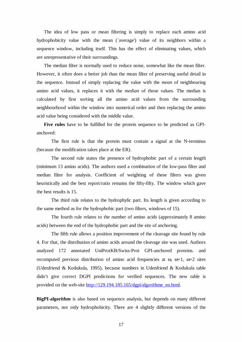
The idea of low pass or mean filtering is simply to replace each amino acid
hydrophobicity value with the mean (`average') value of its neighbors within a
sequence window, including itself. This has the effect of eliminating values, which
are unrepresentative of their surroundings.
The median filter is normally used to reduce noise, somewhat like the mean filter.
However, it often does a better job than the mean filter of preserving useful detail in
the sequence. Instead of simply replacing the value with the mean of neighbouring
amino acid values, it replaces it with the median of those values. The median is
calculated by first sorting all the amino acid values from the surrounding
neighbourhood within the window into numerical order and then replacing the amino
acid value being considered with the middle value.
Five rules have to be fulfilled for the protein sequence to be predicted as GPI-
anchored:
The first rule is that the protein must contain a signal at the N-terminus
(because the modification takes place at the ER).
The second rule states the presence of hydrophobic part of a certain length
(minimum 13 amino acids). The authors used a combination of the low-pass filter and
median filter for analysis. Coefficient of weighting of these filters was given
heuristically and the best report/ratio remains the fifty-fifty. The window which gave
the best results is 15.
The third rule relates to the hydrophylic part. Its length is given according to
the same method as for the hydrophobic part (two filters, windows of 15).
The fourth rule relates to the number of amino acids (approximately 8 amino
acids) between the end of the hydrophobic part and the site of anchoring.
The fifth rule allows a position improvement of the cleavage site found by rule
4. For that, the distribution of amino acids around the cleavage site was used. Authors
analyzed 172 annotated UniProtKB/Swiss-Prot GPI-anchored proteins. and
recomputed previous distribution of amino acid frequencies at ω, ω+1, ω+2 sites
(Udenfriend & Kodukula, 1995), because numbers in Udenfriend & Kodukula table
didn’t give correct DGPI predictions for verified sequences. The new table is
provided on the web-site http://129.194.185.165/dgpi/algorithme_en.html.
BigPI-algorithm is also based on sequence analysis, but depends on many different
parameters, not only hydrophobicity. There are 4 slightly different versions of the

18
program, dedicated to 4 eukaryotic taxonomic groups, metazoa, protozoa, fungi and
plants. The general GPI-terminal signal scheme is the same for all four, but differs in
taxon-specific details.
The GPI modification sequence motif for BigPI appeared to be described in
terms of physical properties such as length requirements and average hydrophobicity
(e.g. for the C-terminal segment), sometimes involving interactions of several
sequence positions (Caras & Weddel, 1987; Moran et al.,1991; Udenfriend &
Kodukula, 1995a,b; Furukawa et al., 1997; Eisenhaber et al., 1998).
Since profile constructions, based on alignments of sequence segments around
ω-site are not successful in terms of their failure to find potential proproteins in the
database, the facts that GPI-sequence signal is not well characterized by amino acid
type preferences, and single residue substitution may completely reverse the
modification, a sophisticated scoring function should be applied.
Therefore, a final scoring function S consists of two parts: S=Sprofile +Sppt.
A profile-dependent section Sprofile evaluates the concordance with the weak
amino acid type preferences in the learning set at single alignment positions. The
relative occurrences of amino acids of particular type at a given motif position were
determined. A new profile extraction technique (PSIC: position-specific independent
counts) which assigns both sequence and alignment position-specific weights
(Eisenhaber et al., 1998; Sunyaev et al., 1999) was applied. The profile score Sprofile is
composed of weighted subscores for specific sequence regions (see Figure 2A) and
two penalties.
Physical property terms, which compose the score Sppt, describe the
conservation of physical properties in the GPI-modification signal arising from the
interaction of few or many sequence positions. Those terms include:
• side-chain volume limitations and mutual volume compensation effects
for residues ω-1 . . . ω+2 ;
• backbone flexibility requirements within the segment ω-1 . . . ω+2;
• propeptide length ranges (from ω+1 to the C end);
• spacer region (ω+3 ... ω+8) hydrophilicity and sequence volume per residue;
• hydrophobicity limits averaged over the C-terminal
hydrophobic region and conditions for even distribution
of hydrophobic residues;

19
• the presence of aliphatic hydrophobic residues (LVI-contents in the tail) and
the absence of long stretches of residues with a flexible backbone in the C-
terminal hydrophobic tail.
Sppt is organized in such a manner that clear deviations from value ranges in the
learning set of proproteins are penalized. The form of physical terms in Sppt reflects
BigPI authors model for requirements of the protein binding site in the transamidase
complex executing the GPI modification.
A scoring function, developed for the animal predictor (Eisenhaber et al, 1999)
was slightly modified for plant and fungi predictor; some terms were introduced and
some were removed. For all predictors, four signal region-specific profile terms are in
Sprofile but they are parameterized using the alignment of C termini of corresponding
taxonomy learning set sequences. For the plant predictor, when the analysis of the
plant-specific C-terminal pattern in learning set sequences was carried out, it was
found out that, in addition to the previously described terms in the Sppt component
(physical property terms; see Eisenhaber et al., 1999), seven new terms for plant-
specific features can be introduced. Seven terms in Sppt have been changed in their
functional form for fungi predictor as well, replaced or newly introduced as compared
with the animal predictor.
When a query sequence was submitted, the predictor scans the C-terminal 55
residues (the same length for all taxonomies), calculates a score for each position
(assuming it being the ω-site) and selects the best scoring sites. A score S ≥ 2
indicates a reliably predicted site (prediction label P). Typically, such a score requires
an almost zero Sppt and a sufficiently positive Sprofile. Scores S ≤ −2 are interpreted as
resulting from sequences without capacity for GPI lipid anchoring (label N). The
remaining scores −2 < S < 2 belong to a twilight zone where clear prediction is
difficult (label S). Nevertheless, low Sppt subscores (Sppt ≤ −12 for positive S and Sppt ≤
−8 for negative S) are used to discriminate unlikely transamidase substrates within
this group (prediction quality I). The scores are translated into probabilities of false
motif detection.
GPI-SOM program is based on different principles then DGPI and BigPI
ones. The web-site of the program is http://www.unibe/gpi.ch. Kohonen neural
networks are known to be powerful tools for classification of hidden information in
large datasets. Learning happens classically by adjusting the weights of the

20
connections (synapses) between units (neurons). But Kohonen self-organizing maps
(SOMs), or Kohonen neural networks (Kohonen, 1988), were employed because they
learn by unsupervised training, in contrast to the classical feed-forward networks.
This means that the network self-organizes during the training, and distributes the
sequences in a map without knowing to which classes they belong. Only at the end of
the training the sequences are labelled with their known classes. The neurons are then
assigned to classes according to the sequences that excited them, and by inspection of
the resulting map it is possible to verify if clustering of the different classes occurred.
It was necessary to evaluate different numerical representation formats for
amino acid sequence. Finally sequence information had to be read by the input layer
of a neural network, and there was a dilemma – either substantial loss of information
or huge amount of data and long computation time.
The authors of GPI-SOM tried several different input formats for
representation of positional transformation of protein sequences. They started with
slightly modified Virtual potential (VP) concept from Aires-de-Sousa et al (2003),
who proposed VP formula for DNA sequences; in GPI-SOM the formula was adapted
to Virtual Potentials for amino acids. But finally GPI-SOM authors used principally
different formula for calculation of each amino acid occurrence and introduced so
called “zentriole”. The zentriole Z of a given amino acid A represents its average
position weighed by its proximity to the C-terminus. For three occurrences of A at
positions pA1, pA2, pA3 counted upwards from 1 starting at distance 32 from the C-
terminus, Z was defined as ((pA1/2+pA2)/2+pA3)/2, which generalizes to
∑=
−−=n
iAi
inPAZ
1
1
22)( , where n is the number of
occurrences for a particular amino acid A within 32-residue fragment considered.
For amino acids not occurring in the input sequence, Z equals zero.
Several techniques were used by GPI-SOM authors to identify and minimize
the number of significant C-terminal residues for GPI-anchoring, and as a result just
22 important amino acid positions in C-terminal protein sequence were selected out of
last 32 positions. For a given protein zentriols were calculated, taking into
consideration only amino acids in these 22 positions. This approach provided 20
zentriols (some value for each amino acid present and zero for the absent ones).
Additional input format for GPI-protein recognition, relative hydrophobicity for those

21
22 important amino acid position at the protein C-terminus was also proposed and
investigated. Finally, the combination of a zentriol for each of the 20 amino acids
(occurring in 22 important amino acid positions at the protein C-terminus) with a
collinear representation of relative hydrophobicity for each of these 22 positions was
accepted as a suitable input format. Already by itself, zentriole input format
performed promisingly well and combined with hydrophobicity values of each
position, it achieved minimal error rates. Further studies and optimization were,
therefore, carried out with this type of input vector (Z + H).
Final input vector contained 44 components : calculated values from the
distribution of all 20 amino acids (zentriols), hydrophobicity values for the 22
important C-terminal amino acid positions and 2 extra units. The two extra-units
were added in order to better distinguish GPI-anchoring signals from transmembrane
domains, one unit for the quality of a putative ω site and one for its position.
Thus, the final GPI-prediction program, GPI-SOM, was implemented as a
Kohonen SOM with an input layer with 44 neurons.

22
2. Materials and methods
2.1 Annotation of GPI-anchor in the UniProtKB/Swiss-Prot
Architecture of the UniProtKB/Swiss-Prot and of a UniProtKB entry is explained in
detail in (Farriol-Mathis et al., 2004) and in the UniProtKB/Swiss-Prot user manual
(http://www.expasy.org/sprot/userman.html). To build the datasets required for the
evaluation of the GPI-anchor prediction programs, we used annotations provided
within entries of the UniProtKB/Swiss-Prot. This work was started with
UniProtKB/Swiss-Prot Release 48.1 (September, 2005) and finally updated with new data
from Release 49.7 (May, 2006). We required only experimentally proven
sequences/entries to be included in datasets.
Entries for GPI-anchored proteins contain information on both the fact of GPI-
anchoring and the attachment site. GPI-annotation is represented in several lines – key
words (KW), comments (CC) and feature table (FT).
Statement in KW-line on GPI-anchoring is independent of the fact of experimental
verification, and no non-experimental qualifiers are added, even if the protein is not
experimentally proven to be GPI-anchored. See the example below.
KW GPI-anchor;
We considered CC-line as the most informative one for protein GPI-status.
Concerning the fact of GPI-anchor modification, CC-line looks as the following:
CC -!-SUBCELLULAR LOCATION: Attached to the membrane by a GPI-
anchor.
This kind of CC-line was considered as experimental proof of GPI-anchoring. The
entries with non-experimental qualifiers in the end of this line we did not consider as
proven, and didn’t include them into the positive dataset.
Cleavage site information is indicated in FT LIPID line. Example is shown below.
FT LIPID 174 174 GPI-anchor amidated asparagine
There are only 43 entries with experimentally proven cleavage sites, others contain
non-experimental qualifiers in FT LIPID-line.
The protein which is supposed to become GPI-anchored, has to be transported to the
endoplasmic reticulum of the cell to be modified with the enzyme. That’s why the
presence of the signal-peptide, which is indicated in FT SIGNAL-line is important to
consider (see example below).
FT SIGNAL 1 36

23
This kind of entry without any non-experimental qualifier in the FT SIGNAL-line was
considered as experimentally verified secreted protein.
2.2 Building datasets
Taking into account considerations above, we built the negative and positive datasets
used later to evaluate the program performance. We used grep-function with indicated
regular expressions for the purpose of database search.
2.2.1 Positive dataset creation:
First we selected entries from the whole SP database with regular expression
KW.*GPI-anchor; (“GPI-anchor biosynthesis.” entries have the expression in their
key-word line, so are not included). To have only experimentally verified proteins
with GPI-anchor, out of those entries we selected ones without:
CC.*GPI-anchor\(By.*\s+.*similarity\)
CC.*GPI-anchor.\*\s+.*\(Potential\)
CC.*GPI-anchor.*\s+.*\(Probable\)
which can be generalized with CC.*GPI-anchor.*\s+.*\( .
In order to avoid introduction of a bias in the evaluation procedure, we subtracted the
entries used for program training from the entries extracted from the
UniProtKB/Swiss-Prot to build the positive datasets. We did not exclude proteins
whose sequence has similarity greater than 90% with proteins of a training dataset, as
it is usually done with datasets, because even a point mutation may prohibit GPI-
anchoring (Eisenhaber et al, 1999). The remaining proteins are those which carry
experimentally verified GPI-anchor and which the considered prediction programs
have not been trained with.
2.2.2 Dataset with experimentally verified cleavage site.
Entries with proven cleavage site were selected in the following way. Dataset with
GPI-anchor key-word KW.*GPI-anchor; was checked for the expression below.
FT.*LIPID.*\(.*\)\.
GPI-entries without the expression were selected (we wanted entries without non-
experimantal qualifiers, which are always in parentheses).
2.2.3 Negative dataset creation.
We required protein N-terminal sequencing on amino acid level to make sure that
leader peptide existed for these proteins and was properly cleaved. We wanted to
make sure, that the protein can be transported to the ER and encounter the proper
enzyme.

24
Again, we started with the whole Swiss-Prot database, and took all entries from SP
with the following expression: FT SIGNAL\s+\d+\s+\d+\s*\n
We wanted to exclude entries if any which were annotated to be GPI-anchored. So
we selected entries without KW.*GPI-anchor;
We wrote a Perl script to find all entries where amino acid protein sequence starts at
(+1) amino acid position relative to the end of signal peptide. The result was
improved with the addition of entries with RP.*Sequence of N-terminus.
In addition, we required proteins in the negative dataset to be secreted. A condition
CC.*SUBCELLULAR LOCATION.*Secreted protein was imposed.
2.2.4 Special case: transmembrane proteins
We investigated the performance of all three programs on the dataset of
transmembrane proteins, which are a known source of false-positive predictions
(Dalley and Bulleid, 2003, Fankhauser et al, 2005). The dataset was made of
proteins, spanning the membrane one time with their N-terminus being extracellular
and intracellular (type I and type II transmembrane proteins, respectively) and of
proteins, having their C-terminal region imbedded to the membrane (type IV
membrane proteins). The following expressions were used for the search:
CC SUBCELLULAR LOCATION: Type I membrane protein\.\n
CC SUBCELLULAR LOCATION: Type II membrane protein\.\n
For type I and II proteins we took only metazoan entries with signal peptide, indicated
in FT-line, although allowing non-experimental qualifiers there.
Type IV metazoan proteins were also selected, using the following expression and
later filtering for metazoan OC line.
CC SUBCELLULAR LOCATION: Type IV membrane protein\.\n Those entries
were not filtered for signal peptide presence, since there is no signal peptide in them,
but importance of these entries for program evaluation is significant.
2.2.5 Automated dataset development and update
Since the grep-search is semi-automated and requires human intervention, in
the end we decided to develop alternative fully automated tool(s) for the search of
GPI-anchored proteins in UniProtKB/Swiss-Prot and for the check of their
experimental verification.
Two scripts were developed for this purpose.
The first script is performing the full-text search in UniProtKB/Swiss-Prot, retrieves
the entries and saves them in a file in Swiss-Prot format. The output is ready to use by

25
the second script, which filters the entries to retain ones with key-word “GPI-anchor”
and to remove afterwards ones with non-experimental qualifiers for GPI-anchor in
CC-line, SUBCELLULAR LOCATION. Its output can be AC-list of verified entries or
verified entries themselves.
Script 1 (for Perl-code, see appendix).
The search is done with LWP::User Agent perl module and implies three steps.
The first step calls one of the expasy-web-sites for the full-text search in
UniProtKB/Swiss-Prot (http://www.expasy.ch/cgi-bin/sprot-search-ful). We propose
to search just for “GPI-anchor”.
The second step saves the entries found in a file in the directory outgoing of the
ExPASy anonymous ftp server.
The third step downloads the file from the ftp-server and saves it on a user-computer.
The second step is possible only if the first one was successful, since the
second step uses the URL, retrieved from the first step output. The third step is
possible only if the second one was successful and the file with entries found was
saved on ftp-server.
Script 2 (for Perl-code, see appendix).
The second script was written to replace multiple calls for grep-function from CRISP-
program, which was used initially to build all the datasets. As an input the script uses
the file with entries in Swiss-Prot format, so it can be merged with the first script
without any modifications.
The major engine of the script is Swiss Knife perl-module. Key element of the script
is analysis of KW-line (an array of key-words) and CC-lines (a hash-table), exactly
the same idea which was used with grep-function initially, but now with different
tools, analyzing several conditions from different entry lines on the same run. We
select entries with KW “GPI-anchor” and filter them for non-experimental qualifiers
in CC-line, SUBCELLULAR LOCATION, mentioning GPI-anchor.
2.3 Datasets used for development of the GPI-prediction programs.
We analyzed all the datasets, which were used for development of the 3 programs.
Those datasets have to be subtracted from the final datasets to give a clean update. We
will discuss their contents in Results section.
2.4 Access to GPI-prediction-programs.

26
Three currently available programs – BigPI, DGPI and GPI-SOM were analyzed. Perl
web-scripts, calling corresponding web-sites and analyzing the output from them were
written. See Figure 3 for the description of the algorithm of the scripts. Perl-script to
call DGPI-program is provided in the appendix.
Figure 3. General algorithm used for accessing web-sites of 3 GPI-predicting
programs.
Call for GPI-prediction program
Read a single entry from the file with SP-entries
Have another entry?
Having the sequence, call remote server of predicting program using LWP::UserAgent.
Extract protein sequence from it with SwissKnife Perl module
Server response: success or not
Parse the output, looking for prediction
Print ‘Error’
Positive GPI Prediction ++
Negative GPI Prediction ++
no
yes
no
yes
Print results
no yes

27
For program evaluation, entries from positive and negative datasets were used as an
input. Sequence and accession number information were retrieved from the entry with
Swiss Knife Perl-module, and then submitted to the web-sites in fasta-format. Slightly
different scripts were written to call each prediction program. The key element of all
the three scripts was LWP::UserAgent module. When we create a User-Agent object,
it connects to a server, sends requests, receives responses and masters the received
data.
2.5 Program performance evaluation
In order to assess each prediction program performance, several statistical values were
calculated. In the following formulas, abbreviations stand for: TP, true positive; FP,
false positive; TN, true negative; FN, false negative.
The sensitivity (Sn) is defined as the proportion of true positive items out of all items
in a positive set and can be calculated with:
FNTP
TPSn
+=
The specificity (Sp) is the proportion of true negatives out of all negatives and can be
calculated with:
FPTN
TNSp
+=
The accuracy (Acc) is the proportion of all true predictions (positive and negative) on
positive and negative examples and is given by:
FNFPTNTP
TNTPAcc
++++=
Finally, Mathews’ correlation coefficient (MCC, Matthews, 1975) is given by:
( )( )( )( )FNTNFPTNFPTPFNTP
FNFPTNTPMCC
++++×−×= or
( )( )( )( )FNTNFPTNFPTPFNTP
FPTPFNTPTPFPTNFNTPMCC
++++++−×+++= ))(()(
It varies between –1 and +1; a value of +1 indicating there is a perfect agreement
between observations and predictions; a value of 0 indicating that predictions are not
better as they were randomly generated; a value of –1 indicating that predictions are
in total disagreement with what is observed.

28
3. Results
3.1 Datasets.
3.1.1 Analysis of the datasets, used for the program training.
Contents of all positive training datasets, filtered for GPI-verified sequences
and UniProtKB/Swiss-Prot accession-numbers, are displayed in Table 1.
BigPI (metazoa/protozoa) dataset consists of 167 sequences, with 136 out of
them being experimentally verified (row 1, Table 1); BigPI (fungal) and BigPI (plant)
datasets are based on mostly non-verified entries:
plant dataset: 254 total/ 42 experimentally verified (Table 1, row 2);
fungal dataset: 219 total/ 19 experimentally verified (Table 1, row 3).
DGPI authors didn’t mention the dataset they used for their program creation,
but it was done in Swiss-Prot with Swiss-Prot entries, and approximately at the same
time when BigPI was written, so we considered DGPI dataset, used for program
development to be the same, as verified BigPI original dataset for metazoa/protozoa,
so only subtraction of BigPI training set from final positive dataset was done.
GPI-SOM required both positive (training and validating ones) and negative
sets. GPI-SOM positive training set and GPI-SOM positive validation set together
consisted of 358 proteins, taken from Genbank, out of which just 87 have 100%
sequence identity with some UniProtKB/Swiss-Prot entries, and just 75 out of these
87 are experimentally verified. (Table 1, row 5). Used sequences were subtracted
from the final positive dataset. We used different principles to create negative dataset,
than those that were used by GPI-SOM authors (for instance, they used cytosolic
protein sequences without signal-peptide), so GPI-SOM negative dataset was not
taken into account at all.
Winding up, we will give a few comments about training datasets quality.
Positive sets used for program creation contain a lot of unverified sequences (see
Fig.4). It immediately created some doubts about programs quality, since this is the
case for all three programs. Besides this, there are other examples of irregularities.
GPI-SOM, non-taxon-specific program, used mostly protein sequences from
Arabidopsis thaliana as a positive set, which is an obvious bias, which should have
been avoided. One of GPI-SOM negative training set consists of proteins without
signal peptide, which is not appropriated. If a protein doesn’t contain a signal peptide,
it cannot be transported to the ER and will never encounter the modifying enzyme.
Whatever C-terminal sequence it has, in nature this protein will never become GPI-

29
anchored, although it may contain a proper C-terminal part for anchoring. This
approach may lead to false predictions.
Table 1. Taxonomic contents of the training datasets:
Row Datasets Metazoa Protozoa Fungi Plants Total Positive datasets, used for the program development
1 BigPI/DGPI meta/prot 96 37 3 0 136 2 BigPI plants 0 0 0 42 42 3 BigPI fungi 0 0 19 0 19 4 Verified ω-site, training 19 15 1 0 35 5 SOM positive 16 14 3 42 75
Table 1 is based only on experimentally verified GPI UniprotKB/Swiss-Prot entries.
The training datasets of BigPI/DGPI and GPI-SOM overlap significantly; in Table 1
we showed just experimentally verified Swiss-Prot entries, but a significant
percentage of entries in the training datasets used are not verified.
3.1.2 Updated datasets.
Since all the datasets had to be updated, we created our own positive and negative
datasets. The goal was to make datasets of protein sequences which programs have
never encountered before.
Positive dataset. First we started with “grep”-search procedure, described in
“Materials and methods”, but finally we wrote two Perl-scripts to make it automatic
(for Perl-code, see Appendix), and updated our previoius results. UniProtKB/Swiss-
Prot release 49.7 contain 389 proven GPI-anchored proteins. Its taxonomic contents,
before and after subtraction of “already seen” entries, is presented in updated dataset
section, Table 2, rows 1 and 3, respectively.
Negative dataset. Proteins with determined N-terminal amino-acid sequence, proven
cleaved signal-peptide, with the comment “Secreted” in CC SUBCELLULAR
LOCATION line and without any mentioning of GPI-anchor anywhere in
corresponding entry were selected. (See row 4, Table 2). The requirement of
secretion will give some additional confidence, that the protein is not GPI-anchored,
since only the presence of signal peptide does not guarantee it. To verify that, we
looked at our positive set of verified metazoan entries – 262. There are 23 entries
with comment “Secreted protein”, but either “After cleavage: Secreted protein” or

30
“Secreted protein” is a comment for a different splice-isoform. So “Secreted protein”
is a proper additional selection criteria, which would imply the absence of GPI-
anchoring.
As we said in the previous section, for negative dataset we used different principles,
than authors of GPI-SOM, that is why our dataset was totally different, and we didn’t
need any subtraction.
Transmembrane proteins dataset. See Table 2, rows 6 and 7.
We made two datasets of eukaryotic transmembrane proteins, as described in
“Materials and methods”. The first one – for non-plant type I and II transmembrane
proteins with signal peptide (1156 entries), the second one – for non-plant type IV
transmembrane proteins without signal peptide (29 entries).
Dataset of proteins with verified GPI-cleavage site. Unfortunately, only 8 entries
out of 43 eukaryotic proved cleavage site entries were not used in a training set, so
this studies don’t really show if the cleavage site prediction is reliable or not. For
taxonomic contents see Table 1, row 4 (training set) and Table 2, row 2 (total set
before subtraction) and row 5 (evaluation set).
Table 2. Taxonomic contents of newly created datasets.
Row Datasets Metazoa Protozoa Fungi Plants Total Updated datasets before subtraction
1 Positive 262 66 19 42 389 2 Verified ω-site 26 16 1 0 43
Updated datasets after subtraction 3 Positive 158 27 0 0 185 4 Negative 227 0 17 13 257 5 Verified ω-site, evaluat 7 1 0 0 8 6 Transmem I & II, SIG 1143 10 3 0 1156 7 Transmem IV, NO SIG 23 3 3 0 29
Table 2 is based only on experimentally verified GPI UniprotKB/Swiss-Prot entries.
Highlighted column in Table 2 shows updated metazoan datasets, later used for
program evaluation. Table 2 clearly demonstrates the lack of verified entries for
plants and fungi, and limited amount of protozoan entries, that is why we decided to
proceed just with metazoan datasets.

31
On the way of creation updated datasets, we encountered a number of difficulties.
There were two main problems for the positive set:
• alternative splicing
Analyzing positive entries with “GPI-anchor” key word, we encountered a problem
with alternative splicing. Sometimes the sequence displayed in the entry was not the
sequence of the spliced isoform, which would become GPI-anchored, there was just a
reference to the anchored isoform inside the entry. We removed these entries from the
positive set. Also it was not stated clearly for a few times which isoform is anchored.
As far as we know, this problem has been already fixed by curators (Nathalie Farriol-
Mathis, personal communications).
• lack of verified entries
The general difficulty, which we encountered – the small number of experimentally
proved entries with GPI-anchor, especially for fungi and protozoa.
The problem with negative set is that no scientist ever states, that the protein is
not GPI-anchored. To conclusively prove that a site is negative under all conditions is
impossible, but to know that it is negative at least in some contexts would be useful.
Since there is no experimental verification of the fact, that the protein is not GPI-
anchored, we based our negative set construction on the entries, where the presence of
signal-peptide was experimentally verified by N-terminal amino acid sequencing and
without any mentioning of GPI-anchoring, and selected only secreted proteins from
these entries. None of those entries contain mass spectrometry measurements, which
would exclude the possibility of GPI-anchoring, which is unfortunate.
3.2 Evaluation of the GPI-anchor prediction programs
GPI-anchor prediction programs are involved in the annotation procedure of
the UniProtKB/Swiss-Prot, so their evaluation is of high importance for providing a
high-quality annotated database. The results obtained are presented in the following
paragraphs. There is different taxonomic specificity for existing GPI-prediction
programs in the way they were developed. BigPI implies different versions for
metazoa, protozoa, plants and fungi, DGPI and GPI-SOM make no difference
between different taxonomies. For protozoa we observed very few entries, and all
known verified entries for fungi and plants were used for the development of at least
one of the three programs, that is another reason why the evaluation was performed

32
only for the most numerous dataset – metazoan. It allowed us to compare conclusions
of metazoan-specialized (BigPI) and non-specialized (DGPI and GPI-SOM) programs
as well, and also to compare results of BigPI programs, written for different
taxonomies than the datasets used.
First we compared three programs performance for the fact of GPI-anchoring
on positive and negative metazoan datasets and calculated sensitivity, specificity,
accuracy and Matthew’s correlation coefficient for the programs (see “Materials and
methods”, section 2.5 for the formulas). Results are presented in Table 3.
Table 3. Prediction of the fact of GPI-anchoring by BigPI, DGPI and GPI-SOM
on metazoan datasets.
POSITIVE SET NEGATIVE SET Sensitivity Specificity Matt cor cf Accuracy TP FN FP TN Sn Sp MCC ACC
BigPI 93 65 0 227 0.589 1.000 0.676 0.831 DGPI 119 39 14 213 0.753 0.938 0.715 0.862
GPI-SOM 122 36 12 215 0.772 0.947 0.743 0.875
Calculations of sensitivity/specificity show, that only BigPI satisfies the
requirements of very low FP-value. Let’s not forget, that BigPI is a specialized
program, which distinguish between taxonomic groups, and it seems to be a clear
advantage. BigPI is currently used in Anabelle (a tool in UniProtKB/Swiss-Prot
annotation platform which gives curators ways to choose the right decision
concerning a particular protein feature) for GPI-anchor prediction. BigPI is the most
specific program, and partially because of that the least sensitive. DGPI and GPI-
SOM are less specific (0.95), but subsequently more sensitive. More than 5% of false
positive predictions is a lot, that is why the other two programs are not used in
UniProtKB/Swiss-Prot curation procedure. Accuracy for all three programs is good
and have about the same value for all three, about 0.85; Matthews correlation
coefficients (MCC) are also similar, about 0.7. MCC is symmetric with respect to
false positive and false negative values, Fp and Fn, and since it is almost equal for all
the programs, we expected, that programs, which gain in specificity, would lose in
sensitivity and visa versa (basic statistical property).
That’s interesting, that DGPI and GPI-SOM have close sensitivity and specificity
values, although are based on different principles.

33
3.3 Cleavage site prediction comparison
All three programs do prediction of the ω-site, and we wanted to proceed with
the evaluation of this feature as well. There are just 26 verified metazoan entries.
Program performance for them is presented in Table 4.
Table 4. Summary of the cleavage site prediction results (metazoa only):
DGPI BigPI GPI-SOM Evaluation set : 7 sequences
Correct site prediction 4 6 4 Incorrect site prediction 2 1 3
NO site 1 0 0 Total 7 7 7
Training set : 19 sequences Correct site prediction 13 17 10
Incorrect site prediction 5 2 9 NO site 1 0 0 Total 19 19 19
Sum of both sets : 26 sequences Correct site prediction 17 23 14
Incorrect site prediction 7 3 12 NO site 2 0 0 Total 26 26 26
We looked at the site prediction regardless of the result of GPI-anchoring
prediction, since they are independent for programs. All three programs may predict
the site correctly or incorrectly or give no site prediction for the protein sequence
(three possibilities). Comparison of the programs results for the training and
evaluation cleavage site sets did not show a dramatic difference. When we look just at
the results for 7 entries, which were not used for the program development (see Table
4, results for evaluation set), we see that all the programs make mistakes in the site
prediction. But there is roughly the same proportion of wrong site predictions for the
training set of BigPI and DGPI as well, and a little bit less than 50% of false
predictions for GPI-SOM (see predictions for training set). That is why we decided to
make conclusions about this feature, looking at the whole dataset of 26 metazoan
proteins with verified ω-site (see predictions for the sum of both sets).
We didn’t calculate sensitivity and specificity for cleavage site predictions,
since there are very few verified sites and it wouldn’t have any statistical meaning, so
we just looked at the percentage of correct answers.
BigPI predicts ω-site correctly in about 90% of the cases. DGPI is also a good
program to predict ω-site (about 65% of correct predictions), but in more than one

34
third of the cases it is wrong, and a couple of times it cannot recognize the sites at all.
GPI-SOM is the worst program out of these three in site predictions, it is wrong in
about 45% of the cases.
Unfortunately, there are not enough entries to state something about statistical
significance of these results, but overall conclusion is clear – more data are needed.
There are some confusing conclusions which all three programs make, since
all three predict the fact of GPI-anchoring and the corresponding cleavage site(s)
independently. So the conclusion of the program can be “No GPI-anchoring”, but it
still predicts the site of cleavage. It is a really useful thing that the programs do,
because it may give some clues why the prediction was negative (BigPI, for example,
gives numerical scores for each physical term), and allow even “in silico” mutations
of the protein. Certainly, the programs have to be upgraded for this kind of studies,
but this is just a very useful initial step.
3.4 Transmembrane proteins
We also checked if three investigated programs make mistakes for transmembrane
proteins, which are not GPI-anchored. The dataset was built, according to the rules
from “Materials and methods” section. Fankhauser et al argued, that since both C-
terminal transmembrane proteins and proteins, carrying GPI-anchor, have highly
hydrophobic C-terminus, GPI-prediction programs sometimes give false positive
predictions for such proteins. To evaluate this assumption, we looked at program
perfomance on typeI/typeII transmembrane protein dataset (see Table 5). Since
transmembrane proteins form another negative set, here we can talk about
“transmembrane specificity”. We cannot add transmembrane proteins to our main
negative dataset (Table 2, line 4), because they were built according to different
principles. The only Fp prediction for BigPI is P10379. In the corresponding Swiss-
Prot entry there is no indication of the fact of GPI-anchoring for this protein.
Table 5. “Specialized” specificity, calculated on transmembrane proteins,
type I and II.
TRANSMEMBRANE NEGATIVE SET SPECIFICITY
FP TN SP BigPI 1 1142 0.9991 DGPI 59 1084 0.9484
GPI-SOM 71 1072 0.9379

35
We also made a dataset of metazoan type IV transmembrane proteins. There are just
23 entries selected, and these proteins don’t carry signal peptide (that is why they are
rejected by DGPI and GPI-SOM). To demonstrate significance for the programs the
presence of signal peptide in the sequence, we attached artificial signal to all 23
sequences and ran prediction programs again. Results are presented on the diagram 1.
BigPI doesn’t look at the presence of signal peptide, just at the C-terminal
region, so predictions are the same with and without the signal. It is interesting, that
three entries, Q9UNK0, O88983 and Q9Z2Q7, are predicted to be GPI-anchored
according to BigPI with good scores. This is the case when closer look is necessary,
since those proteins can potentially carry GPI-anchor despite being transmembrane,
and these are interesting cases when the very good performance program – BigPI –
gives some false positive predictions.
If we compare predictions by DGPI and GPI-SOM on the same sequences, but
with artificial signal and without signal, we see, that these two programs make their
conclusions about GPI-anchoring, based mostly on the presence of N-terminal signal,
and GPI-signal itself is almost ignored.
Diagram 1. Comparison of GPI-predictions on Type IV membrane proteins with
and without artificial signal sequence.
3.5 Comparative analysis of program performance
We thought, it could be useful to see, if there are some tendencies in mutual
mistakes, like –“if one program prediction is wrong for some sequence, the second
program prediction for this sequence is also always wrong”. First we looked at
Done on 23 proteins (with signal).
0%
20%
40%
60%
80%
100%
FP
TN
BigPI DGPI GPI-SOM
Done on 23 proteins (without signal).
0%
20%
40%
60%
80%
100%
FP
TN
BigPI DGPI GPI-SOM

36
comparison of Fp-predictions between 3 programs on metazoan negative set. See
Figure 5 and Table A1 in Appendix for the details.
Figure 5. Relations between programs for Fp-predictions.
BigPI is the best program, no false positive prediction for 227 negative entries.
Although two other programs, GPI-SOM and DGPI, are based on different principles,
total number of Fp-predictions they give is about the same (12 and 14, respectively,
for metazoan negative set). Some entries (7) are the same for both programs, the other
incorrectly predicted entries are different. The reason of both phenomena can be the
target of further investigation and improvement of the programs.
We also looked at comparison of Fn-predictions between three programs on metazoan
positive set. See Figure 6 and Table A2 in Appendix. The goal is to compare Fn
prediction and to figure out, which predictions are shared and try to understand what
it can be useful for.
Figure 6. Relations between programs for Fn-predictions.
37
When we look at the Figure 6 and compare cases of negative answers for the positive
set (see also table A2 in Appendix), we see, that DGPI and GPI-SOM share 21 Fn
predictions with BigPI, and have groups of 9 and 10 Fn-predictions for DGPI and
GPI-SOM, respectively, unshared with any other program. Comparing overlap
between DGPI/BigPI (8) and GPI-SOM/BigPI (4) Fn-predictions, the impression is
that DGPI approach is closer to BigPI one, than GPI-SOM to BigPI. Of course, there
are very few data to make this conclusion, but what we know about the program
algorithms, may suggest that it is right to some extent. What is an interesting fact -
DGPI and GPI-SOM share only 1 Fn-prediction, while sharing many false positives
(Figure 5). It may suggest, that programs accept sequences according to similar
principles, but reject them because of different criteria. Another striking thing, which
was expected, is a lot of Fn predictions by BigPI alone (32), what is a directly linked
to its very high specificity value.
The most interesting thing, which could be done here is an investigation of correlation
between the program conclusions (between scores). Unfortunately, only one program,
BigPI, gives numerical score, so it was not possible.
Another useful thing, which can be derived from this kind of analysis, is the
following. Let’s look at the BigPI case and analyze whether the program for one
taxonomic group can be used for another one.
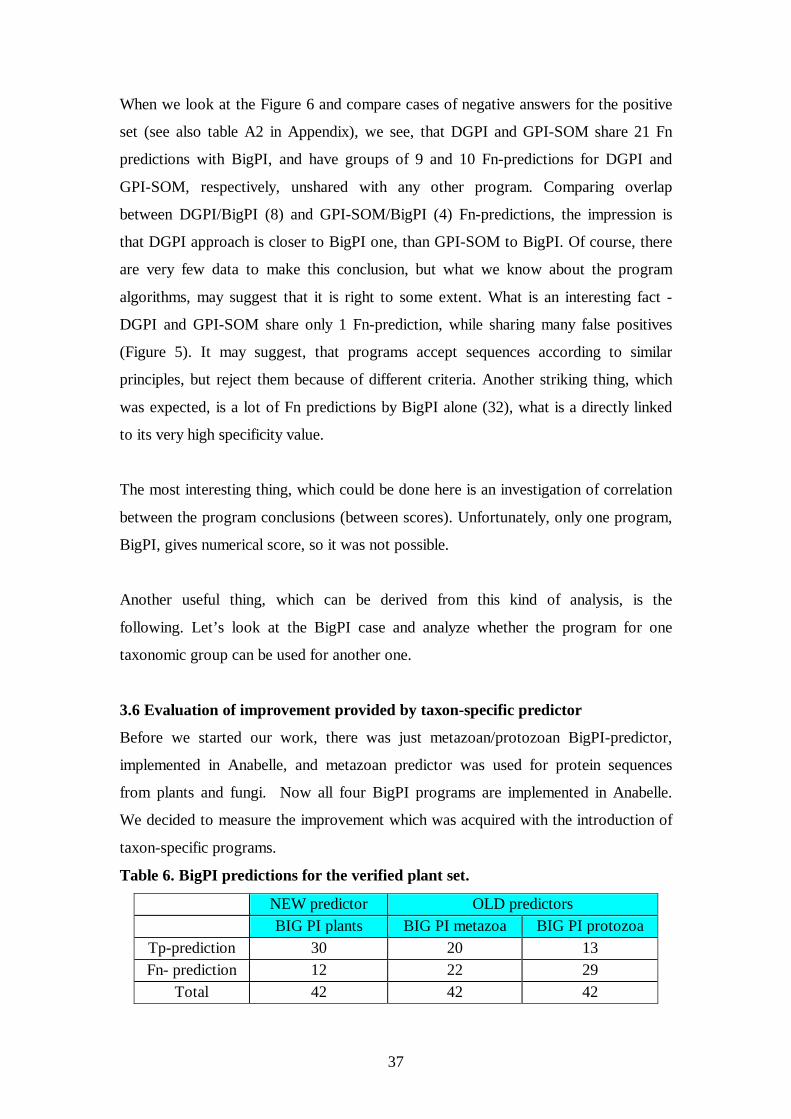
3.6 Evaluation of improvement provided by taxon-specific predictor
Before we started our work, there was just metazoan/protozoan BigPI-predictor,
implemented in Anabelle, and metazoan predictor was used for protein sequences
from plants and fungi. Now all four BigPI programs are implemented in Anabelle.
We decided to measure the improvement which was acquired with the introduction of
taxon-specific programs.
Table 6. BigPI predictions for the verified plant set.
NEW predictor OLD predictors BIG PI plants BIG PI metazoa BIG PI protozoa
Tp-prediction 30 20 13 Fn- prediction 12 22 29
Total 42 42 42

38
We analyzed the relations between taxon-specific BigPI-predictors on 42 verified
plant sequences (Eisenhaber et al, 2003, Borner et al). Metazoan/protozoan (old)
predictor results were compared with plant (new) predictor result for each sequence.
All the plants sequences predicted by metazoan program are predicted by plant
program, but not visa versa – some of the sequences, predicted by plant program, are
not recognized by metazoan one. For this dataset, metazoan predictor never says that
the protein is not GPI-anchored, when plant predictors says that it is anchored, it
verifies former use of metazoan predictor for plants.
Metazoan predictor recognizes 20 of 42 proven sequences, 9 of which are shared with
protozoan predictor. Although protozoan predictor recognizes less sequences as GPI-
anchored (13 total), it does recognize 4 additional sequences which metazoan
predictor doesn’t. What is important – all 24 sequences, predicted by meta/pro BigPI
for plants are recognized by plant BigPI, and 6 additional sequences are identified.
Clear advantage of taxon-specific program use is observed. However it has to be
noticed, that those 42 sequences were in plant-BigPI training set. BigPI is not a
program, based on mashine-learning, so training implies for it just choice of a
threshold to minimize Fp-rate, so we think we can make some conclusions based on
these “training” data.
Nowadays plant BigPI is already implemented in Anabelle, so this investigation is
just confirming that is was useful to do it, because it is a huge improvement
We performed the same evaluation for verified fungi set (Eisenhaber et al,
2003). The problem was that just 19 fungi sequences in UniProtKB/Swiss-Prot are
proven to be GPI-anchored, but let’s see what we had for them.
There are now at least 43 verified fungi-entries known, according to
Eisenhaber et al, 2004, simply in the UniProtKB/Swiss-Prot most of them are still
marked with non-experimental qualifiers in CC-line. To be consistent, we decided to
proceed only with those proven 19 sequences from UniProtKB/Swiss-Prot and look,
as before for plant sequences, what is the difference between old and new BigPI-
predictors.

39
Table 7. BigPI predictions for the verified fungi set.
NEW predictor OLD predictors BIG PI fungi BIG PI metazoa BIG PI protozoa
Tp prediction 18 11 4 Fn prediction 1 8 15
Total 19 19 19
We analyzed the relations between taxon-specific BigPI-predictors on 19 verified
fungal entries. Metazoan/protozoan predictor results were compared with fungal
predictor result for each sequence.
Protozan predictor gives too many false negatives to be applicable for fungi, just 4
fungal sequences are recognized. These 4 sequences are shared with metazoan
predictor, so probably there was no point to use protozoan predictor for fungi. In its
turn, metazoan predictor recognizes 11 sequences from this fungal set. But fungal
predictor performance is very good for the fungal set, it recognizes 18 out of 19
sequences. Those 18 include 11 sequences, recognized by metazoan predictor. Again,
there is a clear advantage in using fungal predictor for fungi – much more true
positive predictions, than would be for metazoan predictor on fungal set. It should be
noticed, that 19 sequences are from the fungal BigPI training set. Fungal predictor is
now also implemented in Anabelle.
4. Discussion and concluding remarks.
Let’s make some overview of all GPI-prediction programs from the user position.
None of the programs seem to be really user friendly.
• BigPI
• Doesn’t allow to download a file with sequences, just cut and paste
fasta-formatted sequences into the on-line form
• Doesn’t check for the presence of a signal peptide in proprotein
sequence
• There is a critical limit of 500 amino acids per line; if more, the
program just ignores the rest of the sequence without further warnings
• No fasta-header in plant- and fungi-output predictions, just the number
of amino acids in the sequence submitted
• DGPI

40
• Allows to download just a single protein sequence, although in variety
of formats
• Doesn’t give the resulting score, according to which the prediction is
made
• GPI-SOM
• Doesn’t give any numerical scores, just a graphical map
• Shows the cleavage site position from the C-terminus of the protein
sequence
The properties listed make the programs inconvenient for users and have to be fixed
sooner or later.
Particular results, especially numerical results, were already discussed in the Result-
section in the end of each sub-section.
The two scripts for the automatic GPI-dataset update are to be integrated into the
generic procedure of updating Biosapiense datasets.
Evaluation of all three programs was performed before just once by Fankhauser et al,
2005, the authors of the program GPI-SOM, The datasets they used for their
evaluation were not composed of verified GPI-anchored proteins.
To conclude, we can list the following items:
Evaluation sets suffer from insufficient number of verified data in the database. We
considered possible to perform the evaluation only for metazoan datasets.
For metazoa the best prediction program is BigPI – no Fp. It has a special web-site,
dedicated to metazoan sequences, which is a clear advantage over non-specialized
programs. Exact cleavage site prediction performance is difficult to evaluate – lack of
independent dataset.
Comparison of metazoan and plant BigPI-predictor performance for verified plant
dataset, and metazoan and fungal BigPI-predictor performance for verified fungal
dataset showed, that taxon-specialized BigPI predictors give a huge improvement in
GPI-anchoring prediction. Metazoan predictor never contradicts with negative
predictions of specialized predictors on corresponding datasets, but gives more false
negative predictions.

41
All the programs make mistakes with transmembrane proteins, BigPI - mostly for
type IV transmembrane proteins, and DGPI and GPI-SOM make a lot of false-
positive predictions for type I and II transmembrane proteins.
The kind of program evaluation performed shows clearly a “chicken and egg”
problem. Good prediction program can be written only if good datasets are provided,
but datasets themselves, at least indirectly, depend on prediction programs. Ideally the
programs should give experimental scientists some hints, which proteins to
investigate to enlarge those datasets.
The Swiss-Prot annotation as we see it is a process of extraction of
information about the protein from scientific literature together with its
gathering/collection from other sources, which lead to the information digestion,
rearrangement and reassessment and finally give rise to a comprehensive description
of the protein. Databases like Swiss-Prot, which are committed to contain annotated
entries, are of great importance to experimental biologist community, since their
existence make scientist life easier. Scientific database contents should be
experimentally proven, or at least can be trusted. The extent of this trust has to be
clearly indicated (an indispensable role which non-experimental qualifiers play in
Swiss-Prot entries). The amount of experimentally verified information available is
growing, but still limited. Prediction programs are supposed to partially fill the
existing gaps, at least for the time being. Evaluation of this kind of programs should
lead to the choice of a proper technique if any, which will allow minimal amount of
false positive predictions, mistakes, which may cause at least a significant loss of
efforts. False negative prediction can be considered just as a temporary loss of
information until new data arrival, and not as a mistake.
Acknowledgements.
Many-many thanks to Dr.Anne-Lise Veuthey (my supervisor), Dr.David Perret,
Dr.Patricia Palagi and Prof. Amos Bairoch, as well as to all the other colleagues from
the Swissprot PTM-group and the whole SwissProt. It was a great pleasure for me to
work inside the community for some period of time (unfortunately, a very short
period!).

42
REFERENCES
Aires-de-Sousa,J. and Aires-de-Sousa,L. (2003) Representation of DNA sequences with virtual potentials and their processing by (seqrep) Kohonen self-organizing maps. Bioinformatics, 19, 30–36.
Apweiler R, Bairoch A, Wu CH, Barker WC, Boeckmann B, Ferro S, Gasteiger E, Huang H, Lopez R, Magrane M, Martin MJ, Natale DA, O'Donovan C, Redaschi N, Yeh LS. (2004) UniProt: the Universal Protein knowledgebase. Nucleic Acids Res. Jan 1;32 (Database issue):D115-9.
Apweiler R., Bairoch A., Wu C.H. (2004) Protein sequence databases. Curr. Opin.Chem. Biol. 8:76-80.
Attwood, T., Int. J. Biochem. Cell Biol. 2000, 32, 139–155. Bailey CA, Howard A, Micanovic R, Berger J, Heimer E, Felix A, Gerber L, Brink L, Udenfriend S. Site-directed antibodies for probing the structure and biogenesis of phosphatidylinositol glycan-linked membrane proteins: application to placental alkaline phosphatase. Anal Biochem. 1988 May 1;170(2):532-41.
Bairoch A, Apweiler R, Wu CH, Barker WC, Boeckmann B, Ferro S, Gasteiger E, Huang H, Lopez R, Magrane M, Martin MJ, Natale DA, O'Donovan C, Redaschi N, Yeh LS. The Universal Protein Resource (UniProt).Nucleic Acids Res. 2005 Jan 1;33 (Database issue):D154-9.
Bairoch A. (2000) Serendipity in bioinformatics, the tribulations of a Swiss bioinformatician through exciting times! Bioinformatics 16:48-64.
Bairoch A., Boeckmann B., Ferro S., Gasteiger E. (2004) Swiss-Prot: juggling between evolution and stability. Brief. Bioinform. 5:39-55.
Beghdadi-Rais C, Schreyer M, Rousseaux M, Borel P, Eisenberg RJ, Cohen GH, Bron C, Fasel N. (1993) Carboxyl terminus structural requirements for glycosylphosphatidylinositol anchor addition to cell surface proteins. J of Cell Science 105 831-840. Blom N, Sicheritz-Ponten T, Gupta R, Gammeltoft S, Brunak S. Prediction of post-translational glycosylation and phosphorylation of proteins from the amino acid sequence. Proteomics. 2004 Jun;4(6):1633-49. Review.
Boeckmann B, Blatter MC, Famiglietti L, Hinz U, Lane L, Roechert B, Bairoch A. Protein variety and functional diversity: Swiss-Prot annotation in its biological context.C R Biol. 2005 Oct-Nov;328(10-11):882-99. Epub 2005 Jul 28. Review.
Boeckmann B., Bairoch A., Apweiler R., Blatter M.C., Estreicher A., Gasteiger E.,Martin M.J., Michoud K., O'Donovan C., Phan I., Pilbout S., Schneider M. (2003) The UniProtKB/Swiss-Prot protein knowledgebase and its supplement TrEMBL in 2003.Nucleic Acids Res. 31:365-370.
Bork, P., Dandekar T., Diaz-Lazcoz, Y., Eisenhaber, F. et al., J. Mol. Biol. 1998, 283, 707–725.
Borner GH, Lilley KS, Stevens TJ, Dupree P. Identification of glycosylphosphatidylinositol-anchored proteins in Arabidopsis. A proteomic and genomic analysis.Plant Physiol. 2003 Jun;132(2):568-77. Epub 2003 May 1. Bucht G, Hjalmarsson K. Residues in Torpedo californica acetylcholinesterase necessary for processing to a glycosyl phosphatidylinositol-anchored form. Biochim Biophys Acta. 1996 Feb 8;1292(2):223-32. Butikofer P, Vassella E, Ruepp S, Boschung M, Civenni G, Seebeck T, Hemphill A, Mookherjee N, Pearson TW, Roditi I. Phosphorylation of a major GPI-

43
anchored surface protein of Trypanosoma brucei during transport to the plasma membrane.J Cell Sci. 1999 Jun;112 ( Pt 11):1785-95. Caras IW, Weddell GN, Davitz MA, Nussenzweig V, Martin DW Jr. Signal for attachment of a phospholipid membrane anchor in decay accelerating factor. Science. 1987 Nov 27;238(4831):1280-3.
Cross G.(1990) Glycolipid anchoring of plasma membrane proteins. Annu Rev Cell Biol 6,1-39
Dalley,J.A. and Bulleid,N.J. (2003) The endoplasmic reticulum (ER) translocon can differentiate between hydrophobic sequences allowing signals for glycosylphosphatidylinositol anchor addition to be fully translocated into the ER lumen. J. Biol.Chem., 278, 51749–51757. Eisenhaber B, Bork P, Eisenhaber F. Post-translational GPI lipid anchor modification of proteins in kingdoms of life: analysis of protein sequence data from complete genomes .Protein Eng. 2001 Jan;14(1):17-25.
Eisenhaber B, Bork P, Eisenhaber F. Prediction of potential GPI-modification sites in proprotein sequences.J Mol Biol. 1999 Sep 24;292(3):741-58.
Eisenhaber B, Eisenhaber F, Maurer-Stroh S, Neuberger G. Prediction of sequence signals for lipid post-translational modifications: insights from case studies. Proteomics. (2004a)Jun;4(6):1614-25.
Eisenhaber B, Schneider G, Wildpaner M, Eisenhaber F. A sensitive predictor for potential GPI lipid modification sites in fungal protein sequences and its application to genome-wide studies for Aspergillus nidulans, Candida albicans, Neurospora crassa, Saccharomyces cerevisiae and Schizosaccharomyces pombe. J Mol Biol. (2004b) Mar 19;337(2):243-53.
Eisenhaber B, Wildpaner M, Schultz CJ, Borner GH, Dupree P, Eisenhaber F. Glycosylphosphatidylinositol lipid anchoring of plant proteins. Sensitive prediction from sequence- and genome-wide studies for Arabidopsis and rice. Plant Physiol. 2003 Dec;133(4):1691-701.
Eisenhaber F, Eisenhaber B, Kubina W, Maurer-Stroh S, Neuberger G, Schneider G, Wildpaner M. Prediction of lipid posttranslational modifications and localization signals from protein sequences: big-Pi, NMT and PTS1. Nucleic Acids Res. 2003 Jul 1;31(13):3631-4.
Eisenhaber,B. et al. (1998) Sequence properties of GPI-anchored proteins near the omega-site: constraints for the polypeptide binding site of the putative transamidase. Protein Eng., 11, 1155–1161.
F. Eisenhaber, B. Eisenhaber, and S. Maurer-Stroh. (2003) Prediction of Post-Translational modifications from Amino Acid Sequence: Problems, Pitfalls and Methodological Hits. In Bioinformatics and Genomes: Current Perspectives. Fankhauser N, Maser P. Identification of GPI anchor attachment signals by a Kohonen self-organizing map. Bioinformatics. 2005 May 1;21(9):1846-52. Epub 2005 Feb 2.
Farriol-Mathis N, Garavelli JS, Boeckmann B, Duvaud S, Gasteiger E, Gateau A, Veuthey AL, Bairoch A. Annotation of post-translational modifications in the Swiss-Prot knowledge base.Proteomics. 2004 Jun;4(6):1537-50.
Ferguson,M.A. (1999) The structure, biosynthesis and functions of glycosylphosphatidylinositol anchors, and the contributions of trypanosome research. J. Cell. Sci., 112 (Pt 17), 2799–2809.
Field MC, Menon AK, Cross GA. A glycosylphosphatidylinositol protein anchor from procyclic stage Trypanosoma brucei: lipid structure and biosynthesis. EMBO J. 1991 Oct;10(10):2731-9.

44
Field MC, Menon AK, Cross GA. Developmental variation of glycosylphosphatidylinositol membrane anchors in Trypanosoma brucei. In vitro biosynthesis of intermediates in the construction of the GPI anchor of the major procyclic surface glycoprotein.J Biol Chem. 1992 Mar 15;267(8):5324-9. Furukawa Y, Tsukamoto K, Ikezawa H. Mutational analysis of the C-terminal signal peptide of bovine liver 5'-nucleotidase for GPI anchoring: a study on the significance of the hydrophilic spacer region. Biochim Biophys Acta. 1997 Sep 4;1328(2):185-96.
Garavelli, J., Nucleic Acids Res. 2003, 31, 499–501. Gerber,L.D. et al. (1992) Phosphatidylinositol glycan (pi-g) anchored
membrane proteins. Amino acid requirements adjacent to the site of cleavage and pi-g attachment in the COOH-terminal signal peptide. J. Biol. Chem., 267, 12168–12173.
Guadiz,G. et al. (1998) The carboxyl terminus of Pneumocystis carinii glycoprotein a encodes a functional glycosylphosphatidylinositol signal sequence. J. Biol. Chem.,273, 26202–26209. Haas R, Jackson BC, Reinhold B, Foster JD, Rosenberry TL. Glycoinositol phospholipid anchor and protein C-terminus of bovine erythrocyte acetylcholinesterase: analysis by mass spectrometry and by protein and DNA sequencing. Biochem J. 1996 Mar 15;314 ( Pt 3):817-25.
Hamburger,D. et al. (1995) Yeast gaa1p is required for attachment of a completed GPI anchor onto proteins. J. Cell Biol., 129, 629–639.
Han, K. K., Martinage, A., Int. J. Biochem. 1992, 24, 1928. Hobohm U, Scharf M, Schneider R and Sander C (1992) Protein science, 1,
409-417. Hooper NM. Detergent-insoluble glycosphingolipid/cholesterol-rich
membrane domains, lipid rafts and caveolae (review). Mol Membr Biol. 1999 Apr-Jun;16(2):145-56. Review. Horejsi V, Drbal K, Cebecauer M, Cerny J, Brdicka T, Angelisova P, Stockinger H. GPI-microdomains: a role in signalling via immunoreceptors. Immunol Today. 1999 Aug;20(8):356-61. Review. Howard AD, Berger J, Gerber L, Familletti P, Udenfriend S. Characterization of the phosphatidylinositol-glycan membrane anchor of human placental alkaline phosphatase.Proc Natl Acad Sci U S A. 1987 Sep;84(17):6055-9.
Jensen, L., Gupta, R., Blom, N., Devos, D. et al., J. Mol. Biol.2002, 319, 1257–1265.
Jensen, L., Ussery, D., Brunak, S., Genome Res. 2003, 13, 2444–2449. Kodukula,K. et al. (1993) Biosynthesis of glycosylphosphatidylinositol (GPI)-
anchored membrane proteins in intact cells: specific amino acid requirements adjacent to the site of cleavage and GPI attachment. J. Cell Biol., 120, 657–664.
Kohonen,T. (2001) Self-Organizing Maps, 3rd edn, Springer Series in Information Sciences, Vol. 30, Springer, Berlin.
Kronegg,J. and Buloz,D. (1999) http://129.194.185.165/dgpi/. Kyte,J. and Doolittle,R.F. (1982) A simple method for displaying the
hydropathic character of a protein. J. Mol. Biol., 157, 105–132. Lacey,S.W. et al. (1989) Complementary DNA for the folate binding protein
correctly predicts anchoring to the membrane by glycosyl-phosphatidylinositol. J. Clin. Invest.,84, 715–720.
Masterson, W. J., Doering, T, L, Hart, G. Wo and Englund, P. (1989) Cell 56, 793-800

45
Meyer,U. et al. (2002) The glycosylphosphatidylinositol (GPI) signal sequence of human placental alkaline phosphatase is not recognized by human gpi8p in the context of the yeast GPI anchoring machinery. Mol. Microbiol., 46, 745–748.
Micanovic R, Bailey CA, Brink L, Gerber L, Pan YC, Hulmes JD, Udenfriend S. Aspartic acid-484 of nascent placental alkaline phosphatase condenses with a phosphatidylinositol glycan to become the carboxyl terminus of the mature enzyme. Proc Natl Acad Sci U S A. 1988 Mar;85(5):1398-402.
Moran,P. and Caras,I.W. (1994) Requirements for glycosylphosphatidylinositol attachment are similar but not identical in mammalian cells and parasitic protozoa. J. CellBiol., 125, 333–343.
Nozaki,M. et al. (1999) Developmental abnormalities of glycosylphosphatidylinositolanchor-deficient embryos revealed by Cre/LoxP system. Lab. Invest., 79, 293–299. Ogata S, Hayashi Y, Misumi Y, Ikehara Y. Membrane-anchoring domain of rat liver 5'-nucleotidase: identification of the COOH-terminal serine-523 covalently attached with a glycolipid. Biochemistry. 1990 Aug 28;29(34):7923-7. Ogata S, Hayashi Y, Takami N, Ikehara Y. Chemical characterization of the membrane-anchoring domain of human placental alkaline phosphatase. J Biol Chem. 1988 Jul 25;263(21):10489-94.
Omaetxebarria MJ, Hagglund P, Elortza F, Hooper NM, Arizmendi JM, Jensen ON. Isolation and Characterization of Glycosylphosphatidylinositol-Anchored Peptides by Hydrophilic Interaction Chromatography and MALDI Tandem Mass Spectrometry. Anal Chem. 2006 May 15;78(10):3335-3341.
Roberts WL, Myher JJ, Kuksis A, Low MG, Rosenberry TL. Lipid analysis of the glycoinositol phospholipid membrane anchor of human erythrocyte acetylcholinesterase. Palmitoylation of inositol results in resistance to phosphatidylinositol-specific phospholipase C. J Biol Chem. 1988 Dec 15;263(35):18766-75.
Roberts WL, Santikarn S, Reinhold VN, Rosenberry TL. Structural characterization of the glycoinositol phospholipid membrane anchor of human erythrocyte acetylcholinesterase by fast atom bombardment mass spectrometry. J Biol Chem. 1988 Dec 15;263(35):18776-84.
Stahl,N., Borchelt,D.R., Hsiao,K. and Prusiner,S.B. (1987) Scrapie prion protein contains a phosphatidylinositol glycolipid. Cell, 51, 229–240. Sunyaev SR, Eisenhaber F, Rodchenkov IV, Eisenhaber B, Tumanyan VG, Kuznetsov EN. PSIC: profile extraction from sequence alignments with position-specific counts of independent observations.Protein Eng. 1999 May;12(5):387-94.
Sutterlin,C. et al. (1998) Saccharomyces cerevisiae gpi10, the functional homologue of human pig-b, is required for glycosylphosphatidylinositol-anchor synthesis. Biochem. J., 332 (Pt 1), 153–159.
Taguchi R, Hamakawa N, Maekawa N, Ikezawa H.J. Application of electrospray ionization MS/MS and matrix-assisted laser desorption/ionization-time of flight mass spectrometry to structural analysis of the glycosyl-phosphatidylinositol-anchored protein. Biochem (Tokyo). (1999a) Aug;126(2):421-9. Taguchi R, Yamazaki J, Takahashi M, Hirano A, Ikezawa H. Identification of a new glycosylphosphatidylinositol-anchored 42-kDa protein and its C-terminal peptides from bovine erythrocytes by gas chromatography-, time-of-flight-, and electrospray-ionization-mass spectrometry. Arch Biochem Biophys. (1999b) Mar 1;363(1):60-7.

46
Takeda,J. and Kinoshita,T. (1995) GPI-anchor biosynthesis. Trends Biochem. Sci., 20, 367–371.
Udenfriend and Kodukula (1995b) How glycosylphosphatidylinosital-anchored membrane proteins are made. Annu. Rev. Biochem. 1995, 64:563-91
Udenfriend,S. and Kodukula,K. (1995a) Prediction of ω site in nascent precursor of glycosylphosphatidylinositol protein. Methods Enzymol., 250, 571–582.
Wang J, Maziarz K, Ratnam M. Recognition of the carboxyl-terminal signal for GPI modification requires translocation of its hydrophobic domain across the ER membrane.J Mol Biol. 1999 Mar 12;286(5):1303-10.
Wang J, Shen F, Yan W, Wu M, Ratnam M. Proteolysis of the carboxyl-terminal GPI signal independent of GPI modification as a mechanism for selective protein secretion. Biochemistry. 1997 Nov 25;36(47):14583-92.
Wu CH, Apweiler R, Bairoch A, Natale DA, Barker WC, Boeckmann B, Ferro S, Gasteiger E, Huang H, Lopez R, Magrane M, Martin MJ, Mazumder R, O'Donovan C, Redaschi N, Suzek B. The Universal Protein Resource (UniProt): an expanding universe of protein information.Nucleic Acids Res. 2006 Jan 1;34(Database issue):D187-91.
Yan W, et al (1998) The hydrophobic domains in the Carboxyl-terminal Signal for GPI modification and in the Amino-terminal leader peptide have similar structural requirements. JMB, 275. 25-33.

47
7.Appendix.
7.1 Perl-scripts
### Script 1. # The program is supposed to provide update to the GPI-anchor # positive datasets. It was written for detection of GPI-anchored sequences, # but can easily be adapted to other protein modifications #!/usr/bin/perl use strict; # Modules used use LWP::UserAgent; use HTTP::Cookies; my $ua; my $line; my $file_number; my $file="new_GPI.txt"; my $file_format="sp"; my $key="GPI-anchor"; my $selection_yes="on"; my $wait; $ua = new LWP::UserAgent; # Cookies $ua->cookie_jar( HTTP::Cookies->new( file => 'mycookies.txt', autosave => 1 ) ); # First round - find references to all the entries in the current release, # which have the expression “GPI-anchor” anywhere. We will filter them # again with another script for experimentally verified entries. my $WEB = 'http://www.expasy.ch/cgi-bin/sprot-search-ful'; my $req = new HTTP::Request POST => $WEB; $req->content_type('application/x-www-form-urlencoded'); $req->content("SEARCH=$key&S=$selection_yes"); my $res = $ua->request($req); if ($res->is_success) { my $output1 = $res->content; # $output1 =~ s/<.*?>//g; print $output1; # parse the output to find the current file number if($output1=~/\/cgi-bin\/sprot_to_ftp\.pl\?(\d+)/) { $file_number=$1; sleep(1); # Second round - saving entries found in SP-format in the file # with provided name on ongoing expasy-server
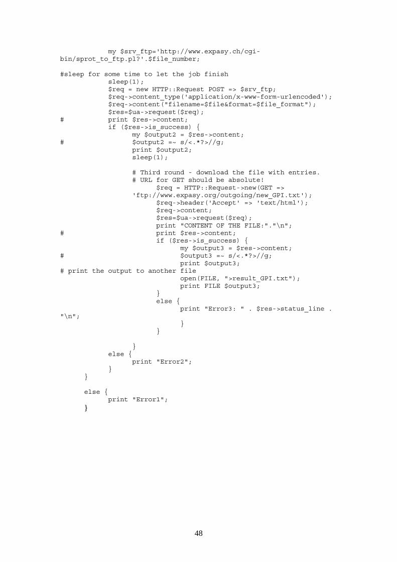
48
my $srv_ftp='http://www.expasy.ch/cgi-bin/sprot_to_ftp.pl?'.$file_number; #sleep for some time to let the job finish sleep(1); $req = new HTTP::Request POST => $srv_ftp; $req->content_type('application/x-www-form-urlencoded'); $req->content("filename=$file&format=$file_format"); $res=$ua->request($req); # print $res->content; if ($res->is_success) { my $output2 = $res->content; # $output2 =~ s/<.*?>//g; print $output2; sleep(1); # Third round - download the file with entries.
# URL for GET should be absolute! $req = HTTP::Request->new(GET =>
'ftp://www.expasy.org/outgoing/new_GPI.txt'); $req->header('Accept' => 'text/html'); $req->content; $res=$ua->request($req); print "CONTENT OF THE FILE:"."\n"; # print $res->content; if ($res->is_success) { my $output3 = $res->content; # $output3 =~ s/<.*?>//g; print $output3; # print the output to another file open(FILE, ">result_GPI.txt"); print FILE $output3; } else { print "Error3: " . $res->status_line . "\n"; } } } else { print "Error2"; } } else { print "Error1";
}

49
### Script 2 ### This program has been written for selecting experimentally # verified entries from the file with entries in Swiss-Prot format # MAIN # Modules used: use SWISS::Entry; use strict; use warnings; print "\n"; # Command line usage print " Usage : perl entry_file [output_file]\n"; my $entry_file; my $output_file; my $metazoa=0; my $protozoa=0; my $ fungi=0; my $plants=0; my $alt_splis=0; my $proven=0; my $non_proven=0; my $taxon; my @metazoa=(); my @protozoa=(); my @fungi=(); my @plants=(); my @alt_splis=(); # manage input/output file $entry_file=shift @ARGV or die "you must provide input file!"; print"\n"; # if output-file name is not provided, default name will be used if (scalar @ARGV>0) { $output_file=shift @ARGV; } else { $output_file="CC_output.txt" } my $count=0; my $positive; # record separator \n is replaced by // $/ = "\n//\n"; open (FILIN, "$entry_file") or die "cannot open file $!\n"; open (FILOUT, ">$output_file") or die "cannot open file $!\n"; # for each Swiss-Prot entry in the input file while (<FILIN>) { # read an entry and store it in the $entry variable my $entry = SWISS::Entry->fromText($_); # store the AC number in the entry_AC variable my $entry_AC = $entry->AC;

50
# store the sequence in the entry_SQ variable my $entry_SQ = $entry->SQ; my $conclusion; # to select just metazoan entries if ($entry->OCs->size) { if ($entry->OCs->head eq "Eukaryota") { $taxon=${$entry->OCs->list}[1]; if ($taxon eq "Metazoa") { $metazoa++; push(@metazoa, $entry_AC); } if ($taxon ne "Metazoa") {next;} } } # Find out if GPI-anchor is a key-word foreach my $kw ($entry->KWs->elements) { if ($kw->text eq 'GPI-anchor') { $conclusion=1; # print $kw->text."\n"; } # select alternative splicing entries, which may need manual verification if($kw->text eq 'Alternative splicing') { print $kw->text."\n"; $alt_splis++; push(@alt_splis, $entry_AC); } } if ($conclusion==0) { next; } # Print all CCs my @CC = $entry->CCs->elements; foreach my $CC (@CC) { print "TOPIC ".$CC->{topic}."\t"; if ($CC->{topic} eq 'SUBCELLULAR LOCATION') { if($CC->{comment}=~/GPI-anchor\s+(\((?:By similarity|Potential|Probable)\))/){
print "------------------------------------------"."\n";
$non_proven++."\n"; print "NON"."\t".$non_proven."\t".$CC->{comment}."\n".$taxon."\t"; print $entry_AC."\t"." NON-EXPERIMENTAL!!!"."\n"; } else { print "------------------------------------------"."\n"; $proven ++; print $proven."\t".$CC->{comment}."\n".$taxon."\t"; print $entry_AC."\t"." Verified experimentally!"."\n"; } }

51
} } print "Metazoa"."\t".$metazoa.”\n”; for (my $i=0; $i<scalar @alt_splis; $i++){ print $alt_splis[$i]."\n"; }
# This script was written to call DGPI web-site # and parse the output. use strict; # Modules used use SWISS::Entry; use SWISS::FTs; use LWP::UserAgent; # record separator \n is replaced by // local $/="//\n"; my $count=0; my $count_posit=0; my $count_negat=0; my $CGI; my $ua; my $line; while(<>) { $CGI = 'http://129.194.185.165/DGPI/DGPI'; # call UserAgent $ua = new LWP::UserAgent; my $entry = SWISS::Entry->fromText($_); my $entry_AC = $entry->AC; my $entry_SQ = $entry->SQ; my $entry_ID = $entry->ID; # print $entry_AC; # print $entry_SQ."\n"; my $fasta_SQ = ">".$entry_AC."\n".$entry_SQ; # send new request my $req = new HTTP::Request POST => $CGI; $req->content_type('application/x-www-form-urlencoded'); $req->content("protein=$entry_SQ"); my $res = $ua->request($req); if ($res->is_success) { my $output = $res->content; $output =~ s/<.*?>//g; print "##################################"."\n"; print $fasta_SQ."\n"; # print $output; # print "###################################"."\n"; #split output into lines foreach $line (split /\n/, $output) { if($line=~/This protein is GPI-anchored/) { print "AC ". $entry_AC." ". "ID ". $entry_ID."\n"; # print $entry_AC." ".$entry_ID."\n"; $count_posit++; $count++;
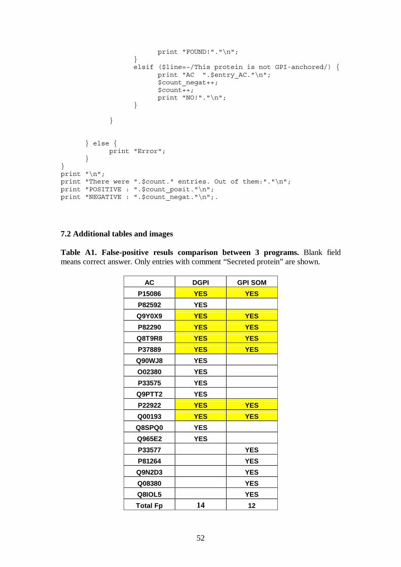
52
print "FOUND!"."\n"; } elsif ($line=~/This protein is not GPI-anchored/) { print "AC ".$entry_AC."\n"; $count_negat++; $count++; print "NO!"."\n"; } } } else { print "Error"; } } print "\n"; print "There were ".$count." entries. Out of them:"."\n"; print "POSITIVE : ".$count_posit."\n"; print "NEGATIVE : ".$count_negat."\n";.
7.2 Additional tables and images
Table A1. False-positive resuls comparison between 3 programs. Blank field means correct answer. Only entries with comment “Secreted protein” are shown.
AC DGPI GPI SOM
P15086 YES YES
P82592 YES Q9Y0X9 YES YES
P82290 YES YES
Q8T9R8 YES YES
P37889 YES YES
Q90WJ8 YES O02380 YES P33575 YES Q9PTT2 YES P22922 YES YES
Q00193 YES YES
Q8SPQ0 YES Q965E2 YES P33577 YES
P81264 YES
Q9N2D3 YES
Q08380 YES
Q8IOL5 YES
Total Fp 14 12

53
Table A2. False negative results comparison for the 3 programs on verified positive set.
DGPI BIGPI GPI-SOM
O08523 NO
O08524 NO
O08602 NO
O08603 NO
O08604 NO
O43895 NO
O46647 NO NO NO
O75015 NO NO NO
O75326 NO NO
O75443 NO NO
O88310 NO NO
O95980 NO
P05186 NO
P06858 NO NO NO
P07140 NO
P08571 NO
P08582 NO
P09487 NO
P10675 NO
P11151 NO NO NO
P11152 NO NO NO
P11153 NO NO NO
P11602 NO NO NO
P13265 NO NO NO
P14384 NO
P22063 NO
P25291 NO NO
P28685 NO NO
P35459 NO
P38566 NO NO
P48733 NO
P48794 NO NO NO
P49060 NO NO NO

54
P49291 NO
P49923 NO NO NO
P51780 NO
P52797 NO
P52961 NO
P54097 NO
P55031 NO NO NO
P56161 NO NO NO
P58019 NO
P58020 NO NO NO
P83408 NO NO NO
P83456 NO NO NO
P91885 NO
P91887 NO
P97527 NO
P97528 NO NO
Q06000 NO NO NO
Q12860 NO
Q14210 NO
Q28106 NO
Q28680 NO
Q28896 NO
Q29524 NO NO NO
Q5R5C1 NO
Q60935 NO NO NO
Q62803 NO NO NO
Q62997 NO NO
Q64389 NO
Q80ZA0 NO NO
Q8JG54 NO
Q8N6Q3 NO
Q8R4F1 NO
Q8R4G0 NO
Q8WWA0 NO NO
Q93070 NO NO
Q95323 NO
Q95333 NO

55
Q96PL2 NO
Q98892 NO
Q9BZM4 NO
Q9BZM6 NO
Q9D733 NO
Q9DA79 NO
Q9JI58 NO
Q9JJT2 NO
Q9NHB0 NO NO NO
Q9QUG3 NO NO
Q9QUR8 NO
Q9R0S3 NO
Q9VN14 NO
Q9YH85 NO NO
Q9Z0J1 NO
TOTAL: 39 65 36

56
Table A3. Program performance for cleavage-site prediction (43 metazoan sequences)
ENTRIES Proved best site BIG PI best site DGPI best site GPI-SOM
Site BIG PI prediction DGPI prediction SOM Prediction
P21589 549 549 549 549
P21588 551 551 551 551
P22748 284 284 284 281
P08174 353 353 353 350 NO GPI
P04058 564 564 563 564
P07692 567 567 566 567
P15328 234 234 234 230
P14207 230 230 230 230
P31997 320 326 325 319
P09326 220 220 206 218
P18181 217 217 217 217
P10252 217 217 217 NO GPI 217
P04216 130 130 NO SITE 139
O62643 130 130 NO SITE 139
P01831 131 131 131 140
P01830 130 130 130 139
P16444 385 385 385 384
P31358 36 36 36 36
Q09553 205 205 205 206
P05187 506 NO GPI 506 506
Q60506 231 231 231 231
Q60468 231 231 231 231
P04273 231 231 231 231
Q95333 649 656 NO GPI 658 649
P13987 102 102 102 121
O62680 98 98 103 98
If a program predicted ω-site, it is indicated, and highlighted in dark-grey if it is the
same as experimentally verified ω-site. In case a program didn’t predict the fact of
GPI-anchoring, it is indicated “NO GPI”. In some cases programs predict cleavage
site differently from a proved one (it is not highlighted in this case) or are not able to
predict it at all (as indicated “NO SITE”). Accsession numbers of 7 entries, which
were not in BigPI-learning set, are highlighted.

57
GPI-anchor detailed chemical structure.
Chemical structure of a GPI anchor typically found attached to proteins at the external surface of mammalian plasma membranes. The glycan core, Man(α1→2)-Man(α1→6)-Man(α1→4)-GlcN, is glycosidically linked to the 6-hydroxyl group of phosphatidylinositol. Possible modifications can occur at R1, R2, R3 and R4, as well as palmitoylation of the inositol ring. The sites of anchor cleavage by the phospholipases PI-PLC and PI-PLD are indicated.
(Taken from http://www.uoguelph.ca/~fsharom/research/gpi.html)