Microenvironments in haemopoietic and lymphoid differentiation
Upload
truongkhanhCategory
view
229download
4
DOI: 10.21276/ijabpt.2016.7.4.14
Volume-7, Issue-4, Oct-Dec-2016 Coden IJABFP-CAS-USA Copyrights@2016
Received: 11th
May 2016 Revised: 19th
July -2016 Accepted: 20th
Aug 2016
Research article
EVALUATION OF ORGANOSOMATIC INDICES AND HISTOPATHOLOGICAL RESPONSE OF
CLARIAS GARIEPINUS JUVENILES FED DIETS CONTAINING GRADED LEVELS OF
MECHANICALLY EXTRACTED SUNFLOWER (HELIANTHUS ANNUUS) SEED MEAL
S. A. Adesina1*
, A. E. Falaye2 and E. K. Ajani
2
1Department of Biological Sciences, Faculty of Science, Ondo State University of Science and Technology,
Okiti-pupa, Nigeria. 2Department of Aquaculture and Fisheries Management, University of Ibadan, Nigeria.
ABSTRACT: Histopathological studies provide useful information on diet quality and metabolism as well as reflect
fish nutritional and physiological status. This study assessed the effects of replacing soybean meal (SBM) with
mechanically extracted sunflower seed meal (MESSM) on the organosomatic indices and histopathological alterations
in the liver, kidney and intestine of Clarias gariepinus juveniles for fifteen weeks. MESSM was substituted for SBM at
0, 20, 40, 60, 80 and 100% in formulating six isonitrogenous and isocaloric diets. Diets were fed twice daily to 360 C.
gariepinus juveniles inside eighteen rectangular tanks (in triplicate treatments). Finally, three fish specimens per
treatment were dissected and their livers, kidneys and intestines removed and processed for histopathological
examinations. Their tissues were fixed, washed and dehydrated with graded alcohol, embedded in paraffin, sectioned,
stained and examined using photomicrography. Data were analysed using descriptive statistics and ANOVA at P =
0.05. The results revealed that hepatosomatic index (HSI) was insignificantly (p>0.05) superior (1.87%) in the fish fed
20% MESSM diet and lowest (1.19%) in those fed 100% inclusion. Kidney-somatic index (KSI) and intestino-somatic
index (ISI) were insignificantly (p>0.05) highest (0.65% and 3.63%) in fish fed 0% MESSM inclusion and least
(0.38% and 2.42%) in those fed 40% inclusion respectively. Fish fed 0% and 20% MESSM inclusions maintained
structurally normal liver, kidney and intestine without visible lesions. However, fish fed above 20% MESSM inclusion
exhibited extensive fatty infiltration, central portal venous congestion and several large cytoplasmic vacuolations of the
hepatocytes. Kidney tubules showed moderate swelling, depletion of haemopoietic and tubular compartments,
conspicuous degeneration and necrosis of tubule epithelia and epithelial cells with pyknotic nuclei. Intestinal changes
included hyperplastic villi, pronounced necrosis and erosion of villi and enterocytes at villi’s tips. The study showed
that inclusion of mechanically extracted sunflower seed meal above 20% in C. gariepinus’ diet could cause severe
physiological alterations, predispose fish to disease and consequently lower aquaculture profitability.
Key words: Clarias gariepinus, Mechanically extracted sunflower seed meal, Histology,
Organosomatic indices, Anti-nutritional factors, Photomicrography.
*Corresponding author: S. A. Adesina, Department of Biological Sciences, Ondo State University of Science
and Technology, Okiti-pupa, Nigeria E-mail: [email protected] Tel: +2348028574784
Copyright: ©2016 S. A. Adesina. This is an open-access article distributed under the terms of the Creative Commons
Attribution License , which permits unrestricted use, distribution, and reproduction in any medium, provided the
original author and source are credited.
International Journal of Applied Biology and Pharmaceutical Technology Page: 118
Available online at www.ijabpt.com

Adesina et al Copyrights@2016, ISSN: 0976-4550
INTRODUCTION The success of aquacultural operation partly depends on the quality and quantity of feed which constitutes about 70%
of the total production cost while protein is the most essential and expensive component of aquaculture diets
(Omitoyin, 2007; Garza de Yta, 2012). The formulation and production of commercial feeds for cultured aquatic
animals have traditionally been based on fishmeal as the principal protein source due to its high protein content and
balanced essential amino acid profile. Fishmeal is also a good source of essential fatty acids, digestible energy,
minerals and vitamins. However, the availability of fishmeal as the main protein component in fish feeds can no longer
be guaranteed because the capture fisheries are levelling off (FAO, 2011). Also, there has always been much demand
for fishmeal, hence its supply is inadequate and it is relatively expensive. As a result, the price of fishmeal
continuously rises and adversely affects the profitability of aquaculture enterprises (Sintayehu et al., 1996). Besides,
the quality of fish meal is often compromised and does not always meet the requirements for proper growth and
development of cultured fish. This has necessitated the aquaculture industry to constantly source for and explore
alternative protein-rich dietary supplements that are cheap, locally available and nutritionally safe for use as fishmeal
replacers in aquafeeds. The decrease in the global production of fishmeal clearly indicates that the growth and
sustainability of this industry will largely depend on the sustained supply of plant proteins for aquafeeds.
Soybean meal has been the main plant protein source used in animal feeds as a replacement for fishmeal because of its
high protein content and relatively well balanced amino acid profile (Sintayehu et al., 1996). However, soybean meal
has been increasingly commercialised and variously used in human, livestock and poultry dietary formulations, hence
its utilisation as the main protein source in fish feeds may longer be economically viable (Siddhuraju and Becker,
2001). Therefore, this has necessitated the need to focus on using less expensive, less competitive and readily available
alternative plant protein sources such as sunflower seed meal to replace soybean meal without reducing the nutritional
quality of the feed (Barros et al., 2002). Sunflower (Helianthus annuus Linnaeus) seed is one of the important annual crops of the world grown for oil.
It has a nutritional quality comparable to most other oilseed proteins including soybean and other conventional
legumes (Sanz et al.,1994; Sintayehu et al., 1996) and its potential as a dietary protein source in animal feeds is well
recognized (Olvera-Novoa et al., 2002). Studies into the use of sunflower seed meal in the feeds of livestock, poultry
birds and some other monogastric animals including fish are not as extensive as for soybean meal. However, for a plant
protein ingredient to be included in aquafeeds, its utilisation should be tested in different fish species because fish
species differ in their sensitivity and response to anti-nutrients present in plant protein sources (Francis et al., 2001;
Gatlin et al., 2007; Chaudhuri et al., 2012). Clariid catfishes are the second most important group of cultured fish in the world (Fasakin et al., 2003). They
feed on a wide range of natural and artificial food items, exhibit high growth rates and always tolerate poor water
quality parameters (Amisah et al., 2009). High activities of protease, lipase and amylase enzymes in the digestive tract
of C. gariepinus often indicate its ability to utilise both animal- and plant-based feed resources (Hlophe et al., 2014).
The intestine and liver are major organs responsible for digestion and absorption of nutrients from food while the
kidney performs excretion of metabolic wastes; therefore, the monitoring of these organs is imperative in nutritional
studies (Raskovic et al., 2011).
Histopathological changes have been widely used as biomarkers in the assessment of fish health status after they have
been exposed to various ontaminants in the laboratory (Thophon et al., 2003) and field studies (Schwaiger et al., 1997;
Teh et al., 1997). One of the main advantages of using histopathological assessment is that the markers allow us to
study the target organs, such as kidney, gill and liver, which are responsible for important physiological functions, such
as deposition and bio-magnification of chemicals as well as excretion in fish (Gernhofer et al., 2001). The
histopathological changes recorded are generally simpler to identify than functional changes (Fanta et al., 2003) and
serve as signs of deleterious effects on animal health (Hinton and Laurén, 1990). Histological studies provide
information on diet quality and metabolism as well as indicate the nutritional status of a fish (Segner and Braunbeck,
1988; Caballero et al., 2004).
Exposure of fish to pollutants and anti-nutritional compounds usually stimulates lesions in different organs to
varying degrees. Gills, liver and gut are suitable organs for histological examination to determine the effects of
pollution, especially in laboratory experiments (Capkin et al., 2009). For an accurate and effective assessment of the
effects of xenobiotic and anti-nutritional compounds in field and experimental studies, the proper monitoring of
histological changes in fish liver is a highly sensitive and accurate approach (Shalaka and Pragna, 2013). The
inspection of liver is pertinent as it plays an important role in the metabolism and excretion of xenobiotic compounds
(Rocha and Monteiro, 1999).
International Journal of Applied Biology and Pharmaceutical Technology Page: 119
Available online at www.ijabpt.com

Adesina et al Copyrights@2016, ISSN: 0976-4550
Fish kidney is an important organ which performs endocrine, reticulo-endothelial, haematopoietic and
excretory functions. The major function of the kidney in fish is the osmotic regulation of salts and water other than the
excretion of nitrogenous wastes as in the case of mammals. The histological alterations in the kidney tissues of
vertebrates subjected to experimental dietary treatments are useful bio-markers in the assessment of the effects of such
dietary treatments. Assessment of histological tissues of fish kidney is a method required to establish the possible
effects of various nutrient raw materials of plant and animal origin (Akhilesh et al., 2014). The lesions in kidneys alone
are not sufficient to reveal the effects of the contaminants and they must be supported by the histopathological results
obtained from the other tissues (Mishra and Mohanty, 2008).
Organosomatic indices also constitute a useful tool in correlating the weight of the visceral organs, such as
liver, kidney and intestine, with the body weight of fish. For instance, hepatosomatic index (HSI) of fish has been used
as an indicator of environmental risk (Pinkney et al., 2001; Yang and Baumann, 2006). They found a positive
correlation between HSI and the concentration of polycyclic aromatic hydrocarbon (PAH) metabolites in fish. Thus,
the aim of this study was to evaluate the effects of substituting mechanically extracted sunflower seed meal (MESSM)
for soybean meal (SBM) on the organosomatic indices and histopathological alterations in the liver, kidney and
intestine of Clarias gariepinus juveniles.
MATERIALS AND METHODS
Collection of organs Effects of dietary treatments on histology of liver, kidney and intestine of C. gariepinus juveniles were investigated. At
the completion of the feeding trial, three fish samples were taken from each dietary treatment, weighed individually
and injected with benzocaine at a concentration of 50 mg/L (Coyle et al., 2004) to anaesthetize them before dissection.
The fish were dissected using a dissecting kit and images of internal organs were taken by means of a digital camera
(Olympus CH XSZ-107BN) during dissection. After gross examination of the internal organs, the entire liver, kidney
and intestine of each fish sample were removed, weighed separately and recorded for evaluation of organosomatic
indices.
Determination of organosomatic indices The ratio of the weight of the liver, kidney and intestine in relation to the body weight of fish was calculated separately
from the following organosomatic index formula as described by Ali (2001):
Organosomatic index (%) = Organ weight (g) x 100
Fish body weight (g)
This formula was used to calculate organosomatic indices of the liver, kidney and intestine respectively as
follows:
Hepatosomatic index (HSI) = weight of liver (g) x 100
fish body weight (g)
Kidney-somatic index (KSI) = weight of kidney (g) x 100
fish body weight (g)
Intestino-somatic index (ISI) = weight of intestine (g) x 100
fish body weight (g)
Histopathological analysis Histopathological examinations were carried out to assess possible alterations in the intestines, livers and kidneys of
the fish fed with the different experimental diets. The examinations were carried out at the Department of Veterinary
Pathology Laboratory, Faculty of Veterinary Medicine, University of Ibadan, Nigeria, following Lynch’s medical
laboratory procedures. At the end of the experiment, three fish samples from each dietary treatment were used for the
diagnostic histological analysis.
International Journal of Applied Biology and Pharmaceutical Technology Page: 120
Available online at www.ijabpt.com

Adesina et al Copyrights@2016, ISSN: 0976-4550
The fish were injected with benzocaine at a concentration of 50 mg/L (Coyle et al., 2004) to anaesthetize them before
dissection. They were then dissected using a dissecting kit and their whole intestines, livers and kidneys were carefully
removed, washed with distilled water to remove blood stain and immediately pre-fixed in Bouin’s fixative solution and
later in 10% formalin solution for 48 hours. The organs were dehydrated in periodic acid Schiff”s reagent (PAS)
following the method of Hughes and Perry (1976) in graded levels of 50%, 70%, 90% and 100% alcohol for 3 days, to
allow paraffin wax to penetrate the tissue during embedding. The organs were then cleaned and embedded in melted
wax and carefully sliced into thin sections with a rotatory microtome (5μm thick).
The cut sections were again cleaned by placing them in warm water (38°C) from where they were transferred
into clean slides and oven-dried at 58°C for 30 minutes to melt the wax and stained with Harris’ haematoxylin–eosin
(H and E) stain (Bancroft and Cook, 1994). The slides containing sectioned tissues were cleaned using xylene and
graded levels of 50%, 70%, 90%, 95% and 100% alcohol for two minutes each. The sections were again stained in
haematoxylin-eosin for ten minutes and mounted in dipterex on glass slides. To obtain their photomicrography, the
stained sections were examined and photographed at different magnifications (x40, x100 and x400) by means of a
binocular light microscope (Olympus Japan 312545) fitted with a digital camera (Olympus CH XSZ-107BN), a
photographic attachment (Olympus C35 AD4) and an automatic light exposure unit (Olympus PM CS5P).
Data analysis Histopathological description of morphological changes and statistical analysis of indices were used to present the
research findings. All data obtained in this work are presented as mean± standard deviation. Comparisons were made
between the control and experimental groups. One-way ANOVA and Duncan’s multiple range test (Duncan, 1955)
were used on SPSS statistical software (Version 16.0 for Windows; SPSS Inc., Chicago, USA) to detect the significant
differences among the control and experimental groups. Differences were considered to be statistically significant at
probability levels below 0.05 (i.e. p<0.05) (Zar, 1984).
RESULTS
Organosomatic indices The values of organosomatic indices of the liver, kidney and intestine of C. gariepinus juveniles are shown in Table 1.
Hepatosomatic index (HSI) was highest (1.87%) in the fish fed 20% MESSM-based diet and least (1.31%) in the fish
fed 60% MESSM-based diet. However, no significant difference (p>0.05) existed in the HSI values among the fish in
the dietary treatments. Kidney-somatic index (KSI) was highest (0.65%) in the fish fed 0% MESSM-based diet and
least (0.38%) in the fish fed 40% MESSM-based diet. However, the values of KSI did not show significant differences
(p>0.05) in the fish among the dietary treatments. Intestino-somatic index (ISI) was highest (3.63%) in the fish fed 0%
MESSM-based diet and lowest (2.42%) in the fish fed 40% MESSM-based diet. The fish fed with 0%, 40% and 80%
MESSM-based diets had ISI values which significantly differed (p<0.05) from the values recorded for the fish fed with
the other MESSM-based diets.
Table 1: Organosomatic indices of C. gariepinus juveniles fed graded levels of mechanically extracted
sunflower seed meal-based diets for 15 weeks
Organosomati
c indices (%)
MESSM 1 (0%)
(Control)
MESSM 2
(20%)
MESSM 3
(40%)
MESSM 4
(60%)
MESSM
5 (80% )
MESSM 6
(100%)
Liver (HSI) 1.67±0.71
a 1.87±0.80
a 1.55±0.15
a 1.31±0.31
a 1.51±0.13
a 1.19±0.29
a
Kidney (KSI) 0.65±0.09a 0.60±0.15
a 0.38±0.04
a 0.46±0.18
a 0.63±0.21
a 0.49±0.17
a
Intestine (ISI) 3.63±0.82a 3.13±0.38
ab 2.42±0.16
b 3.27±0.16
ab 3.61±0.57
a 3.31±0.40
ab
The above values are means of triplicate data. Mean values in each row with similar superscripts are not significantly
different (p>0.05). MESSM = mechanically extracted sunflower seed meal HSI - Hepatosomatic index KSI - Kidney-
somatic index ISI - Intestino-somatic index
International Journal of Applied Biology and Pharmaceutical Technology Page: 121
Available online at www.ijabpt.com
Experimental dietary inclusions

Adesina et al Copyrights@2016, ISSN: 0976-4550
Gross and photomicrographic examination of liver, kidney and intestine At the end of the feeding experiment, the liver, kidney and intestine samples appeared externally normal as no visible
deformity was observed and they retained their normal colour appearance. However, microscopic examination of these
organs revealed varying degrees of histological changes as a result of dietary treatments (Table 2 and Plates 1 to 18).
Photomicrographs of sections of the livers of the fish fed 0% (control diet) and 20% MESSM-based diets showed
moderate diffuse cytoplasmic vacuolations in their hepatocytes (Plates 1 and 2) while the fish fed 40% MESSM-based
diet revealed multiple foci of large cytoplasmic vacuolations of the hepatocytes (Plate 3). Moderate diffuse vacuolar
change and fatty infiltration were observed in the liver sections of the fish fed 60% MESSM-based diet (Plate 4). The
livers of the fish fed 80% MESSM-based diet revealed moderate periportal vacuolar change (thin arrow), extensive
fatty infiltration and central portal venous congestion (thick arrow) (Plate 5) while those fed 100% MESSM-based diet
had severe diffuse cytoplasmic vacuolations (arrows) and central portal venous congestion (Plate 6).
Table 2: Histopathological observations on C. gariepinus juveniles fed graded levels of mechanically extracted
sunflower seed meal-based diets for 15 weeks
Tissues of organs examined
Dietary inclusions Liver Kidney Intestine
MESSM 1 (0%)
(Control)
Moderate diffuse cytoplasmic
vacuolations in the
hepatocytes.
No visible lesion as both
tubular and haemopoietic
compartments appeared
normal in proportion.
No visible lesions as
enterocytes and villi
appeared normal in
numbers and architecture.
MESSM 2 (20%)
Moderate diffuse cytoplasmic
vacuolations in the
hepatocytes.
No visible lesions.
Prominent haemopoietic
compartments occurred in
the midst of the tubular
compartments.
No visible lesions
observed.
MESSM 3 (40%)
Multiple foci of large
cytoplasmic vacuolations of
the hepatocytes.
No visible lesions as
haemopoietic and renal
tubular compartments
retained relative normal
proportions.
No visible lesions except
for numerous villi.
MESSM 4 (60%) Moderate diffuse vacuolar
change and fatty infiltration.
Moderately swollen
tubules. Haemopoietic
compartments were slightly
reduced.
Very long villi appearing
slightly hyperplastic.
MESSM 5 (80%)
Moderate periportal vacuolar
change, extensive fatty
infiltration and central portal
venous congestion.
Marked degeneration and
necrosis of renal tubular
epithelia. A few of the
epithelial cells had pyknotic
nuclei
Moderate sloughing
off/erosion of villi.
MESSM 6 (100%) Moderate diffuse vacuolar
change and fatty infiltration.
Depletion of haemopoietic
and renal tubular
compartments
Marked necrosis and
sloughing off (erosion) of
enterocytes at the tips of
villi.
MESSM = mechanically extracted sunflower seed meal
International Journal of Applied Biology and Pharmaceutical Technology Page: 122
Available online at www.ijabpt.com

Adesina et al Copyrights@2016, ISSN: 0976-4550
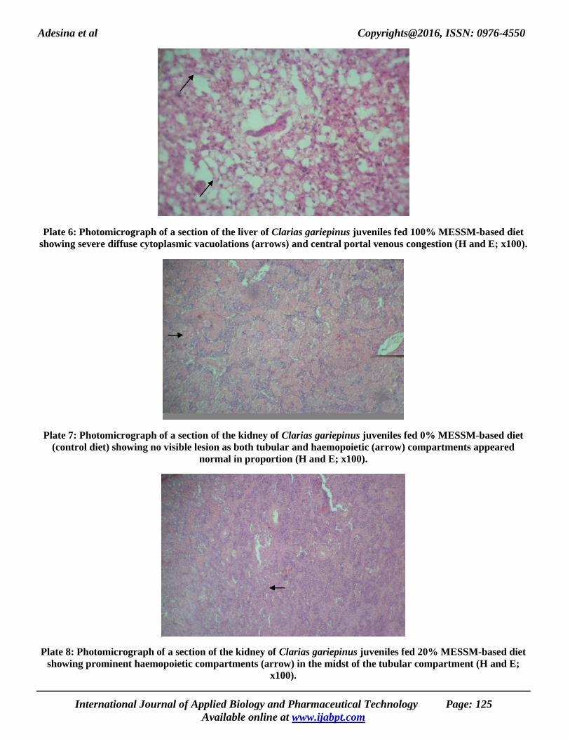
Photomicrograph of sections of the kidneys of the fish fed 0% MESSM-based diet (control diet) showed no visible
lesions as both tubular and haemopoietic compartments appeared normal in proportion (Plate 7). Sections of the
kidneys of the fish fed 20% MESSM-based diet revealed prominent haemopoietic compartments (arrow) in the midst
of the renal tubular compartment (Plate 8). A section of the kidney of the fish fed 40% MESSM-based diet showed no
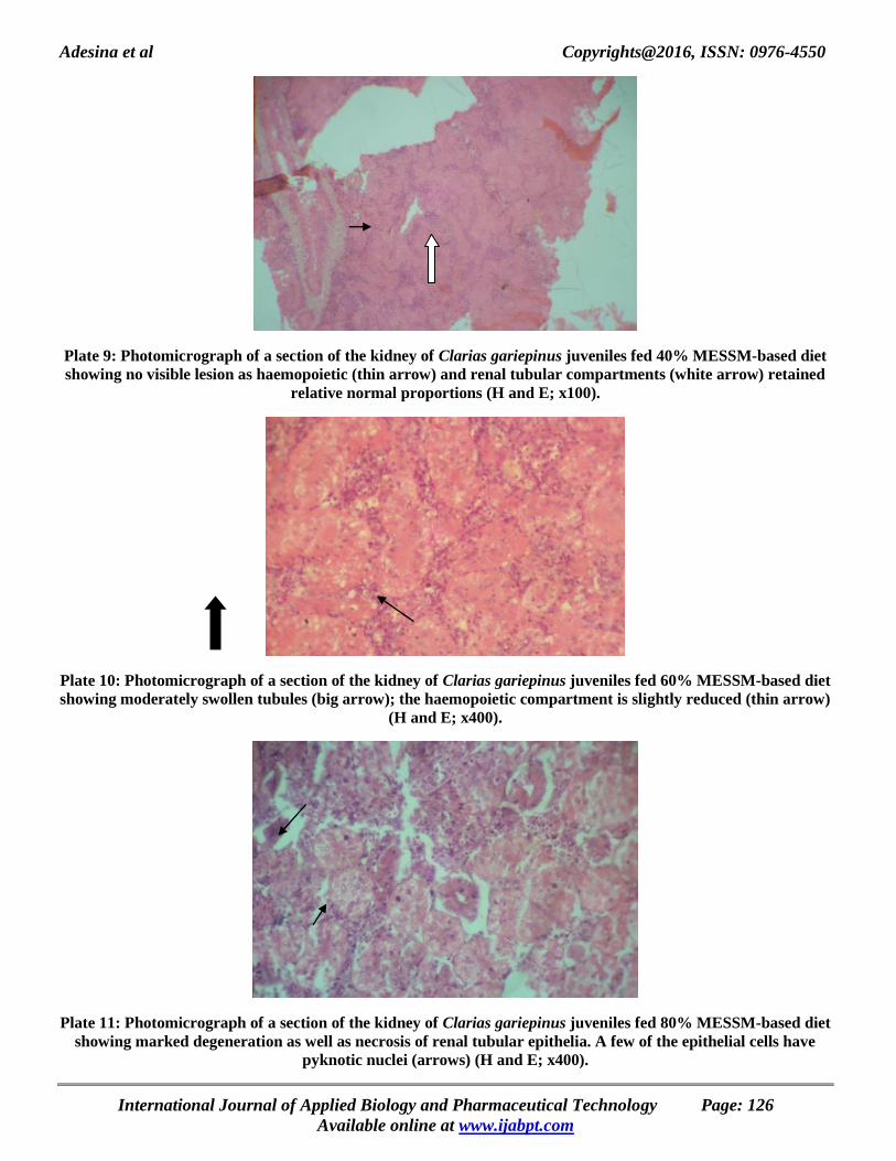
visible lesion as haemopoietic and renal tubular compartments retained their relative normal proportions (Plate 9).
There were moderately swollen tubules (big arrow) and slightly reduced haemopoietic compartment (thin arrow) in the
section of the kidney of the fish fed 60% MESSM-based diet (Plate 10). Sections of the kidneys of the fish fed 80%
MESSM-based diet showed marked degeneration and necrosis of renal tubular epithelia while a few of the epithelial
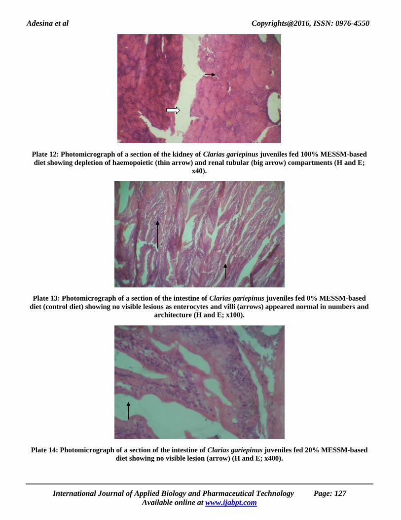
cells had pyknotic nuclei (arrows) (Plate 11). Depletion of haemopoietic and renal tubular compartments were
observed in the kidney sections of the fish fed 100% MESSM-based diet (Plate 12).
Photomicrograph of a section of the intestine of the fish fed 0% MESSM-based diet (control diet) showed no
visible lesions as enterocytes and villi appeared normal in numbers and architecture (Plate 13). There were no visible
lesions in the section of the intestine of the fish fed 20% MESSM-based diet (Plate 14). A section of the intestine of the
fish fed 40% MESSM-based diet showed numerous villi without any visible lesions (Plate 15). A section of the
intestine of the fish fed 60% MESSM-based diet was observed to show very long villi appearing slightly hyperplastic
(Plate 16). There was moderate sloughing off/erosion (arrow) of villi in the section of the intestine of the fish fed 80%
MESSM-based diet (Plate 17). A section of the intestine of the fish fed 100% MESSM-based diet revealed marked
necrosis and sloughing off of enterocytes at the tips of villi (circle) (Plate 18).
Plate 1: Photomicrograph of a section of the liver of Clarias gariepinus juveniles fed 0% MESSM-based diet
(control diet) showing moderate diffuse cytoplasmic vacuolations (arrow) in the hepatocytes (H and E; x40).
Plate 2: Photomicrograph of a section of the liver of Clarias gariepinus juveniles fed 20% MESSM-based diet
showing moderate diffuse cytoplasmic vacuoles (circle) in the hepatocytes (H and E; x100).
International Journal of Applied Biology and Pharmaceutical Technology Page: 123
Available online at www.ijabpt.com

Adesina et al Copyrights@2016, ISSN: 0976-4550
Plate 3: Photomicrograph of a section of the liver of Clarias gariepinus juveniles fed 40% MESSM-based diet
showing multiple foci of large cytoplasmic vacuolations (arrow) of the hepatocytes (H and E; x40).
Plate 4: Photomicrograph of a section of the liver of Clarias gariepinus juveniles fed 60% MESSM-based diet
showing moderate diffuse vacuolar change and fatty infiltration (arrow) (H and E; x100).
Plate 5: Photomicrograph of a section of the liver of Clarias gariepinus juveniles fed 80% MESSM-based diet
showing moderate periportal vacuolar change (thin arrow), extensive fatty infiltration and central portal venous
congestion (thick arrow) (H and E; x400).
International Journal of Applied Biology and Pharmaceutical Technology Page: 124
Available online at www.ijabpt.com
Adesina et al Copyrights@2016, ISSN: 0976-4550
Plate 6: Photomicrograph of a section of the liver of Clarias gariepinus juveniles fed 100% MESSM-based diet
showing severe diffuse cytoplasmic vacuolations (arrows) and central portal venous congestion (H and E; x100).
Plate 7: Photomicrograph of a section of the kidney of Clarias gariepinus juveniles fed 0% MESSM-based diet
(control diet) showing no visible lesion as both tubular and haemopoietic (arrow) compartments appeared
normal in proportion (H and E; x100).
Plate 8: Photomicrograph of a section of the kidney of Clarias gariepinus juveniles fed 20% MESSM-based diet
showing prominent haemopoietic compartments (arrow) in the midst of the tubular compartment (H and E;
x100).
International Journal of Applied Biology and Pharmaceutical Technology Page: 125
Available online at www.ijabpt.com
Adesina et al Copyrights@2016, ISSN: 0976-4550
Plate 9: Photomicrograph of a section of the kidney of Clarias gariepinus juveniles fed 40% MESSM-based diet
showing no visible lesion as haemopoietic (thin arrow) and renal tubular compartments (white arrow) retained
relative normal proportions (H and E; x100).
Plate 10: Photomicrograph of a section of the kidney of Clarias gariepinus juveniles fed 60% MESSM-based diet
showing moderately swollen tubules (big arrow); the haemopoietic compartment is slightly reduced (thin arrow)
(H and E; x400).
Plate 11: Photomicrograph of a section of the kidney of Clarias gariepinus juveniles fed 80% MESSM-based diet
showing marked degeneration as well as necrosis of renal tubular epithelia. A few of the epithelial cells have
pyknotic nuclei (arrows) (H and E; x400).
International Journal of Applied Biology and Pharmaceutical Technology Page: 126
Available online at www.ijabpt.com
Adesina et al Copyrights@2016, ISSN: 0976-4550
Plate 12: Photomicrograph of a section of the kidney of Clarias gariepinus juveniles fed 100% MESSM-based
diet showing depletion of haemopoietic (thin arrow) and renal tubular (big arrow) compartments (H and E;
x40).
Plate 13: Photomicrograph of a section of the intestine of Clarias gariepinus juveniles fed 0% MESSM-based
diet (control diet) showing no visible lesions as enterocytes and villi (arrows) appeared normal in numbers and
architecture (H and E; x100).
Plate 14: Photomicrograph of a section of the intestine of Clarias gariepinus juveniles fed 20% MESSM-based
diet showing no visible lesion (arrow) (H and E; x400).
International Journal of Applied Biology and Pharmaceutical Technology Page: 127
Available online at www.ijabpt.com

Adesina et al Copyrights@2016, ISSN: 0976-4550
Plate 15: Photomicrograph of a section of the intestine of Clarias gariepinus juveniles fed 40% MESSM-based
diet showing numerous villi (arrows) without any visible lesions (H and E; x100).
Plate 16: Photomicrograph of a section of the intestine of Clarias gariepinus juveniles fed 60% MESSM-based
diet showing very long villi (arrow) appearing slightly hyperplastic (H and E; x100).
Plate 17: Photomicrograph of a section of the intestine of Clarias gariepinus juveniles fed 80% MESSM-based
diet showing moderate sloughing off/erosion (arrow) of villi (H and E; x100).
International Journal of Applied Biology and Pharmaceutical Technology Page: 128
Available online at www.ijabpt.com

Adesina et al Copyrights@2016, ISSN: 0976-4550
Plate 18: Photomicrograph of a section of the intestine of Clarias gariepinus juveniles fed 100% MESSM-based
diet showing marked necrosis and sloughing off (erosion) of enterocytes at the tips of villi (circle) (H and E;
x100).
DISCUSSION Hepatosomatic index (HSI) of the liver is a considerable potential tool used by fish biologists to assess the toxicity
situation of the exposure of fish to any toxicant as well as a management tool for evaluating growth or health status of
various fish species in different environments (Hoque et al., 1998). HSI is also a useful biomarker to detect the
hazardous effects of the environmental stressors (Pait and Nelson, 2003). The increase in HSI value in an ideal
environment is related to normal liver growth but, in cases of pollution, liver enlargement is associated with
hyperplasia (Hoque et al., 1998). The higher values of hepatosomatic index (HSI) observed in the fish fed 0, 20 and
40% MESSM-based diets could be attributed to higher feed take at these inclusion levels and indicated normal liver
growth resulting from dietary treatment. Enlargement of organs, such as liver, kidney and heart, has been associated
with dietary factors especially if such diets contain toxins, anti-nutrients or heavy metals (Adejinmi, 2000). High HSI
values have been reported for male Fundulus heteroclitus (Killifish) induced by selected estrogenic compounds (Pait
and Nelson, 2003) and Crucian carp (Carassius carassius) exposed to treated sewage effluent (Diniz et al., 2005).
Barse et al. (2006) also reported superior HSI values for Cyprinus carpio subjected to 4-tert-butylphenol while Abdel-
Hameid (2007) reported elevated HSI values for Oreochromis aureus juveniles due to phenol intoxication and stated
that the observed hepatomegaly might partially reflect the enhancement of the liver size due to destructive changes. In
the same vein, Figueiredo-Fernandes et al. (2006) also obtained increased values of HSI in male and female tilapias,
Oreochromis niloticus, exposed to paraquat.
However, the progressively reduced HSI values recorded for the fish fed 60% to 100% MESSM-based diets could be
linked with lesser feed intake which was probably associated with the presence of anti-nutrients in the mechanically
extracted sunflower seed meal incorporated in the diets at these higher levels. Akerman et al. (2003) also found a
decrease in HSI values after nine weeks in rainbow trout, Oncorhynchus mykiss, injected with paraquat.
Histopathological biomarkers are useful as indicators of the general health of the fish and are considered as a mirror
that reflects the exposure of fish to a variety of anthropogenic pollutants (Van der Oost et al., 2003).
The lack of statistical variation in the values of kidney-somatic index (KSI) recorded for fish in all the
treatments suggests that this index was not affected by both the varying MESSM inclusion levels and the presence of
anti-nutrients in the diets. It can also be suggested that the non-significant variation in the KSI values indicates that the
weight of the kidney and total body weight were not correspondingly affected by the increasing inclusion level of
mechanically extracted sunflower seed meal in the diets. Intestino-somatic index (ISI) was observed to exhibit an
irregular pattern of variation and was not correspondingly affected by the increasing inclusion level of mechanically
extracted sunflower seed meal in the diets. This result disagrees with that of Abdel-Hameid (2007) who obtained
reduced ISI values for O. aureus juveniles and attributed them to a reduction in the total body weight as a result of
reduced appetite caused by phenol intoxication.
International Journal of Applied Biology and Pharmaceutical Technology Page: 129
Available online at www.ijabpt.com

Adesina et al Copyrights@2016, ISSN: 0976-4550
Histopathological observations on Clarias gariepinus fed graded levels of MESSM diets In this study, histopathological examination of the liver, kidney and intestine was carried out because of their
physiological importance during absorption and metabolism of nutrients and chemicals (Roberts, 1989). Evaluation of
histological structure of digestive organs in fish fed new dietary ingredients provides valuable information about their
digestive capacity as well as potential health effects of such new diets (Caballero et al., 2003; Diaz et al., 2006).
Substitution of different inclusion levels of mechanically extracted sunflower seed meal (MESSM) for soybean meal in
the diets has resulted in varying degrees of histopathological changes in the liver cells (hepatocytes) of C. gariepinus
juveniles. Such changes included mild/moderate diffuse vacuolations, periportal congestion, central venous congestion,
mild periportal vacuolar degeneration, severe fatty infiltration, extensive hepatic degeneration and overlapping of liver
tissue. These observations closely support the finding of Hlophe and Moyo (2014) who, in a related feeding trial,
observed that C. gariepinus fed high moringa leaf meal inclusion levels (>50%) showed an increase in the number of
degraded hepatocytes with irregularly shaped cells, small dark pyknotic nuclei, poor fatty deposition and isolated
necrosis. The present observations also agree with those of Uwachukwu et al. (2003) who reported that diets
containing raw beans caused extensive periportal necrosis with some mononuclear cell infiltration in the livers of
broilers while the centrilobular areas showed vacuolation and degeneration of hepatocytes. Vacuolated
hepatocytes are usually accumulated with glycogen and have little or no degenerative and regenerative ability (Nayak
et al., 1996) and the excessive vacuolation of the liver cells would result in abnormal functioning of such liver cells, for
instance, accumulation and immobilization of fat, which could consequently result in fatty infiltration of the hepatic
parenchyma (Adeyemo, 2005). Despite similar protein and energy levels in the experimental diets in the present study,
liver histology showed that C. gariepinus juveniles fed higher MESSM inclusion levels had necrotic signs associated
with poor nutritional status (Ostaszewska et al., 2005; Tusche et al., 2012). The malnutrition signs observed in C.
gariepinus fed higher levels of MESSM might be due to non-availability of protein and amino acids that have bound
with or have formed indigestible complexes with the anti-nutritional compounds in the sunflower seed meal. As a
result of the poor digestibility, a substantial portion of the essential dietary nutrients was not available to the fish and
was subsequently excreted. This could be responsible for the nutritional necrosis observed in the hepatocytes.
Wade et al. (2002) earlier reported that after a 96-hour toxicity bio-assay of cassava (Manihot esculenta
Crantz) effluent on the Nile tilapia, histopathological examination of the liver of the treated fish indicated vacuolation
and necrosis of the liver cells. Adeyemo (2005) also made similar observations in C. gariepinus fed cassava mill
effluent. Similarly, Jha (2004) reported remarkable lesions in the liver of Clarias batrachus exposed to surf and
Omitoyin et al. (2006) observed similar trends in C. gariepinus exposed to Lindane. Ayoola (2008) observed similar
effects of glyphosate in C. gariepinus and opined that vacuolation of liver cells is an evidence of fatty degeneration of
the cells. In this study, histological changes observed in the liver might have been caused by the ingestion of a high
percentage of MESSM-based diets which probably imposed stress on the organ above its physiological capacity to
cope with. The lesions observed in the liver might probably have resulted from the excessive work load done by the
liver of the experimental fish during the processes of detoxification and removal of toxicants from its body.
Hepatocytes in the periportal areas have been reported to suffer most from toxicants. In this situation, anti-nutritional
substances present in sunflower seed meal must have been responsible for the observed histopathological changes in
the liver sections.
Metelev et al. (1971) stated that the liver as the primary organ for detoxification of organic xenobiotics is often prone
to various pollutants and other toxic by-products which tend to accumulate in high concentrations within it and thereby
suffer from harmful effects. Alterations in the liver serve as useful markers of exposure to environmental stress. Both
the liver and kidney have been identified as the sites that are mostly affected by toxic substances in man and various
clinical signs have been associated with liver detoxifying and kidney removing these toxic substances in man
(Benjamin, 2009). The results obtained indicated a sign of toxicity of the diets to the fish at higher inclusion levels and
therefore necessitated further research to explore better and more effective processing methods that will significantly
reduce the levels of anti-nutritional components in sunflower seeds as an alternative feed ingredient.
Substitution of mechanically extracted sunflower seed meal for soybean meal in the diets also caused some histological
alterations in the kidney of C. gariepinus such as marked degeneration and necrosis of renal tubular epithelia at 80%
MESSM inclusion as well as depletion of haemopoietic and renal tubular compartments at 100% MESSM inclusion.
Olasunkanmi (2011) earlier reported a marked congestion in the kidneys of C. gariepinus fed raw, cooked and toasted
mucuna seed meal diets and associated the histological changes in the kidney with ingestion of a high percentage of
mucuna seed meal which probably imposed stress on the organ’s physiological capacity.
International Journal of Applied Biology and Pharmaceutical Technology Page: 130
Available online at www.ijabpt.com

Adesina et al Copyrights@2016, ISSN: 0976-4550
In a related study, Olasunkanmi (2015) also observed that C. gariepinus fed higher inclusion levels of processed velvet
beans showed a marked congestion of the kidney cells which usually impairs their maintenance of constant
homeostatic conditions, thus implying that fish fed with the diets containing higher inclusion levels of processed velvet
bean meal and mechanically extracted sunflower seed meal (in the present study) might have some difficulty with
maintaining a constant osmoregulatory mechanism. Benjamin (2009) stated that congestion of the kidney tubules is the
first stage in the development of kidney disease while Wade et al. (2002) and Adeyemo (2005) earlier observed
histological changes such as oedema in the kidneys of Oreochromis niloticus and C. gariepinus fed cassava mill
effluents respectively.
Fish kidney is one of the most susceptible organs being affected by contaminants in water bodies (Thophon et al.,
2003). Most common alterations found in the kidneys of fishes are tubule degeneration, dilation of capillaries in the
glomerulus and reduction of Bowman’s capsular space (Takashima and Hibya, 1995). Exposure to chemicals often
causes alterations in the glomerulus and renal tubules as described by Thophon et al. (2003) for the perch (Lates
calcarifer). In more severe cases, the degenerative process can lead to tissue necrosis (Takashima and Hibya, 1995).
The occurence of pronounced tubule disruption, degeneration and necrosis of renal tubular epithelia as well as
depletion of haemopoietic and renal tubular compartments observed in the kidneys of fish fed higher MESSM
inclusions in the present study indicates that the kidneys must have suffered some damage which could be attributed to
the presence of anti-nutritional compounds in the MESSM. Lesions observed in kidneys are not enough to verify the
level of contamination or effects of dietary treatments. They must be supported by the histopathological data of the
other organs. The histopathological findings commonly observed in kidneys are necrosis, fibrin and haemorrhage
(Lawrence et al., 2003; Uçar and Atamanalp, 2008).
The examined sections of the intestines of C. gariepinus juveniles fed mechanically extracted sunflower seed meal-
based diets revealed very mild histological changes except for moderate erosion of villi at 80% MESSM inclusion as
well as pronounced necrosis and erosion of enterocytes at the tips of villi at 100% MESSM inclusion. This result
corroborates that of Hlophe and Moyo (2014) who, in a related study, observed that the intestine histology of C.
gariepinus fed diets containing higher moringa leaf meal inclusion levels (>50%) showed significantly shorter villi.
The longer villi found in fish fed lower levels of sunflower seed meal in the diet indicate a larger surface area and
consequently higher efficiency of the intestine in the absorptive process (Caballero et al., 2002; Da Silva et al., 2012).
This was corroborated by the better growth performance of fish fed with these diets at lower inclusion levels as
reported by Adesina et al. (2013). The decrease in villi height resulted in reduced surface area for nutrient absorption
(Da Silva et al., 2012). Necrosis and mucosal degeneration may affect the permeability and absorption of substances
across the stomach (Roberts, 1878) and intestinal walls. Other authors have reported a widening of the central stroma
within the mucosal folding, higher amounts of connective tissue and an infiltration of inflammatory cells in the lamina
propria (Krogdahl et al., 2000; Refstie et al., 2000). This may suggest that C. gariepinus, being an omnivore, is more
capable of utilising plant diets than carnivorous fish.
CONCLUSION Histopathological examinations of the thin sections of the liver, kidney and intestine of C. gariepinus fed graded levels
of MESSM-included diets have revealed changes ranging from mild to severe lesions and few anatomical alterations,
particularly at the higher levels of inclusion of mechanically extracted sunflower seed meal in the formulated diets.
From histological analysis, it was clearly observed that, as the level of inclusion of mechanically extracted sunflower
seed meal in the diets increased, C. gariepinus was subjected to more stress. The presence of residual traces of anti-
nutritional factors (tannin, oxalate and phytate) in the mechanically extracted sunflower seed meal could be most
probably responsible for the poor performance of the MESSM-based diets at higher levels of inclusion. It is suggested
that the specific roles that each of these anti-nutrients plays in the utilisation of nutrients be further investigated. The
results of this study showed a disruption of normal physiological activities in C. gariepinus which was due to the level
of processing adopted in this study that had not completely detoxified the inherent anti-nutrients in the sunflower seed
meal for safe and maximum consumption and utilisation by C. gariepinus. It is therefore concluded that further
processing methods should be explored before incorporating sunflower seed meal in fish diets.
ACKNOWLEDGEMENTS The authors gratefully acknowledge Messrs. I. C. Mpama, O. I. Tunji, J. U. Augustine, Ambrose Nwagbara and Miss
Janet Oni as well as Drs. O. O. Aina and John Ogunsola of the Department of Veterinary Anatomy, University of
Ibadan, for their thorough technical assistance in tissue processing and slide photomicrographic preparation and
interpretation.
International Journal of Applied Biology and Pharmaceutical Technology Page: 131
Available online at www.ijabpt.com

Adesina et al Copyrights@2016, ISSN: 0976-4550
REFERENCES
Abdel-Hameid N. A. H. (2007). Physiological and Histopathological Alterations Induced by Phenol Exposure in
Oreochromis aureus Juveniles. Turkish Journal of Fisheries and Aquatic Sciences: Vol. 7, 131-138.
Adejinmi O. O. (2000). The chemical composition and nutritional potential of soldierfly (Hermetia elucens) larvae in
poultry rations. PhD. Thesis. Department of Animal Science. University of Ibadan. pp: 292.
Adesina S. A., Falaye A. E., Olusola S. E. and Ajani E. K. (2013). Growth performance and nutrient utilization of
Clarias gariepinus juveniles fed graded levels of boiled sunflower (Helianthus annuus L) seed meal-based
diets. Wudpecker Journal of Agricultural Research: Vol. 2, 12, 342-351.
Adeyemo O. K. (2005). Haematological and Histopathological effects of cassava mill effluent in Clarias gariepinus.
Afr. J. of Biomed. Res.: Vol. 8, 179-183.
Akerman G., Amcoff P., Tjarnlund U., Fogelberg K., Torrissen O. and Balk, L. (2003). Paraquat and menadione
exposure of rainbow trout (Oncorhynchus mykiss). Studies of effects on the penthose-phosphate shunt and
thiamine levels in liver and kidney. Chem. Biol. Int.: Vol. 142, 269-283.
Akhilesh K. Y., Prem P. S., Joykrushna J., Pradeep S., Shipra C. and Rajesh D. (2014). Impact of dietary fats on
histological alterations in the kidney tissue of Asian catfish, Clarias batrachus (Linnaeus, 1758). International
Journal of Fisheries and Aquatic Studies: Vol. 1, 3, 147-151.
Ali M. Z. (2001). Dietary protein and energy interactions in African catfish Clarias gariepinus (Burchell, 1822). PhD.
Thesis. Department of Aquaculture. University of Aquaculture, Stirling, United Kingdom. pp: 274.
Amisah S., Oteng M. A. and Ofor J. K. (2009). Growth performance of the African catfish, Clarias gariepinus, fed
varying inclusion levels of Leucaena leucocephala leaf meal. Journal of Applied Sciences and Environmental
Management: Vol. 13, 1, 21-26. doi:10.4314/jasem.v13i1.55257.
Ayoola S. O. (2008). Histopathological Effects of Glyphosate on Juvenile African catfish (Clarias gariepinus).
American-Eurasian J. Agric. & Environ. Sc.: Vol. 4, 3, 362-367.
Bancroft J. D. and Cook H. C. (1994). Manual of Histological Techniques and Their Diagnostic Application. Churchill
Livingstone, London. pp: 305.
Barros M. M., Lim C. and Klesius P. H. (2002). Effect of soybean meal replacement by cottonseed meal and iron
supplementation on growth, immune response and resistance of channel catfish (Ictalurus punctatus) to
Edwarsiella ictaluri challenge. Aqua.: Vol. 207, 263-279.
Barse A. V., Chakrabarti T., Ghosh T. K., Pal A. K. and Jadhao S. B. (2006). One-tenth dose of LC50 of 4-tert butyl
phenol causes endocrine disruption and metabolic changes in Cyprinus carpio. Pesticide Biochem. Physiol.:
Vol. 86, 3, 172-179.
Benjamin H. (2009). Diseases of the liver, gall bladder, kidneys and pancreas. Accessed online from www:
herbdatanz.com/liver gall bladder kidney pancreas harry-benjamin nd.htm on 21/05/2011.
Caballero M. J., Izquierdo M. S. and Kjørsvik E. (2002). Morphological aspects of intestinal cells from gilthead
seabream (Sparus aurata) fed diets containing different lipid sources. Aquaculture: Vol. 225, 325-340.
Caballero M. J., Izquierdo M. S., Kjørsvik E., Fernández A. J. and Rosenlund G. (2004). Histological alterations in the
liver of sea bream, Sparus aurata L., caused by short- or long-term feeding with vegetable oils. Recovery of
normal morphology after feeding fish oil as the sole lipid source. J. Fish Dis.: Vol. 27, 531–541. doi:
10.1111/j.1365- 2761.2004.00572.x.
Capkin E, Birincioglu S, Altinok I. (2009). Histopathological changes in bowtrout (Oncorhynchus mykiss) after
exposure to sublethal composite nitrogen fertilizers. Ecotoxicol. Environ Safety: Vol. 72, 1999-2004.
Chaudhuri A., Mukherjee S. and Homechaudhuri S. (2012). Diet composition and digestive enzymes activity in
carnivorous fishes inhabiting mudflats of Indian Sundarban estuaries. Turkish Journal of Fisheries and Aquatic
Sciences: Vol. 12, 265-275. doi: 10.4194/1303- 2712-v12_2_11.
Coyle S. D., Durborow R. M. and Tidwell J. H. (2004). Anaesthetics in Aquaculture. SRAC Publication, Texas, pp: 6.
Da Silva M. R., Natali M. R. M. and Hahn N. S. (2012). Histology of the digestive tract of Satanoperca pappaterra
(Osteichthyes, Cichlidae). Acta Scientiarum Biological Sciences: Vol. 34, 319-326. doi: 10.4025/ acta
scibiolsci. v34i3.8956.
Diaz A. O, Escalante A. H, Garcìa A. M. and Goldemberg A. L. (2006). Histology and histochemistry of the
pharynxgeal cavity and oesophagus of the Silverside Odontesthes bonariensis (Cuvier and Valenciennes).
Anatomia Histolologia Embryologia: Vol. 35, 42–46.
International Journal of Applied Biology and Pharmaceutical Technology Page: 132
Available online at www.ijabpt.com

Adesina et al Copyrights@2016, ISSN: 0976-4550
Diniz M. S., Peres I., Magalhaes-Antonine I., Falla J. and Pihan J. C. (2005). Estrogenic effects in Crucian carp
(Carassius carassius) exposed to treated sewage effluent. Ecotoxicol. Environ. Saf.: Vol. 62, 3, 427-435.
Duncan D. B. (1955). Multiple range and multiple F tests. Biometrics: Vol. 11, 1 – 42.
Fanta E., Rios F. S., Romão S., Vianna A. C. C. and Freiberger S. (2003). Histopathology of fish Corydoras paleatus
contaminated with sublethal levels of organophosphorus in water and food. Ecotoxicology and Environmental
Safety: 54, 119-130.
FAO (2011). Demand and supply of feed ingredients for farmed fish and crustaceans: trends and prospects.
http://www.fao.org/docrep/015/ba0002e/ba0002e.pdf (accessed January 22, 2014).
Fasakin E. A., Balogun A. M. and Ajayi O. O. (2003). Evaluation of full-fat and defatted maggot meals in the feeding
of clariid catfish Clarias gariepinus fingerlings. Aquaculture Research: Vol. 34, 733-738. doi: 10.1046/j.1365-
2109.2003.00876.x.
Figueiredo-Fernandes A., Fontaínhas-Fernandes A., Peixoto F., Rocha E. and Reis- Henriques M. A. (2006). Effect of
paraquat on oxidative stress enzymes in tilapia Oreochromis niloticus at two levels of temperature. Pest.
Biochem. Physiol.: Vol. 85, 97- 103.
Francis G., Makkar H. P. S. and Becker K. (2001). Antinutritional factors present in plant-derived alternative fish feed
ingredients and their effects in fish. Aquaculture: Vol. 199, 197-227. doi:10.1016/S0044-8486(01)00526-9.
Garza de Yta, A., Davis D. A., Rouse D. B., Ghanawi J. and Saoud I. P. (2012). Evaluation of practical diets
containing various terrestrial protein sources on survival and growth parameters of redclaw crayfish Cherax
quadricarinatus. Aquacult. Res.: Vol. 43, 1, 84- 90.
Gatlin D. B., Barrows F. T., Brown P., Dabrowski K., Gaylord T. G., Hardy R. W., Herman E., Hu G., Krogdahl A.,
Nelson R., Overturf K., Rust M., Sealey W., Skonberg D., Souza E. J., Stone D., Wilson R. and Wurtele E.
(2007). Expanding the utilization of sustainable plant products in aquafeeds: a review. Aquaculture
Research: Vol. 38, 551- 579. doi: 10.1111/j.1365-2109.2007.01704.x.
Gernhofer M., Pawet M., Schramm M., Müller E. and Triebskorn R. (2001). Ultrastructural biomarkers as tools to
characterize the health status of fish in contaminated streams.Journal of Aquatic Ecosystem, Stress and
Recovery: Vol. 8, 241-260.
Hinton D. E. and Laurén D. J. (1990). Liver structural alterations accompanying chronic toxicity in fishes: potential
biomarkers of exposure. Biomarkers of Environmental Contamination. Boca Raton, Lewis Publishers, pp: 51-
65.
Hlophe S. N. and Moyo N. A. G. (2014). Replacing Fishmeal with Kikuyu Grass and Moringa Leaves: Effects on
Growth, Protein Digestibility, Histological and Haematological Parameters in Clarias gariepinus. Turkish
Journal of Fisheries and Aquatic Sciences: Vol. 14, 795-806.
Hlophe S. N. and Moyo N. A. G. (2014). Replacing Fishmeal with Kikuyu Grass and Moringa Leaves: Effects on
Growth, Protein Digestibility, Histological and Haematological Parameters in Clarias gariepinus. Turkish
Journal of Fisheries and Aquatic Sciences: Vol. 14, 795-806.
Hoque M. T., Yusoff F. M., Law A. T. and Syed M. A. (1998). Effect of Hydrogen Sulphide on Liver-Somatic Index
and Fulton Factor in Mystus nemurus. Journal of Fish Biology: Vol. 52, 23-50.
Hughes G. M. and Perry S. F. (1976). Morphometric study of trout gills: A light microscopic method for the evaluation
of pollutant action. J. Exp. Biol.: Vol. 63, 447 – 460.
Jha B. S. (2004). Toxicological Impact of Household Detergent, Surf, on Digestive Tissues of the Freshwater Fish,
Clarias batrachus. Fish Research Vision for 21st Century edited by B.N. Pandey. A.P.H. Publishing
Corporation, New Delhi.
Krogdahl A., Bakke-Mckellep A. M., Roed K. H. and Baeverfjord G. (2000). Feeding Atlantic salmon Salmo salar L.
soybean products: effects on disease resistance (furunculosis), and lysozyme and IgM levels in the intestinal
mucosa. Aquaculture Nutrition: Vol.6, 77-84. doi: 10.1046/j.1365-2095.2000.00129.x.
Lawrence A. J., Arukwe A., Moore M., Sayer M. and Thain J. (2003). Molecular/cellular processes and the
physiological response to pollution. In: Effects of pollution on fish: Molecular effects and population responses
(Eds. Lawrence, A.J., Hemingway KL) Blackwell Sciences Ltd. UK, 83-133.
Metelev V. V., Kanaev A. L. and Diasokhva N. G. (1971). Water Toxicity. Amerind Publishing, New Delhi, India.
Mishra A. K. and Mohanty B. (2008). Histopathological effects of hexavalent chromium in the ovary of a freshwater
fish Channa punctatus (Bloch). Bull Environ Contam Toxicol.: Vol. 80, 6, 507-511.
International Journal of Applied Biology and Pharmaceutical Technology Page: 133
Available online at www.ijabpt.com
Adesina et al Copyrights@2016, ISSN: 0976-4550
Nayak N. C., Sathar S. A., Mughal S., Duttagupta S., Mathur M. and Chopra P. (1996). The nature and significance of
liver cell vacuolation following hepatocellular injury-an analysis based on observations on rats rendered
tolerant to hepatotoxic damage. Virchows Archiv.: Vol. 428, 353-365.
Olasunkanmi J. B. (2011). Nutrient utilisation and growth performance of Clarias gariepinus fed differently processed
Mucuna utilis meals as a replacement for soyabean-based diet. PhD. Thesis, University of Ibadan, Ibadan,
Nigeria. pp:209.
Olasunkanmi J. B. (2015). Histopathological changes in the organs of Clarias gariepinus fed processed velvet beans.
International Journal of Fisheries and Aquatic Studies: Vol. 2, 5, 128-132.
Olvera–Novoa M. A., Olivera–Castillo L. and Martinez-Palacios C. A. (2002). Sunflower seed meal as a protein source
in diets for Tilapia rendalli fingerlings. Aqua. Res.: Vol. 33, 3, 223-230.
Omitoyin B. O. (2007). Introduction to Fish Farming in Nigeria. Ibadan University Press, pp: 90.
Omitoyin B. O., Ajani E. K., Adesina B. T. and Okuagu C. N. F. (2006). Toxicity of Lindane (Gamma hexachloro–
cyclohexane) to Clarias gariepinus (Burchell, 1822). International Digital Organization for Scientific
Information. World Journal of Zoology: Vol. 1, 1, 57-63.
Ostaszewska T., Dabrowski K., Palacios M. E., Olejniczak M. and Wieczorek M. (2005). Growth and morphological
changes in the digestive tract of rainbow trout (Oncorhynchus mykiss) and pacu (Piaractus mesopotamicus)
due to casein replacement with soybean proteins. Aquaculture: Vol. 245, 273-286.
Pait A. S. and Nelson J. O. (2003). Vitellogenesis in male Fundulus heteroclitus (Killifish) induced by selected
estrogenic compounds. Aquat. Toxicol.: Vol. 64, 331-342.
Pinkney A. E., Harshbarger J. C., May E. B. and Melancon M. J. (2001). Tumor prevalence and biomarkers of
exposure in brown bullheads (Ameiurus nebulosus) from the tidal Potomac River, USA, Watershed. Environ.
Toxicol. Chem.: Vol. 20, 1196-1205.
Rašković B. S., Stanković M. B., Marković Z. Z. and Poleksić V. D. (2011). Histological methods in the assessment of
different feed effects on liver and intestine of fish. Journal of Agricultural Sciences: Vol. 56, 87-100. doi:
10.2298/jas1101087r.
Refstie S., Korsoen O. J., Storebakken T., Baeverfjord G., Lein I. and Roem A. J. (2000). Differing nutritional
responses to dietary soybean meal in rainbow trout (Oncorhynchus mykiss) and Atlantic salmon (Salmo salar).
Aquaculture: Vol. 190, 49-63.
Roberts R. J. (1878). The Pathophysiology and Systematic Pathology of Teleosts. In: Fish Pathology, [Roberts, R. J.
(eds)], Baillierre Tendall, London, UK.
Roberts R. J. (1989). The Pathophysiology and Systematic Pathology of Teleosts. In: Fish Pathology, Roberts, R. J.
(Ed.). 2nd Edn., Bailliere Tendall, London, UK., pp: 56-134.Rocha E. and Monteiro R. A. F. (1999). Histology
and Cytology of Fish Liver: A Review. In: Ichthyology: Recent Research Advances (eds) D.N. Saksena
(Science Publishers, Inc., Enfield, NH, USA), pp: 321-344.
Sanz A., Morales A. E., De la Higuera M. and Cardenete G. (1994). Sunflower meal compared with soybean meal as
partial substitutes for fishmeal in rainbow trout (Oncorhynchus mykiss) diets: protein and energy utilisation.
Aquaculture: Vol. 128, 287–300.
Schwaiger J., Wanke R., Adam S., Pawert M., Honnen W. and Triebskorn R. (1997). The use of histopatological
indicators to evaluate contaminant-related stress in fish. Journal of Aquatic Ecosystem, Stress and Recovery:
Vol. 6, 75-86.
Segner H. and Braunbeck T. (1988). Hepatocellular adaptation to extreme nutritional conditions in ide, Leuciscus
idus melanotus L. (Cyprinidae): a morphofunctional analysis. Fish Physiol. Biochem.: Vol. 5, 79–97.
Shalaka S. and Pragna P. (2013). Gonadosomatic and Hepatosomatic Indices of Freshwater Fish Oreochromis
mossambicus in Response to a Plant Nutrient. World Journal of Zoology: Vol. 8, 1, 110-118.
Siddhuraju P. and Becker K. (2001). Preliminary nutritional evaluation of mucuna seed meal (Mucuna pruriens var.
utilis) in common carp (Cyprinus carpio L.): An assessment by growth performance and feed utilization.
Aqua.: Vol. 196, 105-123.
Sintayehu A, Mathies E, Meyer-Burgdorff K.-H., Rosenow H. and Gunther K.-D. (1996). Apparent digestibilities and
growth experiments with tilapia (Oreochromis niloticus) fed soybean, cottonseed meal and sunflower seed
meal. Journal of Applied Ichthyology: Vol. 12, 125-130. doi: 10.1111/j.1439-0426.1996.tb00075.x.
Takashima F. and Hibya T. (1995). An atlas of fish histology: normal and pathological features. Edn 2, Tokyo,
Kodansha.
Teh S. J., Adams S. M. and Hinton D. E. (1997). Histopathological biomarkers in feral freshwater fish populations
exposed to different types of contaminant stress. Aquatic Toxicology: Vol. 37, 51-70.
International Journal of Applied Biology and Pharmaceutical Technology Page: 134
Available online at www.ijabpt.com

Adesina et al Copyrights@2016, ISSN: 0976-4550
Thophon S, Kruatrachue M, Upathan E. S, Pokethitiyook P., Sahaphong S. and Jarikhuan S. (2003). Histopathological
alterations of white seabass, Lates calcarifer in acute and subchronic cadmium exposure. Environmental
Pollution: Vol. 121, 307-320.
Tusche K., Arning S., Wuertz S., Susenbeth A. and Schulz C. (2012). Wheat gluten and potato protein concentrate -
Promising protein sources for organic farming of rainbow trout (Oncorhynchus mykiss). Aquaculture: Vol.
349, 120-125. doi:10.1016/j.aquaculture. 2012.03.009.
Uçar A. and Atamanalp M. (2008). Toxicopathological lesions in fish I. Journal of the Faculty of Agriculture, Atatürk
University: Vol. 39, 2, 255-261.
Uwachukwu S. N., Shoyinka V. O. and Obioha F. C. (2003). Chronic toxicity of raw lyon’s bean (Mucuna
cochinchinensis) in broilers [toxicidad cronica del frijol lyon (Mucuna cochinchinensis) pollos de engorda].
Tropical and Subtropical Agroeco systems 2: 23-30.
Van der Oost R., Beyer J. and Vermeulen N. P. E. (2003). Fish bioaccumulation and biomarkers in environmental risk
assessment: a review. Environ. Toxicol. Pharmacol.: Vol. 13, 57- 149.
Wade J. W., Omoregie E. and Ezenwaka I. (2002). Toxicity of cassava (Manihot esculenta Crantz) effluent on the
Nile tilapia, Oreochromis niloticus (L) under laboratory conditions. Journal of Aquatic Sciences: Vol. 17, 2,
76-88.
Yang X. and Baumann P. C. (2006). Biliary PAH metabolites and the hepatosomatic index of brown bullheads from
Lake Erie tributaries. Ecological Indicators: Vol. 6, 567-574.
International Journal of Applied Biology and Pharmaceutical Technology Page: 135
Available online at www.ijabpt.com