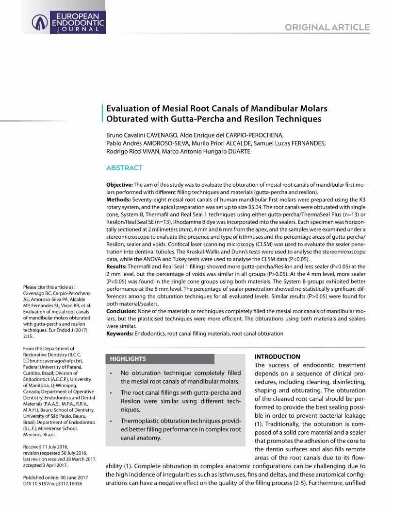
Evaluation of Mesial Root Canals of Mandibular Molars ...
7
Evaluation of Mesial Root Canals of Mandibular Molars Obturated with Gutta-Percha and Resilon Techniques ORIGINAL ARTICLE Bruno Cavalini CAVENAGO, Aldo Enrique del CARPIO-PEROCHENA, Pablo Andrés AMOROSO-SILVA, Murilo Priori ALCALDE, Samuel Lucas FERNANDES, Rodrigo Ricci VIVAN, Marco Antonio Hungaro DUARTE Please cite this article as: Cavenago BC, Carpio-Perochena AE, Amoroso-Silva PA, Alcalde MP, Fernandes SL, Vivan RR, et al. Evaluation of mesial root canals of mandibular molars obturated with gutta-percha and resilon techniques. Eur Endod J (2017) 2:15. From the Department of Restorative Dentistry (B.C.C. [email protected]), Federal University of Paraná, Curitiba, Brazil; Division of Endodontics (A.E.C.P.), University of Manitoba, Q-Winnipeg, Canada; Department of Operative Dentistry, Endodontics and Dental Materials (P.A.A.S., M.P.A., R.R.V., M.A.H.), Bauru School of Dentistry, University of São Paulo, Bauru, Brazil; Department of Endodontics (S.L.F.), Mineirense School, Mineiros, Brazil. Received 11 July 2016, revision requested 30 July 2016, last revision received 28 March 2017, accepted 3 April 2017. Published online: 30 June 2017 DOI 10.5152/eej.2017.16026 ABSTRACT Objective: The aim of this study was to evaluate the obturation of mesial root canals of mandibular first mo- lars performed with different filling techniques and materials (gutta-percha and resilon). Methods: Seventy-eight mesial root canals of human mandibular first molars were prepared using the K3 rotary system, and the apical preparation was set up to size 35.04. The root canals were obturated with single cone, System B, Thermafil and Real Seal 1 techniques using either gutta-percha/ThermaSeal Plus (n=13) or Resilon/Real Seal SE (n=13). Rhodamine B dye was incorporated into the sealers. Each specimen was horizon- tally sectioned at 2 milimeters (mm), 4 mm and 6 mm from the apex, and the samples were examined under a stereomicroscope to evaluate the presence and type of isthmuses and the percentage areas of gutta-percha/ Resilon, sealer and voids. Confocal laser scanning microscopy (CLSM) was used to evaluate the sealer pene- tration into dentinal tubules. The Kruskal-Wallis and Dunn’s tests were used to analyse the stereomicroscope data, while the ANOVA and Tukey tests were used to analyse the CLSM data (P<0.05). Results: Thermafil and Real Seal 1 fillings showed more gutta-percha/Resilon and less sealer (P<0.05) at the 2 mm level, but the percentage of voids was similar in all groups (P>0.05). At the 4 mm level, more sealer (P<0.05) was found in the single cone groups using both materials. The System B groups exhibited better performance at the 6 mm level. The percentage of sealer penetration showed no statistically significant dif- ferences among the obturation techniques for all evaluated levels. Similar results (P>0.05) were found for both material/sealers. Conclusion: None of the materials or techniques completely filled the mesial root canals of mandibular mo- lars, but the plasticised techniques were more efficient. The obturations using both materials and sealers were similar. Keywords: Endodontics, root canal filling materials, root canal obturation INTRODUCTION The success of endodontic treatment depends on a sequence of clinical pro- cedures, including cleaning, disinfecting, shaping and obturating. The obturation of the cleaned root canal should be per- formed to provide the best sealing possi- ble in order to prevent bacterial leakage (1). Traditionally, the obturation is com- posed of a solid core material and a sealer that promotes the adhesion of the core to the dentin surfaces and also fills remote areas of the root canals due to its flow- ability (1). Complete obturation in complex anatomic configurations can be challenging due to the high incidence of irregularities such as isthmuses, fins and deltas, and these anatomical config- urations can have a negative effect on the quality of the filling process (2-5). Furthermore, unfilled HIGHLIGHTS • No obturation technique completely filled the mesial root canals of mandibular molars. • The root canal fillings with gutta-percha and Resilon were similar using different tech- niques. • Thermoplastic obturation techniques provid- ed better filling performance in complex root canal anatomy.
Transcript of Evaluation of Mesial Root Canals of Mandibular Molars ...
Evaluation of Mesial Root Canals of Mandibular Molars Obturated with Gutta-Percha and Resilon Techniques
ORIGINAL ARTICLE
Bruno Cavalini CAVENAGO, Aldo Enrique del CARPIO-PEROCHENA, Pablo Andrés AMOROSO-SILVA, Murilo Priori ALCALDE, Samuel Lucas FERNANDES, Rodrigo Ricci VIVAN, Marco Antonio Hungaro DUARTE
Please cite this article as: Cavenago BC, Carpio-Perochena AE, Amoroso-Silva PA, Alcalde MP, Fernandes SL, Vivan RR, et al. Evaluation of mesial root canals of mandibular molars obturated with gutta-percha and resilon techniques. Eur Endod J (2017) 2:15.
From the Department of Restorative Dentistry (B.C.C. [email protected]), Federal University of Paraná, Curitiba, Brazil; Division of Endodontics (A.E.C.P.), University of Manitoba, Q-Winnipeg, Canada; Department of Operative Dentistry, Endodontics and Dental Materials (P.A.A.S., M.P.A., R.R.V., M.A.H.), Bauru School of Dentistry, University of São Paulo, Bauru, Brazil; Department of Endodontics (S.L.F.), Mineirense School, Mineiros, Brazil.
Received 11 July 2016, revision requested 30 July 2016, last revision received 28 March 2017, accepted 3 April 2017.
Published online: 30 June 2017DOI 10.5152/eej.2017.16026
ABSTRACT
Objective: The aim of this study was to evaluate the obturation of mesial root canals of mandibular first mo-lars performed with different filling techniques and materials (gutta-percha and resilon). Methods: Seventy-eight mesial root canals of human mandibular first molars were prepared using the K3 rotary system, and the apical preparation was set up to size 35.04. The root canals were obturated with single cone, System B, Thermafil and Real Seal 1 techniques using either gutta-percha/ThermaSeal Plus (n=13) or Resilon/Real Seal SE (n=13). Rhodamine B dye was incorporated into the sealers. Each specimen was horizon-tally sectioned at 2 milimeters (mm), 4 mm and 6 mm from the apex, and the samples were examined under a stereomicroscope to evaluate the presence and type of isthmuses and the percentage areas of gutta-percha/Resilon, sealer and voids. Confocal laser scanning microscopy (CLSM) was used to evaluate the sealer pene-tration into dentinal tubules. The Kruskal-Wallis and Dunn’s tests were used to analyse the stereomicroscope data, while the ANOVA and Tukey tests were used to analyse the CLSM data (P<0.05).Results: Thermafil and Real Seal 1 fillings showed more gutta-percha/Resilon and less sealer (P<0.05) at the 2 mm level, but the percentage of voids was similar in all groups (P>0.05). At the 4 mm level, more sealer (P<0.05) was found in the single cone groups using both materials. The System B groups exhibited better performance at the 6 mm level. The percentage of sealer penetration showed no statistically significant dif-ferences among the obturation techniques for all evaluated levels. Similar results (P>0.05) were found for both material/sealers. Conclusion: None of the materials or techniques completely filled the mesial root canals of mandibular mo-lars, but the plasticised techniques were more efficient. The obturations using both materials and sealers were similar.Keywords: Endodontics, root canal filling materials, root canal obturation
INTRODUCTIONThe success of endodontic treatment depends on a sequence of clinical pro-cedures, including cleaning, disinfecting, shaping and obturating. The obturation of the cleaned root canal should be per-formed to provide the best sealing possi-ble in order to prevent bacterial leakage (1). Traditionally, the obturation is com-posed of a solid core material and a sealer that promotes the adhesion of the core to the dentin surfaces and also fills remote areas of the root canals due to its flow-
ability (1). Complete obturation in complex anatomic configurations can be challenging due to the high incidence of irregularities such as isthmuses, fins and deltas, and these anatomical config-urations can have a negative effect on the quality of the filling process (2-5). Furthermore, unfilled
HIGHLIGHTS
• No obturation technique completely filled the mesial root canals of mandibular molars.
• The root canal fillings with gutta-percha and Resilon were similar using different tech-niques.
• Thermoplastic obturation techniques provid-ed better filling performance in complex root canal anatomy.
isthmus areas can house multi-species biofilms, which might lead to the failure of endodontic treatment and consequently to a possible retreatment (6).
Gutta-percha is the most used core in root canal filling because of its adequate dimensional stability. In addition, this material can be plasticised with the application of heat (7). The ther-moplasticisation of gutta-percha can significantly increase its adaptation and filling capacity into the root canals (5). Sever-al thermoplastic techniques have been proposed, and they are widely used to improve the root canal fillings (7-10). Bu-chanan (7) modified the vertical compaction technique using the System B (SybronEndo, Orange, USA) device to promote a continuous wave method for warm vertical condensation of gutta-percha. Thermafil (Dentsply Tulsa, Johnson City, USA) is another well-known method that consists of heated α-phase gutta-percha on a carrier for obturation (9). In line with those findings, an earlier study showed more gutta-percha, less sealer and fewer voids in mesial root canals of mandibular molars ob-turated with System B and Thermafil techniques in comparison to single cone and lateral condensation filling techniques (5).
Gutta-percha and epoxy resin-based root canal sealers are the most common obturation materials, but materials such as Re-silon (Resilon Research LLC, Madison, USA) an polyester poly-mer-based in conjunction with dual-cured methacrylate sealer and self-etching primer have been proposed as substitutes for the traditional root canal filling (11). Resilon is available as cones according to International Organization for Standardization (ISO), similar to gutta-percha. Moreover, Resilon has a significantly higher thermal plasticity in comparison to gutta-percha (12). As was previously shown in a tooth model, this advantageous prop-erty improves its flow into root canal grooves and depressions (13). The gutta-percha and epoxy resin-based sealer can also promote better marginal adaptation of the root canal obturation compared to Resilon and methacrylate sealer, and this might be responsible for the formation of large gaps (14).
Although many studies have investigated the sealing ability of Resilon in single-rooted canals, none have evaluated the unfilled areas when using this material in complex morphologies of me-sial root canals of mandibular molars (10). Additionally, Resilon’s thermoplasticity that allows it to flow into isthmuses during the warm vertical compaction or core carrier-based techniques com-pared to gutta-percha has not been reported so far. Thus, this in vitro study aimed to evaluate the obturation of mesial root canals of mandibular first molars performed with single cone, System B, and core carrier-based techniques using gutta-percha or Resilon.
Two null hypotheses were tested:1) Thermoplastic techniques do not completely fill complex
root canals systems.2) There is no difference between the root canal filling ma-
terials (gutta-percha/epoxy sealer and Resilon/methacry-late sealer) regardless of the obturation technique.
METHODSThis study was approved by the Human Research Ethics Com-mittee of Bauru School of Dentistry, University of São Paulo (Protocol: CEP 122-2009), and the teeth were donated by the Bank of Teeth of the same institution, with consent of the pa-tients allowing the use of their extracted teeth for research purposes.
Seventy-eight mesial roots of extracted human mandibular first molars with complete rhizogenesis, 19 to 21 mm length, curvature degrees between 15° and 30°, with two canals and separate foramina, and without previous endodontic treatment or calcifications were selected. High-speed dia-mond burs were used to form tooth-access cavities, and the working length was established by subtracting 1 mm of the total length up to the apex as measured with a size 10 K-file (Dentsply Maillefer, Ballaigues, Switzerland) (15). The root ca-nals were prepared using K3 NiTi rotary system (SybronEndo) according to the manufacturer recommendations and the api-cal preparation was set up to size 35.04 rotary file. After the use of instruments, the root canals were irrigated with 1 mL of 2.5% sodium hypochlorite (NaOCl). A final irrigation was performed using a passive ultrasonic irrigation with a 20/.01 ultrasonic file (Irrisonic E1; Helse, Santa Rosa de Viterbo, Bra-zil) with intermittent flushing for 1 min. The smear layer was removed with 2 mL of 17% ethylenediaminetetraacetic acid (EDTA) for 3 minutes, then the root canals were flushed with distilled water and dried with paper points.
Root Canal Obturation The teeth were randomly distributed into 6 experimental groups (n=13 for each). ThermaSeal Plus (Dentsply) and Real-Seal SE (SybronEndo) were the sealers used for the gutta-per-cha and Resilon fillings, respectively. Both sealers were mixed with Rhodamine B fluorescent dye (Sigma-Aldrich, St. Louis, USA) at an approximate concentration of 0.1% to allow con-focal microscope visualisation (8, 16). When the Real Seal SE sealer was used, the specimens were light-cured for 40 sec-onds.
The single-cone technique was used for the Groups 1 and 2. For Group 1, 35.04 K3 gutta-percha cones (SybronEndo) with a tug-back were selected. The canals were first filled with Ther-maSeal Plus sealer using a lentulo instrument, and then the selected gutta-percha cones were inserted. A compatible Sys-tem B plugger was placed at the canal orifice and activated so that the gutta-percha was vertically condensed with a plugger at 1 mm below the canal orifice. In Group 2, a 35.04 Resilon cone with RealSeal SE sealer was used in the same manner as described for Group 1.
The vertical compaction technique using the System B device was used in Groups 3 and 4. In the Group 3, the first step of ob-turation was performed similar to Group 1, and the System B
Eur Endod J (2017) 2:15 | Page 2 of 7 Cavenago et al. Evaluation of Gutta-Percha and Resilon Obturation Techniques
Elements were pre-set at 200ºC and the down-pack procedure was performed using a 0.06 plugger inserted within 4 mm of the working length. At the apical level, the gutta-percha was condensed using Buchanan hand pluggers (SybronEndo). The extruder hand piece of the Elements was used as the backfill method. In Group 4, the Resilon and RealSeal SE were used in the same manner as described for Group 3; however, the System B was pre-set at 150°C and the Resilon was used for the backfill. A new layer of sealer was applied before the back-filling procedures in both groups.
For Group 5, the root canal fillings were size 35 Thermafil carri-er points as determined with a Thermafil 35 verifier. The canals were filled with ThermaSeal Plus sealer with the use of a lentu-lo spiral filler. Then the Thermafil obturator was heated in the ThermaPrep Plus oven and inserted into the canal until reach-ing the established working length. In the Group 6, the 0.35 mm RealSeal 1 was used in the same manner as described for Group 5, but the RealSeal SE sealer and the RealSeal 1 oven were used.
All specimens were stored at 37°C with 100% humidity for one week, and a single operator performed the obturation proce-dures.
Sectioning, Isthmus Classification and Microscopy AnalysisEach specimen was horizontally sectioned at 2 mm, 4 mm, and 6 mm from the apex using a 0.3 mm Isomet saw (Buehler, Lake Bluff, USA) under water cooling at 200 rpm. The slices were fixed on a glass plate and then polished with metallograph-ic abrasive grinding with a sequence of SiC abrasive papers (320 grit, 600 grit, 800 grit, and 1200 grit). The specimens were evaluated with a stereomicroscope (Carl Zeiss, Jena, Germany) using 8× magnification and with a confocal laser scanning mi-croscope (CLSM-Leica, Mannheim, Germany).
A high-resolution stereomicroscope camera was used to ac-quire the specimens’ sectional images. From the images, the presence of isthmuses was registered in five categories and their proportions among the experimental groups were system-
atically verified (17). The Axiovision software (Carl Zeiss, Jena, Germany) was used to measure the total area of the two mesial root canals, the gutta-percha or Resilon, and the voids. All ob-tained values were converted into mm2, and the percentages (%) of gutta-percha/Resilon, sealer, and voids were calculated. The measurements were repeated twice to ensure consistency.
Confocal laser scanning microscopy images were obtained at 10 μm below the sample surface by using a 10× lens and a 1 μm step size (8). The images were acquired at 1024×1024 pix-els and were evaluated using the Image J software (National Institutes of Health, Bethesda, USA). The total area of the root canals and the perimeter of dental tubules in which the sealer penetrated were measured to determine the sealer penetra-tion, which was expressed in percentages.
Statistical AnalysisThe D’Agostino and Person normality tests did not show nor-mal distributions in regard to the preliminary analysis of per-centages of gutta-percha/Resilon, sealer, and voids data, so the statistical analysis was performed using the Kruskal-Wallis test with Dunn’s test for post-hoc analysis. The percentage of sealer penetration was compared by ANOVA and Tukey tests. The significance was set at 5%, and the Prism 5.0 software (GraphPad, La Jolla, USA) was used as the analytical tool.
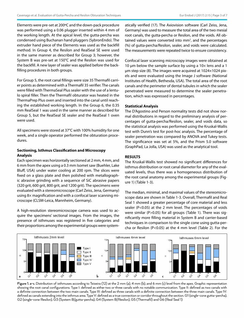
RESULTSThe Kruskal-Wallis test showed no significant differences for isthmus distribution or root canal diameter for any of the eval-uated levels, thus there was a homogeneous distribution of the root canal anatomy among the experimental groups (Fig-ure 1) (Table 1-3).
The median, minimal, and maximal values of the stereomicro-scope data are shown in Table 1-3. Overall, Thermafil and Real Seal 1 showed a greater percentage of core material and less sealer (P<0.05) at the 2 mm level. The percentages of voids were similar (P>0.05) for all groups (Table 1). There was sig-nificantly more filling material in System B and carrier-based techniques in comparison to the single cone using gutta-per-cha or Resilon (P<0.05) at the 4 mm level (Table 2). For the
Eur Endod J (2017) 2:15 | Page 3 of 7Cavenago et al. Evaluation of Gutta-Percha and Resilon Obturation Techniques
Figure 1. a-c. Distribution of isthmuses according to Teixeira (12) at the 2 mm (a), 4 mm (b), and 6 mm (c) level from the apex. Graphic representation showing the root canal configurations. Type I: defined as either two or three canals with no notable communication. Type II: defined as two canals with a definite connection between the two main canals. Type III: defined as three canals with a definite connection between the three main canals. Type IV: defined as canals extending into the isthmus area. Type V: defined as a true connection or corridor throughout the section. G1 (single-cone gutta-percha), G2 (single-cone Resilon), G3 (System B/gutta-percha), G4 (System B/Resilon), G5 (Thermafil) and G6 (Real Seal 1)
a b c
6 mm level, System B techniques showed better obturation performance (P<0.05) in comparison to the single cone tech-niques for both materials (Table 3).
The percentage of sealer penetration showed no statistically significant differences among the obturation techniques for any of the evaluated levels (Table 4). Similar results (P>0.05) were found for both material/sealers with each root canal filling technique. Figure 2 shows representative stereomicro-scope and confocal images of the obturated areas of the root canals at different levels.
DISCUSSIONThe mesial roots of mandibular molars do not follow a con-sistent pattern and have a high incidence of isthmuses, and this can influence the quality of the root canal filling (2, 4, 5, 17). Thus, the distribution of isthmuses in the root canals was verified, and similarity between groups was observed. In addi-
tion, the root canal areas were also similar showing a homo-geneous experimental model. The method of root sectioning for microscopic observations provides relevant data such
Eur Endod J (2017) 2:15 | Page 4 of 7 Cavenago et al. Evaluation of Gutta-Percha and Resilon Obturation Techniques
Group Canal diameter Gutta-Percha/Resilon (%) Sealer (%) Voids (%)
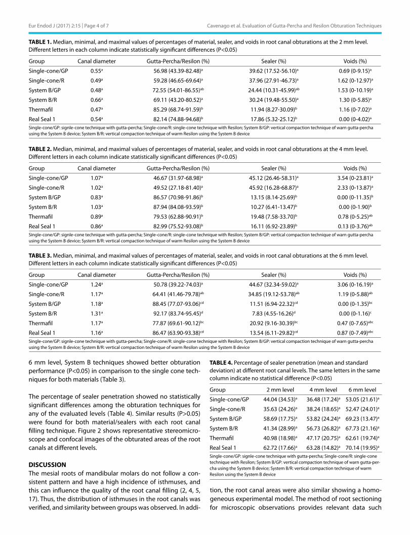
Single-cone/GP 0.55a 56.98 (43.39-82.48)a 39.62 (17.52-56.10)a 0.69 (0-9.15)a
Single-cone/R 0.49a 59.28 (46.65-69.64)a 37.96 (27.91-46.73)a 1.62 (0-12.97)a
System B/GP 0.48a 72.55 (54.01-86.55)ab 24.44 (10.31-45.99)ab 1.53 (0-10.19)a
System B/R 0.66a 69.11 (43.20-80.52)a 30.24 (19.48-55.50)a 1.30 (0-5.85)a
Thermafil 0.47a 85.29 (68.74-91.59)b 11.94 (8.27-30.09)b 1.16 (0-7.02)a
Real Seal 1 0.54a 82.14 (74.88-94.68)b 17.86 (5.32-25.12)b 0.00 (0-4.02)a
Single-cone/GP: signle-cone technique with gutta-percha; Single-cone/R: single-cone technique with Resilon; System B/GP: vertical compaction technique of warn gutta-percha using the System B device; System B/R: vertical compaction technique of warm Resilon using the System B device
TABLE 1. Median, minimal, and maximal values of percentages of material, sealer, and voids in root canal obturations at the 2 mm level. Different letters in each column indicate statistically significant differences (P<0.05)
Group Canal diameter Gutta-Percha/Resilon (%) Sealer (%) Voids (%)
Single-cone/GP 1.07a 46.67 (31.97-68.98)a 45.12 (26.46-58.31)a 3.54 (0-23.81)a
Single-cone/R 1.02a 49.52 (27.18-81.40)a 45.92 (16.28-68.87)a 2.33 (0-13.87)a
System B/GP 0.83a 86.57 (70.98-91.86)b 13.15 (8.14-25.69)b 0.00 (0-11.35)b
System B/R 1.03a 87.94 (84.08-93.59)b 10.27 (6.41-13.47)b 0.00 (0-1.90)b
Thermafil 0.89a 79.53 (62.88-90.91)b 19.48 (7.58-33.70)b 0.78 (0-5.25)ab
Real Seal 1 0.86a 82.99 (75.52-93.08)b 16.11 (6.92-23.89)b 0.13 (0-3.76)ab
Single-cone/GP: signle-cone technique with gutta-percha; Single-cone/R: single-cone technique with Resilon; System B/GP: vertical compaction technique of warn gutta-percha using the System B device; System B/R: vertical compaction technique of warm Resilon using the System B device
TABLE 2. Median, minimal, and maximal values of percentages of material, sealer, and voids in root canal obturations at the 4 mm level. Different letters in each column indicate statistically significant differences (P<0.05)
Group Canal diameter Gutta-Percha/Resilon (%) Sealer (%) Voids (%)
Single-cone/GP 1.24a 50.78 (39.22-74.03)a 44.67 (32.34-59.02)a 3.06 (0-16.19)a
Single-cone/R 1.17a 64.41 (41.46-79.78)ab 34.85 (19.12-53.78)ab 1.19 (0-5.88)ab
System B/GP 1.18a 88.45 (77.07-93.06)cd 11.51 (6.94-22.32)cd 0.00 (0-1.35)bc
System B/R 1.31a 92.17 (83.74-95.45)d 7.83 (4.55-16.26)d 0.00 (0-1.16)c
Thermafil 1.17a 77.87 (69.61-90.12)bc 20.92 (9.16-30.39)bc 0.47 (0-7.65)abc
Real Seal 1 1.16a 86.47 (63.90-93.38)cd 13.54 (6.11-29.82)cd 0.87 (0-7.49)abc
Single-cone/GP: signle-cone technique with gutta-percha; Single-cone/R: single-cone technique with Resilon; System B/GP: vertical compaction technique of warn gutta-percha using the System B device; System B/R: vertical compaction technique of warm Resilon using the System B device
TABLE 3. Median, minimal, and maximal values of percentages of material, sealer, and voids in root canal obturations at the 6 mm level. Different letters in each column indicate statistically significant differences (P<0.05)
Group 2 mm level 4 mm level 6 mm level
Single-cone/GP 44.04 (34.53)a 36.48 (17.24)a 53.05 (21.61)a
Single-cone/R 35.63 (24.26)a 38.24 (18.65)a 52.47 (24.01)a
System B/GP 58.69 (17.75)a 53.82 (24.24)a 69.23 (13.47)a
System B/R 41.34 (28.99)a 56.73 (26.82)a 67.73 (21.16)a
Thermafil 40.98 (18.98)a 47.17 (20.75)a 62.61 (19.74)a
Real Seal 1 62.72 (17.66)a 63.28 (14.82)a 70.14 (19.95)a
Single-cone/GP: signle-cone technique with gutta-percha; Single-cone/R: single-cone technique with Resilon; System B/GP: vertical compaction technique of warn gutta-per-cha using the System B device; System B/R: vertical compaction technique of warm Resilon using the System B device
TABLE 4. Percentage of sealer penetration (mean and standard deviation) at different root canal levels. The same letters in the same column indicate no statistical difference (P<0.05)
as the homogeneity, adaptation to dentin, filling ability, the presence of voids, and the intratubular penetration of sealer, therefore the combined use of stereomicroscope and confo-cal laser scanning microscope methods allows for the collec-tion of accurate data (7, 8, 12, 18, 19).
Regardless of the material used, no technique provided com-plete root canal filling. Thus, the first null hypothesis was ac-cepted. The single cone fillings were tested in this study with the absence of compaction forces to establish a control group. At the 2 mm level, this technique was able to promote a simi-lar filling as the System B technique as previously described (5, 20). However, the single-cone technique generated a signifi-cantly larger amount of sealer, mainly at the 4 mm and 6 mm
levels, in comparison to the other tested techniques. This was probably due to the higher incidence of isthmuses in these sections. However, in these complex anatomic conditions the rotary files do not touch all the root canal walls, thus there is not a completely circular shape and this will certainly change the core:sealer ratio (21). It is widely accepted that an ideal root canal filling requires the maximum of core and the mini-mum amount of sealer.
Both core carrier techniques (Thermafil and Real Seal 1) showed lower amounts of sealer, as reported previously (9, 20). Although the core carrier techniques provide better seal-ing at 2 mm, some studies have reported the risk of root canal filling extrusion. In a previous retrospective in vivo study, the
Eur Endod J (2017) 2:15 | Page 5 of 7Cavenago et al. Evaluation of Gutta-Percha and Resilon Obturation Techniques
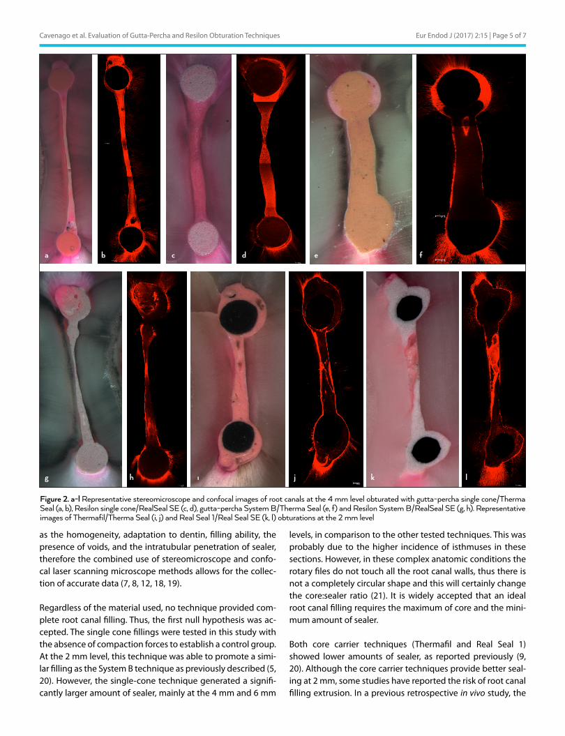
Figure 2. a-l Representative stereomicroscope and confocal images of root canals at the 4 mm level obturated with gutta-percha single cone/Therma Seal (a, b), Resilon single cone/RealSeal SE (c, d), gutta-percha System B/Therma Seal (e, f) and Resilon System B/RealSeal SE (g, h). Representative images of Thermafil/Therma Seal (i, j) and Real Seal 1/Real Seal SE (k, l) obturations at the 2 mm level
a
g h ı j k l
b c d e f
root canal filling performed with Thermafil and using warm vertical compaction showed 80% and 42% extruded filling material, respectively (22).
In this context, the physicochemical properties of the sealers are relevant primarily for single-cone techniques. The flow-ability of both sealers appears to be comparable because they were able to fill areas with isthmuses that are normally difficult to address with the rotary shaping techniques used. In the present study the void areas were similar for all groups, and this was similar to what was previously found in curved root canals (23). Solubility is another critical factor when sin-gle-cone techniques are considered for clinical use because the critical area of fillings is located at the sealer-dentin in-terface (24). According to Resende et al. (25), both the meth-acrylate sealer and epoxy resin sealer used in the present study have a similar solubility. In contrast, De-Deus et al. (18) showed that the Resilon and self-etching sealer induce a high incidence of gaps. In the present study, we found a similar dis-tribution of sealer inside the root canal anatomy using both sealers. The sealer interface under stressed conditions can be expected to be different. In a previous study, the gutta-percha and epoxy resin-based sealer showed better marginal adap-tation in comparison to Resilon and methacrylate sealer filled with single cone and System B techniques (14).
The intensity of heat transferred through a material is signif-icant when considering thermoplastic obturation. According to a previous study, gutta-percha and Resilon cones have a similar thermoplastic ability (26). However, Tanomaru-Filho et al. (12) showed that Resilon has a significantly higher ther-moplasticity in comparison to gutta-percha. Despite the dif-ference in the temperature used to reach the thermoplastic abilities in both materials, we found no significant difference in obturation for the two types of materials or sealers tested, which was in concordance with previous studies (13, 27). The System B technique provided fewer voids at the 4 mm and 6 mm levels compared to the single-cone technique, thus in-creasing the quality of obturations, but in contrast to our re-sults a previous study with a similar methodology found no significant differences between these techniques in terms of the percentage of voids (23).
According to the intratubular sealer penetration parameter found in the CLSM data, both sealers showed similar perfor-mance for the three evaluated techniques. These results repre-sent an important property because sealer penetration inside the dentinal tubules might act as a physical barrier to separate any residual microorganisms from nutrient sources (8).
The second null hypothesis was also confirmed because the results obtained in this study showed that root canal filling techniques using gutta-percha or Resilon had a similar perfor-mance; in other words, both of them presented similar degrees of filling areas using System B or the carrier-based techniques.
The superiority of gutta-percha filling in comparison to Resilon shown in previous studies can be explained by other factors not related to the filling techniques, such as the physicochemical properties of the sealer such as polymerisation shrinkage, ex-tended setting time, or solubility (23, 28-30). These factors show that Resilon fillings reach their weakest point at the sealer inter-face. Regardless of the materials used, better filling quality was obtained with the thermoplasticised techniques that are able to fill the isthmus area. Therefore, such techniques are indicated to fill the complex root canal anatomy.
CONCLUSIONNone of the materials or techniques tested here completely filled the mesial root canals of mandibular molars; however, the plasticised techniques were more efficient. The obturations us-ing both gutta-percha/ThermaSeal Plus and Resilon/Real Seal SE were comparable regardless of the technique used.
Ethics Committee Approval: Ethics committee approval was received for this study from the ethics committee of Bauru School of Dentistry, University of São Paulo (Decision date: 29. 07.2009/Decision No: CEP 122-2009).Informed Consent: N/A.Peer-review: Externally peer-reviewed.Author contributions: Concept - B.C.C., A.E.C.P., M.A.H.D.; Design - B.C.C., P.A.A.S., R.R.V., M.A.H.D.; Supervision - B.C.C., M.A.H.D., R.R.V.; Resource - M.A.H.D., R.R.V.; Materials - B.C.C., P.A.A.S., S.L.F., M.A.H.D.; Data Collection and/or Processing - B.C.C., P.A.A.S., M.P.A., S.L.F.; Analysis and/or Interpretation - B.C.C., A.E.C.P, M.P.A.; Literature Search - B.C.C., M.P.A., S.L.F.; Writing - B.C.C., A.E.C.P.; Critical Reviews - B.C.C., A.E.C.P., P.A.A.S., M.P.A., S.L.F., R.R.V., M.A.H.D.Conflict of Interest: No conflict of interest was declared by the authors.Financial Disclosure: This study was supported by the State of São Paulo Re-search Foundation (FAPESP) (2011/18272-1).
REFERENCES
1. Schilder H. Filling root canals in three dimensions. Dent Clin North Am 1967: 723-44.
2. Villas-Boas MH, Bernardineli N, Cavenago BC, Marciano M, Del Carpio-Per-ochena, de Moraes IG, et al. Micro-computed tomography study of the internal anatomy of mesial root canals of mandibular molars. J Endod 2011; 37(12):1682-6. [CrossRef]
3. Nair PN, Henry S, Cano V, Vera J. Microbial status of apical root canal sys-tem of human mandibular first molars with primary apical periodontitis after “one-visit” endodontic treatment. Oral Surg Oral Med Oral Pathol Oral Radiol Endod 2005; 99(2):231-52. [CrossRef]
4. Mannocci F, Peru M, Sherriff M, Cook R, Pitt Ford TR. The isthmuses of the mesial root of mandibular molars: a micro-computed tomographic study. Int Endod J 2005; 38(8):558-63. [CrossRef]
5. Marciano MA, Ordinola-Zapata R, Cunha TVRN, Duarte MAH, Cavenago BC, Garcia RB, et al. Analysis of four gutta-percha techniques used to fill mesial root canals of mandibular molars. Int Endod J 2011; 44(4):321-9. [CrossRef]
6. Carr GB, Schwartz RS, Schaudinn C, Gorur A, Costerton JW. Ultrastruc-tural examination of failed molar retreatment with secondary apical periodontitis: an examination of endodontic biofilms in an endodontic retreatment failure. J Endod 2009; 35(9):1303-9. [CrossRef]
7. Buchanan LS. The continuous wave of obturation technique: ‘centered’ condensation of warm gutta percha in 12 seconds. Dent Today 1996; 15(1):60-2.
8. Ordinola-Zapata R, Bramante CM, Bernardineli N, Graeff MSZ, Garcia RB, de Moraes IG, et al. A preliminary study of the percentage of sealer pen-etration in roots obturated with the Thermafil and RealSeal-1 obturation techniques in mesial root canals of mandibular molars. Oral Surg Oral Med Oral Pathol Oral Radiol Endod 2009; 108(6):961-8. [CrossRef]
Eur Endod J (2017) 2:15 | Page 6 of 7 Cavenago et al. Evaluation of Gutta-Percha and Resilon Obturation Techniques
9. Gençoğlu N, Garip Y, Baş M, Samani S. Comparison of different gutta-per-cha root filling techniques: Thermafil, Quick-fill, System B, and lateral condensation. Oral Surg Oral Med Oral Pathol Oral Radiol Endod 2002; 93(3):333-6. [CrossRef]
10. De-Deus G, Audi C, Murad C, Fidel S, Fidel RA. Sealing ability of oval-shaped canals filled using the System B heat source with either gutta-per-cha or Resilon: an ex vivo study using a polymicrobial leakage model. Oral Surg Oral Med Oral Pathol Oral Radiol Endod 2007; 104(4):e114-9. [CrossRef]
11. Shipper G, Ørstavik D, Teixeira FB, Trope M. An evaluation of microbial leakage in roots filled with a thermoplastic synthetic polymer-based root canal filling material (Resilon). J Endod 2004; 30(5):342-7. [CrossRef]
12. Tanomaru-Filho M, Silveira GF, Tanomaru JM, Bier CA. Evaluation of the thermoplasticity of different gutta-percha cones and Resilon. Aust En-dod J 2007; 33(1):23-6. [CrossRef]
13. Alicia Karr N, Baumgartner JC, Marshall JG. A comparison of gutta-percha and Resilon in the obturation of lateral grooves and depressions. J Endod 2007; 33(6):749-52. [CrossRef]
14. Cavenago BC, Duarte MA, Ordinola-Zapata R, Marciano MA, Carpio-Per-ochena AE, Bramante CM. Interfacial adaptation of an epoxy-resin sealer and a self-etch sealer to root canal dentin using the System B or the sin-gle cone technique. Braz Dent J 2012; 23(3):205-11. [CrossRef]
15. Schneider SW. A comparison of canal preparations in straight and curved root canals. Oral Surg Oral Med Oral Pathol 1971; 32(2):271-5. [CrossRef]
16. D’Alpino PH, Pereira JC, Svizero NR, Rueggeberg FA, Pashley DH. Use of fluorescent compounds in assessing bonded resin-based restorations: a literature review. J Dent 2006; 34(9):623-34. [CrossRef]
17. Teixeira FB, Sano CL, Gomes BP, Zaia AA, Ferraz CC, Souza-Filho FJ. A preliminary in vitro study of the incidence and position of the root ca-nal isthmus in maxillary and mandibular first molars. Int Endod J 2003; 36(4):276-80. [CrossRef]
18. De-Deus G, Reis C, Di Giorgi K, Brandão MC, Audi C, Fidel RA. Interfacial adaptation of the Epiphany self-adhesive sealer to root dentin. Oral Surg Oral Med Oral Pathol Oral Radiol Endod 2011; 111(3):381-6. [CrossRef]
19. ElAyouti A, Kiefner P, Hecker H, Chu A, Löst C, Weiger R. Homogeneity and adaptation of endodontic fillings in root canals with enlarged api-cal preparation. Oral Surg Oral Med Oral Pathol Oral Radiol Endod 2009; 108(3):e141-6. [CrossRef]
20. De-Deus G, Maniglia-Ferreira CM, Gurgel-Filho ED, Paciornik S, Macha-
do AC, Coutinho-Filho T. Comparison of the percentage of gutta-per-cha-filled area obtained by Thermafil and System B. Aust Endod J 2007; 33(2):55-61. [CrossRef]
21. De-Deus G, Belladonna FG, Silva EJ, Marins JR, Souza EM, Perez R, et al. Micro-CT evaluation of non-instrumented canal areas with different en-largements performed by NiTi Systems. Braz Dent J 2015; 26(6):624-9. [CrossRef]
22. Tennert C, Jungbäck IL, Wrbas KT. Comparison between two thermoplas-tic root canal obturation techniques regarding extrusion of root canal filling--a retrospective in vivo study. Clin Oral Investig 2013; 17(2):449-54.
23. Schäfer E, Nelius B, Bürklein S. A comparative evaluation of gutta-percha filled areas in curved root canals obturated with different techniques. Clin Oral Investig 2012; 16(1):225-30. [CrossRef]
24. Tay F, Loushine R, Lambrechts P, Weller RN, Pashley DH. Geometric fac-tors affecting dentin bonding in root canals: a theoretical modeling ap-proach. J Endod 2005; 31(8):584-9. [CrossRef]
25. Resende LM, Rached-Junior FJ, Versiani MA, Souza-Gabriel AE, Miranda CE, Silva-Sousa YT, et al. A comparative study of physicochemical proper-ties of AH Plus, Epiphany, and Epiphany SE root canal sealers. Int Endod J 2009; 42(9):785-93. [CrossRef]
26. Sant’Anna-Júnior A, Tanomaru-Filho M, Hungaro Duarte MA, Santos Nunes Reis JM, Guerreiro-Tanomaru JM. Temperature changes in gut-ta-percha and Resilon cones induced by a thermomechanical compac-tion technique. J Endod 2009; 35(6):879-82. [CrossRef]
27. Raina R, Loushine RJ, Weller RN, Tay FR, Pashley DH. Evaluation of the quality of the apical seal in Resilon/Epiphany and Gutta-Percha/AH Plus-filled root canals by using a fluid filtration approach. J Endod 2007; 33(8):944-7. [CrossRef]
28. Hirai VH, da Silva Neto UX, Westphalen VP, Perin CP, Carneiro E, Fariniuk LF. Comparative analysis of leakage in root canal fillings performed with gutta-percha and Resilon cones with AH Plus and Epiphany sealers. Oral Surg Oral Med Oral Pathol Oral Radiol Endod 2010; 109(2):e131-5. [Cross-Ref]
29. Onay EO, Ungor M, Unver S, Ari H, Belli S. An in vitro evaluation of the api-cal sealing ability of new polymeric endodontic filling systems. Oral Surg Oral Med Oral Pathol Oral Radiol Endod 2009; 108(2):e49-54. [CrossRef]
30. Kim YK, Grandini S, Ames JM, Gu L, Kim SK, Pashley DH, et al. Critical review on methacrylate resin-based root canal sealers. J Endod 2010; 36(3):383-99. [CrossRef]
Eur Endod J (2017) 2:15 | Page 7 of 7Cavenago et al. Evaluation of Gutta-Percha and Resilon Obturation Techniques