EVALUATION OF GENISTEIN ABILITY TO MODULATE CTGF...
15
Acta Poloniae Pharmaceutica ñ Drug Research, Vol. 71 No. 6 pp. 972ñ986, 2014 ISSN 0001-6837 Polish Pharmaceutical Society Keloid disease is an abnormal cutaneous fibro- proliferative disorder of unknown etiopathogenesis. Keloids present as benign fibroproliferative tumors are considered to be scars that occur when full thick- ness wounds heal abnormally (1ñ6). The molecular abnormalities in keloids that correlate with molecu- lar mechanisms in normal wound healing can be cat- egorized into the synthesis and degradation of extra- cellular matrix (ECM) components, cytokines and growth factors, and apoptotic pathways (6, 7). ECM deposition in any tissue is regulated by a balance between synthesis and degradation of individual components (8). The major component of keloids ECM is mainly either type III collagen (early) or type I collagen (late) because keloid is a type of scar which depends on its maturity (5, 6). The synthesis of type I collagen is highly regu- lated by different cytokines at transcriptional level. Especially, isoforms β1 and β2 of transforming growth factor (TGFβ1 and TGFβ2), members of TGFβ family, enhance type I collagen gene expres- sion. Moreover, they prevent collagen degradation EVALUATION OF GENISTEIN ABILITY TO MODULATE CTGF mRNA/PROTEIN EXPRESSION, GENES EXPRESSION OF TGFβ ISOFORMS AND EXPRESSION OF SELECTED GENES REGULATING CELL CYCLE IN KELOID FIBROBLASTS IN VITRO MAGDALENA JURZAK 1 *, KATARZYNA ADAMCZYK 1 , PAWE£ ANTO—CZAK 1 , AGNIESZKA GARNCARCZYK 1 , DARIUSZ KUåMIERZ 2 and MA£GORZATA LATOCHA 2 Medical University of Silesia, School of Pharmacy with the Division of Laboratory Medicine, 1 Department of Cosmetology, 3 Kasztanowa St., 41-200 Sosnowiec, Poland 2 Department of Cell Biology, 8 Jednoúci St., 41-200 Sosnowiec, Poland Abstract: Keloids are characterized by overgrowth of connective tissue in the skin that arises as a consequence of abnormal wound healing. Normal wound healing is regulated by a complex set of interactions within a net- work of profibrotic and antifibrotic cytokines that regulate new extracellular matrix (ECM) synthesis and remodeling. These proteins include transforming growth factor β (TGFβ) isoforms and connective tissue growth factor (CTGF). TGFβ1 stimulates fibroblasts to synthesize and contract ECM and acts as a central medi- ator of profibrotic response. CTGF is induced by TGFβ1 and is considered a downstream mediator of TGFβ1 action in fibroblasts. CTGF plays a crucial role in keloid pathogenesis by promoting prolonged collagen syn- thesis and deposition and as a consequence sustained fibrotic response. During keloids formation, besides imbalanced ECM synthesis and degradation, fibroblast proliferation and itís resistance to apoptosis is observed. Key genes that may play a role in keloid formation and growth involve: suppressor gene p53, cyclin-depend- ent kinase inhibitor CDKN1A (p21) and BCL2 family genes: antiapoptotic BCL-2 and proapoptotic BAX. Genistein (4í,5,7-trihydroxyisoflavone) exhibits multidirectional biological action. The concentration of genis- tein is relatively high in soybean. Genistein has been shown as effective antioxidant and chemopreventive agent. Genistein can bind to estrogen receptors (ERs) and modulate estrogen action due to its structure simi- larity to human estrogens. Genistein also inhibits transcription factors NFκB, Akt and AP-1 signaling pathways, that are important for cytokines expression and cell proliferation, differentiation, survival and apoptosis. The aim of the study was to investigate genistein as a potential inhibitor of CTGF and TGFβ1, β2 and β3 isoforms expression and a potential regulator of p53, CDKN1A (p21), BAX and BCL-2 expression in normal fibroblasts and fibroblasts derived from keloids cultured in vitro. Real time RT-QPCR was used to estimate transcription level of selected genes in normal and keloid fibroblasts treated with genistein. Secreted/cell-associated CTGF protein was evaluated in cell growthís medium by ELISA. Total protein quantification was evaluated by fluo- rimetric assay in cells lysates (Quant-iT TM Protein Assay Kit). It was found that TGFβ1, β2 and β3 genes expression are decreased by genistein. Genistein suppresses the expression of CTGF mRNA and CTGF protein in a concentration dependent manner. p53 and p21 genes expression are modulated by genistein in concentra- tion dependent manner. The agent also modulates BAX/BCL-2 ratio in examined cells in vitro. Keywords: keloids, fibroproliferative disorders, genistein, connective tissue growth factor, transforming growth factor, cell cycle * Corresponding author: e-mail: [email protected] 972
Transcript of EVALUATION OF GENISTEIN ABILITY TO MODULATE CTGF...

Acta Poloniae Pharmaceutica ñ Drug Research, Vol. 71 No. 6 pp. 972ñ986, 2014 ISSN 0001-6837Polish Pharmaceutical Society
Keloid disease is an abnormal cutaneous fibro-proliferative disorder of unknown etiopathogenesis.Keloids present as benign fibroproliferative tumorsare considered to be scars that occur when full thick-ness wounds heal abnormally (1ñ6). The molecularabnormalities in keloids that correlate with molecu-lar mechanisms in normal wound healing can be cat-egorized into the synthesis and degradation of extra-cellular matrix (ECM) components, cytokines andgrowth factors, and apoptotic pathways (6, 7). ECMdeposition in any tissue is regulated by a balance
between synthesis and degradation of individualcomponents (8). The major component of keloidsECM is mainly either type III collagen (early) ortype I collagen (late) because keloid is a type of scarwhich depends on its maturity (5, 6).
The synthesis of type I collagen is highly regu-lated by different cytokines at transcriptional level.Especially, isoforms β1 and β2 of transforminggrowth factor (TGFβ1 and TGFβ2), members ofTGFβ family, enhance type I collagen gene expres-sion. Moreover, they prevent collagen degradation
EVALUATION OF GENISTEIN ABILITY TO MODULATE CTGF mRNA/PROTEIN EXPRESSION, GENES EXPRESSION OF TGFβ ISOFORMS
AND EXPRESSION OF SELECTED GENES REGULATING CELL CYCLE IN KELOID FIBROBLASTS IN VITRO
MAGDALENA JURZAK1*, KATARZYNA ADAMCZYK1, PAWE£ ANTO—CZAK1, AGNIESZKAGARNCARCZYK1, DARIUSZ KUåMIERZ2 and MA£GORZATA LATOCHA2
Medical University of Silesia, School of Pharmacy with the Division of Laboratory Medicine, 1Department of Cosmetology, 3 Kasztanowa St., 41-200 Sosnowiec, Poland
2 Department of Cell Biology, 8 Jednoúci St., 41-200 Sosnowiec, Poland
Abstract: Keloids are characterized by overgrowth of connective tissue in the skin that arises as a consequenceof abnormal wound healing. Normal wound healing is regulated by a complex set of interactions within a net-work of profibrotic and antifibrotic cytokines that regulate new extracellular matrix (ECM) synthesis andremodeling. These proteins include transforming growth factor β (TGFβ) isoforms and connective tissuegrowth factor (CTGF). TGFβ1 stimulates fibroblasts to synthesize and contract ECM and acts as a central medi-ator of profibrotic response. CTGF is induced by TGFβ1 and is considered a downstream mediator of TGFβ1action in fibroblasts. CTGF plays a crucial role in keloid pathogenesis by promoting prolonged collagen syn-thesis and deposition and as a consequence sustained fibrotic response. During keloids formation, besidesimbalanced ECM synthesis and degradation, fibroblast proliferation and itís resistance to apoptosis is observed.Key genes that may play a role in keloid formation and growth involve: suppressor gene p53, cyclin-depend-ent kinase inhibitor CDKN1A (p21) and BCL2 family genes: antiapoptotic BCL-2 and proapoptotic BAX.Genistein (4í,5,7-trihydroxyisoflavone) exhibits multidirectional biological action. The concentration of genis-tein is relatively high in soybean. Genistein has been shown as effective antioxidant and chemopreventiveagent. Genistein can bind to estrogen receptors (ERs) and modulate estrogen action due to its structure simi-larity to human estrogens. Genistein also inhibits transcription factors NFκB, Akt and AP-1 signaling pathways,that are important for cytokines expression and cell proliferation, differentiation, survival and apoptosis. Theaim of the study was to investigate genistein as a potential inhibitor of CTGF and TGFβ1, β2 and β3 isoformsexpression and a potential regulator of p53, CDKN1A (p21), BAX and BCL-2 expression in normal fibroblastsand fibroblasts derived from keloids cultured in vitro. Real time RT-QPCR was used to estimate transcriptionlevel of selected genes in normal and keloid fibroblasts treated with genistein. Secreted/cell-associated CTGFprotein was evaluated in cell growthís medium by ELISA. Total protein quantification was evaluated by fluo-rimetric assay in cells lysates (Quant-iT TM Protein Assay Kit). It was found that TGFβ1, β2 and β3 genesexpression are decreased by genistein. Genistein suppresses the expression of CTGF mRNA and CTGF proteinin a concentration dependent manner. p53 and p21 genes expression are modulated by genistein in concentra-tion dependent manner. The agent also modulates BAX/BCL-2 ratio in examined cells in vitro.
Keywords: keloids, fibroproliferative disorders, genistein, connective tissue growth factor, transforminggrowth factor, cell cycle
* Corresponding author: e-mail: [email protected]
972

Influence of inositol hexaphosphate on the expression of... 973
by stimulating production of protease inhibitors (9).In contrast, TGFβ3, which is predominantly inducedin the later stages of wound healing, has been foundto reduce connective tissue deposition (10).Connective tissue growth factor (CTGF) is a cys-teine-rich protein (CCN2) induced by TGFβ1 inconnective tissue cells e.g., keloid fibroblasts whichare implicated as mediators of elevated ECM depo-sition (11, 12). Elevated CTGF expression causescell activation and tissue fibrosis (13ñ19). SustainedCTGF synthesis might be responsible for mainte-nance of the fibrotic phenotype in keloids (11, 16,18, 19).
During keloids formation, besides imbalancedECM synthesis and degradation, fibroblast prolifer-ation and itís resistance to apoptosis is observed (2,20ñ23). Key genes that may play a role in keloidformation and growth involve: suppressor gene p53,cyclin-dependent kinase inhibitor p21 (CDKN1A)and BCL2 family genes: antiapoptotic BCL-2 andproapoptotic BAX. The most crucial gene in the net-work of pathways governing cellís division andapoptosis is p53. The p53 tumor suppressor func-tions as transcription factor, by inducing or repress-ing different genes. It can also act as direct inducerof apoptosis by translocation into mitochondria (24).The p21 (also known as p21WAF1/Cip1) is a negative reg-ulator of cell divisions. It inhibits the kinase activityof the cyclin-dependent kinases (CDKs) CDK2 andCDK1 leading to growth arrest at specific stages inthe cell cycle. In addition, by binding to proliferat-ing cell nuclear antigen (PCNA), p21 interferes withPCNA-dependent DNA polymerase activity, there-by inhibiting DNA replication and modulating vari-ous PCNA-dependent DNA repair processes (25).BCL2 family genes are involved in intrinsic, mito-chondrial pathway of apoptosis. In response to stressactivation, BAX forms a homodimer and releasescytochrome c from the mitochondria, which resultsin caspase-9 activation and apoptosis. BCL-2homodimers play opposite, protective role by stabi-lization of mitochondrial membrane (26).
Keloids are important problem in dermatologyand esthetic dermatosurgery. In spite of long-lastingstudies on keloid formation, there is no one satisfac-tory method to be successful in keloid preventionand treatment (14, 27, 28). There are some interest-ing perspectives with the use of genistein, whichpossesses antiproliferative and proapoptotic proper-ties in many human cancer cell lines (29ñ36).
Genistein is a natural component of the plantbelonging to isoflavones class. Soybean is the mostabundant source of naturally occurring genistein.Genistein (4í,5,7-trihydroxyisoflavone) exhibits
antioxidant properties (37ñ40) and multidirectionalaction at the molecular level including i.a., estro-genic and antiestrogenic activity; inhibition of: pro-tein tyrosine kinases (PTKs), topoisomerase II,phosphatidylinositol turnover and proteins involvedin multidrug resistance of cancer cells. Genisteinalso interacts with other potential cellular target pro-teins (36, 41ñ50). Multidirectional molecular actionof genistein led to its use as a modulator of cell pro-liferation, apoptosis, differentiation and cell cycleprogression, particularly cancer cells. Recent reporthas shown that genistein is a potent cancer chemo-preventive agent (51).
The aim of the study was to investigate genis-tein as a potential modulator of CTGF gene expres-sion (mRNA and protein expression) and TGFβ1,β2 and β3 genes expression (mRNAs expressions)and was to enquire whether genistein exerts antipro-liferative or apoptotic activity through effect on p53,p21 (CDKN1A), BCL-2 and BAX genes expressionin keloid fibroblast and normal dermal fibroblastscultured in vitro.
EXPERIMENTAL
Cell culture
Normal human dermal fibroblasts line (NHDF,Adult) was obtained from the tissue culture collec-tion of CloneticsÆ (Lonza Walkersville Inc.,Walkersville, MD, USA). Keloid fibroblasts line(KEL FIB) was obtained from the tissue culture col-lection of ATCCÆ (LGC Standards, Teddingtin,UK). Dulbeccoís modified Eagleís medium(DMEM) with L-glutamine and fetal bovine serum(FBS) were purchased from ATCCÆ (LGCStandards, Teddington, UK). Antibiotic (penicillin-streptomycin solution), Dulbeccoís phosphatebuffered saline without Mg2+ and Ca2+ and 1xTrypsin with EDTA were purchased from SigmaAldrich (St. Louis, USA). Nunc EasYFlasks withfilter 25 cm2 culture surface area was purchasedfrom Thermo Scientific NuncÆ (Roskilde,Denmark); 96-wells microplates and other plasticmaterials were purchased from Sarstedt(N¸mbrecht, Germany).
Normal human dermal fibroblasts (NHDF) andkeloid fibroblasts (KEL FIB) were cultured inDMEM with L-glutamine supplemented with 10%FBS and 1% pen/strep solution (100 U/mL peni-cillin, 200 µg/mL streptomycin). Cells were main-tained at 37OC and 5% CO2 atmosphere in a humid-ified incubator. At confluence, cells were routinelypassaged. Cells from passages 4 to 6 were used inthe study. For the quantitative determination of

974 MAGDALENA JURZAK et al.
examined mRNAs expression, the cells were seededat the density of 5 ◊ 105 per 5 mL medium in 25 cm2
surface area culture flasks. For the quantitativedetermination of CTGF protein expression and totalprotein quantity, the cells were seeded at the densityof 5 ◊ 103/well per 300 µL medium in a 96-wellsmicroplate. After 24 h, when cells adhered to thebottom of wells and started growing, the media werechanged to media containing genistein.
Preparation of genistein dilutions
Genistein from Glycine max (soybean) used inthe study was supplied by Sigma AldrichÆ (cat. no.G6776). Genistein was dissolved in dimethyl sul-foxide (DMSO) as 1% stock solution (concentra-tion of genistein 37 mM) and diluted in the growthmedium (DMEM) with fetal bovine serum (FBS)and antibiotics for normal dermal fibroblast andkeloid fibroblast cultures. Genistein in: 370, 185,92.5, 37, 18.5, 3.7, 1.85, 0.37 µM and 0 µM (con-trol) concentrations was used in the study to evalu-ate CTGF mRNA/protein expression and genesexpression of TGFβ isoforms. For evaluation of theeffect of soybean genistein on the expression ofselected genes regulating cell cycle genistein in:370, 37, 3.7, 0.37 µM and 0 µM (control) concen-trations was used.
The expression of selected genes regulating cell
cycle (p53, p21, BCL-2, BAX) and CTGF,
TGFββ1, ββ2, ββ3 isoforms mRNAs expression in
NHDF and KEL FIB cells treated with genistein
For the analysis of selected mRNAs expres-sion, NHDF and KEL FIB cells were seeded at thedensity of 5 ◊ 105 per 5 mL medium in 25 cm2 sur-face area culture flasks. After 24 h of incubationunder standard conditions (37OC, 5% carbon dioxideand 95% humidity), DMEM with L-glutamine, 10%FBS and 1% pen/strep in fibroblasts cultures werechanged to proper mediums supplemented withgenistein. Cells were treated with genistein for 72 hunder standard conditions. Control groups of cellsconstituted NHDF and KEL FIB cells cultured 72 hin standard culture media described above.
RNA extraction and real-time RT-QPCR assay
Total RNA was extracted from control cell cul-tures and cell cultures incubated with genistein withthe use of TRIZOLÆ reagent (Invitrogen, Carlsbad,CA, USA) according to the manufacturerís protocol.RNA concentration was determined spectrophoto-metrically, on the basis of absorbance values at thewavelength of 260 nm, using a Gene Quant IIRNA/DNA Calculator (Pharmacia Biotech, USA).
All RNA extracts were treated with DNase I (MBIFermentas) according to the manufacturerís instruc-tion.
Transcriptional activity of p53, p21, BCL-2,BAX, CTGF, TGFβ1, β2, β3 genes was evaluatedby the use of real-time RT-QPCR technique with aSYBR Green I chemistry (SYBR Green QuantitectRT-PCR Kit, QIAGEN, Valencia, CA, USA). Theanalysis was carried out using an OpticonTM DNAEngine Sequence Detector (MJ Research, USA).Real-time RT-PCR assay was performed in tripli-cate for each sample. The thermal cycling condi-tions for one-step RT-QPCR assay were as follows:50OC for 30 min for reverse transcription, 95OC for15 min followed by 45 cycles at 94OC for 15 s, 60OCfor 30 s and 72OC for 10 min.
mRNA (RT-QPCR) quantitative detection ofCTGF, p53, p21, BCL-2, BAX were conducted onthe basis of primer pairs designed for referencesequences (Gene Bank www.ncbi.nlm.nih.gov) withthe following sequences: CTGF mRNA: 5í-GTgACgAgCCCAAggACCAAACCT-3í (forward),5í-TggACCAggCAgTTggCTCTAATCATAgTTg-3í(reverse);p53 mRNA: 5í-TAACAgTTCCTgCATgggCggC-3í (for-ward), 5í-AggACAggCACAAACACg CACC-3í (reverse); p21 mRNA: 5í-CACTCCAAACgC CggCTgATCTTC-3í(forward),5í-TgTAgAgCgggCCTTTgAggCCCTC-3í (reverse); BCL-2 mRNA: 5í-TTgTggCCTTCTTTgAgTTCggTg-3í(forward), 5í-ggTgCCggTTC AggTACTCAgTCA-3í (reverse); BAX mRNA: 5í- CCTgTgCACCAA ggTgCCggAACT-3í (forward),5í-CCACCCTggTCT TggATCCAgCCC-3í (reverse).
mRNA (RT-QPCR) quantitative detection ofTGFβ1, β2 and β3 isoforms was conducted based onthe published sequences (52).
The generally used housekeeping genes, β-actin and glyceraldehyde-3-phosphate dehydroge-nase (GAPDH) were used as a potential, accessibleendogenous controls for experimental conditions.The PCR primers for β-actin and GAPDH were pur-chased from PE Applied Biosystems (PE AppliedBiosystems, Inc., Foster, CA, USA).
The mRNA copy numbers of examined geneswere obtained by amplifying commercially avail-able standard of β-actin cDNA (TaqManÆ DNATemplate Reagents Kit; PE Applied Biosystems,Inc., Foster, CA, USA). The control genes wereamplified in triplicate under conditions applied forthe target genes.

Influence of inositol hexaphosphate on the expression of... 975
Following RT-PCR, the samples were subject-ed to temperature ramp from 60 to 95OC at the rateof 0.2OC/s with continuous fluorescence monitoringfor melting curve analysis.
Specificity of real-time RT-QPCR reactionsfor analyzed genes was experimentally confirmedby 8% polyacrylamide gel electrophoresis and deter-mination of the amplimer melting temperatures. Noprimer-dimers or unspecific by-products were gen-erated during applied RT-QPCR amplificationcycles.
Finally, specificity of RT-PCR reaction wasconfirmed by determining characteristic tempera-ture of melting for each amplimer (p53 = 81.8OC;p21 = 83.3OC, BCL-2 = 82.6OC; BAX = 82.0OC;CTGF = 82.7OC; TGFβ1 = 85.5OC; TGFβ2 =80.4OC; TGFβ3 = 80.8OC) and by 8% polyacry-lamide gel electrophoresis of RT-PCR products withtheir visualization using silver staining (121 basepair (bp) for p53 amplimer, 101 bp for p21amplimer, 114 bp for BCL-2 amplimer, 99 bp forBAX amplimer, 109 bp for CTGF amplimer, 152 bpfor TGFβ1 amplimer, 201 bp for TGFβ2 amplimerand 121 bp for TGFβ3 amplimer).
Enzyme-linked immunosorbent assay (ELISA)
quantitative measurement of CTGF protein
CTGF protein expression was measured byELISA in cell culture media (NHDF and KEL FIB)according to the manufacturerís protocol (USCNLife Science Inc., Wuhan, P.R. China). The kit is asandwich enzyme immunoassay for in vitro quanti-tative measurement of CTGF protein in humanserum, plasma, cell culture medium and other bio-logical fluids. ELISA was performed in triplicate foreach sample and in duplicate for each standard andcontrol. Optical density readings of each well (stan-dards and samples) were taken at 450 ± 10 nm onthe Asys UVM 340 Microplate Reader (Biogenet).The concentration of CTGF protein in the sampleswas determined by comparing the O.D. of the sam-ples to the standard curve. The standard curve con-centrations used for the ELISAís were: 30, 15, 7.5,3.75, 1.88, 0.94 and 0.47 ng/mL. The minimumdetectable concentration of human CTGF protein istypically less than 0.21 ng/mL.
The total protein concentration measurement in
cell cultures
Total protein was extracted from control cellsand cells treated with genistein (NHDF and KELFIB) with the use of CellLyticTMM kit (SigmaAldrichÆ, Saint Louis, MO, USA) following the pro-tocol of manufacturer. Total protein concentration
(µg/mL) was determined in triplicate using a Qubitprotein assay kit (no. Q3321; Invitrogen) on a Qubit2.0 fluorometer (Invitrogen).
Statistical analysis
Statistical analysis was carried out usingStatistica PL 8.0 software. The Shapiro-Wilk test fordata normality was applied. Statistical analysis ofthe data showed their normality, therefore all theresults are represented as the means ± SD. The one-way ANOVA followed by post hoc Dunnettís testwas used to determine the significant differencesbetween a control group (cells untreated with genis-tein) mean and the remaining genistein treatmentgroup means in an analysis of variance setting. Thedifference between control cells (NHDF vs. KELFIB) was analyzed using the Studentís t-test. Valuesof p < 0.05 were considered statistically significant.
RESULTS
Housekeeping genes ββ-actin and glyceraldehyde-
3-phosphate dehydrogenase GAPDH as potential
candidates for endogenous control
The generally used housekeeping genes, β-actin and glyceraldehyde-3-phosphate dehydroge-nase (GAPDH) were used as potential, accessibleendogenous controls for experimental conditions.The transcriptional activity of β-actin (ACTB) andGAPDH genes were detected in both cell lines(NHDF and KEL FIB) untreated genistein.
The expression of β-actin gene exhibited sig-nificant differences between normal and keloidfibroblasts (p = 0.004, Studentís t-test) and waslower in NHDF cells (3.44 ◊ 107 ± 4.81 ◊ 106 copynumber of mRNA per 1 µg of total RNA) comparedto KEL FIB cells (4.12 ◊ 107 ± 2.03 ◊ 106 copy num-ber of mRNA per 1 µg of total RNA). Statisticalanalysis of the experimental data with the use ofANOVA revealed statistically significant differ-ences (p < 0.05) in β-actin gene expression betweencontrol cells and cells treated with genistein, simi-larly as described previously (53). Moreover,expression of β-actin mRNAs changed, dependingon genistein concentration, which excludes its appli-cation as endogenous control that might serve tonormalize expression results of examined genes(54).
The expression of GAPDH mRNA exhibitedno significant differences between normal andkeloid fibroblasts (p = 0.272087, Studentís t-test)and was lower in KEL FIB cells (4.16 ◊ 104 ± 3.5 ◊103 copy number of mRNA per 1 µg of total RNA)compared to NHDF cells (1.94 ◊ 105 ± 2.04 ◊ 104

976 MAGDALENA JURZAK et al.
copy number of mRNA per 1 µg of total RNA) (Fig.1). Statistical analysis of the experimental datarevealed no statistically significant differences inGAPDH mRNA expression between NHDF controlcells and NDHF cells treated with genistein (p =0.39094, ANOVA). Statistical analysis of the exper-imental data with the use of ANOVA revealed sta-
tistically significant differences in GAPDH geneexpression between KEL FIB control cells and KELFIB cells treated with genistein (p = 0.000022,ANOVA). Expression of GAPDH mRNA in KELFIB cells exposed to genistein in concentration 0.37and 370 µM compared to control KEL FIB cellsshowed statistical significance (p = 0.00059 and p =
Figure 1. Comparison of the copy number of GAPDH mRNAs per 1 µg of total RNA in NHDF/KEL FIB cells treated with various genis-tein concentration vs. respective control NHDF/KEL FIB cells. * p < 0.01
Figure 2. The native expression of CTGF, TGFβ1, β2 and β3 isoforms in NHDF control cells and KEL FIB control cells. * p < 0.05 (com-parison between control NHDF cells and KEL FIB cells)
Figure 3. Expression of CTGF mRNA in NHDF cells and KEL FIB cells determined by real-time PCR. Comparison of the copy numberof CTGF mRNAs per 1 µg of total RNA in NHDF cells treated with various genistein concentration vs. control NHDF cells (*p) and inKEL FIB cells treated with various genistein concentration vs. control KEL FIB cells (#p). */# p < 0.01, **/## p < 0.001, ***/### p <0.0001 (post hoc Dunnettís test)

Influence of inositol hexaphosphate on the expression of... 977
0.005634, respectively; post hoc Dunnettís test)(Fig.1).
The results of the statistical analysis showed nopossibility of using GAPDH as endogenous controlto normalize the results of examined genes expres-sion.
The native mRNA expression of CTGF, TGFββ1,
ββ2 and ββ3 isoforms in NHDF control cells and
KEL FIB control cells
The transcriptional activity of all examinedgenes was detected in unstimulated (control) NDHFcells and KEL FIB cells. The expression of CTGFmRNA exhibited significant differences betweennormal and keloid fibroblasts (p = 0.0154, Studentíst-test) and was lower in NHDF cells (7.06 ◊ 104 ±7.92 ◊ 103 copy number of mRNA per 1 µg of total
RNA) compared to KEL FIB cells (1.07 ◊ 105 ± 1.26◊ 104 copy number of mRNA per 1 µg of totalRNA). Likewise, statistical analysis revealed signif-icant differences in TGFβ1 mRNA (p = 0.0106,Studentís t-test) and TGFβ3 mRNA (p < 0.0001,Studentís t-test) between normal and keloid fibrob-lasts. The expression of TGFβ1 mRNA and TGFβ3mRNA was lower in NHDF cells (8.21 ◊ 105 ± 8.67◊ 104 and 4.0 ◊ 104 ± 7.04 ◊ 103 copy number ofmRNA per 1 µg of total RNA, respectively), com-pared to KEL FIB cells (1.11 ◊ 106 ± 1.03 ◊ 105 and1.49 ◊ 105 ± 1.87 ◊ 104 copy number of mRNA per1 µg of total RNA, respectively). No markedchanges in TGFβ2 mRNA were demonstratedbetween control NHDF cells (1.50 ◊ 105 ± 5.99 ◊ 104
copy number of mRNA per 1 µg of total RNA) andcontrol KEL FIB cells (9.96 ◊ 104 ± 1.36 ◊ 104 copy
Table 1. The results of the statistical analysis (post hoc Dunnettís test) used to determine the differencesbetween TGFβ1, β2, β3 mRNAs expression between control cells (cells untreated genistein) and cells treatedwith genistein in NHDF cells.
Genistein The results of post hoc Dunnettísconcentration test presented as p value
[µM]TGFββ1 TGFββ2 TGFββ3
0.37 0.000007 0.000005 0.000007
1.85 0.000005 0.012149 0.000416
3.7 0.000014 0.005199 0.007349
18.5 0.000023 0.000423 0.000246
37 0.000006 0.000006 0.000005
92.5 0.000044 0.000237 0.000124
185 0.000007 0.000042 0.000336
370 0.000005 0.000005 0.000006
Table 2. The results of the statistical analysis (post hoc Dunnettís test) used to determine the differencesbetween TGFβ1, β2, β3 mRNAs expression between control cells (cells untreated with genistein) and cellstreated with genistein in KEL FIB cell cultures.
Genistein The results of post hoc Dunnettísconcentration test presented as p value
[µM]TGFββ1 TGFββ2 TGFββ3
0.37 0.000006 0.000005 0.000005
1.85 0.000005 0.000005 0.000005
3.7 0.000006 0.000005 0.000005
18.5 0.000005 0.000005 0.000005
37 0.000005 0.000005 0.000005
92.5 0.000005 0.000005 0.000005
185 0.000005 0.000005 0.000005
370 0.000009 0.000005 0.000005

978 MAGDALENA JURZAK et al.
number of mRNA per 1 µg of total RNA) (p > 0.05,Studentís t-test) (Fig. 2).
CTGF mRNA expression in NHDF and KEL FIB
exposed to genistein
Treatment of NHDF cells and KEL FIB cellswith genistein in each applied concentration result-ed in downregulation of CTGF mRNA expression ascompared with untreated cells in both lines (p =0.000004, p = 0.001009, respectively; ANOVA)(Fig. 3).
Statistical analysis revealed no significantdecrease in CTGF mRNA expression in NHDF cellsexposed to genistein in concentration of 37 µM (p =0.064446, post hoc Dunnettís test) (Fig. 3) and inKEL FIB cells exposed to genistein in concentration370 µM (p = 0.992769, post hoc Dunnettís test)compared to the respective control cells, only. Theother concentration of genistein showed significantdifferences compared to control NHDF cells andcontrol KEL FIB cells, respectively (Fig. 3).
CTGF protein expression in NHDF and KEL FIB
exposed to genistein
Statistical analysis revealed no significancedifferences between obtained CTGF protein concen-tration (ng/mL) (Fig. 4a) and CTGF protein concen-tration recalculated per total amount of protein in thecell lysates (Fig. 4b) (p > 0.05, Studentís t-test), inboth cells lines (NHDF and KEL FIB).
The expression of CTGF protein exhibited sig-nificant differences between normal and keloidfibroblasts (p = 0.00001, Studentís t-test) and wasabout 5 times lower in NHDF cells (3.511 ± 1.046ng/µL) compared to KEL FIB cells (26.133 ± 2.202ng/µL).
Exposure of NHDF cells to genistein in eachapplied concentration resulted in downregulation ofCTGF protein expression as compared with untreat-ed NHDF cells (p = 0.0261, ANOVA). Statisticalanalysis revealed significant decrease in the CTGFprotein expression in NHDF cells exposed to genis-tein in concentration 1.85 µM (p = 0.024373, post
Figure 4. Expression of CTGF protein in NHDF cells and KEL FIB cells determined by ELISA. a Comparison of CTGF protein (µg/µL)between: control normal dermal fibroblasts (NHDF) and control keloid fibroblasts (KEL FIB) (# p < 0.05, Studentís t-test), NHDF controlcells and genistein treated NHDF cells (^ p < 0.05, Dunnettís test) and KEL FIB control cells and genistein treated KEL FIB cells (* p <0.00001, Dunnettís test). b Comparison of CTGF protein (µg/µL) and CTGF protein (µg/µL) recalculated per µg of total protein (in eachconcentration p > 0.05, Student t-test)
Influence of inositol hexaphosphate on the expression of... 979
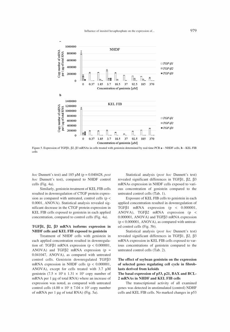
hoc Dunnettís test) and 185 µM (p = 0.040428, posthoc Dunnettís test), compared to NHDF controlcells (Fig. 4a).
Similarly, genistein treatment of KEL FIB cellsresulted in downregulation of CTGF protein expres-sion as compared with untreated, control cells (p <0.0001, ANOVA). Statistical analysis revealed sig-nificant decrease in the CTGF protein expression inKEL FIB cells exposed to genistein in each appliedconcentration, compared to control cells (Fig. 4a).
TGFββ1, ββ2, ββ3 mRNA isoforms expression in
NHDF cells and KEL FIB exposed to genistein
Treatment of NHDF cells with genistein ineach applied concentration resulted in downregula-tion of: TGFβ1 mRNA expression (p < 0.000001,ANOVA) and TGFβ2 mRNA expression (p =0.041047, ANOVA), as compared with untreatedcontrol cells. Genistein downregulated TGFβ3mRNA expression in NHDF cells (p < 0.000001,ANOVA), except for cells treated with 3.7 µMgenistein (7.5 ◊ 104 ± 1.31 ◊ 104 copy number ofmRNA per 1 µg of total RNA) where an increase ofexpression was noted, as compared with untreatedcontrol cells (4.00 ◊ 104 ± 7.04 ◊ 103 copy numberof mRNA per 1 µg of total RNA) (Fig. 5a).
Statistical analysis (post hoc Dunnettís test)revealed significant differences in TGFβ1, β2, β3mRNAs expression in NHDF cells exposed to vari-ous concentration of genistein compared to theuntreated control cells (Tab. 1).
Exposure of KEL FIB cells to genistein in eachapplied concentration resulted in downregulation ofTGFβ1 mRNA expression (p < 0.000001,ANOVA), TGFβ2 mRNA expression (p <0.000001, ANOVA) and TGFβ3 mRNA expression(p < 0.000001, ANOVA), as compared with untreat-ed control cells (Fig. 5b).
Statistical analysis (post hoc Dunnettís test)revealed significant differences in TGFβ1, β2, β3mRNA expression in KEL FIB cells exposed to var-ious concentrations of genistein compared to theuntreated control cells (Tab. 2).
The effect of soybean genistein on the expression
of selected genes regulating cell cycle in fibrob-
lasts derived from keloids
The basal expression of p53, p21, BAX and BCL-
2 mRNAs in NHDF and KEL FIB cells
The transcriptional activity of all examinedgenes was detected in unstimulated (control) NDHFcells and KEL FIB cells. No marked changes in p53
Figure 5. Expression of TGFβ1, β2, β3 mRNAs in cells treated with genistein determined by real-time PCR a ñ NHDF cells, b ñ KEL FIBcells
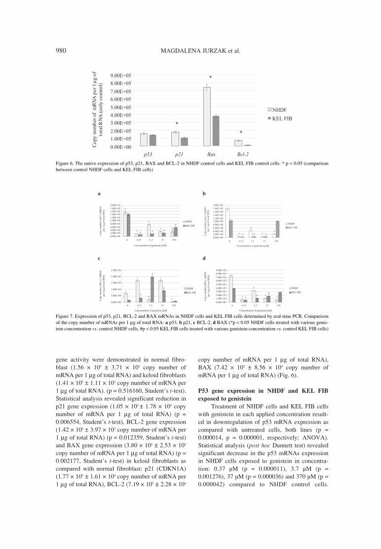
980 MAGDALENA JURZAK et al.
gene activity were demonstrated in normal fibro-blast (1.56 ◊ 105 ± 3.71 ◊ 103 copy number ofmRNA per 1 µg of total RNA) and keloid fibroblasts(1.41 ◊ 105 ± 1.11 ◊ 103 copy number of mRNA per1 µg of total RNA). (p = 0.516160, Studentís t-test).Statistical analysis revealed significant reduction inp21 gene expression (1.05 ◊ 105 ± 1.78 ◊ 104 copynumber of mRNA per 1 µg of total RNA) (p =0.006554, Studentís t-test), BCL-2 gene expression(1.42 ◊ 104 ± 3.97 ◊ 103 copy number of mRNA per1 µg of total RNA) (p = 0.012359, Studentís t-test)and BAX gene expression (3.80 ◊ 104 ± 2.53 ◊ 104
copy number of mRNA per 1 µg of total RNA) (p =0.002177, Studentís t-test) in keloid fibroblasts ascompared with normal fibroblast: p21 (CDKN1A)(1.77 ◊ 105 ± 1.61 ◊ 104 copy number of mRNA per1 µg of total RNA), BCL-2 (7.19 ◊ 105 ± 2.28 ◊ 104
copy number of mRNA per 1 µg of total RNA),BAX (7.42 ◊ 105 ± 8.56 ◊ 104 copy number ofmRNA per 1 µg of total RNA) (Fig. 6).
P53 gene expression in NHDF and KEL FIB
exposed to genistein
Treatment of NHDF cells and KEL FIB cellswith genistein in each applied concentration result-ed in downregulation of p53 mRNA expression ascompared with untreated cells, both lines (p =0.000014, p = 0.000001, respectively; ANOVA).Statistical analysis (post hoc Dunnett test) revealedsignificant decrease in the p53 mRNAs expressionin NHDF cells exposed to genistein in concentra-tion: 0.37 µM (p = 0.000011), 3.7 µM (p =0.001276), 37 µM (p = 0.000036) and 370 µM (p =0.000042) compared to NHDF control cells.
Figure 6. The native expression of p53, p21, BAX and BCL-2 in NHDF control cells and KEL FIB control cells. * p < 0.05 (comparisonbetween control NHDF cells and KEL FIB cells)
Figure 7. Expression of p53, p21, BCL-2 and BAX mRNAs in NHDF cells and KEL FIB cells determined by real-time PCR. Comparisonof the copy number of mRNAs per 1 µg of total RNA: a p53, b p21, c BCL-2, d BAX (*p < 0.05 NHDF cells treated with various genis-tein concentration vs. control NHDF cells; #p < 0.05 KEL FIB cells treated with various genistein concentration vs. control KEL FIB cells)

Influence of inositol hexaphosphate on the expression of... 981
Genistein in concentrations: 0.37 µM (p = 000005,Dunnettís test), 3.7 µM (p = 000005, Dunnettís test),37 µM (p = 000005, Dunnettís test) and 370 µM (p= 0.000019, Dunnettís test) downregulated constitu-tive transcriptional activity of p53 gene in KEL FIBcells (Fig. 7a).
P21 gene expression in NHDF and KEL FIB
exposed to genistein
Exposure of NHDF cells and KEL FIB cells togenistein in each applied concentration resulted indownregulation of p21 mRNA expression as com-pared with untreated cells, both lines (p = 0.000002,p = 0.000001, respectively; ANOVA). Statisticalanalysis (post hoc Dunnett test) revealed significantdecrease in the p21 mRNAs expression both inNHDF cells exposed to genistein in concentration:0.37 µM (p = 0.000006), 3.7 µM (p = 0.000014), 37µM (p = 0.000006) and KEL FIB cell exposed togenistein at the same concentration (p = 0.000005),(p = 0.000005), (p = 0.000006), respectively.Genistein in 370 µM concentration decreasedexpression of p21 mRNA in NHDF cells (p =0.000069) compared to the NHDF control cells.Genistein in 370 µM concentration induced oppositeeffect in keloid fibroblasts cultured in vitro, wherewas noted increase of p21 expression (1.01 ◊ 105 ±1.22 ◊ 104 copy number of mRNA per 1 µg of totalRNA) in comparison to control KEL FIB cells (1.05◊ 105 ± 1.78 ◊ 104 copy number of mRNA per 1 µgof total RNA). Nevertheless, no statistically signifi-cant differences were revealed (p > 0.05, Dunnettístest) (Fig. 7b).
BCL-2 gene expression in NHDF and KEL FIB
exposed to genistein
Exposure of NHDF cells to genistein in theconcentration: 0.37, 3.7, 37 and 370 µM changed
expression of BCL-2 gene in normal dermal fibrob-last (p = 0.000003, ANOVA) and in keloid fibrob-lasts (p = 0.000002, ANOVA). The expression ofBCL-2 revealed similar quantity in cells treated with0.37 µM (1.72 ◊ 105 ± 3.32 ◊ 104) and 37 µM genis-tein (1.71 ◊ 105 ± 3.41 ◊ 104) to control cells (7.19 ◊105 ± 2.28 ◊ 104) (p > 0.05, Dunnettís test). Theexpression of BCL-2 was significantly lower in cellstreated with 3.7 µM genistein (2.06 ◊ 104 ± 8.28 ◊103) and 370 µM genistein (2.43 ◊ 103 ± 3.75 ◊ 102
copy number of mRNA per 1 µg of total RNA) com-pared to control NHDF cells (p = 0.025015, p =0.00024, respectively; post hoc Dunnettís test).Expression of BCL-2 revealed similar quantity incells treated with 370 µM genistein (1.05 ◊ 104 ±1.20 ◊ 103) compared to control KEL FIB cells (p >0.05, Dunnettís test). Treatment of KEL FIB cellswith 0.37, 3.7 and 370 µM genistein resulted in sig-nificant decrease (post hoc Dunnettís test) in BCL-2expression in comparison to control KEL FIB cells(p = 0.042235, p = 0.000012, p = 0.00007, respec-tively) (Fig. 7c).
BAX gene expression in NHDF and KEL FIB
exposed to genistein
Treatment of NHDF cells and KEL FIB cellswith genistein in each applied concentration result-ed in downregulation of BAX mRNA expression ascompared with untreated cells, both lines (p =0.000001, p = 0.000006, respectively; ANOVA).The expression of BAX in NHDF cells was signifi-cantly lower in cells treated with 0.37 µM genistein(6.60 ◊ 104 ± 1.94 ◊ 104), 3.7 µM genistein (3.19 ◊105 ± 4.64 ◊ 104), 37 µM genistein (6.60 ◊ 104 ± 1.94◊ 104) and 370 µM genistein (1.13 ◊ 105 ± 1.22 ◊ 104
copy number of mRNA per 1 µg of total RNA) com-pared to control NHDF cells (p = 0.000005, p =0.000482, p = 0.000006, p = 0.000005, respectively;
Table 3. BAX/BCL-2 ratio for mRNA levels in normal fibroblasts (NHDF line) and fibroblast derived fromkeloid (KEL FIB) treated with genistein *p < 0.05 NHDF cells treated with various genistein concentration vs.control NHDF cells; #p < 0.05 KEL FIB cells treated with various genistein concentration vs. control KEL FIBcells.
Genistein BAX/BCL-2 mRNA
concentration ratio
[µM]NHDF KEL FIB
0 10.31 ± 3.76 26.67 ± 6.40
0.37 0.38 ± 0.07 * 2.00 ± 0.22 #
3.7 15.48 ± 5.60 0.58 ± 0.07 #
37 0.66 ± 0.04 * 2.00 ± 0.09 #
370 25.76 ± 2.15 * 27.04 ± 1.89

982 MAGDALENA JURZAK et al.
post hoc Dunnettís test).The expression of BAX inKEL FIB cells was significantly lower in cells treat-ed with 0.37 µM genistein (6.04 ◊ 104 ± 1.26 ◊ 104),3.7 µM genistein (1.15 ◊ 105 ± 8.15 ◊ 103) and 37µM genistein (1.79 ◊ 105 ± 7.73 ◊ 104) compared tocontrol KEL FIB cells (p = 0.000008, p = 0.000145,p = 0.002822, respectively; post hoc Dunnettís test).Although, expression of BAX gene was lower incells treated with 370 µM genistein (6.26 ◊ 104 ±8.07 ◊ 103) compared to control KEL FIB cells, sta-tistical analysis revealed no significant differences(p > 0.05, Dunnettís test) (Fig. 7d).
Proapoptotic ratio BAX/BCL-2 in normal and
keloid fibroblasts
The BAX/BCL-2 ratio determines cell suscep-tibility to apoptosis. The BAX/BCL-2 mRNA ratiowas determined based on real time RT-PCR results(Tab. 3).
Statistical analysis revealed significantincrease in BAX/BCL-2 mRNA ratio in controlfibroblasts derived from keloid (p = 0.03621,Studentís t-test) compared to control normal fibrob-lasts. Treatment of NHDF cells with genisteinresulted in statistically significant decrease ofBAX/BCL-2 ratio in cells treated with 0.37, 37 and370 µM genistein (p = 0.003651, p = 0.004211, p =0.002644, respectively; Dunnettís test). No markedchanges in BAX/BCL-2 mRNA ratio were demon-strated with 3.7 µM genistein stimulated cells (p >0.05; Dunnettís test).
Keloid fibroblasts exposed to 0.37, 3.7 and 37µM genistein revealed significant decrease ofBAX/BCL-2 mRNA ratio compared to control KELFIB cells (p = 0.000211, p = 0.000055, p =0.000200, respectively; Dunnettís test). Keloidfibroblast exposed to 370 µM genistein revealed nodifferences in BAX/BCL-2 mRNA ratio in compar-ison to control fibroblast derived from keloids (p >0.05; Dunnettís test).
DISCUSSION
Keloids are abnormal wound responses in pre-disposed individuals and represent a connective tis-sue response to skin injury. Treatment of keloidsincludes surgical, pharmacological and physicalmethods. Each of these methods can be applied asmonotherapy, but combining them gives betterresults. Moreover, various modalities of therapymay be applied, but none of them has been provedentirely successful (55, 56). Keloids are abnormalcutaneous fibroproliferative disorders which growbeyond the confines of the original wound, invading
the normal surrounding skin and they rarely regressover time (1, 6, 55). Cutaneous fibroproliferativedisorders other than keloids include systemic sclero-sis and hypertrophic scars (56, 57) and CTGF istheir effective marker (57). Moreover, expression ofCTGF in keloid fibroblast is dependent on fibroblastlocation and is more concentrated in keloid fibrob-lasts at the expanding invasive border of the keloidscar (6, 58). In the study, the keloid fibroblast cul-ture revealed an increase of CTGF mRNA and anincrease of CTGF protein expression compared tonormal fibroblast confirming contribution of CTGFin keloid fibroblast pathology.
CTGF is up-regulated at the transcriptionallevel by a number of specific factors such as TGFβand endothelin-1 (ET-1) or nonspecific factor(mechanicals stress, hypoxia) (16ñ19, 37, 57).Several studies describe the role of TGFβ in keloidsand hypertrophic scars. CTGF expression increasedmore than 100-fold after stimulation with TGFβ1and more than 75-fold after the addition of TGFβ2and TGF-β3 (10).
TGFβ1 stimulation induces collagen synthesisin keloid and hypertrophic scar fibroblasts, butTGFβ1 upregulation alone seems not to be sufficientfor excessive scarring (59, 60). In animal models,TGFβ2 induces cell proliferation and collagen pro-duction in scars (61, 62). The role of TGFβ3 inwound healing and pathological scarring is not fullyunderstood, but TGF-β3 is both scar inducing andscar reducing in animals (63).
Our data revealed increased expression ofTGFβ1 mRNA in keloid fibroblasts compared tonormal dermal fibroblast, confirming results fromother studies (64, 65). Lee et al. (64) describeincreased TGF-β2 protein expression in keloidderived fibroblasts compared to normal skin fibro-blasts. They did not define the biopsy site fromwhich the fibroblasts were harvested and the timepoints for analysis of mRNA and protein expressionmay diverge. Our data reveal a decrease (no statisti-cal significance) of TGFβ2 mRNA expression inkeloids fibroblast compared to normal dermalfibroblasts, confirming Seiferts results (66). Thestudy revealed increased expression of TGFβ3mRNA in keloid fibroblasts compared to normaldermal fibroblast, in contrast to Leeís et al. studywhich revealed no difference between keloidderived fibroblasts and normal human skin fibrob-lasts. Seifert results revealed significantly decreasedexpression of TGFβ3 mRNA in keloids compared tohypertrophic scars to control skin (66).
Our study seems to confirm the thesis that theincreased expression of CTGF by keloid fibroblasts

Influence of inositol hexaphosphate on the expression of... 983
could be a response to TGFβ1 stimulation. TheTGFβ signal is mediated by SMADs intracellularproteins. Changes in this signaling pathway regulateTGFβ expression and induce gene expression in thecell. The SMAD signaling pathway is crucial forsimultaneous activation of several collagen genes byTGFβ and other ECM-related genes are identified asgene targets downstream of TGFβ (67). Moreover,functional Smad element resides within the CTGFpromoter (68). However, the ability of TGFβ tofully induce the CTGF promoter and protein alsorequires protein kinase C and the ras/MEK/ERKMAP kinase cascade (69, 70).
Inhibitors of CTGF expression may be used asanti-fibrotic therapies (71) via TGFβ-dependent orTGFβ-independent mechanism. Inhibitors of TGFβor endothelin receptors may be used to reduce CTGFexpression in skin fibroproliferative disease such asscleroderma (72, 73). Ilprost, synthetic prostacyclinand 9-cis-retinoic acid reduced CTGF expression inscleroderma fibroblasts (74). One of the mechanismsof ilprostís action is antagonizing MEK/ERK signal-ing (75). U0126 is a highly selective inhibitor of bothMEK1 and MEK2, a type of MAPK/ERK kinase, itreduces CTGF (CCN2) expression in response toTGFβ and ET-1 in scleroderma fibroblasts as well asconstitutive over-expression of CTGF in the pancre-atic cancer cell line (76). TNFα suppresses TGFβ-induced gene expression in fibroblasts but has noappreciable effect on the constitutive CTGF expres-sion in scleroderma fibroblasts (77). Caffeine alsoreduces TGFβ-induced CTGF expression in hepato-cytes by blocking Smad activation (78). Many invitro, epidemiological and animal model studiesproved application of genistein in the prophylaxisand treatment of i.a., menopause, cancer, cardiovas-cular disease and cystic fibrosis (79). Recent studiesare focused on application of this natural isoflavonein fibroproliferative diseases. The results of the studyrevealed that genistein decreased mRNA and proteinexpression of CTGF in keloid fibroblast in a concen-tration-dependent manner. Moreover, genisteindecreases TGFβ1, β2 and β3 genes expression(mRNA level) in keloid fibroblast. Han et al. (80)showed that genistein can effectively inhibit TGFβ1-induced invasion and metastasis in pancreatic cancercell line Panc-1 and could partly suppress the expres-sion of CTGF gene and its protein stimulated TGFβ1in rat renal mesangial cells and probably decreasesthe accumulation of extracellular matrix (ECM) andhas the potential ability of anti-fibrosis (81).
Keloid derived fibroblasts have a greater pro-liferative capacity than normal dermal fibroblasts(21, 82). Furthermore, the proliferation profile of
fibroblasts in keloid in vivo has been properly docu-mented (83) and clearly demonstrated differences inthe proliferation of cells between the center and theedge of the lesion. Besides, proliferation, apoptosis,and necrosis occur simultaneously in keloids andthese processes are distinctly compartmentalized,too. When keloid matures, apoptosis and necrosisresult in selective removal of certain cellular popu-lations resulting in the characteristic avascularfibrotic collagenous lesion, whereas proliferation offibroblasts in the keloid dermis propagates the fibro-sis (21). Moreover, keloid fibroblasts fail to undergoapoptosis and thus, continue to produce and secreteextracellular matrix components (20).
The induction of apoptosis is partly mediated,intracellularly, by several genes, such as p53, BCL-2, BAX, and p21. The p53 tumor suppressor gene isa cell cycle regulator able to induce cell cycle arrestto allow DNA repair or apoptosis. p21 is activatedby the p53 protein, and an increased level of p21 isassociated in cyclin-containing complexes withdecreasing cyclin-dependent activity in damagedcells destined to apoptosis. BCL-2 functions as asuppressor of apoptotic death triggered by a varietyof signals, and is negatively regulated by wild typep53. BCL-2 overexpression has been shown toinhibit apoptosis induced by a variety of stimuli,whereas, a predominance of BAX over BCL-2accelerates apoptosis upon apoptotic stimuli (84).The aim of the study was to evaluate the influence ofgenistein on transcriptional activity of genes encod-ing p53, p21, BCL-2 and BAX in keloid fibroblasts.Results of the study reveal no marked changes inp53 gene expression between normal dermal fibrob-last and fibroblast derived from keloid and markeddecrease of p21 gene expression in keloid fibroblastcompared to normal fibroblasts. De Felice et al.found that in keloid fibroblasts a p53 (TP53) underexpression, due to the sequence mutations, in con-cert with ∆Np63 (an isoform of the p63 gene) acti-vation, is central in the mechanism involved inkeloid proliferation (85).
In the study, exposure of NHDF cells and KELFIB cells to genistein in each applied concentrationresulted in downregulation of p53 mRNA and p21expression as compared with untreated cells, bothlines. The effects of genistein may depend on the lev-els of endogenous p53 in the cells. p53 is known toinduce DNA repair enzymes, and cells containingwildtype p53 may have repair of the DNA damagecaused by genistein treatment. Tumor cells, eitherdeficient in p53 or with very low doses of it, replicatethrough the damage and are more susceptible togenistein effects (34). The obtained results may indi-

984 MAGDALENA JURZAK et al.
cate that fibroblast derived from keloid compartmentwhere cells proliferation and apoptosis were bal-anced, and keloid fibroblast not existed as malignantcells. Besides, in normal cells p53 is found at verylow levels (86). The increase in the level of active p53protein leads to an inhibition of entry into S-phase orthe induction of apoptosis. Genistein has been identi-fied as a protein tyrosine kinases (PTKs) inhibitor,which play a key role in oncogenesis, control of cellgrowth and apoptosis, therefore genistein is a potentinhibitor of cell proliferation, oncogenesis and clono-genic ability in animal and human cells (84).
P21 expression is usually controlled at the tran-scriptional level by both p53-dependent and p533-independent mechanisms. Cells lacking p21 appearto undergo apoptosis normally. The results of thestudy (decrease of p21 gene expression in cells KELFIB cells and NHDF cells treated with genistein) arecontrary to Upadhyay et al. results (87), whichrevealed that p21 is up-regulated by genistein treat-ment, and greatly induced at RNA and protein lev-els in normal breast epithelial cells, whereas its levelwas only slightly induced in malignant MDA-MB-231 breast epithelial cells and not detectable inmalignant MCF10CA1a breast epithelial cells.Therefore, p21 is responsible for differential sensi-tivity of genistein among these cell lines (87). Li etal. revealed up-regulation of p21 and BAX expres-sions and down- regulation of p53 and BCL-2expression in genistein-treated breast cancer cellsMDA-MB-231 (84).
BAX and BCL-2 have been reported to play amajor role in determining whether cells will under-go apoptosis under experimental conditions that pro-mote cell death. Increased expression of BAX caninduce apoptosis, while BCL-2 protects cells fromapoptosis. Ratio of BAX/BCL-2 is important for thesurvival of drug-induced apoptosis in leukemia celllines (84, 88). The study revealed significant reduc-tion in BCL-2 and BAX genes expression in keloidfibroblasts as compared with normal fibroblast.Moreover, treatment of NHDF cells and KEL FIBcells with genistein in each applied concentrationresulted in downregulation of BAX mRNA expres-sion as compared with untreated cells, both lines.Expression of BCL-2 was dependent of genisteinconcentration. Genistein decreases BAX/BCL-2ratio both, in normal fibroblast and keloid fibroblast,which may indicate its structural similarity to 17β-estradiol.
Genistein and 17β-estradiol may act byincreasing the expression of BCL-2 and decreasingthe expression of BAX, resulting in a protectiveeffect (89). This protective effect may also come
from the stimulation of estrogen receptor β, whichactivates the estrogen response element of the BCL-2 gene, and then increases transcription and transla-tion to upregulate the expression of BCL-2 (90).
CONCLUSION
Summarizing the results of the study, genisteinin vitro suppresses the expression of CTGF mRNAand CTGF protein probably in TGFβ-dependentmechanism in keloid fibroblasts but its potentialapplication as a antifibrotic factor in keloids treat-ment requires further research.
Genistein does not induce the p53 and p21genes expression, therefore it seems that it does notinduce apoptosis in monoculture of keloids fibrob-last. Genistein revealed cytoprotective effect stimu-lating BCL-2 gene expression in fibroblasts derivedfrom keloids.
REFERENCES
1. Wolfram D., Tzankov A., P¸lzl P., Piza-KatzerH.: Dermatol. Surg. 35, 171 (2009).
2. Gauglitz G.G., Korting H.C., Pavicic T.,Ruzicka T., Jeschke M.G.: Mol. Med. 17, 113(2011).
3. Barrientos S., Stojadinovic O., Golinko M.S.,Brem H., Tomic-Canic M.: Wound. Rep. Reg.16, 585 (2008).
4. Atiyeh B.S., Costagliola M., Hayek S.N.: Ann.Plast. Surg. 54, 676 (2005).
5. Marneros A.G., Krieg T.: J. Dtsch. Dermatol.Ges. 11, 905 (2004).
6. Ashcroft K.J., Syed F., Bayat A.: PLoS One 8,e75600 (2013).
7. Baum C.L., Arpey C.J.: Dermatol. Surg. 31,674 (2005).
8. McCarty S.M., Percival S.L.: Adv. Wound Care(New Rochelle) 2, 438 (2013).
9. Leask A., Abraham D.J.: FASEB J. 18, 816(2004).
10. Colwell A.S., Phan T.T., Kong W., LongakerM.T., Lorenz P.H.: Plast. Reconstr. Surg. 116,1387 (2005).
11. Grotendorst G.R., Rahmanie H., Duncan M.R.:FASEB J. 18, 469 (2004).
12. Butler P.D., Longaker M.T., Yang G.P.: J. Am.Coll. Surg. 206, 731 (2008).
13. Leask A., Abraham D.J.: Biochem. Cell Biol.81, 355 (2003).
14. Yang Q., Ma Y., Zhu R., Huang G., Guan M.,Avram M.M., Lu Z.: Lasers Surg. Med. 44, 377(2012).

Influence of inositol hexaphosphate on the expression of... 985
15. Moussad E.E., Brigstock D.R.: Mol. Genet.Metab. 71, 276 (2000).
16. Gojniczek K., Jurzak M., Garncarczyk A.: Adv.Cell Biol. 1, 1 (2008).
17. Holbourn K.P, Acharya K.R., Perbal B.: TrendsBiochem. Sci. 33, 461 (2008).
18. Shi-Wen X., Leask A., Abraham D.: CytokineGrowth Factor Rev. 19, 133 (2008).
19. Cicha I., Goppelt-Struebe M.: Biofactors 35,200 (2009).
20. Sayah D.N., Soo C., Shaw W.W., Watson J.,Messadi D., Longaker M.T., Zhang X., Ting K.:J. Surg. Res. 87, 209 (1999).
21. Appleton I., Brown N.J., Willoughby D.A.:Am. J. Pathol. 149, 1441 (1996).
22. Teofoli P., Barduagni S., Ribuffo M.,Campanella A., Pita O.D., Puddu P.: J.Dermatol. Sci. 22, 33 (1999).
23. Chen W., Fu X., Sun X., Sun T., Zhao Z., ShengZ.: J. Surg. Res. 113, 208 (2003).
24. Chumakov P.M.: Biochemistry (Mosc) 72,1399 (2007).
25. Abbas T., Dutta A.: Nat. Rev. Cancer 9, 400(2009).
26. Haupt S., Berger M., Goldberg Z., Haupt Y.: J.Cell Sci. 116, 4077 (2003).
27. Kelly A.P.: Dermatol. Ther. 17, 212 (2004). 28. Al-Attar A., Mess S., Thomassen J.M.,
Kauffman C.L., Davison S.P.: Reconstr. Surg.117, 286 (2006).
29. Constantinou A.I., Kamath N., Murley J.S.:Eur. J. Cancer 34, 1927 (1998).
30. Davis J.N., Singh B., Bhuiyan M., Sarkar F.H.:Nutr. Cancer 32, 123 (1998).
31. Constantinou A.I., Kiguchi K., Huberman E.:Cancer Res. 50, 2618 (1990).
32. Lian F., Li Y., Bhuiyan M., Sarkar F.H.: Nutr.Cancer 33, 125 (1999).
33. Salti G.I., Grewal S., Mehta R.R., Das GuptaT.K., Boddie Jr A.W. Constantinou A.I.: Eur. J.Cancer 36, 796 (2000).
34. Rauth S., Kichina J., Green A.: Br. J. Cancer 75,1559 (1997).
35. Markovits J., Linassier C., Fosse P., Couprie J.,Pierre J., Jacquemin-Sablon A., Saucier J. et al.:Cancer Res. 49, 5111 (1989).
36. Schmidt F., Knobbe Ch.B., Frank B., WolburgH., Weller M.: Oncol. Rep. 19, 1061 (2008).
37. Cai Q., Wei H.: Nutr. Cancer 25, 1 (1996).38. Giles D., Wei H.: Nutr. Cancer 29, 77 (1997).39. Ibrahim W.H., Habib H.M., Chow C.K., Bruck-
ner G.G.: Int. J. Vitam. Nutr. Res. 78, 217 (2008).40. Rasche M., Rowland I.R., Magee P.J., Pool-
Zobel B.L.: Carcinogenesis 27, 2322 (2006).
41. Kraszewska O., Nynca A., Kaminska B.,Ciereszko R.: Post. Biol. Kom. 34, 189 (2007)(Polish).
42. Muller F.J., Diel P., Zieran O., Hertrampf T.,Maab J., Vollemer G.: Toxicol. Lett. 196, 142(2010).
43. Schleipen B., Hertrampf T., Fritzemeir K.H.,Kluxen F.M., Lorenz A., Molzbergen A.,Velders M., Diel P.: Carcinogenesis 32, 1675(2011).
44. Wang T.T.Y., Sathyamoorthy N., Phang J.M.:Carcinogenesis 17, 271 (1996).
45. Constantinou A., Huberman E.: Proc. Soc. Exp.Biol. Med. 208, 109 (1995).
46. Li Z., Li J., Mo B., Hu Ch., Liu H., Qi H., WangX., Xu J.: Cell. Biol. Toxicol. 24, 401 (2008).
47. Park S.-Sh., Kim Y.-N., Jean Y.K., Kim Y.A.,Kim J.E., Kim H., Kim Ch.W.: CancerChemother. Pharmacol. 56, 271 (2005).
48. Sandoval M.J., Cutini P.H., RauschembergerMassheimer V.L.: Br. J. Nutr. 104, 171 (2010).
49. Marini H., Bitto A., Altavilla D., Burnett B.P.,Polito F., Di Stefano V., Minutoli L. et al.: J.Clin. Endocrinol. Metab. 93, 4787 (2008).
50. Okura A., Arakawa H., Oka H., Yoshinari T.,Monden Y.: Biochem. Biophys. Res. Commun.157, 183 (1988).
51. Taylor Ch.K., Levy R.M., Elliott J.C., BurnettB.P.: Nutr. Rev. 67, 398 (2009).
52. Kapral M., Strza≥ka B., Kowalczyk M., JurzakM., Mazurek U., Gierek T., Paluch J. et al.: Am.J. Otolaryngol. 29, 233 (2008).
53. Jurzak M., Adamczyk K.: Acta Pol. Pharm.Drug. Res. 70, 205 (2013).
54. Romanowski T., Markiewicz A., Bednarz N.,Bielawski K.P.: Postepy Hig. Med. Dosw. 61,500 (2007).
55. Slemp A.E., Kirschner R.E.: Curr. Opin.Pediatr. 18, 396 (2006).
56. Huang C., Ogawa R.: Connect. Tissue Res. 53,187 (2012).
57. Leask A., Parapuram S.K., Shi-wen X.,Abraham D. J.: J. Cell Commun. Signal. 3, 89(2009).
58. Igarashi A., Nashiro K., Kikuchi K., Sato S.,Ihn H., Fujimoto M., Grotendorst GR.,Takehara K.: J. Invest. Dermatol. 106, 729(1996).
59. Bettinger D.A., Yager D.R., Diegelmann R.F.,Cohen I.K.: Plast. Reconstr. Surg. 98, 827(1996).
60. Younai S., Nichter L.S., Wellisz T., Reinisch J.,Nimni M.E., Tuan T.L.: Ann. Plast. Surg. 33,148 (1994).

986 MAGDALENA JURZAK et al.
61. Polo M., Smith P.D., Kim Y.J., Wang X., Ko F.,Robson M.C.: Ann. Plast. Surg. 43, 185 (1999).
62. Wang X., Smith P., Pu L.L., Kim Y.J., Ko F.,Robson M.C.: J. Surg. Res. 87, 194 (1999).
63. Shah M., Foreman D.M., Ferguson M.W.: J.Cell Sci. 108, 985 (1995).
64. Lee T.Y., Chin G.S., Kim W.J. Chau D., GittesG.K., Longaker M.T.: Ann. Plast. Surg. 43, 179(1999).
65. Fujiwara M., Muragaki Y., Ooshima A.: Arch.Dermatol. Res. 297, 161 (2005).
66. Seifert O.: Linkˆping University MedicalDissertations no. 1023 (2008).
67. Verrecchia F., Chu M.L., Mauviel A.: J. Biol.Chem. 276, 17058 (2001).
68. Holmes A., Abraham D.J., Sa S., Shiwen X.,Black C.M., Leask A.: J. Biol. Chem. 276,10594 (2001).
69. Chen Y., Blom I.E., Sa S., Goldschmeding R.,Abraham D.J., Leask A.: Kidney Int. 62, 1149(2002).
70. Leask A., Holmes A., Black C.M., AbrahamD.J.: J. Biol. Chem. 278, 13008 (2003).
71. Blom I.E., Goldschmeding R., Leask A.: MatrixBiol. 21, 473 (2002).
72. Leask A., Chen S., Pala D., Brigstock D.R.: J.Cell Commun. Signal. 2, 49 (2008).
73. Shi-Wen X., Renzoni E.A., Kennedy L., HowatS., Chen Y., Pearson J.D., Bou-Gharios G. etal.: Matrix Biol. 26, 625 (2007).
74. Stratton R., Shiwen X., Martini G., Holmes A.,Leask A., Haberberger T., Martin G.R. et al.: J.Clin. Invest. 108, 241 (2001).
75. Stratton R., Rajkumar V., Ponticos M., NicholsB., Shiwen X., Black C.M., Abraham D.J.,Leask A.: FASEB J. 16, 1949 (2002).
76. Pickles M., Leask A.: J. Cell Commun. Signal.1, 85 (2007).
77. Abraham D.J., Shiwen X., Black C.M., Sa S.,Xu Y., Leask A.: J. Biol. Chem. 275, 15220(2000).
78. Gressner O.A., Lahme B., Rehbein K.,Siluschek M., Weiskirchen R., Gressner A.M.:J. Hepatol. 49, 758 (2008).
79. Polkowski K. Mazurek A.P.: Acta Pol. Pharm.Drug Res. 57, 2 (2000).
80. Han L., Zhang H.W., Zhou W.P., Chen G.M.,Guo K.J.: Chin. Med. J. 125, 2032 (2012).
81. Guo Y.., Zhang A., Ding Y., Wang Y., YuanW.: Arch. Med. Sci. 9: 724 (2013).
82. Calderon M., Lawrence W.T., Banes A.J.: J.Surg. Res. 61, 343 (1996).
83. Nakaoka H., Miyauchi S., Miki Y.: Acta. Derm.Venereol. 75, 102 (1995).
84. Li Y., Upadhyay S., Bhuiyan M., Sarkar F.H.:Oncogene 18, 3166 (1999).
85. De Felice B., Garbi C., Wilson R.R., SantorielloM., Nacca M.: Genomics 97, 265 (2011).
86. Toledo F., Wahl G.M.: Nat. Rev. Cancer. 6, 909(2006).
87. Upadhyay S., Neburi M., Chinni S.R., AlhasanS., Miller F., Sarkar F.H.: Clin. Cancer Res. 7,1782 (2001).
88. Hammes S.R., Levin E.R.: Endocr. Rev. 28,726 (2007).
89. Peng Y., Jiang B., Huiling W., Ruchun D.,Liming T.: Neural Regen. Res. 36, 2874 (2012).
90. Salomons G.S., Brady H.J., Verwijs-JanssenM., Van J.D., Hart A.A., Van H., Behren K.,Smets L.A.: Int. J. Cancer 71, 959 (1997).


![Journal of Neurological Sciences [Turkish] ...josorge.com/publications/Citations/GT/075.pdf · effects of siRNA, CTGF protein was detected by immunoblotting. VSMCs transfected with](https://static.fdocuments.us/doc/165x107/5c9d646c88c99397348cb50d/journal-of-neurological-sciences-turkish-effects-of-sirna-ctgf-protein.jpg)