COPD Dyspnea Palliation Project: Dyspnea Palliation in End ...
▲
Patient information: A handout on shortness of breath, written by the authors of this article, is provided on page 1538.
See page 1465 for strength-of-recommenda-tion labels.
Dyspnea is a common symptom in patients presenting to the pri-mary care office. The proportion of office visits for this symp-
tom rises with age, with a peak incidence in patients who are 55 to 69 years of age.1 Many patients will have a likely cause of dyspnea, such as exacerbation of known asthma, chronic obstructive pulmonary disease (COPD), or heart failure; however, many other patients will require a thorough diagnostic evaluation to establish the under-lying cause. This article reviews the salient features of the history, physical examination, laboratory testing, office spirometry, and imaging in patients with dyspnea, as well as more specialized testing that is required if the cause remains unexplained after initial evaluation.
DefinitionIn a consensus statement,2 the American Thoracic Society defined dyspnea as “a sub-jective experience of breathing discomfort
that consists of qualitatively distinct sensa-tions that vary in intensity.” Chronic dyspnea is defined as dyspnea lasting longer than one month.
Dyspnea is a subjective phenomenon based on the variation in severity for a given degree of functional impairment.3 This con-dition also is considered a “synthetic” sensa-tion (like thirst) because it is composed of a variety of afferent sources. These sources arise from the automatic centers in the brain stem and the motor cortex, as well as from receptors in the upper airway, lungs, and chest wall.2,4 Other factors that contribute to the variability of dyspnea are the type of stimulus involved, the situational context, behavioral influences, and the patient’s abil-ity to describe the sensation. Some of the more common descriptors include: “I can-not get enough air,” or “My chest feels tight.” However, several studies5-7 have failed to establish an association between the type of descriptors used by patients and the under-lying pathophysiology of dyspnea.
Chronic dyspnea is defined as dyspnea lasting more than one month. In approximately two thirds of patients presenting with dyspnea, the underlying cause is cardiopulmonary disease. Establishing an accu-rate diagnosis is essential because treatment differs depending on the underlying condition. Asthma, congestive heart failure, chronic obstructive pulmonary disease, pneumonia, cardiac ischemia, inter-stitial lung disease, and psychogenic causes account for 85 percent of patients with this principal symptom. The history and physical examination should guide selection of initial diagnostic tests such as electrocardiogram, chest radiograph, pulse oximetry, spirometry, complete blood count, and metabolic panel. If these are inconclusive, additional testing is indicated. Formal pulmonary function testing may be needed to establish a diagnosis of asthma, chronic obstruc-tive pulmonary disease, or interstitial lung disease. High-resolution computed tomography is particularly useful for diagnosing inter-stitial lung disease, idiopathic pulmonary fibrosis, bronchiectasis, or pulmonary embolism. Echocardiography and brain natriuretic peptide levels help establish a diagnosis of congestive heart failure. If the diagnosis remains unclear, additional tests may be required. These include ventilation perfusion scans, Holter monitoring, cardiac catheterization, esophageal pH monitoring, lung biopsy, and cardiopulmonary exercise testing. (Am Fam Physician 2005;71:1529-37, 1538. Copyright© 2005 American Academy of Family Physicians.)
Evaluation of Chronic DyspneaNEEL G. KARNANI, M.D., GARY M. REISFIELD, M.D., and GEORGE R. WILSON, M.D.University of Florida Health Science Center, Jacksonville, Florida
April 15, 2005 ◆ Volume 71, Number 8 www.aafp.org/afp American Family Physician 1529
ILLU
STR
ATI
ON
BY
FLO
YD
E. H
OSM
ER
Downloaded from the American Family Physician Web site at www.aafp.org/afp. Copyright© 2005 American Academy of Family Physicians. For the private, noncommercial use of one individual user of the Web site. All other rights reserved. Contact [email protected] for copyright questions and/or permission requests.
1530 American Family Physician www.aafp.org/afp Volume 71, Number 8 ◆ April 15, 2005
Differential DiagnosisThe differential diagnosis of chronic dyspnea in adults is presented in Table 1.8 The under-lying cause of dyspnea cannot be determined by the duration or severity.9 Approximately two thirds of cases of dyspnea are caused by a pulmonary or cardiac disorder.10 Asthma,
congestive heart failure, COPD, pneumonia, cardiac ischemia, interstitial lung disease, and psy-chogenic conditions (e.g., gen-eralized anxiety disorder, panic disorders, post-traumatic stress disorder) are the cause of dys-pnea in 85 percent of patients with this principal symptom.9,11 In one study9 of patients with dyspnea that was unexplained
by history, physical examination, chest radi-ography, and spirometry, the most common causes of chronic dyspnea were COPD, con-gestive heart failure, psychogenic causes, and deconditioning.
The first step in the evaluation of patients with suspected chronic dyspnea is to estab-lish the primary organ system involved: pul-
monary, cardiac, both, or neither. Studies11
have shown the diagnosis of dyspnea to be multifactorial in approximately one third of patients. When a patient continues to expe-rience breathlessness despite maximal ther-apy, the presence of a coexisting factor, such as deconditioning or emotional response to illness, should be considered.2 Patients with chronic cardiopulmonary disease may gradually limit their activities because of dyspnea associated with exertion. However, a sedentary lifestyle leads to further cardio-vascular deconditioning that will worsen the effects of exertional dyspnea.
Asthma, congestive heart failure, chronic obstructive pulmonary disease, pneu-monia, cardiac ischemia, interstitial lung disease, and psychogenic conditions are the cause of dyspnea in 85 percent of patients.
Strength of Recommendations
Key clinical recommendation Label References
During the initial evaluation, consider the following tests as guided by the clinical examination: electrocardiogram, chest radiograph, complete blood count, metabolic panel, spirometry, and pulse oximetry.
C 10, 22
If pulmonary hypertension is suspected, two-dimensional echocardiography is the most useful initial test.
C 25
Brain natriuretic peptide and echocardiography should be ordered if heart failure is suspected.
C 26, 27
Cardiopulmonary exercise testing should be considered when there is no apparent cause for dyspnea after a thorough diagnostic evaluation.
C 29
High-resolution computed tomographic scanning should be considered when chronic pulmonary emboli, interstitial lung disease, or bronchiectasis are suspected.
C
30
A = consistent, good-quality patient-oriented evidence; B = inconsistent or limited-qual-ity patient-oriented evidence; C = consensus, disease-oriented evidence, usual practice, opinion, or case series. See page 1465 for more information.
TABLE 1
Differential Diagnosis of Chronic Dyspnea
Cardiac
Congestive heart failure
Coronary artery disease
Cardiac arrhythmias
Pericardial disease
Valvular heart disease
Pulmonary
Chronic obstructive pulmonary disease
Asthma
Interstitial lung disease
Pleural effusion
Malignancy (primary or metastatic)
Bronchiectasis
Noncardiac or nonpulmonary (less common)
Thromboembolic disease
Psychogenic causes (GAD, PTSD, panic disorders)
Deconditioning
Pulmonary hypertension
Obesity (massive)
Severe anemia
Gastroesophageal reflux disease
Metabolic conditions (acidosis, uremia)
Liver cirrhosis
Thyroid disease
Neuromuscular disorders (myasthenia gravis, amyotrophic lateral sclerosis)
Chest wall deformities (kyphoscoliosis)
Upper airway obstruction (laryngeal disease, tracheal stenosis)
GAD = generalized anxiety disorder; PTSD = post-traumatic stress disorder.
Adapted with permission from Morgan WC, Hodge HL. Diagnostic evaluation of dyspnea. Am Fam Physi-cian 1998;57:712.
April 15, 2005 ◆ Volume 71, Number 8 www.aafp.org/afp American Family Physician 1531
Chronic Dyspnea
Clinical AssessmentKey features of the history and physical examination may provide diagnostic clues or suggest an investigative pathway (Table 28). In at least one half of patients, the diagnosis can be made based on the history.12 In a study1 of dyspnea in a pulmonary specialty clinic, the history, physical examination, and chest radiography were 81 percent accu-rate for the four most common diagnoses.
The Global Initiative for Chronic Obstruc-tive Lung Disease (GOLD) workshop report13 states that chronic productive cough, chronic sputum production, progressive and persis-tent dyspnea that is exacerbated by respira-tory infections, and exposure to risk factors
(primarily smoking and occupational dust) are key indicators for considering a diag-nosis of COPD. However, the sensitivity of the physical examination for detecting mild to moderate COPD is relatively poor.14 The most useful findings of the clinical examination for a diagnosis of COPD are: wheezing (positive likelihood ratio [LR+] = 15.0); smoking for at least 40 pack-years (LR+ = 8.0); rhonchi (LR+ = 8.0); hyperresonance to percussion (LR+ = 5.3); and forced expiratory time greater than 9 seconds (LR+ = 4.8). A global physician impression of moderate to severe COPD also was fairly accurate (LR+ = 4.2).15,16
In patients with more severe COPD, the
TABLE 2
History and Physical Examination Clues to Causes of Dyspnea
Findings Clinical conditions
Intermittent breathlessness; triggering factors; allergic rhinitis; nasal polyps; prolonged expiration; wheezing
Asthma
Significant tobacco consumption; barrel chest; prolonged expiration; wheezing
Chronic obstructive pulmonary disease
History of hypertension, coronary artery disease, or diabetes mellitus; orthopnea; paroxysmal nocturnal dyspnea; pedal edema; jugular vein distention; S3 gallop; bibasilar rales; wheezing
Congestive heart failure
History of generalized anxiety disorder, post-traumatic stress disorder, obsessive-compulsive disorder, panic disorder; intermittent symptoms; sighing breathing
Anxiety disorder; hyperventilation
Postprandial dyspnea Gastroesophageal reflux disease; aspiration; food allergy
Hemoptysis Lung neoplasm; pneumonia; bronchiectasis; mitral stenosis; arteriovenous malformation
Recurrent pneumonia Lung cancer; bronchiectasis; aspiration
Drug exposure Beta blockers aggravating obstructive airway diseaseAmiodarone (Cordarone)/nitrofurantoin (Furadantin): pneumonitisMethotrexate (Rheumatrex): lung fibrosisIllicit drugs (e.g., heroin): talcosis
History of immunosuppressive disease or therapy; acquired immunodeficiency syndrome
Opportunistic infections: protozoal (Pneumocystis carinii pneumonia); bacterial (tuberculosis; Legionella); viral (cytomegalovirus); or fungal (Aspergillus)
Exposure to inorganic dust, asbestos, or volatile chemicals Pneumoconiosis; silicosis; berylliosis; coal workers lung; asbestosis
Organic exposure to dust (birds, mushrooms) Hypersensitivity pneumonitis (bird fancier’s lung)
Accentuated P2; right ventricular heave; murmurs Pulmonary hypertension
Abnormal inspiratory or expiratory sounds heard best over the trachea
Central airway obstruction; vocal cord paralysis; laryngeal tumor; tracheal stenosis
Localized, decreased, or absent breath sounds Pleural effusion; atelectasis; pneumothorax
Adapted with permission from Morgan WC, Hodge HL. Diagnostic evaluation of dyspnea. Am Fam Physician 1998;57:713.
1532 American Family Physician www.aafp.org/afp Volume 71, Number 8 ◆ April 15, 2005
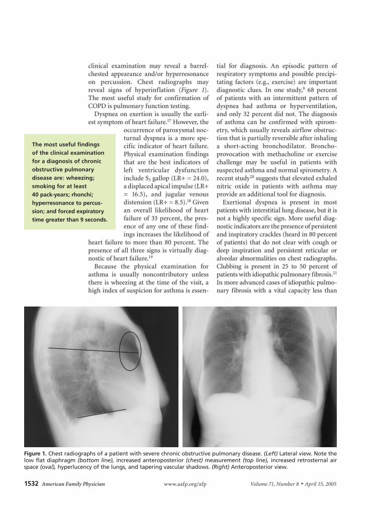
clinical examination may reveal a barrel-chested appearance and/or hyperresonance on percussion. Chest radiographs may reveal signs of hyperinflation (Figure 1). The most useful study for confirmation of COPD is pulmonary function testing.
Dyspnea on exertion is usually the earli-est symptom of heart failure.17 However, the
occurrence of paroxysmal noc-turnal dyspnea is a more spe-cific indicator of heart failure. Physical examination findings that are the best indicators of left ventricular dysfunction include S3 gallop (LR+ = 24.0), a displaced apical impulse (LR+ = 16.5), and jugular venous distension (LR+ = 8.5).18 Given an overall likelihood of heart failure of 33 percent, the pres-ence of any one of these find-ings increases the likelihood of
heart failure to more than 80 percent. The presence of all three signs is virtually diag-nostic of heart failure.19
Because the physical examination for asthma is usually noncontributory unless there is wheezing at the time of the visit, a high index of suspicion for asthma is essen-
tial for diagnosis. An episodic pattern of respiratory symptoms and possible precipi-tating factors (e.g., exercise) are important diagnostic clues. In one study,9 68 percent of patients with an intermittent pattern of dyspnea had asthma or hyperventilation, and only 32 percent did not. The diagnosis of asthma can be confirmed with spirom-etry, which usually reveals airflow obstruc-tion that is partially reversible after inhaling a short-acting bronchodilator. Broncho-provocation with methacholine or exercise challenge may be useful in patients with suspected asthma and normal spirometry. A recent study20 suggests that elevated exhaled nitric oxide in patients with asthma may provide an additional tool for diagnosis.
Exertional dyspnea is present in most patients with interstitial lung disease, but it is not a highly specific sign. More useful diag-nostic indicators are the presence of persistent and inspiratory crackles (heard in 80 percent of patients) that do not clear with cough or deep inspiration and persistent reticular or alveolar abnormalities on chest radiographs. Clubbing is present in 25 to 50 percent of patients with idiopathic pulmonary fibrosis.21 In more advanced cases of idiopathic pulmo-nary fibrosis with a vital capacity less than
The most useful findings of the clinical examination for a diagnosis of chronic obstructive pulmonary disease are: wheezing; smoking for at least 40 pack-years; rhonchi; hyperresonance to percus-sion; and forced expiratory time greater than 9 seconds.
Figure 1. Chest radiographs of a patient with severe chronic obstructive pulmonary disease. (Left) Lateral view. Note the low flat diaphragm (bottom line), increased anteroposterior (chest) measurement (top line), increased retrosternal air space (oval), hyperlucency of the lungs, and tapering vascular shadows. (Right) Anteroposterior view.
April 15, 2005 ◆ Volume 71, Number 8 www.aafp.org/afp American Family Physician 1533
Chronic Dyspnea
50 percent, signs of secondary pulmonary hypertension may be present. These include an accentuated P2, right ventricular heave, and tricuspid regurgitation murmur.22
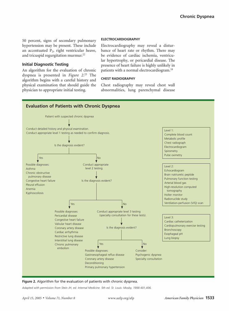
Initial Diagnostic TestingAn algorithm for the evaluation of chronic dyspnea is presented in Figure 2.23 The algorithm begins with a careful history and physical examination that should guide the physician to appropriate initial testing.
ELECTROCARDIOGRAPHY
Electrocardiography may reveal a distur-bance of heart rate or rhythm. There may be evidence of cardiac ischemia, ventricu-lar hypertrophy, or pericardial disease. The presence of heart failure is highly unlikely in patients with a normal electrocardiogram.18
CHEST RADIOGRAPHY
Chest radiography may reveal chest wall abnormalities, lung parenchymal disease
Evaluation of Patients with Chronic Dyspnea
Figure 2. Algorithm for the evaluation of patients with chronic dyspnea.
Adapted with permission from Stein JH, ed. Internal Medicine. 5th ed. St. Louis: Mosby, 1998:401,406.
Patient with suspected chronic dyspnea
Conduct detailed history and physical examination.Conduct appropriate level 1 testing as needed to confirm diagnosis.
Is the diagnosis evident?
Level 1:Complete blood countMetabolic profileChest radiographElectrocardiogramSpirometryPulse oximetry
Possible diagnoses:AsthmaChronic obstructive
pulmonary diseaseCongestive heart failurePleural effusionAnemiaKyphoscoliosis
Conduct appropriate level 2 testing.
Possible diagnoses:Gastroesophageal reflux diseaseCoronary artery diseaseDeconditioningPrimary pulmonary hypertension
Level 3:Cardiac catheterizationCardiopulmonary exercise testingBronchoscopyEsophageal pHLung biopsy
Level 2:EchocardiogramBrain natriuretic peptidePulmonary function testingArterial blood gasHigh-resolution computed
tomographyHolter monitorRadionuclide studyVentilation-perfusion (V/Q) scan
Possible diagnoses:Pericardial diseaseCongestive heart failureValvular heart diseaseCoronary artery diseaseCardiac arrhythmiaRestrictive lung diseaseInterstitial lung diseaseChronic pulmonary
embolismConsider:Psychogenic dyspneaSpecialty consultation
Is the diagnosis evident?
Conduct appropriate level 3 testing (specialty consultation for these tests).
Yes No
Yes No
Is the diagnosis evident?
Yes No
1534 American Family Physician www.aafp.org/afp Volume 71, Number 8 ◆ April 15, 2005
(e.g., COPD), neoplastic lesions, congestive heart failure, or pleural disease.
COMPLETE BLOOD COUNT AND METABOLIC PANEL
Profound anemia is an unusual but impor-tant cause of chronic dyspnea. Secondary erythrocytosis may be present in patients with advanced COPD. Changes in acid-base balance, reflected by the bicarbonate level in a metabolic panel, may provide a clue to dyspnea. Respiratory acidosis, occurring in patients with severe cases of COPD, inter-stitial lung disease, and neuromuscular dis-orders, leads to a metabolic compensation resulting in an elevated bicarbonate level.
SPIROMETRY
This test is useful in distinguishing obstructive lung disorders from restrictive lung disorders. It is highly effort dependent, which can be challenging in older or impaired patients, especially when measuring forced vital capac-ity (FVC). However, forced expiratory vol-ume in six seconds (FEV6) has been shown to be an acceptable surrogate for FVC in the spi-rometric diagnosis of obstructive disease and possibly in restrictive lung disease.24 Most of the newer spirometers are equipped to mea-sure FEV6. Although the FEV in one second (FEV1) and FVC are reduced proportionately in patients with restrictive lung disease, the FEV1 is reduced more than the FVC in those with obstructive lung disease. Therefore, a ratio of FEV1/FVC less than 0.7 or 0.8 is a common diagnostic criterion for COPD.
PULSE OXIMETRY
Desaturation at rest or after exercise is a sensi-tive indicator of gas exchange abnormalities. If abnormal, consideration should be given to obtaining arterial blood gas measurements.
Selective TestingPULMONARY FUNCTION TESTING
The primary limitation of spirometry is its inability to measure lung volumes, including the total amount of air in the lungs at full inspiration (total lung capacity [TLC]), the amount of air remaining in the lungs at the end of passive expiration (functional resid-
ual capacity [FRC]), or the amount of air remaining after maximal expiration (residual volume [RV]). The TLC is reduced in restric-tive disorders and normal or increased in obstructive disorders as a result of air trap-ping. In restrictive disorders caused by lung parenchymal disease, all lung volumes are proportionately reduced. In contrast, with other restrictive diseases (e.g., neuromuscu-lar disease or chest wall restriction) the RV and the RV/TLC ratio are increased.
With the use of carbon monoxide, which is a highly diffusible gas, the gas-trans-fer function of the lung can be estimated by measuring the diffusing capacity of the lung for carbon monoxide (DLCO). This is reduced in patients with diseases affecting the lung parenchyma, vascular abnormali-ties, anemia, and conditions where there is a reduction of effective lung volume (e.g., after lung resection). DLCO may be elevated in conditions where there is an increased effective pulmonary blood volume, such as asthma, obesity, left-to-right cardiac shunts, and polycythemia. Performing a broncho-provocation challenge using methacho-line can identify airway hyperreactivity. A 20 percent reduction in FEV1 is considered diagnostic of asthma.
TESTS FOR PULMONARY VASCULAR DISEASE
Pulmonary hypertension may be primary (rare) or secondary to a pulmonary, cardiac, or extrathoracic pathology. Two-dimensional echocardiography with Doppler flow studies is the most useful imaging modality to dem-onstrate elevated pulmonary artery pressures and the resultant tricuspid regurgitation.25
If the etiology of pulmonary hypertension remains unexplained after appropriate test-ing, chronic thromboembolism should be suspected. Ventilation-perfusion scanning or spiral computed tomography of the chest may be used to confirm this diagnosis. Right heart catheterization may be required to confirm or diagnose less common causes of pulmonary hypertension.
TESTS FOR CARDIAC DISEASE
The test of choice for diagnosing most cardiac causes of chronic dyspnea is echocardiogra-
April 15, 2005 ◆ Volume 71, Number 8 www.aafp.org/afp American Family Physician 1535
Chronic Dyspnea
phy, especially if heart failure is suspected. However, as many as 40 percent of patients with clinical evidence of congestive heart failure have diastolic dysfunction with pre-served left ventricular systolic function.26 In such cases, the diagnosis is suggested by the findings of left ventricular hypertrophy, dilated left atrium, and reversal of the nor-mal pattern of flow velocity across the mitral valve. Other cardiac pathologies that can be demonstrated on echocardiography include valvular dysfunction, atrial tumors, and peri-cardial disease.
Brain natriuretic peptide (BNP), also known as B-type natriuretic peptide, is a neurohormone synthesized by ventricular myocytes that is useful in the diagnosis of heart failure. It is released in response to pres-sure/volume overload resulting in increased wall tension. The magnitude of elevation is proportional to the severity of heart failure and the New York Heart Association func-tional classification.27 Using a threshold of 100 pg per mL, the test is 82 percent sensitive and 99 percent specific.27 Table 328 compares the specificity, sensitivity, and post-test prob-abilities of different cutoffs for an abnormal BNP test with echocardiographic diagnoses of left ventricular dysfunction (systolic or diastolic). This test is available as a point-of-care assay. Although an absolute standard for the diagnosis of congestive heart failure does not exist, the BNP test may be helpful, espe-
cially in patients who have coexisting cardiac and pulmonary disease and if there is uncer-tainty about the primary cause of dyspnea.
Additional Testing for Difficult CasesCardiopulmonary exercise testing is a sophis-ticated procedure that helps quantify cardiac function, pulmonary gas exchange, ventila-tion, and physical fitness.29 It is especially useful in cases where no apparent cause for dyspnea is found after a thorough evaluation or in patients who have multiple poten-tial causes for dyspnea.29 Parameters that are measured by computerized systems are blood pressure, electrocardiography, heart rate, ventilation, oxygen saturation, oxygen uptake, and carbon dioxide output.
Patients who have obstructive lung disease generally will display a decrease in maxi-mal oxygen uptake. Patients with intersti-tial restrictive disease have abnormalities of gas exchange and pulmonary mechanics. Patients who have cardiac disease exhibit a lower than predicted maximal heart rate. Low cardiac output is reflected by decreased maximal oxygen uptake and anaerobic metabolism at low workloads. Additionally, there may be abnormalities of the electro-cardiogram and the blood pressure response may be blunted. Deconditioning results in a decreased maximal oxygen uptake but normal gas exchange and breathing reserve. In these patients, the heart rate, cardiac out-
TABLE 3
Operating Characteristics for Various Cutoff Points of BNP Levels
Cutoff to define abnormal BNP (pg per mL)
Sensitivity (%)
Specificity (%)
Probability of heart failure with BNP
Positive (%)* Negative (%)†
> 400 63 91 88 29
> 300 73 89 87 23
> 200 81 85 84 18
> 100 90 73 77 12
BNP = brain natriuretic peptide.
*—Percentage of patients with BNP above cutoff who have heart failure.†—Percentage of patients with BNP below cutoff who do not have heart failure.
Information from reference 28.
1536 American Family Physician www.aafp.org/afp Volume 71, Number 8 ◆ April 15, 2005
put, and blood pressure rise appropriately in response to exercise.
Cardiac arrhythmias (most commonly atrial fibrillation) may be the sole cause of dyspnea or may exacerbate other cardiac causes, such as cardiomyopathy. Intermittent arrhythmias can be diagnosed using a Holter monitor or an
event recorder. In some patients with coronary artery disease, dyspnea may represent an angi-nal equivalent. Noninvasive car-diovascular testing (e.g., stress thallium, stress echocardiogra-phy, cardiac magnetic resonance
imaging) cardiac catheterization should be considered for these patients.
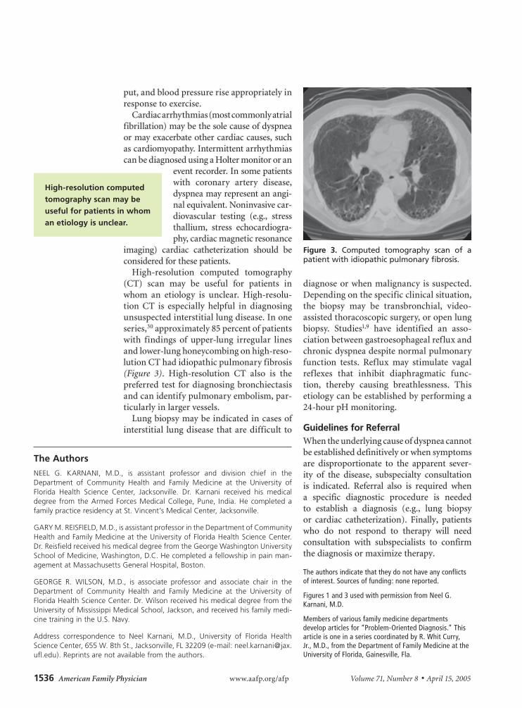
High-resolution computed tomography (CT) scan may be useful for patients in whom an etiology is unclear. High-resolu-tion CT is especially helpful in diagnosing unsuspected interstitial lung disease. In one series,30 approximately 85 percent of patients with findings of upper-lung irregular lines and lower-lung honeycombing on high-reso-lution CT had idiopathic pulmonary fibrosis (Figure 3). High-resolution CT also is the preferred test for diagnosing bronchiectasis and can identify pulmonary embolism, par-ticularly in larger vessels.
Lung biopsy may be indicated in cases of interstitial lung disease that are difficult to
diagnose or when malignancy is suspected. Depending on the specific clinical situation, the biopsy may be transbronchial, video-assisted thoracoscopic surgery, or open lung biopsy. Studies1,9 have identified an asso-ciation between gastroesophageal reflux and chronic dyspnea despite normal pulmonary function tests. Reflux may stimulate vagal reflexes that inhibit diaphragmatic func-tion, thereby causing breathlessness. This etiology can be established by performing a 24-hour pH monitoring.
Guidelines for ReferralWhen the underlying cause of dyspnea cannot be established definitively or when symptoms are disproportionate to the apparent sever-ity of the disease, subspecialty consultation is indicated. Referral also is required when a specific diagnostic procedure is needed to establish a diagnosis (e.g., lung biopsy or cardiac catheterization). Finally, patients who do not respond to therapy will need consultation with subspecialists to confirm the diagnosis or maximize therapy.
The authors indicate that they do not have any conflicts of interest. Sources of funding: none reported.
Figures 1 and 3 used with permission from Neel G. Karnani, M.D.
Members of various family medicine departments develop articles for “Problem-Oriented Diagnosis.” This article is one in a series coordinated by R. Whit Curry, Jr., M.D., from the Department of Family Medicine at the University of Florida, Gainesville, Fla.
High-resolution computed tomography scan may be useful for patients in whom an etiology is unclear.
Figure 3. Computed tomography scan of a patient with idiopathic pulmonary fibrosis.
The Authors
NEEL G. KARNANI, M.D., is assistant professor and division chief in the Department of Community Health and Family Medicine at the University of Florida Health Science Center, Jacksonville. Dr. Karnani received his medical degree from the Armed Forces Medical College, Pune, India. He completed a family practice residency at St. Vincent’s Medical Center, Jacksonville.
GARY M. REISFIELD, M.D., is assistant professor in the Department of Community Health and Family Medicine at the University of Florida Health Science Center. Dr. Reisfield received his medical degree from the George Washington University School of Medicine, Washington, D.C. He completed a fellowship in pain man-agement at Massachusetts General Hospital, Boston.
GEORGE R. WILSON, M.D., is associate professor and associate chair in the Department of Community Health and Family Medicine at the University of Florida Health Science Center. Dr. Wilson received his medical degree from the University of Mississippi Medical School, Jackson, and received his family medi-cine training in the U.S. Navy.
Address correspondence to Neel Karnani, M.D., University of Florida Health Science Center, 655 W. 8th St., Jacksonville, FL 32209 (e-mail: [email protected]). Reprints are not available from the authors.
April 15, 2005 ◆ Volume 71, Number 8 www.aafp.org/afp American Family Physician 1537
Chronic Dyspnea
REFERENCES
1. Pratter MR, Curley FJ, Dubois J, Irwin RS. Cause and evaluation of chronic dyspnea in a pulmonary disease clinic. Arch Intern Med 1989;149:2277-82.
2. American Thoracic Society. Dyspnea. Mechanisms, assessment, and management: a consensus statement. Am J Respir Crit Care Med 1999;159:321-40.
3. Burns BH, Howell JB. Disproportionately severe breath-lessness in chronic bronchitis. Q J Med 1969;38:277-94.
4. Manning HL, Schwartzstein RM. Pathophysiology of dyspnea. N Engl J Med 1995;333:1547-53.
5. Elliott MW, Adams L, Cockcroft A, MacRae KD, Murphy K, Guz A. The language of breathlessness. Use of verbal descriptors by patients with cardiopulmonary disease. Am Rev Respir Dis 1991;144:826-32.
6. Mahler DA, Harver A, Lentine T, Scott JA, Beck K, Schwartzstein RM. Descriptors of breathlessness in cardiorespiratory diseases. Am J Respir Crit Care Med 1996;154:1357-63.
7. Simon PM, Schwartzstein RM, Weiss JW, Fencl V, Teghtsoonian M, Weinberger SE. Distinguishable types of dyspnea in patients with shortness of breath. Am Rev Respir Dis 1990;142:1009-14.
8. Morgan WC, Hodge HL. Diagnostic evaluation of dys-pnea. Am Fam Physician 1998;57:711-6.
9. DePaso WJ, Winterbauer RH, Lusk JA, Dreis DF, Spring-meyer SC. Chronic dyspnea unexplained by history, physical examination, chest roentgenogram, and spi-rometry. Analysis of a seven-year experience. Chest 1991;100:1293-9.
10. Gillespie DJ, Staats BA. Unexplained dyspnea. Mayo Clin Proc 1994;69:657-63.
11. Michelson E, Hollrah S. Evaluation of the patient with shortness of breath: an evidence based approach. Emerg Med Clin North Am 1999;17:221-37,x.
12. Faul JL, Lillington GA. Dyspnea. Best practice of medicine. January 2002. Accessed online February 22, 2005, at: http://merck.micromedex.com/index.asp?page=bpm_report&article_id=BPM01PU04§ion=report.
13. Pauwels RA, Buist AS, Ma P, Jenkins CR, Hurd SS; GOLD Scientific Committee. Global strategy for the diagnosis, management, and prevention of chronic obstructive pulmonary disease. Respir Care 2001;46:798-825.
14. Sharma S. Chronic obstructive pulmonary disease. Accessed online February 22, 2005, at: http://www.emedicine.com/med/topic373.htm.
15. Holleman DR Jr, Simel DL, Goldberg JS. Diagnosis of obstructive airways disease from the clinical examina-tion. J Gen Intern Med 1993;8:63-8.
16. Badgett RG, Tanaka DJ, Hunt DK, Jelley MJ, Feinberg LE, Steiner JF, et al. Can moderate chronic obstruc-tive pulmonary disease be diagnosed by historical and physical findings alone? Am J Med 1993;94:188-96.
17. Buchter CM. Chronic heart failure. Best practice of medicine. Accessed online February 22, 2005, at: http://merck.micromedex.com/index.asp?page=bpm_report&article_id=BPM01CA06§ion=report.
18. Davie AP, Francis CM, Caruana L, Sutherland GR, McMurray JJ. Assessing diagnosis in heart failure: which features are any use? QJM 1997;90:335-9.
19. Konstam MA. Heart failure: evaluation and care of patients with left ventricular systolic dysfunction. Rock-ville, Md.: U.S. Department of Health and Human Services, 1994. AHCPR publication no. 94-0612.
20. Dupont LJ, Demedts MG, Verleden GM. Prospective evaluation of the validity of exhaled nitric oxide for the diagnosis of asthma. Chest 2003;123:751-6.
21. American Thoracic Society. Idiopathic pulmonary fibro-sis: diagnosis and treatment. International consensus statement. American Thoracic Society (ATS), and the European Respiratory Society (ERS). Am J Respir Crit Care Med 2000;161(2 pt 1):646-64.
22. Fessler MB, Brown KK. Interstitial lung disease. Best Practice of Medicine April 2002. Accessed online March 9, 2005, at: http://merck.micromedex.com/index.asp?page=bpm_report&article_id=BPM01PU05§ion=report.
23. Stein JH, ed. Internal Medicine. 5th ed. St. Louis: Mosby, 1998:401,406.
24. Swanney MP, Jensen RL, Crichton DA, Beckert LE, Cardno LA, Crapo RO. FEV(6) is an acceptable sur-rogate for FVC in the spirometric diagnosis of airway obstruction and restriction. Am J Respir Crit Care Med 2000;162(3 pt 1):917-9.
25. Nauser TD, Stites SW. Diagnosis and treatment of pulmo-nary hypertension. Am Fam Physician 2001;63:1789-98.
26. Farmer J, Torre G. Congestive heart failure. Patient guide. Accessed online February 22, 2005, at: http://merck.micromedex.com / index.asp?page = bhg_report&article_id=BHG01CA06§ion=report.
27. Wieczorek SJ, Wu AH, Christenson R, Krishnaswamy P, Gottlieb S, Rosano T, et al. A rapid B-type natriuretic peptide assay accurately diagnoses left ventricular dys-function and heart failure: a multicenter evaluation. Am Heart J 2002;144:834-9.
28. Maisel AS, McCord J, Nowak RM, Hollander JE, Wu AH, Duc P, et al; Breathing Not Properly Multinational Study Investigators. Bedside B-type natriuretic peptide in the emergency diagnosis of heart failure with reduced or preserved ejection fraction. Results from the Breathing Not Properly Multinational Study. J Am Coll Cardiol 2003;41:2010-7.
29. ATS/ACCP Statement on cardiopulmonary exercise testing. American Thoracic Society; American Col-lege of Chest Physicians. Am J Respir Crit Care Med 2003;167:211-77.
30. Hunninghake GW, Lynch DA, Galvin JR, Gross BH, Muller N, Schwartz DA, et al. Radiologic findings are strongly associated with a pathologic diagnosis of usual interstitial pneumonia. Chest 2003;124:1215-23.