EVALUATION OF AUTOMATIC CLASS III DESIGNATION FOR ...The FilmArray Meningitis/Encephalitis (ME)...
45
1 EVALUATION OF AUTOMATIC CLASS III DESIGNATION FOR FilmArray ® Meningitis/Encephalitis (ME) Panel DECISION SUMMARY A. DEN Number: DEN150013 B. Purpose for Submission: De Novo request for evaluation of automatic class III designation for the FilmArray Meningitis/Encephalitis (ME) Panel C. Measurands: The assay detects and identifies nucleic acids of the following organisms: Escherichia coli K1, Haemophilus influenzae, Listeria monocytogenes, Neisseria meningitidis (encapsulated), Streptococcus agalactiae, Streptococcus pneumoniae, Cytomegalovirus, Enterovirus, Herpes simplex virus 1, Herpes simplex virus 2, Human herpesvirus 6, Human parechovirus, Varicella zoster virus, Cryptococcus neoformans/gattii D. Type of Test: The FilmArray Meningitis/Encephalitis (ME) Panel (“FilmArray ME Panel”), performed with FilmArray and FilmArray 2.0 systems, is a nucleic acid-based test for the detection of the above listed bacteria, viruses, and yeast from cerebrospinal fluid (CSF) specimens obtained from patients with signs and symptoms of meningitis or encephalitis. E. Applicant: BioFire Diagnostics, LLC F. Proprietary and Established Names: FilmArray Meningitis/Encephalitis (ME) Panel G. Regulatory Information: 1. Regulation section: 21 CFR 866.3970, Device to detect and identify microbial pathogen nucleic acids in cerebrospinal fluid 2. Classification: Class II (Special Controls)
Transcript of EVALUATION OF AUTOMATIC CLASS III DESIGNATION FOR ...The FilmArray Meningitis/Encephalitis (ME)...

1
EVALUATION OF AUTOMATIC CLASS III DESIGNATION FOR
FilmArray® Meningitis/Encephalitis (ME) Panel
DECISION SUMMARY
A. DEN Number:
DEN150013
B. Purpose for Submission:
De Novo request for evaluation of automatic class III designation for the FilmArray
Meningitis/Encephalitis (ME) Panel
C. Measurands:
The assay detects and identifies nucleic acids of the following organisms: Escherichia coli
K1, Haemophilus influenzae, Listeria monocytogenes, Neisseria meningitidis (encapsulated),
Streptococcus agalactiae, Streptococcus pneumoniae, Cytomegalovirus, Enterovirus, Herpes
simplex virus 1, Herpes simplex virus 2, Human herpesvirus 6, Human parechovirus,
Varicella zoster virus, Cryptococcus neoformans/gattii
D. Type of Test:
The FilmArray Meningitis/Encephalitis (ME) Panel (“FilmArray ME Panel”), performed
with FilmArray and FilmArray 2.0 systems, is a nucleic acid-based test for the detection of
the above listed bacteria, viruses, and yeast from cerebrospinal fluid (CSF) specimens
obtained from patients with signs and symptoms of meningitis or encephalitis.
E. Applicant:
BioFire Diagnostics, LLC
F. Proprietary and Established Names:
FilmArray Meningitis/Encephalitis (ME) Panel
G. Regulatory Information:
1. Regulation section:
21 CFR 866.3970, Device to detect and identify microbial pathogen nucleic acids in
cerebrospinal fluid
2. Classification:
Class II (Special Controls)

2
3. Product code(s):
PLO, OOI, NSU
4. Panel:
83- Microbiology
H. Intended Use:
1. Intended use(s):
The FilmArray Meningitis/Encephalitis (ME) Panel is a qualitative multiplexed nucleic
acid-based in vitro diagnostic test intended for use with FilmArray and FilmArray 2.0
systems. The FilmArray ME Panel is capable of simultaneous detection and identification
of multiple bacterial, viral, and yeast nucleic acids directly from cerebrospinal fluid
(CSF) specimens obtained via lumbar puncture from individuals with signs and/or
symptoms of meningitis and/or encephalitis. The following organisms are identified
using the FilmArray ME Panel:
Bacteria:
Escherichia coli K1
Haemophilus influenzae
Listeria monocytogenes
Neisseria meningitidis (encapsulated)
Streptococcus agalactiae
Streptococcus pneumoniae
Viruses:
Cytomegalovirus
Enterovirus
Herpes simplex virus 1
Herpes simplex virus 2
Human herpesvirus 6
Human parechovirus
Varicella zoster virus
Yeast:
Cryptococcus neoformans/gattii
The FilmArray ME Panel is indicated as an aid in the diagnosis of specific agents of
meningitis and/or encephalitis and results are meant to be used in conjunction with other
clinical, epidemiological, and laboratory data.
Results from the FilmArray ME Panel are not intended to be used as the sole basis for
diagnosis, treatment, or other patient management decisions. Positive results do not rule
out co-infection with organisms not included in the FilmArray ME Panel. The agent

3
detected may not be the definite cause of the disease. Negative results do not preclude
central nervous system (CNS) infection. Not all agents of CNS infection are detected by
this test and sensitivity in clinical use may differ from that described in the package
insert.
The FilmArray ME Panel is not intended for testing of specimens collected from
indwelling CNS medical devices.
The FilmArray ME Panel is intended to be used in conjunction with standard of care
culture for organism recovery, serotyping, and antimicrobial susceptibility testing.
2. Indication(s) for use:
Same as Intended Use
3. Special conditions for use statement(s):
For prescription use only.
4. Special instrument requirements:
The FilmArray ME Panel is performed on the FilmArray and FilmArray 2.0 systems.
I. Device Description:
The FilmArray ME Panel is a multiplex nucleic acid-based test designed to be used with
FilmArray or FilmArray 2.0 system (“FilmArray systems” or “FilmArray instruments”). The
FilmArray ME panel includes a FilmArray ME Panel pouch (pouch) which contains freeze-dried
reagents to perform nucleic acid purification and nested, multiplex polymerase chain reaction
(PCR) with DNA melt analysis. The FilmArray ME Panel simultaneously conducts 14 tests for
the identification of potential CNS pathogens from CSF specimens obtained via lumbar
puncture. Results from the FilmArray ME Panel are available within about one hour.
A test is initiated by loading Hydration Solution into one port of the pouch and a CSF sample
mixed with the provided Sample Buffer ampoules into the other port of the pouch and placing it
in the FilmArray Instrument. The pouch contains all of the reagents required for specimen testing
and analysis in a freeze-dried format; the addition of Hydration Solution and the Sample Buffer
rehydrates the reagents. After the pouch is prepared, the FilmArray Software on the FilmArray
systems guides the user though the steps of placing the pouch into the instrument, scanning the
pouch barcode, entering the sample identification, and initiating the run on the FilmArray
systems.
The FilmArray instruments contain a coordinated system of inflatable bladders and seal points,
which act on the pouch to control the movement of liquid between the pouch blisters. When a
bladder is inflated over a reagent blister, it forces liquid from the blister into connecting
channels. Alternatively, when a seal is placed over a connecting channel it acts as a valve to open
or close a channel.
b(4)
b(4)
4
Nucleic acid extraction occurs within the pouch using mechanical and chemical lysis followed
by purification using standard magnetic bead technology. After extracting and purifying nucleic
acids from the unprocessed sample, a nested multiplex PCR is executed in two stages.
. The solution is then distributed to each well of the array. Array
wells contain sets of primers designed specifically to amplify sequences internal to the PCR
products generated during the first stage PCR reaction. The 2nd
stage PCR, or nested PCR, is
performed in each well of the array. At the conclusion of the 2nd
stage PCR, the array is
interrogated by melt curve analysis for the detection of signature amplicons denoting the
presence of specific targets. A digital camera placed in front of the array captures fluorescent
images of the PCR2 reactions and software interprets the data.
The FilmArray software automatically interprets the results of each DNA melt curve analysis
and combines the data with the results of the internal pouch controls to provide a test result for
each organism on the panel.
Materials provided in each FilmArray ME Panel kit:
Individually packaged FilmArray ME Panel pouches
Single-use (1.0 mL) Sample Buffer ampoules
Single-use pre-filled (1.5 mL) Hydration Injection Vials
Single-use Sample Injection Vials
Individually packaged Transfer Pipettes
Materials required but not provided: FilmArray system including:
FilmArray or FilmArray 2.0 instrument and software
FilmArray Pouch Loading Station
J. Standard/Guidance Document Referenced (if applicable):
CLSI EP12-A2, User Protocol for Evaluation of Qualitative Test Performance, 2008
CLSI EP07-A2, Interference Testing in Clinical Chemistry, 2005
CLSI MM03-A2, Molecular Diagnostic Methods for Infectious Diseases, 2006
EN ISO 14971:2012, ‘Medical devices – Application of risk management to medical
devices’
Guidance for Sponsors, Institutional Review Boards, Clinical Investigators and FDA
Staff – Guidance on Informed Consent for In Vitro Diagnostic Device Studies Using
Leftover Human Specimens that are Not Individually Identifiable, April 25, 2005
Guidance for Industry and Food and Drug Administration Staff – Assay Migration
Studies for In Vitro Diagnostic Devices, April 25, 2013
Guidance for Industry and Food and Drug Administration Staff – Highly Multiplexed
b(4)
b(4)
5
Microbiological/Medical Countermeasure In Vitro Nucleic Acid Based Diagnostic
Devices, August 27, 2014
Statistical Guidance on Reporting Results from Studies Evaluating Diagnostic Tests,
FDA Guidance Document, March 13, 2007
Class II Special Controls Guidance Document: Nucleic Acid Amplification Assay for the
Detection of Enterovirus RNA, January 2, 2009
K. Test Principle:
The FilmArray ME Panel pouch (pouch) is a closed system disposable that houses all the
chemistry required to isolate, amplify and detect nucleic acid from multiple meningitis and
encephalitis pathogens within a single CSF specimen obtained from a lumbar puncture. The rigid
plastic component (fitment) of the pouch contains reagents in freeze-dried form. The flexible
plastic portion of the pouch is divided into discrete segments (blisters) where the required
chemical processes are carried out. The user of the FilmArray ME Panel loads the sample into
the pouch, places the pouch into the FilmArray systems, and starts the run. All other operations
are automated. Operations and processes that occur during a FilmArray run include the
following:
Nucleic Acid Purification -
The sample is lysed by agitation (bead beating) and the liberated nucleic acid
is captured, washed and eluted using magnetic bead technology. These steps require
approximately ten minutes and the bead-beater apparatus can be heard as a high-pitched
whine during the first minute of operation.
Reverse Transcription and 1st Stage Multiplex PCR - Some pathogens identified by
the pouch are RNA viruses, and a reverse transcription (RT) step is performed to convert
the viral RNA into cDNA prior to amplification.
for multiplex PCR. The effect of 1st stage PCR is to enrich for the target
nucleic acids present in the sample.
2nd
Stage PCR - The products of 1st stage PCR are diluted and mixed with fresh PCR
reagents containing an intercalating fluorescent DNA dye Plus, BioFire
Defense, LLC). This solution is distributed over the 2nd
stage PCR array. The individual
wells of the array contain primers for different assays (each present in triplicate) that
target specific nucleic acid sequences from each of the pathogens detected, as well as
control template material. These primers are ‘nested’ or internal to the specific products
of the 1st stage multiplex reaction, which enhances both the sensitivity and specificity of
the reactions.
DNA Melting Analysis – After 2nd
stage PCR, the temperature is slowly increased and
fluorescence in each well of the array is monitored and analyzed to generate a melt curve.
The temperature at which a specific PCR product melts (melting temperature or Tm) is
consistent and predictable and the FilmArray software automatically evaluates the data
from replicate wells for each assay to report results.
b(4)
b(4)
b(4)

6
The FilmArray software controls the operation of the instrument, collects and analyzes data
and automatically generates a test report at the end of the run.
L. Performance Characteristics:
1. Analytical performance:
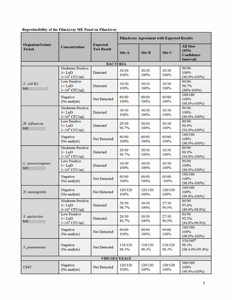
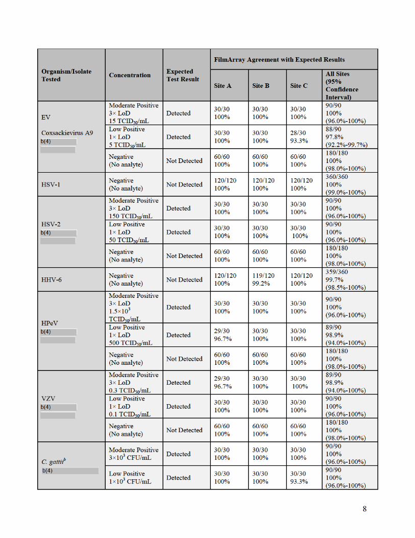
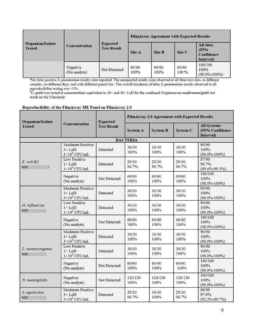
a. Reproducibility
Reproducibility studies were performed with the FilmArray ME Panel on both the
FilmArray and FilmArray 2.0 systems. Testing for the FilmArray system was performed
using multiple instruments at three different testing sites, Biofire Diagnostics and two
external laboratories. Testing for the FilmArray 2.0 system was performed internally at
BioFire Diagnostics using multiple instruments at three different locations within BioFire
Diagnostics.
Assay reproducibility was evaluated for both the FilmArray and FilmArray 2.0 system
using a panel of contrived CSF samples prepared in artificial CSF matrix (aCSF) and
spiked with combinations of nine different ME Panel analytes, including at least one
representative gram-negative bacterium, gram-positive bacterium, yeast, DNA virus and
RNA virus. Each spiked analyte was evaluated at three different concentrations: Negative
(no analyte), Low Positive (1× the limit of detection (LoD)) and Moderate Positive (3×
LoD). Testing on both FilmArray systems incorporated a range of potential testing
variables including different operators, three different pouch lots, and different FilmArray
Instruments. Samples were tested on five different days with a total of 90 replicates tested
per panel member.
For the FilmArray system, 366 runs were initiated and 360 runs were completed (98.4%
initially valid results). Of the six initially invalid runs, one invalid run was due to a
control failure and five invalid tests were due to instrument or software errors. Retesting
of initially invalid specimens gave valid results for all six specimens. There were seven
unexpected false positive results observed in the study: six for Streptococcus
pneumoniae (6/360 = 1.7%) and one for Human herpesvirus 6 (HHV-6) (1/360 = 0.3%).
For the FilmArray 2.0 system, 365 runs were initiated and 360 runs were completed
(98.6% initially valid results). Of the five initially invalid runs, three runs were invalid
because they were aborted by the operator due to being run on an incorrect instrument.
The two other invalid results were due to a software error (1/365 = 0.3%) and due to an
incomplete result caused by a data transfer error (1/365 = 0.3%).
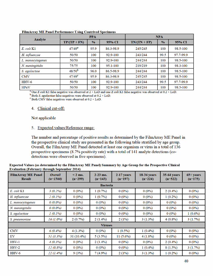
A summary of qualitative results from both reproducibility studies (percent agreement
with the expected result) for each analyte and organism concentration is provided in the
following tables.


13
PCR2 Control: The PCR2 Control assay detects a DNA target
. A positive result
indicates that the 2nd stage PCR was successful.
Both internal control assays must be positive for the test run to pass. When either control
fails, the Controls field of the test report will display "Failed" and all results will be listed
as Invalid. If the controls fail, the user is instructed to repeat the test using a new pouch.
Of the six pouch control failures observed in the prospective clinical study, five were
attributed to both RNA Process Control and PCR 2 Control failures and one was
attributed to RNA Process Control failure only.
Recommended External Controls:
External controls are not provided with the FilmArray ME Panel, but are recommended
in the package insert. Molecular grade water or artificial CSF can be used as an external
negative control. Previously characterized CSF specimens or negative matrix spiked with
well-characterized organisms can be used as external positive controls. External controls
should be used in accordance with the appropriate accrediting organization requirements,
as applicable.
d. Detection Limit:
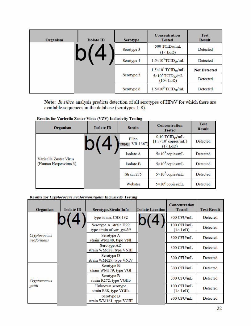
Evaluation of the Limit of Detection:
The limit of detection (LoD) for FilmArray ME Panel analytes was estimated by testing
dilutions of contrived samples containing known concentrations of bacteria, viruses or
yeast detected by the FilmArray ME Panel. Representative strains were chosen in order to
obtain positive results for every assay on the panel and multiple strains were evaluated to
cover clinically important species or variants for some analytes.
Samples for LoD testing were prepared in aCSF (artificial CSF obtained from a
commercial provider) matrix and contained one or up to five targeted organisms.
.
The LoD was confirmed for all
analytes on both FilmArray and FilmArray 2.0 systems. Data presented in the following
table are from LoD confirmation testing on the FilmArray system.
b(4)
b(4)b(4)


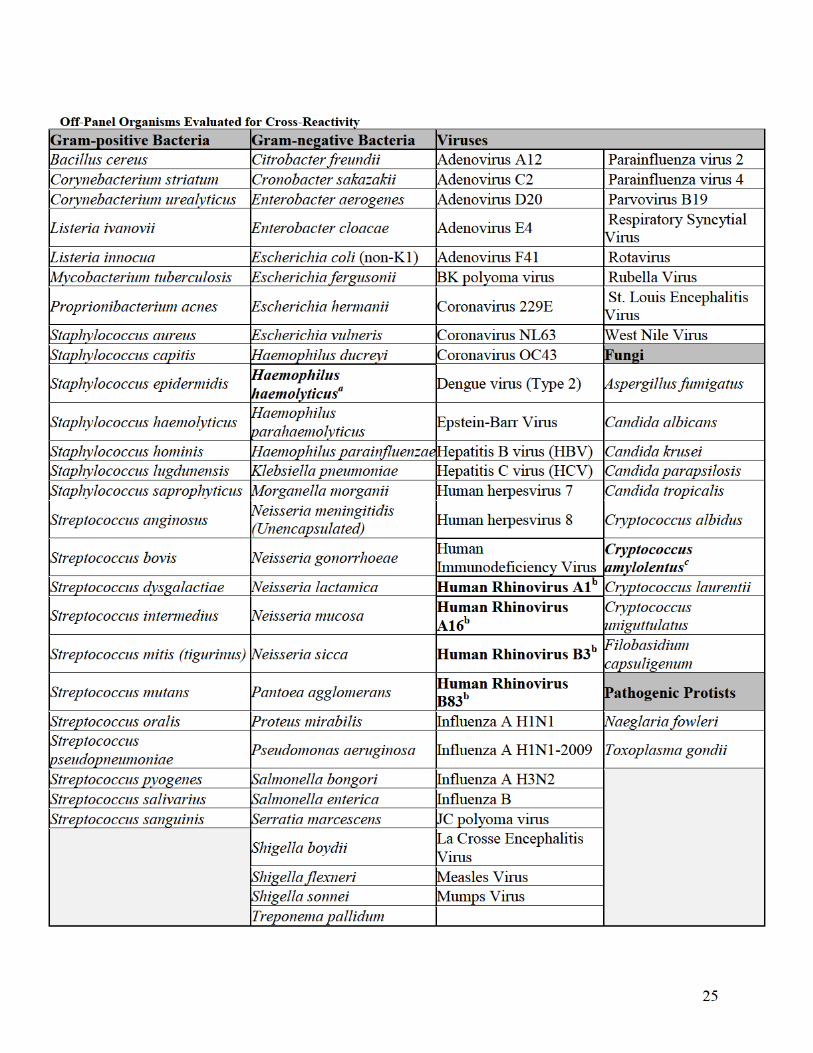
26
a Detected by the FilmArray ME Panel as Haemophilus influenzae. H. haemolyticus is a commensal bacterium of the upper
respiratory tract, rarely isolated from CSF. Cross-reactivity was observed only at concentrations > 1×10⁵ CFU/mL. b Detected by the FilmArray ME Panel as Enterovirus. Human Rhinoviruses are respiratory pathogens and rarely isolated from
CSF. c Detected by the FilmArray ME Panel as Cryptococcus neoformans/gattii. C. amylolentus is not isolated from humans (normal
habitat is insect frass).
h. Interfering Substances
A study was performed to evaluate the FilmArray ME Panel for potential interference
with the assay from substances that could be present in CSF specimens at the time of
collection or introduced into CSF specimens during specimen processing. Positive
samples contained mixes of 10 different organisms detected by the FilmArray ME Panel
with each targeted organism present at concentrations equivalent to approximately 3×
LoD. The concentration of each potentially interfering substance added to each sample
was equal to or greater than the highest level expected to be present in CSF. Contrived
samples without potentially interfering substances added served as positive controls and a
potentially interfering substance in negative sample matrix served as a negative or
substance-only control. Samples containing each substance were evaluated for effects of
the substance on the internal pouch control assays as well as effects on the ability of the
FilmArray ME Panel to provide accurate organism test results compared to the control
samples.
Interference was observed in the form of false negative results for E. coli and Enterovirus
in samples containing high protein concentrations (albumin >15 mg/mL). This observed
interference was further supported by overall higher Cp values for the yeast RNA process
control (internal control). Additional testing was performed to evaluate samples with
lower protein levels and results showed that interference was not observed at
concentrations of 15mg/mL and lower.
Interference was also observed in the form of false negative results for several FilmArray
ME analytes in samples containing bleach at a concentration > 0.1% (v/v).
All other substances evaluated did not interfere with FilmArray ME Panel results. The
substances tested and study results are shown in the following table.

28
different strains/isolates/variants of targeted organisms as well as data from testing of
clinical specimens and known isolates.
After completion of the analytical and clinical studies on the FilmArray ME Panel, a final
validation of the melt ranges was performed and included review of data from the
Inclusivity study and clinical studies. The observed sensitivity and specificity rates for
the individual melt curves and assay calls as compared to expert annotation was greater
than 99.2% and 99.9% respectively. The sensitivity, specificity, and accuracy for the
validation data was determined to be well above the acceptance criteria.
j. Specimen Stability
Stability of CSF specimens was evaluated to support labeling recommendations for
storage of CSF samples at room temperature for up to 24 hours or at 2 - 8°C for up to
seven days prior to testing.
3× LoD for each analyte, with the exception of
C. neoformans and C. gattii which were evaluated at 15× and 30× LoD. Ten replicates
were tested for each sample mix and storage condition. The analyses performed were
both qualitative (percent agreement to the expected result) as well as numerical (change
in Cp values). Study acceptance criteria required a minimum of 9/10 replicates detected.
Storage conditions were considered acceptable for use with the FilmArray ME Panel if
accurate test results (equivalent to the non-stored samples (Day 0)) were obtained for at
least nine of the ten replicates tested after storage.
As shown in the table below, study results demonstrated 10/10 positive results for all
storage conditions for nine of ten analytes evaluated. For HSV-1, 9/10 replicates were
positive, meeting the study acceptance criteria. Mean Cp values for all analytes were
consistent between the control and stored samples, thereby further supporting the storage
claims.
Additional testing was performed to evaluate room temperature storage for samples
containing C. neoformans at an adjusted concentration of 300 CFU/mL (3× LoD). C.
neoformans was detected as expected for 10/10 replicates.
The study data support the specimen handling recommendations provided in the
FilmArray ME package insert.
b(4)


30
assay LoD. Each analyte was represented by six different strains with the exception of
HHV6 for which two strains were evaluated. Following spiking, specimens were
aliquoted and either tested fresh, or immediately frozen at <-70°C for at least 12 hours
before testing.
Study results demonstrated 100% concordance for fresh and frozen samples for H.
influenzae, N. meningitidis, and HPeV. S. pneumoniae demonstrated positive percent
agreement (PPA) of 88.9% (one missed detection in a frozen specimen spiked at 2× LoD)
and negative percent agreement (NPA) of 96% (one missed detection in a fresh specimen
spiked at 0.2× LoD and one additional detection in an un-spiked frozen specimen). Two
analytes demonstrated NPA of 98%: C. neoformans/gattii (one missed detection in a
fresh specimen spiked at 2× LoD) and HHV-6 (one additional detection in an un-spiked
frozen specimen). Analysis of overall Cp values between paired fresh and frozen samples
demonstrated that differences in mean Cp values were within the expected system
variability (typically within two cycles) and no significant trend toward higher or lower
Cp values was observed between fresh and frozen samples.
An additional 22 clinical specimens that were tested fresh during the prospective clinical
study were also re-tested after storage at -70°C. Retesting showed 100% detection after
frozen storage. Similar to what was observed in the contrived specimens, the difference
in average and median Cp values between fresh and frozen specimen testing was within
the expected variability of the system.
The study results support inclusion of frozen specimens in the clinical and reproducibility
studies.
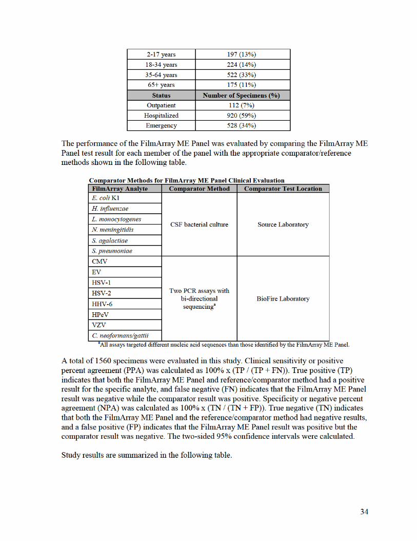
2. Comparison Studies:
a. Clinical Comparison between FilmArray and FilmArray 2.0 systems:
To demonstrate that performance of the FilmArray ME Panel when used with the
FilmArray 2.0 system is equivalent to FilmArray system, a combination of residual, de-
identified CSF specimens and contrived CSF specimens covering all 14 analytes on the
FilmArray ME Panel were evaluated. Specimens were identified as positive for
FilmArray ME analytes at source laboratories or by culture (bacterial analytes) or PCR
comparator methods (yeast and viruses) during the prospective FilmArray ME panel
clinical study. The specimens were not chosen based on analyte levels but were chosen
only for their previous analyte-specific positive test results and availability of sufficient
volume for testing.
Contrived specimens were prepared as follows: For each analyte, leftover negative CSF
specimens (negative as determined by FilmArray and comparator methods) were spiked
with isolates of the organisms of interest at low concentrations (~3× LoD or below).
Multiple strains for each organism were represented.
A total of 149 specimens were tested; 21 positive clinical specimens and128 contrived
specimens. Positive contrived specimens were spiked with FilmArray ME Panel analytes
at approximately 3× LoD or below. Specimens were split into two different aliquots for


42
centrifuged. The operator places a Hydration Injection Vial and a Sample Injection Vial
into the FilmArray Pouch Loading Station. The operator hydrates the test pouch with the
Hydration Injection Vial and then using a transfer pipette, adds~200 µl of CSF into the
Sample Injection Vial. The operator removes the Sample Injection Vial containing the
CSF from the Loading Station, inverts the vial at least three times to mix, and then inserts
it into the Loading Station port where the proper amount of specimen is pulled into the
FilmArray ME Panel pouch by vacuum. The FilmArray ME Panel is then placed onto the
FilmArray instrument for testing.
5. Calibration:
Not applicable
6. Quality Control:
See Quality Control Section above (L.1.c “Traceability, Stability, Expected Values
(controls, calibrators, or methods)”)
O. Other Supportive Instrument Performance Characteristics Data Not Covered In The
“Performance Characteristics” Section above:
Not Applicable
P. Proposed Labeling:
The labeling is sufficient and it satisfies the requirements of 21 CFR Parts 801 and 809 and
the specials controls for this device type.
Q. Identified Risks and Required Mitigations:
Identified Risks to Health Required Mitigations (See Section S below
for Special Controls)
Incorrect identification or lack of identification
of a pathogenic microorganism by the device
can lead to improper patient management
Special Controls (1), (2), (3), (4), and (5)
Failure to correctly interpret test results Special Controls (6), (7), (8), and (9)
Failure to correctly operate the instrument Special Control (10)


45
encephalitis when used in conjunction with clinical signs and symptoms and other
clinical and laboratory findings.
(b) Classification. Class II (special controls). A device to detect and identify microbial
pathogen nucleic acids in cerebrospinal fluid must comply with the following special
controls:
1) Premarket notification submissions must include detailed device description
documentation, including the device components, ancillary reagents required
but not provided, and a detailed explanation of the methodology including
primer/probe sequence, design, and rationale for sequence selection.
2) Premarket notification submissions must include detailed documentation from
the following analytical studies: Analytical sensitivity (Limit of Detection),
inclusivity, reproducibility, interference, cross reactivity, and specimen
stability.
3) Premarket notification submissions must include detailed documentation from
a clinical study. The study, performed on a study population consistent with
the intended use population, must compare the device performance to results
obtained from well-accepted comparator methods.
4) Premarket notification submissions must include detailed documentation for
device software, including, but not limited to, software applications and
hardware-based devices that incorporate software.
5) The Intended Use statement in the device labeling must include a statement
that the device is intended to be used in conjunction with standard of care
culture.
6) A detailed explanation of the interpretation of results and acceptance criteria
must be included in the device’s 21 CFR 809.10(b)(9) compliant labeling.
7) The device labeling must include a limitation that negative results do not
preclude the possibility of central nervous system infection.
8) The device labeling must include a limitation that device results are not
intended to be used as the sole basis for diagnosis, treatment, or other patient
management decisions.
9) The device labeling must include a limitation stating that positive results do
not mean that the organism detected is infectious or is the causative agent for
clinical symptoms.
10) As part of the risk management activities performed as part of your 21 CFR
820.30 design controls, you must document an appropriate end user device
training program that will be offered as part of your efforts to mitigate the risk
of failure to correctly operate the instrument.