Evaluation of a monoclonal antibody-based latex agglutination test
7
Vol. 30, No. 10 JOURNAL OF CLINICAL MICROBIOLOGY, OCt. 1992, p. 2544-2550 0095-1137/92/102544-07$02.00/0 Copyright © 1992, American Society for Microbiology Evaluation of a Monoclonal Antibody-Based Latex Agglutination Test for Diagnosis of Cryptococcosis: Comparison with Two Tests Using Polyclonal Antibodies ALAIN TEMSTET,' PATRICIA ROUX,' JEAN-LOUIS POIROT,2 OLIVIER RONIN,3 AND FRANQOISE DROMER3,4* Laboratoire de Parasitologie et Mycologie, H6pital Tenon, '1 Laboratoire de Parasitologie et Mycologie, H6pital Saint Antoine,2 Unites de Mycologie, Institut Pasteur,3 and Institut National de la Sante et de la Recherche Medicale, Unite 283, Hopital Cochin,4 Paris, France Received 5 March 1992/Accepted 3 July 1992 Cryptococcal antigen detection has become a routine biological test performed for patients with AIDS. The poor prognosis of cryptococcosis explains the need for reliable tests. We evaluated the performances of a newly commercialized agglutination test that uses a monoclonal antibody specific for cryptococcal capsular polysac- charide (Pastorex Cryptococcus; Sanofi-Diagnostics Pasteur, Marnes-la-Coquette, France) and compared them with those of tests that use polyclonal immune sera (Cryptococcal Antigen Latex Agglutination System, Meridian Diagnostics, Inc., Cincinnati, Ohio; and Crypto-LA, International Biological Labs Inc., Cranbury, N.J.). The sensitivities and specificities of the tests were compared by using purified polysaccharides and yeast suspensions. Clinical specimens (131 serum samples, 41 cerebrospinal fluid samples, 34 urine samples, and 19 bronchoalveolar lavage samples) from 87 human immunodeficiency virus-positive subjects with (40 patients) and without (47 patients) culture-proven cryptococcosis were retrospectively tested during a blinded study. The effect of pronase treatment of samples was assessed for Pastorex Cryptococcus and the Cryptococcal Antigen Latex Agglutination System, and the antigen titers were compared. Our results show that (i) during the screening, concordance among the three tests was 97%; (ii) the use of pronase enhanced both the sensitivities and specificities of the Pastorex Cryptococcus test; (iii) titers agreed for 67% of the cerebrospinal fluid samples and 60% of the serum samples; and (iv) cryptococcosis was detected equally well with Pastorex Cryptococcus and with the other tests, whatever the infecting serotype (A, B, or D). The meaning of in vitro sensitivity and the relationship between titers and sensitivity are discussed. The results show that Pastorex Cryptococcus is a rapid and reliable test for the detection of cryptococcal antigen in body fluids and suggest that kits cannot be used interchangeably to monitor antigen titers in patients. The need for sensitive tests for the diagnosis of crypto- coccosis has been particularly acute since the beginning of the AIDS epidemic. Cryptococcosis is the most common life-threatening fungal infection in patients with AIDS (4, 6). For several years now, physicians have included the detec- tion of cryptococcal antigen, at least in serum, as a routine screening test whenever a human immunodeficiency virus (HIV)-positive patient has meningitis, unexplained pneumo- nia, or fever (20). Titration of antigen is used for the diagnosis of cryptococcosis as well as for monitoring anti- fungal therapy (6). Physicians and biologists are aware of the necessity for a reliable and rapid test. In all but one of the commercialized tests (8), the detection of capsular antigen from Cryptococcus neoformans is based on the agglutination of latex beads covered with polyclonal antibodies usually raised in rabbits. A newly commercialized test uses a murine monoclonal antibody specific for cryptococcal polysaccha- ride (7) to sensitize the beads. The purpose of the study described here was to report on the performances of this new test, Pastorex Cryptococcus (Sanofi-Diagnostics Pasteur, Marnes-La-Coquette, France), and to compare them with those of two other tests that are currently used in several laboratories and for which some comparative studies have already been published (8, 11, 14, 24). The two other tests are the Cryptococcal Antigen Latex * Corresponding author. Agglutination System (CALAS; Meridian Diagnostics, Inc., Cincinnati, Ohio) and Crypto-LA (International Biological Labs Inc., Cranbury, N.J.). We compared the sensitivities and specificities of these tests for the detection of crypto- coccal antigen in various clinical specimens from patients with AIDS and with or without culture-proven cryptococco- sis. For Pastorex Cryptococcus, we also checked the use- fulness of pronase treatment of the samples, since this procedure has been reported to eliminate the false aggluti- nation reactions and to improve the sensitivity of the test (9, 11). We tested the influence of the serotype of the infecting C. neoformans strain on the ability of Pastorex Cryptococ- cus to diagnose the infection. We assessed the efficacies of the tests for the detection of cryptococcal antigen in speci- mens other than serum and cerebrospinal fluid (CSF) spec- imens by testing bronchoalveolar lavage (BAL) and urine samples. The data show that Pastorex Cryptococcus is a rapid, sensitive, specific, and reliable assay for the detection of cryptococcal antigen in clinical specimens. MATERIALS AND METHODS Study design. Sera collected from 87 HIV-positive patients in four different clinical microbiology laboratories from university hospitals in Paris, France, were used for the retrospective blinded study. These sera were initially sent for a routine screening for cryptococcal antigen by Crypto-LA and were kept frozen for subsequent assays. The 2544 on January 29, 2019 by guest http://jcm.asm.org/ Downloaded from
Transcript of Evaluation of a monoclonal antibody-based latex agglutination test

Vol. 30, No. 10JOURNAL OF CLINICAL MICROBIOLOGY, OCt. 1992, p. 2544-25500095-1137/92/102544-07$02.00/0Copyright © 1992, American Society for Microbiology
Evaluation of a Monoclonal Antibody-Based Latex AgglutinationTest for Diagnosis of Cryptococcosis: Comparison with
Two Tests Using Polyclonal AntibodiesALAIN TEMSTET,' PATRICIA ROUX,' JEAN-LOUIS POIROT,2 OLIVIER RONIN,3
AND FRANQOISE DROMER3,4*Laboratoire de Parasitologie et Mycologie, H6pital Tenon,'1 Laboratoire de Parasitologie et Mycologie,H6pital Saint Antoine,2 Unites de Mycologie, Institut Pasteur,3 and Institut National de la Sante et de la
Recherche Medicale, Unite 283, Hopital Cochin,4 Paris, France
Received 5 March 1992/Accepted 3 July 1992
Cryptococcal antigen detection has become a routine biological test performed for patients with AIDS. Thepoor prognosis of cryptococcosis explains the need for reliable tests. We evaluated the performances of a newlycommercialized agglutination test that uses a monoclonal antibody specific for cryptococcal capsular polysac-charide (Pastorex Cryptococcus; Sanofi-Diagnostics Pasteur, Marnes-la-Coquette, France) and compared themwith those of tests that use polyclonal immune sera (Cryptococcal Antigen Latex Agglutination System,Meridian Diagnostics, Inc., Cincinnati, Ohio; and Crypto-LA, International Biological Labs Inc., Cranbury,N.J.). The sensitivities and specificities of the tests were compared by using purified polysaccharides and yeastsuspensions. Clinical specimens (131 serum samples, 41 cerebrospinal fluid samples, 34 urine samples, and 19bronchoalveolar lavage samples) from 87 human immunodeficiency virus-positive subjects with (40 patients)and without (47 patients) culture-proven cryptococcosis were retrospectively tested during a blinded study. Theeffect of pronase treatment of samples was assessed for Pastorex Cryptococcus and the Cryptococcal AntigenLatex Agglutination System, and the antigen titers were compared. Our results show that (i) during thescreening, concordance among the three tests was 97%; (ii) the use of pronase enhanced both the sensitivitiesand specificities of the Pastorex Cryptococcus test; (iii) titers agreed for 67% of the cerebrospinal fluid samplesand 60% of the serum samples; and (iv) cryptococcosis was detected equally well with Pastorex Cryptococcusand with the other tests, whatever the infecting serotype (A, B, or D). The meaning of in vitro sensitivity andthe relationship between titers and sensitivity are discussed. The results show that Pastorex Cryptococcus is arapid and reliable test for the detection of cryptococcal antigen in body fluids and suggest that kits cannot beused interchangeably to monitor antigen titers in patients.
The need for sensitive tests for the diagnosis of crypto-coccosis has been particularly acute since the beginning ofthe AIDS epidemic. Cryptococcosis is the most commonlife-threatening fungal infection in patients with AIDS (4, 6).For several years now, physicians have included the detec-tion of cryptococcal antigen, at least in serum, as a routinescreening test whenever a human immunodeficiency virus(HIV)-positive patient has meningitis, unexplained pneumo-nia, or fever (20). Titration of antigen is used for thediagnosis of cryptococcosis as well as for monitoring anti-fungal therapy (6). Physicians and biologists are aware of thenecessity for a reliable and rapid test. In all but one of thecommercialized tests (8), the detection of capsular antigenfrom Cryptococcus neoformans is based on the agglutinationof latex beads covered with polyclonal antibodies usuallyraised in rabbits. A newly commercialized test uses a murinemonoclonal antibody specific for cryptococcal polysaccha-ride (7) to sensitize the beads.The purpose of the study described here was to report on
the performances of this new test, Pastorex Cryptococcus(Sanofi-Diagnostics Pasteur, Marnes-La-Coquette, France),and to compare them with those of two other tests that arecurrently used in several laboratories and for which somecomparative studies have already been published (8, 11, 14,24). The two other tests are the Cryptococcal Antigen Latex
* Corresponding author.
Agglutination System (CALAS; Meridian Diagnostics, Inc.,Cincinnati, Ohio) and Crypto-LA (International BiologicalLabs Inc., Cranbury, N.J.). We compared the sensitivitiesand specificities of these tests for the detection of crypto-coccal antigen in various clinical specimens from patientswith AIDS and with or without culture-proven cryptococco-sis. For Pastorex Cryptococcus, we also checked the use-fulness of pronase treatment of the samples, since thisprocedure has been reported to eliminate the false aggluti-nation reactions and to improve the sensitivity of the test (9,11). We tested the influence of the serotype of the infectingC. neoformans strain on the ability of Pastorex Cryptococ-cus to diagnose the infection. We assessed the efficacies ofthe tests for the detection of cryptococcal antigen in speci-mens other than serum and cerebrospinal fluid (CSF) spec-imens by testing bronchoalveolar lavage (BAL) and urinesamples. The data show that Pastorex Cryptococcus is arapid, sensitive, specific, and reliable assay for the detectionof cryptococcal antigen in clinical specimens.
MATERIALS AND METHODS
Study design. Sera collected from 87 HIV-positive patientsin four different clinical microbiology laboratories fromuniversity hospitals in Paris, France, were used for theretrospective blinded study. These sera were initially sentfor a routine screening for cryptococcal antigen byCrypto-LA and were kept frozen for subsequent assays. The
2544
on January 29, 2019 by guesthttp://jcm
.asm.org/
Dow
nloaded from

MONOCLONAL ANTIBODY FOR CRYPTOCOCCAL ANTIGEN TESTING 2545
results of this first antigen detection in the sera allowed us toselect specimens from 40 patients with culture-proven cryp-tococcosis (84 serum samples, 30 CSF samples, 12 BALsamples, and 10 urine samples) and 47 patients without aknown cryptococcal infection (47 serum samples, 1 CSFsample, and 1 BAL sample). After selection, the sampleswere divided into aliquots and were randomly assigned anumber, their origin being kept from the investigators whodid the tests. All samples were stored at -20°C for 3 monthsto 2 years before testing, without intermediary thawing andfreezing cycles. Since there were no control samples otherthan sera, 24 urine samples collected from HIV-negativesubjects and 10 CSF samples (labeled CSF samples 1 to 10)and 6 BAL samples from HIV-positive patients, all of whomwere hospitalized for diseases other than cryptococcosis,were subsequently added. All the patients were assigned anumber. Those that begin with the letter M indicate thosewith cryptococcosis; those that begin with the letter Tindicate patients in the control group.
Test procedures. (i) Screening. All sera were heat inacti-vated at 56°C for 30 min. The samples were tested by thethree tests by using the procedures recommended by themanufacturers. For the new test (Pastorex Cryptococcus),the influence of enzymatic treatment of the specimens priorto antigen testing on both the sensitivity and the specificityof the test was evaluated. We therefore used in parallel fivemethods for the screening of undiluted samples, as follows:direct testing with the three different reagents and testingafter enzymatic treatment recommended by Sanofi-Diagnos-tics Pasteur and Meridian. When the available volumes weresmall (especially for CSF and BAL samples), only directtesting was performed. Specimens were always tested by thethree tests on the same day. The procedures were as follows.For Crypto-LA, 50 ,ul of the sample was mixed with the
same volume of sensitized beads. The kits were from lotnumber 3864.For CALAS, 100 ,ul of the sample was incubated for 15
min at 56°C with the same volume of pronase solution. Thereaction was stopped by heating at 100°C for 5 min. Of thismixture, 25 IL was removed and mixed with 1 drop of thesensitized latex beads. Direct testing was performed in thesame way with 25 ,ul of the untreated sample. Lot numbers404A089, 404A097, and 404A100 were used.For Pastorex Cryptococcus, the enzymatic treatment was
slightly different. The sample (100 ,ul) was mixed with 35 ,ulof pronase and heated for 15 min at 56°C. The enzyme wasthen instantaneously inactivated by the addition of protein-ase inhibitor (35 RI). Ten microliters of the sensitized beadswas then added to 40 ,ul of the mixture or untreated sample.Different kits were used, corresponding to lot numbersCN26, CN30, CN36, and 1K104.
(ii) Titrations. Depending on the quantities left afterscreening, serum and CSF samples that gave a positiveagglutination reaction were then serially diluted (twofolddilutions) in the buffer provided with the kits to determinethe end point of agglutination. Only 48 serum samples and 9CSF samples were still available for titration by PastorexCryptococcus and CALAS. Titer determination on pronase-treated samples was done on the aliquots that were treatedand frozen after the screening. For 14 different samples, wechecked that the procedure (pronase reaction) and subse-quent storage did not alter antigen detection by PastorexCryptococcus and CALAS. Titers were not changed bymore than 1 dilution (data not shown). In all reports onantigen titration, changes of ±+1 dilution are always consid-ered to be insignificant and within the limit of experimental
error. We also assessed, as has been done by others (11),that the procedure of freezing and thawing did not changethe antigen titers (data not shown). No titration was done onBAL or urine samples. Volumes of samples treated withpronase according to the protocol of Sanofi-DiagnosticsPasteur were adjusted (addition of 30 ,ul of buffer) to give a1:2 starting dilution.Comparison of sensitivities. The sensitivities of the three
kits were assessed by using cryptococcal antigen (positivecontrols) supplied in the Meridian and the InternationalBiological Labs kits. The limit of detection for the fourserotypes was determined by using capsular polysaccharides(CPSs) purified from C. neofornans serotypes A, B, C, andD (kindly provided by J. E. Bennett, National Institute ofAllergy and Infectious Diseases, Bethesda, Md.). Thepolysaccharides were serially diluted in buffer and wereallowed to react with the three different sensitized latexbeads until the end point of agglutination was reached. Theminimal amount of antigen detected by each kit was re-corded.The in vitro sensitivities of Pastorex Cryptococcus and
Crypto-LA were further assessed on suspensions of liveyeasts. C. neofornans serotypes A (CDC B551, CDC B236,NIH 68, NIH 271), B (NIH 112, NIH 444, CDC B237), C(NIH 18, NIH 191, CDC B238), and D (NIH 52, NIH 3501,NIH 3502, CBS 132) and Trichosporon beigelii (CBS 2936)and Candida albicans (NIHB 311) were used. Yeasts weregrown in Sabouraud broth in an orbital shaker (for 15 h at28°C). Cells were then enumerated and diluted in phosphate-buffered saline (PBS; 10 mM; pH 7.4). Agglutination waschecked with 10-fold dilutions of the suspensions; the nega-tive control was PBS alone.
Statistical analysis. All calculations were done with aMacintosh SE/30 computer and Statview II software (Aba-cus Concepts, Inc., Berkeley, Calif.).
RESULTS
Comparison of in vitro sensitivities. The in vitro sensitivi-ties of the tests used in this study are summarized in Table 1.The sensitivity of Pastorex Cryptococcus was similar tothose of Crypto-LA and CALAS for reactivity with CPSserotype A and the control CALAS antigen and lower forpurified CPS serotypes D, B, and C and the controlCrypto-LA antigen. Results were different with yeast sus-pensions. We first noted a prozone phenomenon when thesuspensions were greater than 107/ml. The threshold ofdetection was then determined with 10-fold dilutions startingat 107/ml (Table 1). It varied according to the serotype of thestrains and the reference strain used (serotype D for Pas-torex Cryptococcus and serotypes B, C, and D for Crypto-LA). Overall, the sensitivities of the tests were similar.Cross-reactivities with T. beigelii and C. albicans werelower with Pastorex Cryptococcus than they were withCrypto-LA.
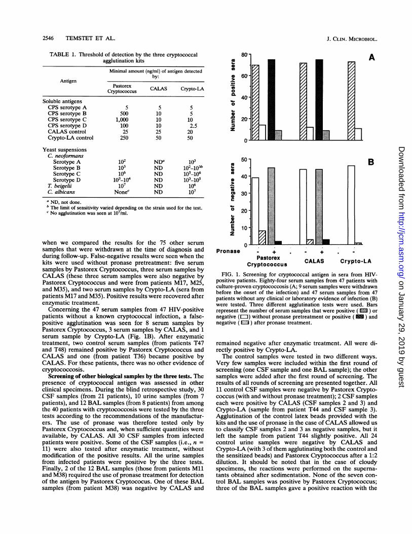
Screening of the sera by the three tests. After completion ofthe screening, the results of screening of sera by the threetests were interpreted as positive, negative, or false positiveand false negative according to the known infection status ofeach patient. Results for 84 serum samples from 40 patientswith culture-proven cryptococcosis are shown in Fig. 1A.Nine of these serum samples were obtained months beforethe onset of infection; eight were negative by the three tests,and one serum sample (from patient M24) gave a false-positive reaction by the Pastorex Cryptococcus test, whichwas resolved by enzymatic treatment. Differences appeared
VOL. 30, 1992
on January 29, 2019 by guesthttp://jcm
.asm.org/
Dow
nloaded from
2546 TEMSTET ET AL.
TABLE 1. Threshold of detection by the three cryptococcalagglutination kits
Minimal amount (ng/ml) of antigen detectedby:
AntigenPastorex CALAS Crypto-LA
Cryptococcus
Soluble antigensCPS serotype A 5 5 5CPS serotype B 500 10 5CPS serotype C 1,000 10 10CPS serotype D 100 10 2.5CALAS control 25 25 20Crypto-LA control 250 50 50
Yeast suspensionsC. neoformans
Serotype A 102 NDa 103Serotype B 103 ND 102_103bSerotype C 106 ND 105_106Serotype D 103-104 ND 103_-15
T. beigelii 107 ND 106C. albicans Nonec ND 107a ND, not done.b The limit of sensitivity varied depending on the strain used for the test.c No agglutination was seen at 107/ml.
when we compared the results for the 75 other serumsamples that were withdrawn at the time of diagnosis andduring follow-up. False-negative results were seen when thekits were used without pronase pretreatment: five serumsamples by Pastorex Cryptococcus, three serum samples byCALAS (these three serum samples were also negative byPastorex Cryptococcus and were from patients M17, M25,and M35), and two serum samples by Crypto-LA (sera frompatients M17 and M35). Positive results were recovered afterenzymatic treatment.Concerning the 47 serum samples from 47 HIV-positive
patients without a known cryptococcal infection, a false-positive agglutination was seen for 8 serum samples byPastorex Cryptococcus, 3 serum samples by CALAS, and 1serum sample by Crypto-LA (Fig. 1B). After enzymatictreatment, two control serum samples (from patients T47and T48) remained positive by Pastorex Cryptococcus andCALAS and one (from patient T36) became positive byCALAS. For these patients, there was no other evidence ofcryptococcosis.
Screening of other biological samples by the three tests. Thepresence of cryptococcal antigen was assessed in otherclinical specimens. During the blind retrospective study, 30CSF samples (from 21 patients), 10 urine samples (from 7patients), and 12 BAL samples (from 8 patients) from amongthe 40 patients with cryptococcosis were tested by the threetests according to the recommendations of the manufactur-ers. The use of pronase was therefore tested only byPastorex Cryptococcus and, when sufficient quantities wereavailable, by CALAS. All 30 CSF samples from infectedpatients were positive. Some of the CSF samples (i.e., n =11) were also tested after enzymatic treatment, withoutmodification of the positive results. All the urine samplesfrom infected patients were positive by the three tests.Finally, 2 of the 12 BAL samples (those from patients Mlland M38) required the use of pronase treatment for detectionof the antigen by Pastorex Cryptococus. One of these BALsamples (from patient M38) was negative by CALAS and
0
ob-
ea
0
0.o0'o0
.0
Ez
co
0
00
0:0
0
.0
Ez
50
40
30
20
10
0Pronase - +
PastorexCryptococcus
A
B
C + A
CALAS Crypto-LA
FIG. 1. Screening for cryptococcal antigen in sera from HIV-positive patients. Eighty-four serum samples from 47 patients withculture-proven cryptococcosis (A; 9 serum samples were withdrawnbefore the onset of the infection) and 47 serum samples from 47patients without any clinical or laboratory evidence of infection (B)were tested. Three different agglutination tests were used. Barsrepresent the number of serum samples that were positive ( ) ornegative (El1) without pronase pretreatment or positive ( M ) andnegative (m ) after pronase treatment.
remained negative after enzymatic treatment. All were di-rectly positive by Crypto-LA.The control samples were tested in two different ways.
Very few samples were included within the first round ofscreening (one CSF sample and one BAL sample); the othersamples were added after the first round of screening. Theresults of all rounds of screening are presented together. All11 control CSF samples were negative by Pastorex Crypto-coccus (with and without pronase treatment); 2 CSF sampleseach were positive by CALAS (CSF samples 2 and 3) andCrypto-LA (sample from patient T44 and CSF sample 3).Agglutination of the control latex beads provided with thekits and the use of pronase in the case of CALAS allowed usto classify CSF samples 2 and 3 as negative samples, but itleft the sample from patient T44 slightly positive. All 24control urine samples were negative by CALAS andCrypto-LA (with 3 of them agglutinating both the control andthe sensitized beads) and Pastorex Cryptococcus after a 1:2dilution. It should be noted that in the case of cloudyspecimens, the reactions were performed on the superna-tants obtained after sedimentation. None of the seven con-trol BAL samples was positive by Pastorex Cryptococcus;three of the BAL samples gave a positive reaction with the
J. CLIN. MICROBIOL.
on January 29, 2019 by guesthttp://jcm
.asm.org/
Dow
nloaded from

MONOCLONAL ANTIBODY FOR CRYPTOCOCCAL ANTIGEN TESTING
TABLE 2. Discrepancies among the three tests during screening
Sample Patient Result by the foliowing test: Clinicalno. Pastorex CALAS Crypto-LA diagnosis
Serum M17 + + - +Serum M35 + + - +BAL M38 + - + +Serum T47 + + - -
Serum T36 - + - -
CSF T44 - - + -
antibody-sensitized and control beads provided with theCALAS and Crypto-LA kits.
Discrepancies among the three kits at the end of screening.Overall, of the 131 serum samples, 31 CSF samples, and 13BAL samples tested during the blinded study, only 6 (3%)discrepancies were noted by the three tests, ifwe consideredthe results of both Pastorex Cryptococcus and CALAS afterenzymatic treatment. Discordant results are summarized inTable 2. They concerned three samples from patients withcryptococcosis and three samples from control subjects. Forpatients M17 and M35, the negative reaction seen withCrypto-LA can be attributed to the lack of enzymatictreatment, since both were negative by Pastorex Cryptococ-cus and CALAS before the pronase reaction. The BALsample from patient M38 was negative by CALAS, despite apositive antigen determination in CSF and serum samples.Concerning the control samples, no explanation is availablefor the faint positivity seen in the serum sample from patientT36 by CALAS (titer, 1:2 after pronase treatment) and in theserum sample from patient T47 by Pastorex Cryptococcusand CALAS (titer, 1:8 whatever procedure was used). Thesepatients had no clinical or laboratory evidence of cryptococ-cosis (a serum sample withdrawn from patient T47 almost 10months after the first serum sample was obtained wasnegative by Pastorex Cryptococcus even after pronase treat-ment). The weak positivity seen by Crypto-LA for the CSFsample from patient T44 has not been confirmed as specificby follow-up of the patient (one serum sample withdrawn onthe same day as the CSF sample was obtained as well asseveral additional serum samples were negative by the threetests). One possible explanation is a cross-reactivity (18)with T. beigelii (cultured from the BAL sample), but therewas no dissemination (negative blood cultures).The sensitivity of Pastorex Cryptococcus was thus 100%,
whereas it was 99% for CAILAS and 98% for Crypto-LAwhen all of the results are summarized. The specificities ofthe tests were 97, 96, and 93%, respectively.Comparison of antigen titers determined by Pastorex Cryp-
tococcus and CALAS. We titrated 48 of the 75 positive serumsamples (from 25 different patients) by Pastorex Cryptococ-cus and CALAS. Correlations between titers before andafter pronase treatment were similar for Pastorex Crypto-coccus and CALAS (r2 = 0.856 and 0.821, respectively) (Fig.2). Pronase treatment did not modify the antigen titers (±1dilution) in 80 and 59% of the serum samples tested byPastorex Cryptococcus and CALAS, respectively. The mod-ifications were seen only for sera with an initial titer of lessthan 1:256; the geometric means before and after pronasetreatment were 1:28 and 1:46, respectively, by PastorexCryptococcus (P = 0.02 by the Wilcoxon signed-rank test)and 1:17 and 1:52, respectively, by CALAS (P = 0.001). Thesmall numbers of serum samples with increased titers afterpronase treatment determined by Pastorex Cryptococcus
04 21qm 2020
19.* 18* 1 7C 160° 15
14; 13a 12
11; 10
9.c 8
76
a 5- 4o 3
21S.
o 0neg
A
BCM50
e
SQ
C
0
a.
0U
0
C0
a
0
er0a..00
Reciprocal antigen titer before pronase (log2)
FIG. 2. Antigen titers (reciprocal titers in log2) in sera fromHIV-positive patients with culture-proven cryptococcosis. Serawere tested by Pastorex Cryptococcus (A) or CALAS (B) with andwithout pronase pretreatment.
compared with those determined by CALAS were not re-lated to the activity of the enzyme provided by the manu-facturers. An increase in the pronase concentration (two tothree times) did not enhance further the titer for 12 differentserum samples (data not shown).
Titers determined with both kits agreed (within 1 dilution)for 61 and 57% of serum specimens, respectively, before andafter pronase treatment. For falsely negative specimens,titers after pronase treatment were within 1 dilution of eachother when determined by Pastorex Cryptococcus and CA-LAS and were equal to or less than 1:64. Titers determinedfor the nine CSF samples (range, 1:2 to 1:16,384) agreed for67% of the samples after pronase treatment. For threedifferent patients, we had additional samples that allowed usto repeat a comparison of the antigen levels over time. Inthese three patients, the evolution of titers was similar byboth tests (data not shown).
Contribution of pronase treatment to antigen detection. Itshould be noted that one prozone phenomenon was not
VOL. 30, 1992 2547
on January 29, 2019 by guesthttp://jcm
.asm.org/
Dow
nloaded from

2548 TEMSTET ET AL.
resolved by pronase treatment, a clear-cut positive reactionbeing seen only after dilution of this serum sample (frompatient M21).As shown in Fig. 1, pronase treatment uncovered all the
false-negative and most of the false-positive reactions insera. As noted above, unexplained positive results remainedfor two patients in the control group (patients T47 and T48),which was disturbing in view of the other results. Pronasetreatment uncovered two false-negative results (for serumsamples from patients M17 and M35), showing that thesepatients were already infected with C. neofornans 232 and153 days, respectively, prior to mycological proof of infec-tion or the positivity of the Crypto-LA.Concerning the other biological samples, pronase treat-
ment also seemed to be beneficial for the detection ofcryptococcal antigen in both BAL and CSF samples frominfected patients. However, use of pronase should beavoided for urine samples, since it constantly gave false-positive results (data not shown). Urine testing required onlya 1:2 dilution of clarified supernatant to prevent nonspecificinterference with the sensitized beads. This dilution wouldnot have prevented the agglutination on specimens frominfected patients, since the titers were 21:2 for two urinesamples and >1:10 for the other eight urine samples thatwere titrated.
Overall, the use of pronase enhanced the sensitivities andthe specificities of the tests. When looking at the results forthe 127 positive samples corresponding to patients withculture-proven cryptococcosis and the 98 negative samples,the sensitivity increased from 94 to 100% for PastorexCryptococcus and from 97 to 99% for CALAS. The speci-ficity increased from 86 to 97% for Pastorex Cryptococcusand from 89 to 96% for CALAS.
Influence of the serotype on the ability of Pastorex Crypto-coccus to detect soluble antigen in patients. We assessed theperformances of Pastorex Cryptococcus with samples with-drawn from patients for whom knew the serotypes of theinfecting strains. Strains of C. neoformans corresponding to14 patients in the present study were available for serotyping(12): all were C. neoformans var. neoformans, 10 wereserotype A, 3 were serotype D (patients M9, M16, and M22),and 1 was untypeable (patient M13). There was no discrep-ancy between Pastorex Cryptococcus and the other tests forsamples from these three patients. Four other patients witha serotype D infecting strain were further tested by one ofthe polyclonal antibody-based tests and Pastorex Cryptococ-cus without a false-negative result and with concordantantigen titers (data not shown). We also tested three serumsamples from one patient infected with a serotype B strain(kindly provided by B. Dupont, Unite de Mycologie, InstitutPasteur, Paris, France). The titers determined by PastorexCryptococcus and Crypto-LA were 1:32 and 1:16, 1:1,024and 1:1,024, and 1:256 and 1:32, respectively.
DISCUSSION
We assessed the performances of a newly commercializedtest that uses a monoclonal antibody to detect cryptococcalantigen in clinical specimens and compared them with thoseof two other kits using polyclonal antibodies. Purifiedpolysaccharides and a total of 131 serum samples, 41 CSFsamples, 34 urine samples, and 19 BAL samples were testedby the three tests.For Pastorex Cryptococcus, we found a sensitivity similar
to those of the other tests for polysaccharide serotype A andlower than those for serotypes B, C, and D. When tested
with yeast suspensions, the performances of Pastorex Cryp-tococcus and Crypto-LA with strains of the four serotypesvaried with the reference strain used and were very close toeach other. The differences seen with antigens that wereeither chemically purified or present on live cells suggest avariability in antigen expression on yeasts of the sameserotype. It might partly explain the variability in antigentiters during the infection. However, the restricted specific-ity of the monoclonal antibody compared with those of thepolyclonal antibodies did not prevent an accurate detectionof cryptococcal antigen. First, in our study, Pastorex Cryp-tococcus was even more sensitive than Crypto-LA forsamples from two patients and could have allowed thediagnosis of the infection several months before theCrypto-LA test detected it. Second, the serotype of theinfecting strains did not seem to influence the efficiency ofthe test. At present, the vast majority of patients withcryptococcosis have AIDS (17). According to publishedreports, probably more than 98% are infected with C.neoformans var. neoformans, i.e., serotypes A and D, witha higher proportion of serotype A (2, 15, 17, 19, 22). Wechecked that Pastorex Cryptococcus as well as polyclonalantibody-based tests detected the infection in patients in-fected with serotype D strains. Among the patients infectedwith C. neofonnans var. gattii, i.e., serotypes B and C, allthe reported cases were of serotype B and, when noted,antigen was not detectable by conventional tests (1, 5, 13,21, 23). For one patient infected with a serotype B strain,soluble antigen was detected by Pastorex Cryptococcus aswell as Crypto-LA. To our knowledge, infections withserotype C strains have rarely been diagnosed except inSouthern California before the AIDS epidemic (16). Unfor-tunately, we did not have the opportunity to test samplesfrom such patients. The results of the study described hereshowed that the in vitro sensitivity and specificity of Pas-torex Cryptococcus should allow the detection of cryptococ-cal antigen in all infected patients.We compared the performances of the tests on various
clinical specimens. Discrepancies were found for only sixspecimens (3%). The performances of Pastorex Cryptococ-cus were excellent and very similar (97% specificity, 100%sensitivity) to those found for CALAS (96% specificity, 99%sensitivity) and Crypto-LA (93% specificity, 98% sensitivity)in this and previous (8, 9, 11) studies. For Pastorex Crypto-coccus as well as for CALAS, the use of pronase enhancedboth the sensitivity and the specificity, as reported previ-ously (9, 11). However, it did not resolve one prozonephenomenon that showed the need for sample dilution inview of clinical symptoms suggestive of cryptococcosis (10).Pronase resolved all the other false-negative reactions (6%for Pastorex Cryptococcus and 4% for CALAS). Thesevalues are lower than those presented in a previous study(11), in which 19% of the sera gave false-negative reactionsthat were uncovered by pronase treatment. Despite pronasetreatment, two false-positive reactions were unresolved,since clinical and laboratory evidence was against the diag-nosis of cryptococcosis. However, it is noteworthy that intwo patients a positive antigen in serum samples was pre-dictive of a later infection. Pronase was also effective forother biological samples, especially BAL samples, in ourhands. Results for none of the positive CSF samples weremodified by pronase treatment (a finding already noted byothers [11]). Pronase pretreatment should be avoided forurine samples. A 1:2 dilution of clarified supernatants pre-vented all false-positive reactions. For none of these differ-ent specimens was there a false-positive reaction by Pas-
J. CLIN. MICROBIOL.
on January 29, 2019 by guesthttp://jcm
.asm.org/
Dow
nloaded from

MONOCLONAL ANTIBODY FOR CRYPTOCOCCAL ANTIGEN TESTING 2549
torex Cryptococcus that would have been clarified by theuse of control globulin-coated latex beads as provided byMeridian or International Biological Labs. The use of a veryspecific reagent and the systematic treatment of all speci-mens except urine with pronase therefore seem sufficient fordetecting all positive and negative samples. In our opinion,manipulations with this test are not more fraught withproblems than those with a polyclonal antibody-based test,owing to a simplified procedure for enzymatic treatment.The antigen titers determined by Pastorex Cryptococcus
and CALAS agreed (within 1 dilution) for 61 and 57% ofserum specimens, respectively, before and after pronasetreatment and for 67% of the titrated CSF samples, a valueclose to that given in a recent report (11). The fact that titersare usually higher after pronase treatment when tests likeCrypto-LA (9, 11) or CALAS (11; this study) are used butare not higher by Pastorex Cryptococcus, even at higherpronase concentrations, suggests that the monoclonal anti-body may be responsible. One hypothesis is that this anti-body detects an antigenic determinant that is less hidden bythe interfering substances (whatever they are). Neverthe-less, in our opinion, there is a misunderstanding between theantigen titer determined by a test and the sensitivity of thetest. A reliable assay should be able to detect the infectionearly and to monitor antifungal therapy and the evolution ofthe disease. Both Pastorex Cryptococcus and CALASproved to be efficient in our hands.
This is the first study of cryptococcal antigen detection inclinical specimens other than CSF and serum specimens.Urine samples are easy to test, and positive antigen detec-tion in samples diluted 1:2 was, in this study, complementaryevidence of cryptococcosis. It is often necessary to obtainBAL samples from patients with AIDS. An abnormal chestX-ray and the occurrence of cryptococcal pneumonia havebeen reported several times in patients with AIDS (3).Testing of the BAL sample for the presence of cryptococcalantigen seems easy and may be helpful (unpublished data):first, in cases in which culture is negative but antigendetection in serum and CSF samples is positive, a positiveBAL sample could reinforce the presumption and, second,during pneumonia caused by C. neofonnans and anotherorganism which can prevent C. neoformans from growing inculture. We therefore think that testing of BAL and urinesamples can be recommended when cryptococcosis is sus-pected. However, other studies are needed to determinewhether the hypothesis based on our experience is fullyjustified.We conclude from the results of this study that Pastorex
Cryptococcus provides a perfectly adequate, rapid, andreliable means of diagnosing and monitoring cryptococcosisin patients. Our results show the need for pronase pretreat-ment to prevent false-positive and false-negative reactionsfor all specimens but urine samples. These data suggest thevalue of a systematic screening for cryptococcal antigen ona regular basis in patients with AIDS. The data also under-line the fact, as has been done previously (11), that kits forthe detection of cryptococcal antigen cannot be used inter-changeably. Finally, when confronted with a negative result,clinicians and biologists should keep in mind the need forrepeated cultures and antigen testing whenever the evidenceof infection is strong enough.
ACKNOWLEDGMENTSWe thank Bertrand Dupont (Institut Pasteur, Paris) for allowing
us to test serum from a patient infected with C. neoformans serotypeB. We also thank Anne-Marie Deluol (H6pital Bichat), Nicole
Bardin (Hopital Rothschild) for providing some of the serum sam-ples, and M. L. Garrigues and L. Meulemans (Sanofi-DiagnosticsPasteur) for providing the kits used in this study.
REFERENCES1. Bottone, E. J., P. A. Kirschner, and I. F. Salkin. 1986. Isolation
of highly encapsulated Cryptococcus neoformans serotype Bfrom a patient in New York City. J. Clin. Microbiol. 23:186-188.
2. Bottone, E. J., I. F. Salkin, N. J. Hurd, and G. P. Wormser.1987. Serogroup distribution of Cryptococcus neoformans inpatients with AIDS. J. Infect. Dis. 156:242.
3. Cameron, M. L., J. A. Bartlett, H. A. Gallis, and H. A. Waskin.1991. Manifestations of pulmonary cryptococcosis in patientswith the acquired immunodeficiency syndrome. Rev. Infect.Dis. 13:64-67.
4. Chuck, S. L., and M. A. Sande. 1989. Infections with Crypto-coccus neoformans in the acquired immunodeficiency syn-drome. N. Engl. J. Med. 321:794-799.
5. Clancy, M. N., J. Fleischman, D. H. Howard, K. J. Kwon-Chung, and R. Y. Shimizu. 1990. Isolation of Cryptococcusneoformans gattii from a patient with AIDS in Southern Cali-fornia. J. Infect. Dis. 161:809.
6. Dismukes, W. E. 1988. Cryptococcal meningitis in patients withAIDS. J. Infect. Dis. 157:624-628.
7. Dromer, F., J. Salamero, A. Contrepois, C. Carbon, and P. Yeni.1987. Production, characterization, and antibody specificity of amouse monoclonal antibody reactive with Cryptococcus neofor-mans capsular polysaccharide. Infect. Immun. 55:742-748.
8. Gade, W., S. W. Hinnefeld, L. S. Babcock, P. Gilligan, W. Kelly,K. Wait, D. Greer, M. Pinilla, and R. L. Kaplan. 1991. Com-parison of the PREMIER cryptococcal antigen enzyme immu-noassay and the latex agglutination assay for the detection ofcryptococcal antigens. J. Clin. Microbiol. 29:1616-1619.
9. Gray, L. D., and G. D. Roberts. 1988. Experience with the useof pronase to eliminate interference factors in the latex aggluti-nation test for cryptococcal antigen. J. Clin. Microbiol. 26:2450-2451.
10. Haldane, D. J. M., D. S. Bauman, A. W. Chow, P. Doyle, G.Garber, J. Ngui-Yen, and J. A. Smith. 1986. False negative latexagglutination test in cryptococcal meningitis. Ann. Neurol.19:413.
11. Hamilton, J. R., A. Noble, D. W. Denning, and D. A. Stevens.1991. Performance of Cryptococcus antigen latex agglutinationkits on serum and cerebrospinal fluid specimens of AIDSpatients before and after pronase treatment. J. Clin. Microbiol.29:333-339.
12. Ikeda, R., T. Shinoda, Y. Fukazawa, and L. Kaufnan. 1982.Antigenic characterization of Cryptococcus neoformans sero-types and its application to serotyping of clinical isolates. J.Clin. Microbiol. 16:22-29.
13. Kapend'a, K., K. Komichelo, D. Swinne, and J. Vandepitte.1987. Meningitis due to Cryptococcus neoformans biovar gattiiin a Zairean AIDS patient. Eur. J. Clin. Microbiol. 6:320-321.
14. Kaufman, C. A., A. G. Bergman, P. J. Severance, and K. D.McClatchey. 1981. Detection of cryptococcal antigen. Compar-ison of two latex agglutination tests. Am. J. Clin. Pathol.75:106-109.
15. Kwon-Chung, K. J., and J. E. Bennett. 1984. Epidemiologicdifferences between the two varieties of Cryptococcus neofor-mans. Am. J. Epidemiol. 120:123-130.
16. Kwon-Chung, K. J., A. K. Varma, and D. H. Howard. 1988.Ecology and epidemiology of Cryptococcus neoformans: arecent study of isolates in the United States, p. 107-112. In J. M.Torres-Rodriguez (ed.), Proceedings of the 10th Congress of theInternational Society for Human and Animal Mycology, Barce-lona, 27 June to 1 July 1988. J. R. Prous Science Publisher,Barcelona, Spain.
17. Levitz, S. M. 1991. The ecology of Cryptococcus neofonnansand the epidemiology of cryptococcosis. Rev. Infect. Dis.13:1163-1169.
18. Melcher, G. P., M. G. Rinaldi, C. L. Frey, and D. J. Drutz. 1988.Demonstration, by immunoelectronmicroscopy, of a cell wallantigen in Trichosporon beigelii that cross-reacts with Crypto-
VOL. 30, 1992
on January 29, 2019 by guesthttp://jcm
.asm.org/
Dow
nloaded from

J. CLIN. MICROBIOL.
coccus neoformans capsular polysaccharide. J. Infect. Dis.158:901-902.
19. Rinaldi, M. G., D. J. Drutz, A. Howell, M. A. Sande, C. B.Wofsy, and W. K. Hadley. 1986. Serotypes of Cryptococcusneoformans in patients with AIDS. J. Infect. Dis. 153:642.
20. Roux, P., J. L. Touboul, M. Feuilhade de Chauvin, T. Delacour,J. Revuz, D. Basset, C. Mayaud, and F. Lancastre. 1986.Disseminated cryptococcosis diagnosed in AIDS patient byscreening for soluble serum antigens. Lancet i:1154.
21. Rozenbaum, R., A. J. R. Gonsalves, B. Wanke, and W. Vieira.1990. Cryptococcus neoformans var. gattii in a Brazilian AIDS
patient. Mycopathologia 112:33-34.22. Shimizu, R. Y., D. H. Howard, and M. N. Clancy. 1986. The
variety of Cryptococcus neoformans in patients with AIDS. J.Infect. Dis. 154:1042.
23. St-Germain, G., G. Noel, and K. J. Kwon-Chung. 1988. Dissem-inated cryptococcosis due to Cryptococcus neoformans varietygattii in a Canadian patient with AIDS. Eur. J. Clin. Microbiol.Infect. Dis. 7:587-588.
24. Wu, T. C., and S. Y. Koo. 1983. Comparison of three commer-cial cryptococcal latex kits for detection of cryptococcal anti-gen. J. Clin. Microbiol. 18:1127-1130.
2550 TEMSTET ET AL.
on January 29, 2019 by guesthttp://jcm
.asm.org/
Dow
nloaded from