Evaluation and Management of...
21
Kathryn Hassell, M.D. Professor of Medicine, Division of Hematology University of Colorado Denver Evaluation and Management of Thrombocytopenia Disclosures No Financial Conflicts No Off-Label Discussion Objectives Integrate the understanding of mechanisms of thrombocytopenia when deciding on evaluation and intervention Analyze available clinical and initial laboratory information to develop a differential diagnosis for thrombocytopenia Initiate and monitor therapy for severe thrombocytopenic syndromes, in collaboration with hematology consultation as needed Recognize situations for which platelet transfusion is beneficial or potentially harmful
Transcript of Evaluation and Management of...

Kathryn Hassell, M.D.
Professor of Medicine, Division of Hematology University of Colorado Denver
Evaluation and Management of Thrombocytopenia
Disclosures � No Financial Conflicts
� No Off-Label Discussion
Objectives � Integrate the understanding of mechanisms of
thrombocytopenia when deciding on evaluation and intervention
� Analyze available clinical and initial laboratory information to develop a differential diagnosis for thrombocytopenia
� Initiate and monitor therapy for severe thrombocytopenic syndromes, in collaboration with hematology consultation as needed
� Recognize situations for which platelet transfusion is beneficial or potentially harmful

Thrombocytopenia: Definitions � Reduction in the number of platelets <100K � Terminology varies, but generally:
� “Pseudothrombocytopenia” occurs in 1 in 1000 healthy individuals – check the smear! � In vitro agglutination of platelets in EDTA (purple top) � “Satellitism”: adhesion of plts to PMNs
Platelet count (x109/L) Description
50-100 (150) “Mild”
30-50 “Moderate”
10-30 “Severe”
<10 “Very Severe”
Veneri, Blood Transfus 7:75, 2009; Stasi Hematology 2012:191,2012
Thrombocytopenia: Incidence � Seen in ~1% of adult acute care hospital inpts
� <30% of these patients manifest with bleeding
� More common in surgical and trauma patients than in medical patients
� In ICU patients, associated with higher morbidity and mortality � Mortality increased 4-6X if thrombocytopenia sustained >4
days � Independent of bleeding events: marker of serious medical
illness, failure to reverse underlying cause � More worrisome if it occurs after initial recovery
Rice, Chest 136:1622, 2009; Akca, Crit Care Med 30:753, 2002 Thiele, Sem Hematol 50:239, 2013
Thrombocytopenia in the ICU � Most common coagulation issue in ICU patients
� 15-68%, depending on the definition used, 2-15% have plts <50K � Half present with low platelets, remainder develop it in ICU
� Most common medical condition: sepsis
Rice, Chest 136:1622, 2009; Akca, Crit Care Med 30:753, 2002; Thiele, Sem Hematol 50:239, 2013; Stasi, Hematology 2012, 191

Platelets and Thrombopoietin (TPO)
Thiele, Sem Hematol 50:239, 2013
Liver disease (not just cirrhosis) → ↓ TPO
BM suppression - drugs - infection - toxins Primary BM disease
Dilution Sequestration - spleen or liver Destruction - thrombotic microangioapathies - immune-mediated
Takes at least 3-4 days to see rise in plts after suppression alleviated
Thrombocytopenia: Causes � Most common etiology of thrombocytopenia in a
hospitalized patient is MULTIFACTORIAL � Often a combination of compromised production and increased
consumption/destruction
� Drug-induced thrombocytopenia affects up to 25% of acutely ill patients � Can suppress bone marrow production of platelets � Platelet counts commonly <50,000, nadirs may be <20,000 � Occurs over days to weeks
� Infections/toxins: similar pattern
Rice, Chest 136:1622, 2009; Pluta, Adv Med Sci 55:16, 2010; Priziola, Crit Care Med 38:S145, 2010; Stasi, Hematology 2012, 191
Louie, J Viral Hep doi:10/j.1365-2893.2010.01366x
Thrombocytopenia: Causes � Classes of agents:
� Viruses: possible with any, but most reported with certain infections, including:
� Toxin Injury � Ethanol (bone marrow suppression, liver disease) � Radiation therapy
Antineoplastics Antivirals Thiazides
Daptomycin Tolbutamide Linezolid
Antifungals Digoxin Haloperidol
Nitrfurantoin Meropenem Quinolones
HIV Hepatitis C Parvovirus
Epstein-Barr Virus Varicella CMV

Typical Hospital Thrombocytopenia � Common in the acutely ill
� Fever/SIRS � Medications, medications, medications � Co-morbidities (e.g. liver disease, history of chemo/XRT) create
“vulnerability”
� Counts of 20-30,000 not uncommon � Knowledge of baseline status useful
� If counts normal within last days/weeks or on presentation, then unlikely a primary BM disorder
� Significance of fall from low baseline less clear, e.g. from 60K to 40K in ESLD
� Patience: may take days to weeks to resolve, even when contributing factors have improved
How Many Platelets Do You Need? Not Many � Platelet counts of ≥10,000 well-tolerated
� Based on studies of chemotherapy (AML) patients � 3% of days complicated by any bleeding
Rebulla, NEJM 337:1870,1997
� Platelet counts of ≥5,000 may be adequate � Based on studies in aplastic anemia
� Loss of blood in stool increases at platelet count of <5,000
Schlichter, Clin Hem 1978
� 3 nonlethal major bleeding events over >18,000 pt-days in chronic severe aplastic anemia
Sagmeister, Blood 93:3124, 1999
5 10 15 20
ml/
days
100
80
60
40
20
51Cr-labeled RBCs in 20 pts
Plt count
Thrombocytopenia: Treat Patients, Not Numbers � In the absence of bleeding symptoms and interventions, platelet
counts of 5-10,000 may be well-tolerated � Especially true for ITP � Need to weigh confounding factors e.g. other coagulation
system changes, platelet dysfunction � Only data for prophylaxis are in chemotherapy-induced
thrombocytopenia � Typically maintain platelets ≥10,000
� NO proven benefit for ANY transfusion threshold in other settings

Reason to Care: Thrombocytopenia as an Indication of a Bigger Problem
Specific Severe Conditions Marked by Thrombocytopenia � Characterized by increased consumption or destruction of
platelets � Platelets may be (initially) innocent by-standers
� Recognition of the specific condition necessary to provide appropriate management
� Treatment is based on reversing the underlying pathophysiology � Correcting the platelet count is not the goal: harm often comes from
the underlying condition NOT the thrombocytopenia � Correcting the platelet count (e.g. transfusion) does not improve the
condition and may create harm
Important Thrombocytopenic Syndromes � Associated with microangiopathy
� Disseminated intravascular coagulation (DIC) � Thrombotic microangiopathies (TMAs)
� Thrombotic thrombocytopenia purpura (TTP) � Complement-mediated TMA (HUS) � Hemolysis elevated liver enzymes low platelets (HELLP)
� Associated with immune destruction � Immune thrombocytopenia purpura (ITP)
� Associated with heparin (HIT)

Sorting It Out: Key Clinical/Laboratory Parameters � Platelet count: severity, rate of decrease
� Medication reconciliation: type of drug, timing
� Evidence of hemolysis: microangiopathy � Elevated reticulocyte count � Falling Hb, especially if retic count is not high � AST, LDH, Indirect bilirubin � Schistocytes (mechanical) or spherocytes (immune)
� Basic coagulation parameters: PT/INR, (aPTT)
� Markers of fibrin(ogen)olysis: FSPs/D-dimer
� Creatinine: severity of increase
Hemolysis: Laboratory Clues
↑ AST (SGOT)
↑ LDH
↑ Indirect Bilirubin
↑ Reticulocytes
ALT (SGPT) and direct bilirubin should be normal
RED CELL ENZYMES BONE MARROW RESPONSE
↑ Urobilinogen in Urine Hemoglobinuria not RBCS
Functional Evidence of RBC Loss (Hemolysis)
Monday Tuesday Wednesday
Patient #1 Evidence of hemolysis (partially compensated)
Hb 8.9 8.3 7.9
Retic 15% 16% 13%
Patient #2 Evidence of hemolysis (compensated)
Hb 8.9 8.8 9.0
Retic >23% >23% >23%
Patient #3 Active RBC destruction unlikely: Hb stable without increased retic
Hb 8.9 8.8 9.0
Retic 2% 1.8% 3%

� How many are too many? � Everyone may have scattered schistocytes (0-0.2%) � Can be seen in metastatic cancer, sepsis, chronic renal failure,
infection, preterm infants, pre-clampsia, acute renal failure without TMA
� Attempts to standardize assessment: ICHS Guideline 2012 Zini, Int J Lab Hematol 34:107, 2012 � Should be the main finding and >1% � Recommend counting 1000 cells (microscopy) � Smaller than normal RBC, sharp edges, various shapes
Schistocytes and Microangiopathy
Schistocytes: Not So Easy � Single site clinical lab study:
� Review of 282 smears, estimated % schistocytes
� Median in DIC 0.70%, TTP 2.45%, HUS, 2.90 � Inter-rater confidence: 0.836
Diagnosis 0-1% 1-3% 3-6% 6-12% DIC 21 8 1 0
TTP/HUS 0 2 0 0
HELLP 1 1 0 0
Autoimmune HA 0 2 1 0
Metastatic cancer 33 23 1 1
Acute renal failure* 3 3 2 1
Huh, Int J Lab Hem 35:542, 2013
*Not TMA
Next Step: Coagulation Parameters � Classical findings
� Normal in TTP/HUS, ITP, [HIT] � Abnormal in DIC
� Not always so easy – confounding factors � Vitamin K deficiency (antibiotics, NPO/poor nutrition): PT,
when severe also aPTT � Antiphospholipid antibody syndrome: aPTT � Anticoagulation: aPTT, PT � Heparinized lines: aPTT � Liver disease: aPTT, PT, fibrinogen, FSP, D-dimer
� D-dimer is non-specific ≠ active clot, DIC

Disseminated Intravascular Coagulation: Definition & Concepts � Acquired syndrome of coagulation with loss of localization
arising from different causes � Can originate from and cause damage to the
microvasculature; if severe, causes organ damage � Characterized by the generation of fibrin-related products
(e.g. FSP, D-dimer) and acquired inflammatory and non-inflammatory vessel injury
� Two general types: � Overt, often decompensated � Non-overt, more often compensated
Kaneko and Wada, J Clin Exp Hematopath 51:67, 2011
DIC: Most Common Settings Diagnosis
(Prospective Japanese Study) DIC/PreDIC
Cases Total Cases
Frequency of DIC
Infectious disease 77 219 35.1%
Solid cancer 61 142 42.9%
Hematopoietic cancer 60 114 52.6%
Aneurysm (e.g. aortic) 15 29 51.7%
Obstetrics disease 6 10 60.0%
Trauma 10 26 38.4%
Digestive disease 5 18 27.7%
Collagen vascular disease 1 10 10.0%
Other 10 45 22.2%
Kaneko and Wada, J Clin Exp Hematopath 51:67, 2011
Microvascular Fibrin Deposition
Additional Consumption
of Platelets
↓ platelet count
Consumption of
Clotting Factors
↑ PT, aPTT, ↓ fibrinogen
Additional Consumption
of Anticoagulants
↓ Protein C, AT
Increased Fibrinolysis
↓ fibrinogen, ↑ FSP
DIC: Pathophysiology
“Fibrinolytic phenotype”
“Thrombotic” phenotype
Gando, Crit Care Med 38:S35, 2010

DIC: Clinical Manifestations
Wada, J Intensive Care 2:15, 2014
Infection
Post-op OB Leukemia
Aortic aneurysm
Trauma OB
DIC: Recognition→Intervention(?) � Temporal changes over time
� No single test or single time point “diagnostic” of DIC: dynamic complex process
� Elevated D-dimer is not specific or diagnostic � Just an expensive way to measure fibrin split products
� Morbidity, mortality associated with microvascular occlusion, organ damage � Ideally, introduce anticoagulation in non-overt stage
� Prophylactic dosing preferred over treatment doses unless overt VTE
� Efforts to control of thrombin (mixed results to date) � Soluble thrombomodulin, antithrombin concentrates
Thachil, Am J Med 123:17, 2010; Gando, Crit Care Med 38:S35, 2010; Hook and Abrams, Clin Trans Sci 5:85, 2012; Toh Hematology 266, 2013
If Intervention is Attempted:
Wada, J Intensive Care 2:15, 2014
*
*not available in the US
(aPC concentrat, thromobumodulin, antithrombin

Mainstay of Treatment of DIC � Treat the underlying cause � Reverse the underlying cause � Eliminate the underlying cause � Get rid of the underlying cause � Fix the underlying cause � Stop the underlying cause � Eradicate the underlying cause
Monitor coag panel (plts, PT, fibrinogen) for stability, improvement
Primary Thrombotic Microangiopathy (TMA) � Microangiopathy and thrombocytopenia but normal coagulation
studies � Oklahoma registry: systemic infection (bacterial, viral,fungal)
ultimately accounted for symptoms thought to be TTP (n=31 of 415) � All had MAHA and low platelets
� 87% neuro changes (65% “major”),90% AKI, 68% fever
� Classic pentad (and coma) more common in systemic infection than in apparent TTP cases
� 56% had low ADAMTS13 activity (levels 12-47%) � 4 had levels <10%, 2 with inhibitors
Booth, Am J Hematol 86:743, 2011
Primary Thrombotic Microangiopathy (TMA) � Acquired TTP more common in adults (2.9 per 1 million) � Hereditary TMA, HUS more common in children � Clinical considerations
� Diarrheal illness, AKI: Shiga-toxin HUS (outbreaks) � Sudden onset severe systemic symptoms and anuric AKI, drug
exposure to quinine, gemctyabine, quetiapine: drug- induced immune TMA
� Gradual onset of renal failure over wks/mos, CyA or tacrolimus: drug-induced toxic TMA
Verbeke, Blood Coag Fibrin 21:3, 2010; Kavanagh, Curr Opin Hem 17:432, 2010 George, NEJM 371:7 2014; Booth, Am J Hematol 86:743, 2011

Primary Thrombotic Microangiopathy (TMA)
Endotheliosis with intraluminal and vessel wall fibrin
George NEJM 371:7
2014
(rare, pediatric, inherited)
(rare, pediatric, inherited)
TMA: Pathophysiologies � TTP
� Complement-mediated (HUS)
Sadler, Blood 112:11, 2008
Fakhouri, Nat Rev Nephrol 3:679, 2007; George, NEJM 371:7 2014
Deficiency of ADAMTS-13 Acquired: autoantibody Congenital: mutations (90+ known), <5% of cases
Increased C’ activity Familial: CFH mutations other C’ genes Acquired: autoantibody to Factor H
Barbour, Nephrol Dial Transplant 27:2673, 2012
TMA: Role of ADAMTS-13 Testing � Testing options
� ADAMTS-13 activity and antigen � Detection of anti-ADAMTS-13 antibodies � Genetic characterization of ADAMTS-13 gene
� Hereditary TTP � Severe deficiency (usually undetectable, may be 5-6%) without
antibodies during remission
� Acquired TTP � Highly variable activity levels, usually not severely low � Values of <10% more common in idiopathic, relapsing
� Severe deficiencies in other TMAs (consumption?) � 13% in C’-mediated TMA (HUS), 16% in overt DIC Peyvandi, J Thromb Haemost 8:631, 2010; Clark, Sem Dialysis 25:214, 2012

TMA: Additional Diagnostic Labs Shiga toxin (STEC) Stool culture for E coli Toxicology for shiga toxin or PCR of shiga toxin gene Urine culture for E coli
ADAMTS13 deficiency ADAMTS13 activity ADAMTS13 autoantibodies ADAMTS13 gene mutations
Complement dysregulation Plasma/serum C3, factor H, factor I, factor B MCP (CD46) expression PBMCs Factor H autoantibodies Specific mutations
Other Associations Pregnancy test Liver and pancreatic enzymes B12/homocysteine assay, MMA HIV and other viral serologies (e.g. varicella, echovirus, coxsackie A&B) ANA, antiphospholipid Ab testing PCR for influenza A Evaluate for C difficle, Salmonella
S. Pneumoniae T antigen expression on RBC PCR of blood and/or secretions Blood culture
Barbour, Nephrol Dial Transplant 27:2673, 2012; Bambauer, Ther Apher Dialy 15:10, 2010
TTP: Initial Management � Hereditary/Acquired TTP
� Fresh frozen plasma therapy: replacement of ADAMTS-13, removal of antibodies � Infusion therapy (25 mg/kg total daily) has benefit
� 52% complete remission , 57% overall survival � Therapeutic plasma exchange (TPE) preferred and (probably)
superior to infusion � Return of donor plasma; cryo-poor preferred � 80% complete remission, 85% overall survival
� Immunosuppressive therapy: assumes antibodies present, no randomized trials � Usually steroids (e.g. methylprednisolone 1-2 mg/kg)
Marques, Clin Lab Med 29:321, 2009; Verbeke, Blood Coag Fibrin 21:3, 2010; Clark, Sem Dialysis 25:214, 2012
TTP: Initial Management � Daily monitoring (just before TPE) to look for:
� Improving Hb with falling reticulocyte count � Normalization of hemolytic markers (LDH, bili) � Normalization of creatinine
� Continue until “in remission” x 2 days; no data to support “5 days of exchange” � Might be considered a minimum; if still worsening, reconsider the
diagnosis (e.g. type of TMA) � Why stop if they’re still getting better but not yet stabilized? � Some require exchanges for weeks to attain stability and eventual
recovery
Verbeke, Blood Coag Fibrin 21:3, 2010; Kavanagh, Curr Opin Hem 17:432, 2010

Other TMAs: Initial Management � Shiga-toxin mediated TMA: supportive � Drug-mediated TMA
� Immune (quinine): supportive, discontinue drug � Toxic (CyA, tacrolimus): supportive, dose reduction may be
sufficient
� Complement-mediated TMA : � Immune suppression if antibody-mediated (anti-Factor H) � Anti-complement therapy (ecluzimab)
� But at the start can be difficult to discern so often plasma exchange and steroids are initiated
George, NEJM 371:7 2014
Anti-complement Therapy: Eculizumab � Inhibitor of C5 activation, used for PNH � Successful use in Shiga-toxin HUS and complement-mediated
TMA � Considered when initial response to TPE is lost or initially poor
� Initial dosing (600 mg/wk x 4), 900 mg week 5, then every 2 weeks maintenance
� May see results within first week � Meningococcal vaccine needed 2 weeks before first dose:
significant inhibition of ability of complement to clear meningococcus
Barbour, Nephrol Dial Transplant 27:2673, 2012; Westra, Neth J Med 70:121, 2012 George, NEJM 371:7 2014
ITP: Definitions and Clinical Settings � Immune Thrombocytopenia Purpura
� No longer “idiopathic” when using the term
� Characterized by duration � Acute: <3 months; severe, often abrupt, profound � Persistent: 3-12 months � Chronic: >12 months; more often moderate
� Secondary – “primary” a diagnosis of exclusion � Autoimmune disorders (e.g. SLE,RA,APS,Sjorgrens) � Infections (e.g. Hepatitis C, HIV, H. pylori) � Lymphoproliferative disorders (often CLL, rarely NHL) � Recent vaccination (e.g. MMR) � Drugs, drugs, drugs
Provan, Blood 115:168, 2010; Cines, Hem Onc Clin NA 23:1155, 2009

ITP: Drug-Induced � Up to 25% of acutely ill have drug-induced
thrombocytopenia (immune + non-immune) � Multiple mechanisms, multiple drugs Hapten-dependent Penicillin, Cephalosporins,
Fluoroquinolones
Drug-GP complex Quinine, NSAIDs, Quinidine, Sulfonamides, Rifampin, Ranitidine
Ligand-induced binding Eptifibatide, Tirofiban
Drug-specific antibody Abciximab
Autoantibody Gold, Procainamide Etanercept, Efalizumab
Immune complex Heparins
Aster, J Thomb Haemost 7:911, 2009; Priziola, Crit Care Med 38:S145, 2010
ITP: Pathophysiology � Immune-mediated
� IgG Ab often directed at specific GP receptors � Platelet destruction
� RE system � Spleen
� Damage to megakaryocytes
� Diminished TPO � Inappropriately low response, plt production
Arnold, Exp Opin Invest Drug 18:805, 2009 Cines, Hem Onc Clin NA 23:1155, 2009; Veneri, Blood Transfus 7:75, 2009
ITP: Diagnosis � Clinical characteristics
� Very severe, onset over days (vs. weeks/months) � Associated risk factors, e.g.autoimmune disease, infections, drugs � NO splenomegaly (primary ITP) � Isolated thrombocytopenia, large platelets
� Testing: BM bx not recommended if typical � Basic evaluation: HIV, Hep C, H pylori, quantitative immunoglobulins
(CVID), Direct Coombs, Rh � Potential utility: specific GP antibodies, ANA, APS testing, anti-
thyroid Ab, other viral infections e.g. CMV � No proven benefit: TPO level, bleeding time, platelet antibodies,
serum complement, platelet survival
Provan, Blood 115:168, 2010; Lo, Autoimmunity Rev 13:577, 2014

ITP: Initial Management � Risk of fatal events: 0.0162 – 0.0389 cases per adult patient-
year at risk � Thought more likely if <5-20K plts, bleeding sx
� If secondary, fix the underlying cause if possible (e.g. stop the drug)
� Initial therapy: immune modulation may take 5+ days � Corticosteroid therapy
� Prednisone: 0.5-2 mg/kg daily � Dexamethsone: 40 mg/day x 4 days, repeat every 14 days
� IvIG or IV anti-D (if Rh+)
� Typical next step: splenectomy vs. rituxan Provan, Blood 115:168, 2010; Pels, Sem Thromb Hemost 621, 2011 Lo, Autoimmunity Rev 13:577, 2014
Refractory ITP: TPO-Receptor Agonists � Stimulation of platelet production
� Romiplostim (Nplate): s.q. weekly � Eltrombopag (Promacta): oral once daily
� Most often used third-line, or as a bridge while awaiting effect of rituximab or other immune-modulating therapy
� Immediate side effects include HA, nausea and vomiting � May cause marrow fibrosis
� Of interest in Hep C-related thrombocytopenia to permit full-dose, full-duration anti-viral therapy
Basciano and Bussei, Curr Opin Hematol 19:392,. 2012
Other ITP Scenarios � H pylori (+): antibodies cross react with plt glycoproteins
� Platelet response to antibiotic therapy 40-50% if successfully eradicate infection
Franchini, Sem Thromb Hemost 38:463, 2012
� HIV-associated: in the HAART era � <50K in 1-9% of HIV-infected; <150K in 10-30% � Respond to usual therapy, with or without HAART
Ambler, Adv in Hem 1, 2012
� Coronary disease and ITP (case series): low platelets not protective � PCI and CABG performed at plt counts as low as 5K � 46% discharged on ASA and clopidogrel
Russo, Interact Cardiovasc Thorac Surg : 153, 2011

A Hematologist’s Plea: Stop “HIT-ting” People! � Heparin-induced thrombocytopenia is a clinical syndrome
� Most common in those receiving UFH � Rare in OB, peds, dialysis, uncommon with LMWH
� Many humans make antibodies to heparin(s), including fondaparinux: this doesn’t mean they have HIT
� The diagnosis cannot be made by laboratory testing: must have appropriate clinical criteria
Arepally, Ann Rev Med 61:77, 2010; Cuker, Sem Thromb Hemost 40:106, 2014
HIT: Not Just an Antibody � Antibodies without clinical HIT are common:
Adult cardiac by-pass* 50% Blood 96:1703 Pediatric by-pass 1.7-16% Anesth Analg 107:371 Ortho prophylaxis (UFH) 15% Blood 96:1703 Ortho prophylaxis (LWMH) 8% Blood 96:1703 Med prophylaxis (UFH) 3% Blood 101:2955, 2003 Neurology prophylaxis (UFH) 20% Neurology 62:657, 2004 PCI (cath-UFH) 12% Thromb Res 115:475, 2005 Chronic hemodialysis 12% Kidney Int 73:713, 2008 Vascular surgery 32% J Vasc Surg 48:377 ED for chest pain/VTE (in hosp in last 6 months) 6.9/9.2% Am J Emer Med 25:279
*including positive SRA in 30%
HIT: Pathophysiology � IgG antibody to platelet factor 4 (PF4) neoantigen exposed
after binding to heparin
- takes 4-5 days to make IgG antibodies - manifestations (↓ plts) BEGIN to occur 4-5 days after antigen exposure - presence of antibody is not sufficient to create clinical HIT
Arepally, Ann Rev Med 61:77
MOST
FEW
HIT: Diagnosis � Clinical Criteria: the “4 Ts” score
Points (0, 1, or 2), Maximum = 8 2 1 0
Thrombocytopenia Nadir (x109/L) 20-100 10-19 <10 Fall >50% 30-50% <30% Timing Recent heparin ≤ 1 dy ≤ 1 dy No prev heparin 5-10 dys Unclear or >10 dys ≤4 days Thrombosis, ASR, skin lesions
New event after heparin
Progressive or new thrombosis/event
None
Other cause for ↓ plt None Possible Definite
Probability of HIT: High: 6-8 Moderate: 4-5 Low: 0-3 Arepally, Ann Rev Med 61:77, 2010
0
50
100
150
200
250
300
350
400
Day 1 Day 2 Day 3 Day 4 Day 5 Day 6 Day 7
Classic HIT HIT with Reexposure Unlikely HIT
Time Course for HIT
γ
γ
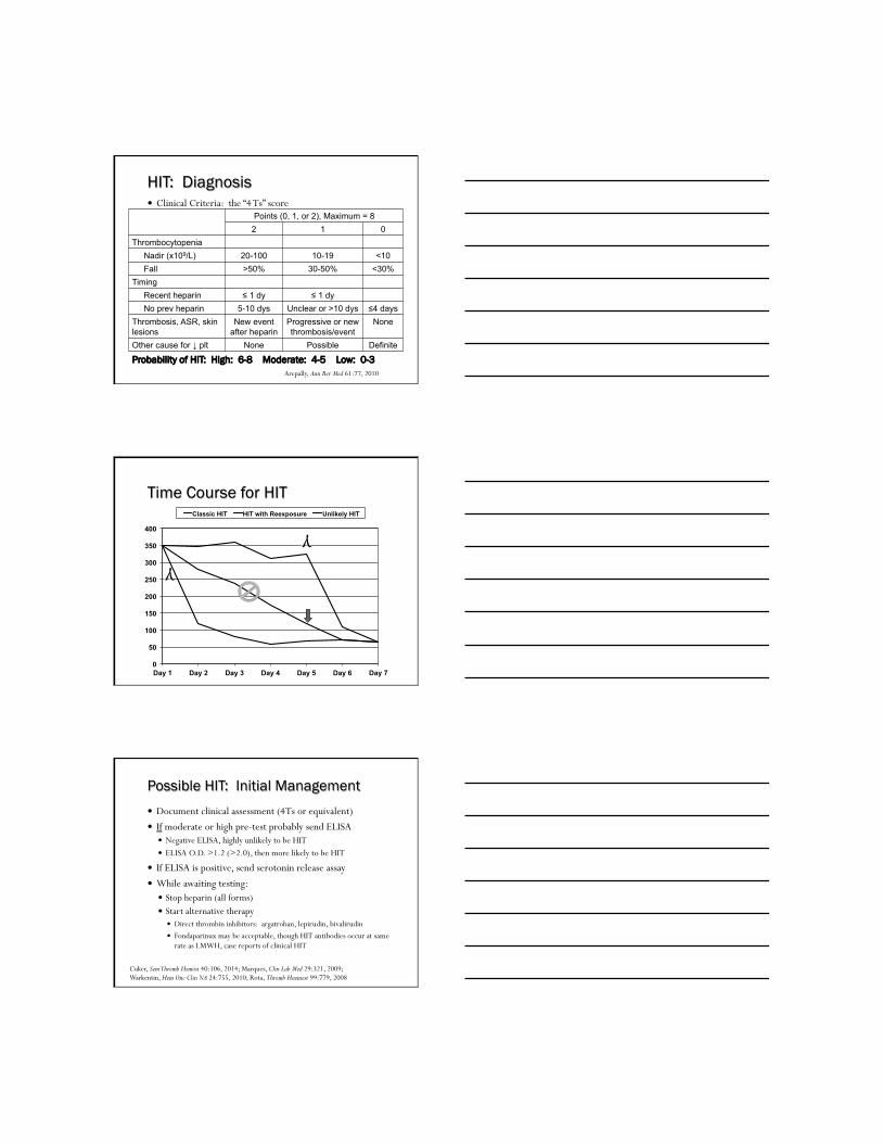
Possible HIT: Initial Management � Document clinical assessment (4Ts or equivalent) � If moderate or high pre-test probably send ELISA
� Negative ELISA, highly unlikely to be HIT � ELISA O.D. >1.2 (>2.0), then more likely to be HIT
� If ELISA is positive, send serotonin release assay � While awaiting testing:
� Stop heparin (all forms) � Start alternative therapy
� Direct thrombin inhibitors: argatroban, lepirudin, bivalirudin � Fondaparinux may be acceptable, though HIT antibodies occur at same
rate as LMWH, case reports of clinical HIT
Cuker, Sem Thromb Hemost 40:106, 2014; Marques, Clin Lab Med 29:321, 2009; Warkentin, Hem Onc Clin NA 24:755, 2010; Rota, Thromb Haemost 99:779, 2008

Practical Considerations “Bad” Thrombocytopenia Syndromes � Overt DIC vs. Liver Disease
� If the parameters are stable, then the DIC compensated or it’s “just” the liver disease
� Regarding MAHA in these syndromes � If the retic count is low and the Hb is stable, then it’s not MAHA
or any form of significant RBC destruction � If there are spherocytes it may be a really bad case of SLE or other
autoimnmune condition, not MAHA
� The TTP “pentad” is more common in non-TTP � The only thing that makes the platelet count <10, in the
absence of an obviously fulminant condition, is autoimmune destruction
Platelets and Hemostasis � Adhesion and aggregation of platelets to area of vessel
injury
� Control is local: minimum number of functional platelets needed at the vessel wall may not be assessed by CBC
How Many Platelets Are Enough? � Cochrane analysis of prophylaxis in chemotherapy/stem
cell transplantation � No evidence prophylaxis prevents bleeding
� Major bleeding the same (RR 1.66, 0.9-3.04) � Longer time to first bleeding with prophylaxis
� If threshold is used, current evidence supports use of 10K vs. 20K � Significant bleeding similar (RR 1.35, 0.95-1.9) � More days with bleeding (RR 1.72, 1.33-2.22)
� Low plt utilization with lower/no threshold (50%)
Estcourt, The Cochrane Library 5, 2012

PLADO – Effects of Prophylactic Platelet Dose on Transfusion Outcomes � Increased bleeding at/below 5K in 1102 adult and pediatric
heme/onc patients
Slichter, NEJM 362:600, 2010; ; Kaufman, Transfusion 55:144, 2015
How Many Platelets Are Enough? � TOPPS: No prophylaxis is NOT non-inferior
� 600 chemo/SCT patients: “prn” vs 10K
Without prophylaxis: - shorter time to first bleed - more days with Grade 2 bleeding (1.7 vs. 1.2)
Stanworth, NEJM 368:1771, 2013
But My Patients Aren’t on Chemo � No data to address threshold for prophylactic platelet
transfusion in hospitalized patients (borrow from oncology) � No data to support ANY specific platelet count to control
bleeding or for procedures � Single administration of 6-8 U platelets (or single donor unit)
provides adequate hemostasis for individuals with inherited platelet dysfunction Tosetto, Thromb Res 124:e13, 2009
� In other settings, usually multiple factors contribute to bleeding despite adequate platelet count
van Veen, Br J Haem 148:1S, 2009

Platelet Count Needed For Interventions � Dogma: >50K (>100K for CNS, heart surgery) � However:
� Actual experience: Lumbar puncture 20-40K; Epidural/spinal 50-80K van Veen, Br J Haem 148:1S, 2009
� 6-8 U platelets (or single donor unit) provides adequate hemostasis for inherited platelet dysfn
Tosetto, Thromb Res 124:e13, 2009
� No increased bleeding with central line placement at 20K Zeidler, Transf 51:2269, 2011
� Surgery/procedures for autoBMT pts at 20 K Wandt, Bone Marrow Transpl 37:387, 2006
� Thoracentsis: no correlation withbleeding even at plts <20K Ault, Thorax 70:127, 2015
Potential Benefits of Platelet Transfusion � Expect 20-60,000 ↑ in plt count from a “4 pack” of random
donor platelets or single donor unit � Assumes no sequestration, activation/consumption or immune
destruction � Failure to increase suggests these processes are present
� No increase in up to 50% of critically ill patients
Arnold, Transf 46:1286, 2006 Goodnough, Lancet 381:1845, 2013
Increased Numbers May Not Equate with Outcomes � May get local hemostatic effect without improving the
circulating number measured by CBC
� No benefit (mortality, function) when plts given for ICH associated with antiplatelet therapy Martin, Ann Emerg Med 61:56, 2013
� Platelet transfusion associated with worse outcomes if used traumatic brain injury patients with 50-107K Anglin, J Neurosurg 118:676, 2013

Potential Harms of Platelet Transfusion � Transfusion-related acute lung injury (TRALI)
� Potentiation of thrombosis: dataset analysis � 96,000 admissions for TTP/HIT/ITP (2007-2011) � Platelet transfusions for 10%/7%/25% � Associated in TTP and HIT with
� Arterial thrombosis: OR 5.8 and 3.4, respectively � Mortality: OR 2.0 and 5.2, respectively
� No association with ITP Goel, Blood 125:1470, 2015
� Further immune stimulation (ITP)
Spiess, Best Prac Res Clin Anes 24:65, 2010
Summary � Thrombocytopenia is common in hospitalized patients
� Frequently multi-factorial � Often stimulated by infections, medications � Baseline may be compromised by underlying conditions (e.g. liver
disease, chronic infection)
� Severe disorders – DIC/TMA/ITP/HIT - are marked by thrombocytopenia � Recognition is necessary to institute specific management to
correct underlying process � Specific “platelet” therapy may not be needed
� Most thrombocytopenia is well-tolerated and will not require platelet transfusion
Thank you!
Questions?