European Resuscitation Council Guidelines for Resuscitation
16
Resuscitation 81 (2010) 1277–1292 Contents lists available at ScienceDirect Resuscitation journal homepage: www.elsevier.com/locate/resuscitation European Resuscitation Council Guidelines for Resuscitation 2010 Section 2. Adult basic life support and use of automated external defibrillators Rudolph W. Koster a,∗ , Michael A. Baubin b , Leo L. Bossaert c , Antonio Caballero d , Pascal Cassan e , Maaret Castrén f , Cristina Granja g , Anthony J. Handley h , Koenraad G. Monsieurs i , Gavin D. Perkins j , Violetta Raffay k , Claudio Sandroni l a Department of Cardiology, Academic Medical Center, Amsterdam, The Netherlands b Department of Anaesthesiology and Critical Care Medicine, University Hospital Innsbruck, Innsbruck, Austria c Department of Critical Care, University of Antwerp, Antwerp, Belgium d Department of Emergency Medicine, Hospital Universitario Virgen del Rocío, Sevilla, Spain e European Reference Centre for First Aid Education, French Red Cross, Paris, France f Department of Clinical Science and Education, Karolinska Institute, Stockholm, Sweden g Department of Emergency and Intensive Medicine, Hospital Pedro Hispano, Matosinhos, Portugal h Colchester Hospital University NHS Foundation Trust, Colchester, UK i Emergency Medicine, Ghent University Hospital, Ghent, Belgium j Department of Critical Care and Resuscitation, University of Warwick, Warwick Medical School, Warwick, UK k Emergency Medicine, Municipal Institute for Emergency Medicine Novi Sad, Novi Sad, AP Vojvodina, Serbia l Department of Anaesthesiology and Intensive Care, Catholic University School of Medicine, Policlinico Universitario Agostino Gemelli, Rome, Italy Basic life support (BLS) refers to maintaining airway patency and supporting breathing and the circulation, without the use of equipment other than a protective device. 1 This section con- tains the guidelines for adult BLS and for the use of an automated external defibrillator (AED). It also includes recognition of sudden cardiac arrest, the recovery position and management of choking (foreign-body airway obstruction). Guidelines for the use of man- ual defibrillators and starting in-hospital resuscitation are found in Sections 3 and 4. 2,3 Summary of changes since 2005 Guidelines Many of the recommendations made in the ERC Guide- lines 2005 remain unchanged, either because no new studies have been published or because new evidence since 2005 has merely strengthened the evidence that was already available. Examples of this are the general design of the BLS and AED algorithms, the way the need for cardiopulmonary resuscitation (CPR) is recognised, the use of AEDs (including the shock pro- tocols), the 30:2 ratio of compressions and ventilations, and the recognition and management of a choking victim. In contrast, new evidence has been published since 2005 that necessitates changes to some components of the 2010 Guidelines. The 2010 changes in comparison with the 2005 Guidelines are summarised here: ∗ Corresponding author. E-mail address: [email protected] (R.W. Koster). • Dispatchers should be trained to interrogate callers with strict protocols to elicit information. This information should focus on the recognition of unresponsiveness and the quality of breathing. In combination with unresponsiveness, absence of breathing or any abnormality of breathing should start a dispatch protocol of suspected cardiac arrest. The importance of gasping as sign of car- diac arrest should result in increased emphasis on its recognition during training and dispatch interrogation. • All rescuers, trained or not, should provide chest compressions to victims of cardiac arrest. A strong emphasis on delivering high quality chest compressions remains essential. The aim should be to push to a depth of at least 5 cm at a rate of at least 100 com- pressions per minute, to allow full chest recoil, and to minimise interruptions in chest compressions. Trained rescuers should also provide ventilations with a compression–ventilation ratio of 30:2. Telephone-guided CPR is encouraged for untrained rescuers who should be told to deliver uninterrupted chest compressions only. • In order to maintain high-quality CPR, feedback to rescuers is important. The use of prompt/feedback devices during CPR will enable immediate feedback to rescuers, and the data stored in rescue equipment can be used to monitor the quality of CPR per- formance and provide feedback to professional rescuers during debriefing sessions. • When rescuers apply an AED, the analysis of the heart rhythm and delivery of a shock should not be delayed for a period of CPR; however, CPR should be given with minimal interruptions before application of the AED and during its use. • Further development of AED programmes is encouraged—there is a need for further deployment of AEDs in both public and resi- dential areas. 0300-9572/$ – see front matter © 2010 European Resuscitation Council. Published by Elsevier Ireland Ltd. All rights reserved. doi:10.1016/j.resuscitation.2010.08.009
Transcript of European Resuscitation Council Guidelines for Resuscitation

ES
RMGa
b
c
d
e
f
g
h
i
j
k
l
aotec(uS
S
lhmEa(trncch
0d
Resuscitation 81 (2010) 1277–1292
Contents lists available at ScienceDirect
Resuscitation
journa l homepage: www.e lsev ier .com/ locate / resusc i ta t ion
uropean Resuscitation Council Guidelines for Resuscitation 2010ection 2. Adult basic life support and use of automated external defibrillators
udolph W. Kostera,∗, Michael A. Baubinb, Leo L. Bossaertc, Antonio Caballerod, Pascal Cassane,aaret Castrénf, Cristina Granjag, Anthony J. Handleyh, Koenraad G. Monsieurs i,avin D. Perkins j, Violetta Raffayk, Claudio Sandroni l
Department of Cardiology, Academic Medical Center, Amsterdam, The NetherlandsDepartment of Anaesthesiology and Critical Care Medicine, University Hospital Innsbruck, Innsbruck, AustriaDepartment of Critical Care, University of Antwerp, Antwerp, BelgiumDepartment of Emergency Medicine, Hospital Universitario Virgen del Rocío, Sevilla, SpainEuropean Reference Centre for First Aid Education, French Red Cross, Paris, France
Department of Clinical Science and Education, Karolinska Institute, Stockholm, SwedenDepartment of Emergency and Intensive Medicine, Hospital Pedro Hispano, Matosinhos, PortugalColchester Hospital University NHS Foundation Trust, Colchester, UKEmergency Medicine, Ghent University Hospital, Ghent, BelgiumDepartment of Critical Care and Resuscitation, University of Warwick, Warwick Medical School, Warwick, UKEmergency Medicine, Municipal Institute for Emergency Medicine Novi Sad, Novi Sad, AP Vojvodina, Serbiacine, P
Department of Anaesthesiology and Intensive Care, Catholic University School of MediBasic life support (BLS) refers to maintaining airway patencynd supporting breathing and the circulation, without the usef equipment other than a protective device.1 This section con-ains the guidelines for adult BLS and for the use of an automatedxternal defibrillator (AED). It also includes recognition of suddenardiac arrest, the recovery position and management of chokingforeign-body airway obstruction). Guidelines for the use of man-al defibrillators and starting in-hospital resuscitation are found inections 3 and 4.2,3
ummary of changes since 2005 Guidelines
Many of the recommendations made in the ERC Guide-ines 2005 remain unchanged, either because no new studiesave been published or because new evidence since 2005 haserely strengthened the evidence that was already available.
xamples of this are the general design of the BLS and AEDlgorithms, the way the need for cardiopulmonary resuscitationCPR) is recognised, the use of AEDs (including the shock pro-ocols), the 30:2 ratio of compressions and ventilations, and theecognition and management of a choking victim. In contrast,
ew evidence has been published since 2005 that necessitateshanges to some components of the 2010 Guidelines. The 2010hanges in comparison with the 2005 Guidelines are summarisedere:∗ Corresponding author.E-mail address: [email protected] (R.W. Koster).
300-9572/$ – see front matter © 2010 European Resuscitation Council. Published by Elsoi:10.1016/j.resuscitation.2010.08.009
oliclinico Universitario Agostino Gemelli, Rome, Italy
• Dispatchers should be trained to interrogate callers with strictprotocols to elicit information. This information should focus onthe recognition of unresponsiveness and the quality of breathing.In combination with unresponsiveness, absence of breathing orany abnormality of breathing should start a dispatch protocol ofsuspected cardiac arrest. The importance of gasping as sign of car-diac arrest should result in increased emphasis on its recognitionduring training and dispatch interrogation.
• All rescuers, trained or not, should provide chest compressionsto victims of cardiac arrest. A strong emphasis on delivering highquality chest compressions remains essential. The aim should beto push to a depth of at least 5 cm at a rate of at least 100 com-pressions per minute, to allow full chest recoil, and to minimiseinterruptions in chest compressions. Trained rescuers shouldalso provide ventilations with a compression–ventilation ratio of30:2. Telephone-guided CPR is encouraged for untrained rescuerswho should be told to deliver uninterrupted chest compressionsonly.
• In order to maintain high-quality CPR, feedback to rescuers isimportant. The use of prompt/feedback devices during CPR willenable immediate feedback to rescuers, and the data stored inrescue equipment can be used to monitor the quality of CPR per-formance and provide feedback to professional rescuers duringdebriefing sessions.
• When rescuers apply an AED, the analysis of the heart rhythmand delivery of a shock should not be delayed for a period of CPR;
however, CPR should be given with minimal interruptions beforeapplication of the AED and during its use.• Further development of AED programmes is encouraged—thereis a need for further deployment of AEDs in both public and resi-dential areas.
evier Ireland Ltd. All rights reserved.

1 citatio
I
E3a(Ilfitrpvit
bactdwbv
T
vta
1
2
278 R.W. Koster et al. / Resus
ntroduction
Sudden cardiac arrest (SCA) is a leading cause of death inurope. Depending on the way SCA is defined, it affects about50,000–700,000 individuals a year.4,5 On initial heart-rhythmnalysis, about 25–30% of SCA victims have ventricular fibrillationVF), a percentage that has declined over the last 20 years.6–10
t is likely that many more victims have VF or rapid ventricu-ar tachycardia (VT) at the time of collapse but, by the time therst electrocardiogram (ECG) is recorded by ambulance personnel,heir rhythm has deteriorated to asystole.11,12 When the rhythm isecorded soon after collapse, in particular by an on-site AED, theroportion of victims in VF can be as high as 59%13 to 65%.14 Manyictims of SCA can survive if bystanders act immediately while VFs still present, but successful resuscitation is much less likely oncehe rhythm has deteriorated to asystole.
The recommended treatment for VF cardiac arrest is immediateystander CPR (combined chest compression and rescue breathing)nd early electrical defibrillation. Most cardiac arrests of non-ardiac origin are from respiratory causes such as drowning (amonghem many children) and asphyxia. In many areas in the worldrowning is a major cause of death (see http://www.who.int/ater sanitation health/diseases/drowning/en/), and rescue
reaths are even more critical for successful resuscitation of theseictims.
he chain of survival
The following concept of the Chain of Survival summarises theital steps needed for successful resuscitation (Fig. 2.1). Most ofhese links apply to victims of both primary cardiac and asphyxialrrest.15
. Early recognition of cardiac arrest: This includes recognition ofthe cardiac origin of chest pain; recognition that cardiac arresthas occurred; and rapid activation of the ambulance service bytelephoning 112 or the local emergency number. Recognizingcardiac chest pain is particularly important, since the probabilityof cardiac arrest occurring as a consequence of acute myocar-dial ischaemia is at least 21–33% in the first hour after onsetof symptoms.16,17 When a call to the ambulance service is madebefore a victim collapses, arrival of the ambulance is significantly
sooner after collapse and survival tends to be higher.18. Early bystander CPR: Immediate CPR can double or triple sur-vival from VF SCA.18–21 Performing chest-compression-only CPRis better than giving no CPR at all.22,23 When a caller has notbeen trained in CPR, the ambulance dispatcher should strongly
Fig. 2.1. Chain o
n 81 (2010) 1277–1292
encourage him to give chest compression-only CPR while await-ing the arrival of professional help.24–27
3. Early defibrillation: CPR plus defibrillation within 3–5 min of col-lapse can produce survival rates as high as 49–75%.28–35 Eachminute of delay before defibrillation reduces the probability ofsurvival to discharge by 10–12%.19,36
4. Early advanced life support and standardised post-resuscitationcare: The quality of treatment during the post-resuscitationphase affects outcome.37–39 Therapeutic hypothermia is now anestablished therapy that greatly contributes to improved sur-vival with good neurological outcome.40–42
In most communities, the median time from ambulance call toambulance arrival (response interval) is 5–8 min,13,14 or 11 min toa first shock.43 During this time the victim’s survival is dependenton bystanders who initiate BLS and use an AED for defibrillation.
Victims of cardiac arrest need immediate CPR. This provides asmall but critical blood flow to the heart and brain. It also increasesthe likelihood that a defibrillatory shock will terminate VF andenable the heart to resume an effective rhythm and cardiac out-put. Chest compression is especially important if a shock cannotbe delivered sooner than the first few minutes after collapse.44
After defibrillation, if the heart is still viable, its pacemaker activityresumes and produces an organised rhythm followed by mechani-cal contraction. In the first few minutes after successful terminationof VF, the heart rhythm may be slow, and the force of contrac-tions weak; chest compressions must be continued until adequatecardiac function returns.45
Lay rescuers can be trained to use automated external defibril-lators (AEDs), which are increasingly available in public places. AnAED uses voice prompts to guide the rescuer, analyses the cardiacrhythm and instructs the rescuer to deliver a shock if VF or rapidventricular tachycardia (VT) is detected. AEDs are extremely accu-rate and will deliver a shock only when VF (or rapid VT) is present.46
AED function and operation are discussed in Section 3.Several studies have shown the benefit on survival of immedi-
ate CPR, and the detrimental effect of delay before defibrillation.For every minute delay in defibrillation, survival from witnessedVF decreases by 10–12%.19,36 When bystander CPR is provided,the decline in survival is more gradual and averages 3–4% perminute.12,36,47 Overall, bystander CPR doubles or triples survivalfrom witnessed cardiac arrest.19,47,48
Adult BLS sequence
Throughout this section, the male gender implies both malesand females.
f survival.
R.W. Koster et al. / Resuscitation 81 (2010) 1277–1292 1279
3
3
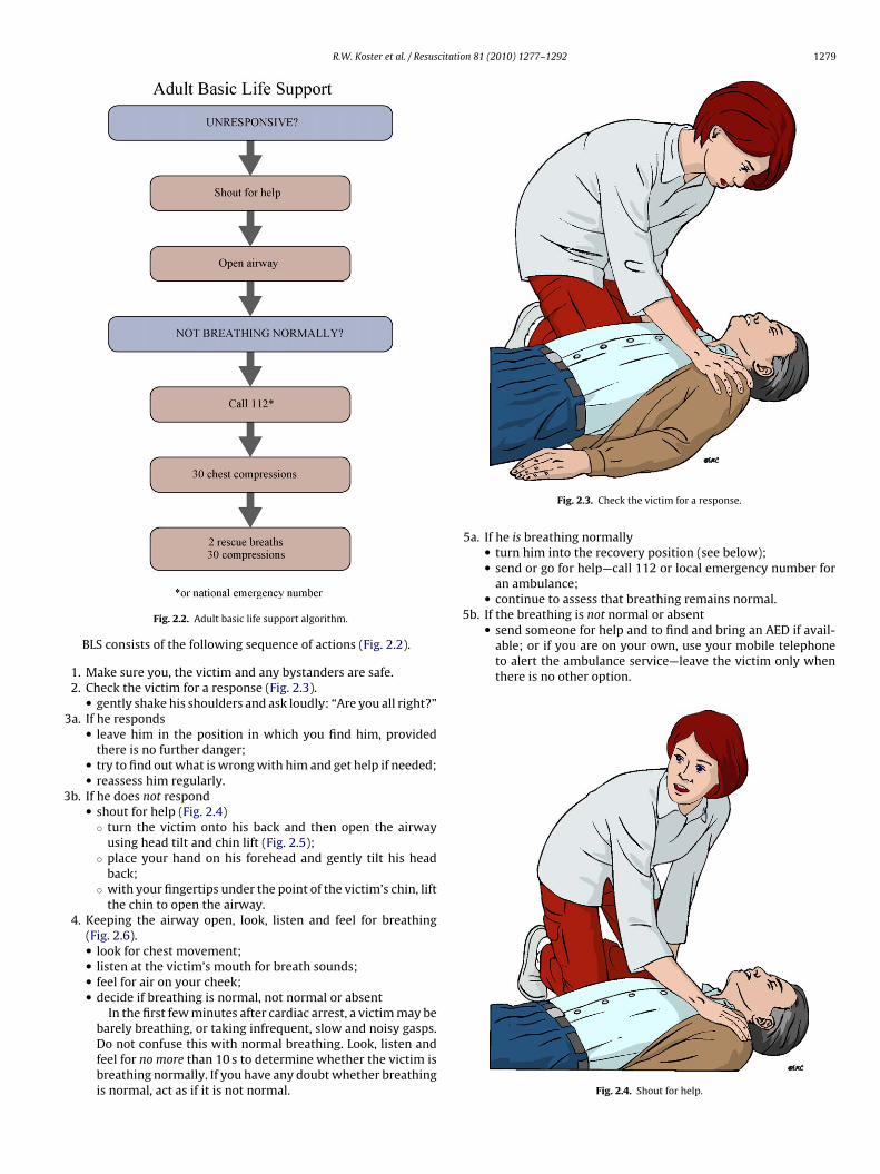
5b. If the breathing is not normal or absent• send someone for help and to find and bring an AED if avail-
able; or if you are on your own, use your mobile telephoneto alert the ambulance service—leave the victim only whenthere is no other option.
Fig. 2.2. Adult basic life support algorithm.
BLS consists of the following sequence of actions (Fig. 2.2).
1. Make sure you, the victim and any bystanders are safe.2. Check the victim for a response (Fig. 2.3).
• gently shake his shoulders and ask loudly: “Are you all right?”a. If he responds
• leave him in the position in which you find him, providedthere is no further danger;
• try to find out what is wrong with him and get help if needed;• reassess him regularly.
b. If he does not respond• shout for help (Fig. 2.4)
◦ turn the victim onto his back and then open the airwayusing head tilt and chin lift (Fig. 2.5);
◦ place your hand on his forehead and gently tilt his headback;
◦ with your fingertips under the point of the victim’s chin, liftthe chin to open the airway.
4. Keeping the airway open, look, listen and feel for breathing(Fig. 2.6).• look for chest movement;• listen at the victim’s mouth for breath sounds;• feel for air on your cheek;• decide if breathing is normal, not normal or absent
In the first few minutes after cardiac arrest, a victim may be
barely breathing, or taking infrequent, slow and noisy gasps.Do not confuse this with normal breathing. Look, listen andfeel for no more than 10 s to determine whether the victim isbreathing normally. If you have any doubt whether breathingis normal, act as if it is not normal.Fig. 2.3. Check the victim for a response.
5a. If he is breathing normally• turn him into the recovery position (see below);• send or go for help—call 112 or local emergency number for
an ambulance;• continue to assess that breathing remains normal.
Fig. 2.4. Shout for help.

1280 R.W. Koster et al. / Resuscitation 81 (2010) 1277–1292
Fig. 2.5. Head tilt and chin lift.
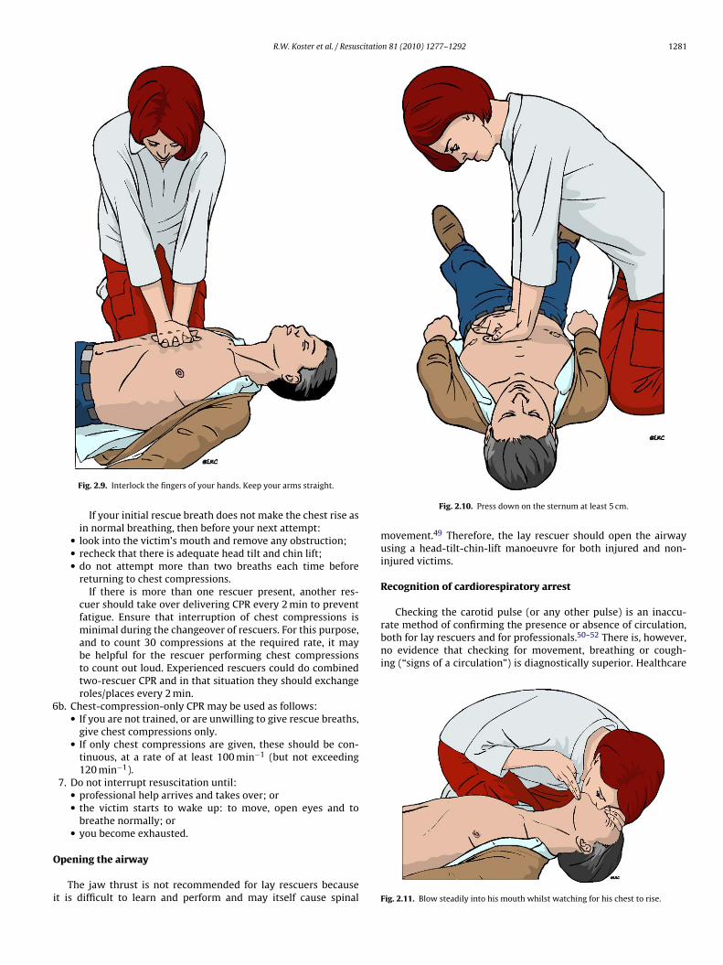
• start chest compression as follows:◦ kneel by the side of the victim;◦ place the heel of one hand in the centre of the victim’s
chest; (which is the lower half of the victim’s breastbone(sternum)) (Fig. 2.7);
◦ place the heel of your other hand on top of the first hand(Fig. 2.8);
◦ interlock the fingers of your hands and ensure that pres-sure is not applied over the victim’s ribs. Keep your armsstraight (Fig. 2.9). Do not apply any pressure over the upperabdomen or the bottom end of the bony sternum (breast-bone);
◦ position yourself vertically above the victim’s chest andpress down on the sternum at least 5 cm (but not exceeding6 cm) (Fig. 2.10);
◦ after each compression, release all the pressure on thechest without losing contact between your hands and the
Fig. 2.6. Look, listen and feel for normal breathing.
Fig. 2.7. Place the heel of one hand in the centre of the victim’s chest.
sternum; repeat at a rate of at least 100 min−1 (but notexceeding 120 min−1);
◦ compression and release should take equal amounts oftime.
6a. Combine chest compression with rescue breaths.• After 30 compressions open the airway again using head tilt
and chin lift (Fig. 2.5).• Pinch the soft part of the nose closed, using the index finger
and thumb of your hand on the forehead.• Allow the mouth to open, but maintain chin lift.• Take a normal breath and place your lips around his mouth,
making sure that you have a good seal.• Blow steadily into the mouth while watching for the chest to
rise (Fig. 2.11), taking about 1 s as in normal breathing; this isan effective rescue breath.
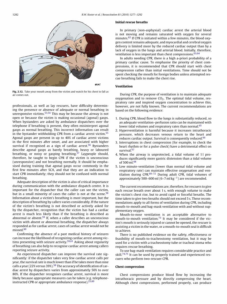
• Maintaining head tilt and chin lift, take your mouth away fromthe victim and watch for the chest to fall as air comes out(Fig. 2.12).
• Take another normal breath and blow into the victim’s mouthonce more to achieve a total of two effective rescue breaths.The two breaths should not take more than 5 s in all. Thenreturn your hands without delay to the correct position onthe sternum and give a further 30 chest compressions.
• Continue with chest compressions and rescue breaths in a
ratio of 30:2.• Stop to recheck the victim only if he starts to wake up: tomove, open eyes and to breathe normally. Otherwise, do notinterrupt resuscitation.
Fig. 2.8. Place the heel of your other hand on top of the first hand.
R.W. Koster et al. / Resuscitation 81 (2010) 1277–1292 1281
6
O
i
Checking the carotid pulse (or any other pulse) is an inaccu-rate method of confirming the presence or absence of circulation,both for lay rescuers and for professionals.50–52 There is, however,no evidence that checking for movement, breathing or cough-ing (“signs of a circulation”) is diagnostically superior. Healthcare
Fig. 2.9. Interlock the fingers of your hands. Keep your arms straight.
If your initial rescue breath does not make the chest rise asin normal breathing, then before your next attempt:
• look into the victim’s mouth and remove any obstruction;• recheck that there is adequate head tilt and chin lift;• do not attempt more than two breaths each time before
returning to chest compressions.If there is more than one rescuer present, another res-
cuer should take over delivering CPR every 2 min to preventfatigue. Ensure that interruption of chest compressions isminimal during the changeover of rescuers. For this purpose,and to count 30 compressions at the required rate, it maybe helpful for the rescuer performing chest compressionsto count out loud. Experienced rescuers could do combinedtwo-rescuer CPR and in that situation they should exchangeroles/places every 2 min.
b. Chest-compression-only CPR may be used as follows:• If you are not trained, or are unwilling to give rescue breaths,
give chest compressions only.• If only chest compressions are given, these should be con-
tinuous, at a rate of at least 100 min−1 (but not exceeding120 min−1).
7. Do not interrupt resuscitation until:• professional help arrives and takes over; or• the victim starts to wake up: to move, open eyes and to
breathe normally; or• you become exhausted.
pening the airway
The jaw thrust is not recommended for lay rescuers becauset is difficult to learn and perform and may itself cause spinal
Fig. 2.10. Press down on the sternum at least 5 cm.
movement.49 Therefore, the lay rescuer should open the airwayusing a head-tilt-chin-lift manoeuvre for both injured and non-injured victims.
Recognition of cardiorespiratory arrest
Fig. 2.11. Blow steadily into his mouth whilst watching for his chest to rise.
1282 R.W. Koster et al. / Resuscitatio
Fa
piuoWtgiAisdbt(sfisb
dibIdobaavrm
ctor
nycd8li
Chest compression
ig. 2.12. Take your mouth away from the victim and watch for his chest to fall asir comes out.
rofessionals, as well as lay rescuers, have difficulty determin-ng the presence or absence of adequate or normal breathing innresponsive victims.53,54 This may be because the airway is notpen or because the victim is making occasional (agonal) gasps.hen bystanders are asked by ambulance dispatchers over the
elephone if breathing is present, they often misinterpret agonalasps as normal breathing. This incorrect information can resultn the bystander withholding CPR from a cardiac arrest victim.55
gonal gasps are present in up to 40% of cardiac arrest victimsn the first minutes after onset, and are associated with higherurvival if recognised as a sign of cardiac arrest.56 Bystandersescribe agonal gasps as barely breathing, heavy or labouredreathing, or noisy or gasping breathing.57 Laypeople should,herefore, be taught to begin CPR if the victim is unconsciousunresponsive) and not breathing normally. It should be empha-ised during training that agonal gasps occur commonly in therst few minutes after SCA, and that they are an indication totart CPR immediately; they should not be confused with normalreathing.
Adequate description of the victim is also of critical importanceuring communication with the ambulance dispatch centre. It is
mportant for the dispatcher that the caller can see the victim,ut in a small minority of cases the caller is not at the scene.58
nformation about a victim’s breathing is most important, but theescription of breathing by callers varies considerably. If the naturef the victim’s breathing is not described or actively asked fory the dispatcher, recognition that the victim has had a cardiacrrest is much less likely than if the breathing is described asbnormal or absent.59 If, when a caller describes an unconsciousictim with absent or abnormal breathing, the dispatcher alwaysesponded as for cardiac arrest, cases of cardiac arrest would not beissed.60
Confirming the absence of a past medical history of seizuresan increase the likelihood of recognizing cardiac arrest among vic-ims presenting with seizure activity.59,61 Asking about regularityf breathing can also help to recognise cardiac arrest among callerseporting seizure activity.
An experienced dispatcher can improve the survival rate sig-ificantly: if the dispatcher takes very few cardiac arrest calls perear, the survival rate is much lower than if he takes more than ninealls a year (22% versus 39%).58 The accuracy of identification of car-
iac arrest by dispatchers varies from approximately 50% to over0%. If the dispatcher recognises cardiac arrest, survival is moreikely because appropriate measures can be taken (e.g. telephone-nstructed CPR or appropriate ambulance response).25,60
n 81 (2010) 1277–1292
Initial rescue breaths
In primary (non-asphyxial) cardiac arrest the arterial bloodis not moving and remains saturated with oxygen for severalminutes.62 If CPR is initiated within a few minutes, the blood oxy-gen content remains adequate, and myocardial and cerebral oxygendelivery is limited more by the reduced cardiac output than by alack of oxygen in the lungs and arterial blood. Initially, therefore,ventilation is less important than chest compressions.63,64
In adults needing CPR, there is a high a-priori probability of aprimary cardiac cause. To emphasise the priority of chest com-pressions, it is recommended that CPR should start with chestcompression rather than initial ventilations. Time should not bespent checking the mouth for foreign bodies unless attempted res-cue breathing fails to make the chest rise.
Ventilation
During CPR, the purpose of ventilation is to maintain adequateoxygenation and to remove CO2. The optimal tidal volume, res-piratory rate and inspired oxygen concentration to achieve this,however, are not fully known. The current recommendations arebased on the following evidence:
1. During CPR, blood flow to the lungs is substantially reduced, soan adequate ventilation–perfusion ratio can be maintained withlower tidal volumes and respiratory rates than normal.65
2. Hyperventilation is harmful because it increases intrathoracicpressure, which decreases venous return to the heart andreduces cardiac output. Survival is consequently reduced.66
3. Interruptions in chest compression (for example, to check theheart rhythm or for a pulse check) have a detrimental effect onsurvival.67
4. When the airway is unprotected, a tidal volume of 1 l pro-duces significantly more gastric distension than a tidal volumeof 500 ml.68
5. Low minute-ventilation (lower than normal tidal volume andrespiratory rate) can maintain effective oxygenation and ven-tilation during CPR.69–72 During adult CPR, tidal volumes ofapproximately 500–600 ml (6–7 ml kg−1) are recommended.
The current recommendations are, therefore, for rescuers to giveeach rescue breath over about 1 s, with enough volume to makethe victim’s chest rise, but to avoid rapid or forceful breaths. Thetime taken to give two breaths should not exceed 5 s. These recom-mendations apply to all forms of ventilation during CPR, includingmouth-to-mouth and bag-mask ventilation with and without sup-plementary oxygen.
Mouth-to-nose ventilation is an acceptable alternative tomouth-to-mouth ventilation.73 It may be considered if the vic-tim’s mouth is seriously injured or cannot be opened, the rescuer isassisting a victim in the water, or a mouth-to-mouth seal is difficultto achieve.
There is no published evidence on the safety, effectiveness orfeasibility of mouth-to-tracheostomy ventilation, but it may beused for a victim with a tracheostomy tube or tracheal stoma whorequires rescue breathing.
To use bag-mask ventilation requires considerable practice andskill.74,75 It can be used by properly trained and experienced res-cuers who perform two-rescuer CPR.
Chest compressions produce blood flow by increasing theintrathoracic pressure and by directly compressing the heart.Although chest compressions, performed properly, can produce

citatio
srdci
pcR
1
23
4
5
6
7
H
tthopUr
C
srt1erstqe
C
qrathntbe6
fii
R.W. Koster et al. / Resus
ystolic arterial pressure peaks of 60–80 mm Hg, diastolic pressureemains low and mean arterial pressure in the carotid artery sel-om exceeds 40 mm Hg.76 Chest compressions generate a small butritical amount of blood flow to the brain and myocardium andncrease the likelihood that defibrillation will be successful.
Since the 2005 Guidelines were published, chest compressionrompt/feedback devices have generated new data from victims inardiac arrest that supplement animal and manikin studies.77–81
ecommendations based on this evidence are:
. Each time compressions are resumed, place your hands withoutdelay ‘in the centre of the chest’.
. Compress the chest at a rate of at least 100 min−1.
. Ensure that the full compression depth of at least 5 cm (for anadult) is achieved.
. Allow the chest to recoil completely after each compression, i.e.do not lean on the chest during the relaxation phase of the chestcompression.
. Take approximately the same amount of time for compressionas relaxation.
. Minimise interruptions in chest compression in order to ensurethe victim receives at least 60 compressions in each minute.
. Do not rely on feeling the carotid or other pulse as a gauge ofeffective arterial flow during chest compressions.50,82
and position
For adults receiving chest compressions, rescuers should placeheir hands on the lower half of the sternum. It is recommendedhat this location be taught in a simplified way, such as, “place theeel of your hand in the centre of the chest with the other handn top.” This instruction should be accompanied by demonstratinglacing the hands on the lower half of the sternum on a manikin.se of the internipple line as a landmark for hand placement is not
eliable.83,84
ompression rate
There is a positive relationship between the number of compres-ions actually delivered per minute and the chance of successfulesuscitation.81 While the compression rate (the speed at whichhe 30 consecutive compressions are given) should be at least00 min−1, the actual number of compressions delivered duringach minute of CPR will be lower due to interruptions to deliverescue breaths and allow AED analysis, etc. In one out-of-hospitaltudy, rescuers recorded compression rates of 100–120 min−1 buthe mean number of compressions was reduced to 64 min−1 by fre-uent interruptions.79 At least 60 compressions should be deliveredach minute.
ompression depth
Fear of doing harm, fatigue and limited muscle strength fre-uently result in rescuers compressing the chest less deeply thanecommended. There is evidence that a compression depth of 5 cmnd over results in a higher rate of return of spontaneous circula-ion (ROSC), and a higher percentage of victims admitted alive toospital, than a compression depth of 4 cm or below.77,78 There iso direct evidence that damage from chest compression is relatedo compression depth, nor has an upper limit of compression deptheen established in studies. Nevertheless, it is recommended that,
ven in large adults, chest compression depth should not exceedcm.CPR should be performed on a firm surface when possible. Air-lled mattresses should be routinely deflated during CPR.85 There
s no evidence for or against the use of backboards,86,87 but if one
n 81 (2010) 1277–1292 1283
is used, care should be taken to avoid interruption in CPR and dis-lodging intravenous lines or other tubes during board placement.
Chest decompression
Allowing complete recoil of the chest after each compressionresults in better venous return to the chest and may improvethe effectiveness of CPR.88,89 The optimal method of achievingthis goal, without compromising other aspects of chest compres-sion technique such as compression depth, has not, however, beenestablished.
Feedback on compression technique
Rescuers can be assisted to achieve the recommended compres-sion rate and depth by prompt/feedback devices that are either builtinto the AED or manual defibrillator, or are stand-alone devices. Theuse of such prompt/feedback devices, as part of an overall strategyto improve the quality of CPR, may be beneficial. Rescuers should beaware that the accuracy of devices that measure compression depthvaries according to the stiffness of the support surface upon whichCPR is being performed (e.g. floor/mattress), and may overestimatecompression depth.87 Further studies are needed to determine ifthese devices improve victim outcomes.
Compression–ventilation ratio
Animal data supported an increase in the ratio of compressionto ventilation to >15:2.90–92 A mathematical model suggests thata ratio of 30:2 provides the best compromise between blood flowand oxygen delivery.93,94 A ratio of 30 compressions to 2 venti-lations was recommended in the Guidelines 2005 for the singlerescuer attempting resuscitation of an adult or child out of hospi-tal, an exception being that a trained healthcare professional shoulduse a ratio of 15:2 for a child. This decreased the number of inter-ruptions in compression and the no-flow fraction,95,96 and reducedthe likelihood of hyperventilation.66,97 Direct evidence that sur-vival rates have increased from this change, however, is lacking.Likewise, there is no new evidence that would suggest a change inthe recommended compression to ventilation ratio of 30:2.
Compression-only CPR
Some healthcare professionals as well as lay rescuers indicatethat they would be reluctant to perform mouth-to-mouth ventila-tion, especially in unknown victims of cardiac arrest.98,99 Animalstudies have shown that chest-compression-only CPR may be aseffective as combined ventilation and compression in the first fewminutes after non-asphyxial arrest.63,100 If the airway is open,occasional gasps and passive chest recoil may provide some airexchange, but this may result in ventilation of the dead spaceonly.56,101–103 Animal and mathematical model studies of chest-compression-only CPR have shown that arterial oxygen storesdeplete in 2–4 min.92,104
In adults, the outcome of chest compression without venti-lation is significantly better than the outcome of giving no CPRat all in non-asphyxial arrest.22,23 Several studies of human car-diac arrest have suggested equivalence of chest-compression-onlyCPR and chest compressions combined with rescue breaths, butnone excluded the possibility that chest-compression-only is infe-
rior to chest compressions combined with ventilations.23,105 Onestudy suggested superiority of chest-compression-only CPR.22 Allthese studies have significant limitations because they were basedon retrospective database analyses, where the type of BLS wasnot controlled and did not include CPR according to Guidelines
1 citatio
2scliinc
tacra
C
ts
R
tpbcfCeth
R
P
hsmTaueg(s
R
dsrbodr
R
tA
Recovery position
284 R.W. Koster et al. / Resus
005 (30:2 compressions to ventilation ratio). Chest compres-ion alone may be sufficient only in the first few minutes afterollapse. Professional help can be expected on average 8 min orater after a call for help, and chest compression only will resultn insufficient CPR in many cases. Chest-compression-only CPRs not as effective as conventional CPR for cardiac arrests ofon-cardiac origin (e.g., drowning or suffocation) in adults andhildren.106,107
Chest compression combined with rescue breaths is, therefore,he method of choice for CPR delivered by both trained lay rescuersnd professionals. Laypeople should be encouraged to performompression-only CPR if they are unable or unwilling to provideescue breaths, or when instructed during an emergency call to anmbulance dispatcher centre.26,27
PR in confined spaces
Over-the-head CPR for single rescuers and straddle-CPR forwo rescuers may be considered for resuscitation in confinedpaces.108,109
isks to the victim during CPR
Many rescuers, concerned that delivering chest compressionso a victim who is not in cardiac arrest will cause serious com-lications, do not initiate CPR. In a study of dispatch-assistedystander CPR, however, where non-arrest victims received chestompressions, 12% experienced discomfort but only 2% suffered aracture: no victims suffered visceral organ injury.110 BystanderPR extremely rarely leads to serious harm in victims who areventually found not to be in cardiac arrest. Rescuers should not,herefore, be reluctant to initiate CPR because of concern of causingarm.
isks to the rescuer during training and during real-life CPR
hysical effects
Observational studies of training or actual CPR performanceave described rare occurrences of muscle strain, back symptoms,hortness of breath, hyperventilation, and case reports of pneu-othorax, chest pain, myocardial infarction and nerve injury.111,112
he incidence of these events is very low, and CPR training andctual performance is safe in most circumstances.113 Individualsndertaking CPR training should be advised of the nature andxtent of the physical activity required during the training pro-ramme. Learners and rescuers who develop significant symptomse.g. chest pain or severe shortness of breath) during CPR traininghould be advised to stop.
escuer fatigue
Several manikin studies have found that chest compressionepth can decrease as little as 2 min after starting chest compres-ions. An in-hospital patient study showed that, even while usingeal-time feedback, the mean depth of compression deterioratedetween 1.5 and 3 min after starting CPR.114 It is therefore rec-mmended that rescuers change about every 2 min to prevent aecrease in compression quality due to rescuer fatigue. Changingescuers should not interrupt chest compressions.
isks during defibrillation
A large randomised trial of public access defibrillation showedhat AEDs can be used safely by laypeople and first responders.115
systematic review identified eight papers that reported a total
n 81 (2010) 1277–1292
of 29 adverse events associated with defibrillation.116 The causesincluded accidental or intentional defibrillator misuse, device mal-function and accidental discharge during training or maintenanceprocedures. Four single-case reports described shocks to rescuersfrom discharging implantable cardioverter defibrillators (ICDs), inone case resulting in a peripheral nerve injury. There are no reportsof harm to rescuers from attempting defibrillation in wet environ-ments.
Injury to the rescuer from defibrillation is extremely rare. Never-theless, rescuers should not continue manual chest compressionsduring shock delivery. Victims should not be touched during ICDdischarge. Direct contact between the rescuer and the victim shouldbe avoided when defibrillation is carried out in wet environments.
Psychological effects
One large, prospective trial of public access defibrillationreported a few adverse psychological effects associated with CPRor AED use that required intervention.113 Two large, retrospec-tive, questionnaire-based reports relating to performance of CPRby a bystander reported that nearly all respondents regarded theirintervention as a positive experience.117,118 The rare occurrencesof adverse psychological effects in rescuers after CPR should berecognised and managed appropriately.
Disease transmission
There are only very few cases reported where performing CPRhas been linked to disease transmission, implicating Salmonellainfantis, Staphylococcus aureus, severe acute respiratory syn-drome (SARS), meningococcal meningitis, Helicobacter pylori,Herpes simplex virus, cutaneous tuberculosis, stomatitis, tracheitis,Shigella and Streptococcus pyogenes. One report described her-pes simplex virus infection as a result of training in CPR. Onesystematic review found that in the absence of high-risk activ-ities, such as intravenous cannulation, there were no reports oftransmission of hepatitis B, hepatitis C, human immunodeficiencyvirus (HIV) or cytomegalovirus during either training or actualCPR.119
The risk of disease transmission during training and actual CPRperformance is extremely low. Wearing gloves during CPR is rea-sonable, but CPR should not be delayed or withheld if gloves are notavailable. Rescuers should take appropriate safety precautions if avictim is known to have a serious infection (e.g. HIV, tuberculosis,hepatitis B virus or SARS).
Barrier devices
No human studies have addressed the safety, effectiveness orfeasibility of using barrier devices (such as a face shield or facemask) to prevent victim contact during rescuer breathing. Twostudies showed that barrier devices decreased transmission of bac-teria in controlled laboratory settings.120,121 Because the risk ofdisease transmission is very low, initiating rescue breathing with-out a barrier device is reasonable. If the victim is known to have aserious infection (e.g. HIV, tuberculosis, hepatitis B virus, or SARS)a barrier device is recommended.
There are several variations of the recovery position, eachwith its own advantages. No single position is perfect for allvictims.122,123 The position should be stable, near to a true lateralposition with the head dependent, and with no pressure on the

R.W. Koster et al. / Resuscitation 81 (2010) 1277–1292 1285
Fig. 2.13. Place the arm nearest to you out at right angles to his body, elbow bentwith the hand palm uppermost.
Ft
cs
••
•
•
•
•
••
•
to
Fig. 2.15. With your other hand, grasp the far leg just above the knee and pull it up,keeping the foot on the ground.
important to ask the conscious victim “Are you choking?”
Table 2.1Differentiation between mild and severe foreign body airway obstruction (FBAO).a
Sign Mild obstruction Severe obstruction
ig. 2.14. Bring the far arm across the chest, and hold the back of the hand againsthe victim’s cheek nearest to you.
hest to impair breathing.124The ERC recommends the followingequence of actions to place a victim in the recovery position:
Kneel beside the victim and make sure that both legs are straight.Place the arm nearest to you out at right angles to the body, elbowbent with the hand palm uppermost (Fig. 2.13).Bring the far arm across the chest, and hold the back of the handagainst the victim’s cheek nearest to you (Fig. 2.14).With your other hand, grasp the far leg just above the knee andpull it up, keeping the foot on the ground (Fig. 2.15).Keeping the hand pressed against the cheek, pull on the far leg toroll the victim towards you onto his side.Adjust the upper leg so that both hip and knee are bent at rightangles.Tilt the head back to make sure the airway remains open.Adjust the hand under the cheek, if necessary, to keep the headtilted and facing downwards to allow liquid material to drainfrom the mouth (Fig. 2.16).Check breathing regularly.
If the victim has to be kept in the recovery position for morehan 30 min, turn him to the opposite side to relieve the pressuren the lower arm.
Fig. 2.16. The recovery position completed. Keep the head tilted to keep the airwayopen. Keep the face downward to allow fluids to go out.
Foreign-body airway obstruction (choking)
Foreign-body airway obstruction (FBAO) is an uncommon butpotentially treatable cause of accidental death.125 As most chokingevents are associated with eating, they are commonly witnessed.Thus, there is often the opportunity for early intervention while thevictim is still responsive.
Recognition
Because recognition of airway obstruction is the key to success-ful outcome, it is important not to confuse this emergency withfainting, myocardial infarction, seizure or other conditions that maycause sudden respiratory distress, cyanosis or loss of consciousness.Foreign bodies may cause either mild or severe airway obstruction.The signs and symptoms enabling differentiation between mildand severe airway obstruction are summarised in Table 2.1. It is
“Are you choking?” “Yes” Unable to speak, may nodOther signs Can speak, cough,
breatheCannot breathe/wheezybreathing/silent attempts tocough/unconsciousness
a General signs of FBAO: attack occurs while eating; victim may clutch his neck.
1286 R.W. Koster et al. / Resuscitation 81 (2010) 1277–1292
way o
As(
1
2
3
F
masso
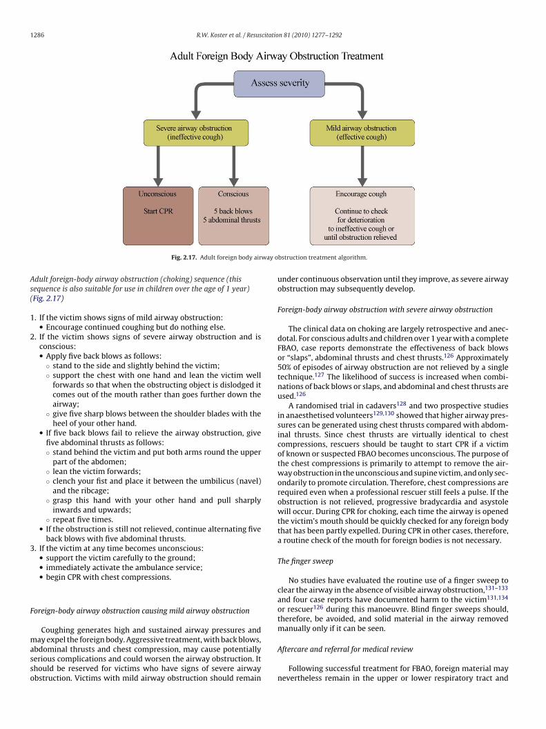
Fig. 2.17. Adult foreign body air
dult foreign-body airway obstruction (choking) sequence (thisequence is also suitable for use in children over the age of 1 year)Fig. 2.17)
. If the victim shows signs of mild airway obstruction:• Encourage continued coughing but do nothing else.
. If the victim shows signs of severe airway obstruction and isconscious:• Apply five back blows as follows:
◦ stand to the side and slightly behind the victim;◦ support the chest with one hand and lean the victim well
forwards so that when the obstructing object is dislodged itcomes out of the mouth rather than goes further down theairway;
◦ give five sharp blows between the shoulder blades with theheel of your other hand.
• If five back blows fail to relieve the airway obstruction, givefive abdominal thrusts as follows:◦ stand behind the victim and put both arms round the upper
part of the abdomen;◦ lean the victim forwards;◦ clench your fist and place it between the umbilicus (navel)
and the ribcage;◦ grasp this hand with your other hand and pull sharply
inwards and upwards;◦ repeat five times.
• If the obstruction is still not relieved, continue alternating fiveback blows with five abdominal thrusts.
. If the victim at any time becomes unconscious:• support the victim carefully to the ground;• immediately activate the ambulance service;• begin CPR with chest compressions.
oreign-body airway obstruction causing mild airway obstruction
Coughing generates high and sustained airway pressures anday expel the foreign body. Aggressive treatment, with back blows,
bdominal thrusts and chest compression, may cause potentiallyerious complications and could worsen the airway obstruction. Ithould be reserved for victims who have signs of severe airwaybstruction. Victims with mild airway obstruction should remain
bstruction treatment algorithm.
under continuous observation until they improve, as severe airwayobstruction may subsequently develop.
Foreign-body airway obstruction with severe airway obstruction
The clinical data on choking are largely retrospective and anec-dotal. For conscious adults and children over 1 year with a completeFBAO, case reports demonstrate the effectiveness of back blowsor “slaps”, abdominal thrusts and chest thrusts.126 Approximately50% of episodes of airway obstruction are not relieved by a singletechnique.127 The likelihood of success is increased when combi-nations of back blows or slaps, and abdominal and chest thrusts areused.126
A randomised trial in cadavers128 and two prospective studiesin anaesthetised volunteers129,130 showed that higher airway pres-sures can be generated using chest thrusts compared with abdom-inal thrusts. Since chest thrusts are virtually identical to chestcompressions, rescuers should be taught to start CPR if a victimof known or suspected FBAO becomes unconscious. The purpose ofthe chest compressions is primarily to attempt to remove the air-way obstruction in the unconscious and supine victim, and only sec-ondarily to promote circulation. Therefore, chest compressions arerequired even when a professional rescuer still feels a pulse. If theobstruction is not relieved, progressive bradycardia and asystolewill occur. During CPR for choking, each time the airway is openedthe victim’s mouth should be quickly checked for any foreign bodythat has been partly expelled. During CPR in other cases, therefore,a routine check of the mouth for foreign bodies is not necessary.
The finger sweep
No studies have evaluated the routine use of a finger sweep toclear the airway in the absence of visible airway obstruction,131–133
and four case reports have documented harm to the victim131,134
or rescuer126 during this manoeuvre. Blind finger sweeps should,therefore, be avoided, and solid material in the airway removedmanually only if it can be seen.
Aftercare and referral for medical review
Following successful treatment for FBAO, foreign material maynevertheless remain in the upper or lower respiratory tract and
citatio
cciAst
Rv
c2pcsreir
R.W. Koster et al. / Resus
ause complications later. Victims with a persistent cough, diffi-ulty swallowing or the sensation of an object being still stuckn the throat should, therefore, be referred for a medical opinion.bdominal thrusts and chest compressions can potentially causeerious internal injuries, and all victims successfully treated withhese measures should be examined afterwards for injury.
esuscitation of children (see also Section 6)134a andictims of drowning (see also Section 8c)134b
For victims of primary cardiac arrest who receive chest-ompression-only CPR, oxygen stores become depleted about–4 min after initiation of CPR.92,104 The combination of chest com-ressions with ventilation then becomes critically important. Afterollapse from asphyxial arrest, a combination of chest compres-
ions with ventilations is important immediately after the start ofesuscitation. Previous guidelines have tried to address this differ-nce in pathophysiology, and have recommended that victims ofdentifiable asphyxia (drowning, intoxication) and children shouldeceive 1 min of CPR before the lone rescuer leaves the victim to getFig. 2.18. Algorithm for use of an automate
n 81 (2010) 1277–1292 1287
help. The majority of cases of SCA out of hospital, however, occur inadults, and although the rate of VF as the first recorded rhythm hasdeclined over recent years, the cause of adult cardiac arrest remainsVF in most cases (59%) when documented in the earliest phase by anAED.13 In children, VF is much less common as the primary cardiacarrest rhythm (approximately 7%).135 These additional recommen-dations, therefore, added to the complexity of the guidelines whileaffecting only a minority of victims.
It is important to be aware that many children do not receiveresuscitation because potential rescuers fear causing harm if theyare not specifically trained in resuscitation for children. This fearis unfounded; it is far better to use the adult BLS sequence forresuscitation of a child than to do nothing. For ease of teaching andretention laypeople should be taught that the adult sequence mayalso be used for children who are not responsive and not breathingor not breathing normally.
The following minor modifications to the adult sequence willmake it even more suitable for use in children.
• Give 5 initial rescue breaths before starting chest compressions(adult sequence of actions, 5b).
d external defibrillator. © 2010 ERC.

1 citation 81 (2010) 1277–1292
•
•
bvt(tpvB
U
aAiaoRdi
Fttifylcc
S
12
3
4
5
5
• the victim starts to wake up: moves, open eyes and breathesnormally;
• you become exhausted.
288 R.W. Koster et al. / Resus
A lone rescuer should perform CPR for approximately 1 minbefore going for help.Compress the chest by at least one third of its depth; use 2 fingersfor an infant under 1 year; use 1 or 2 hands for a child over 1 yearas needed to achieve an adequate depth of compression.
The same modifications of 5 initial breaths and 1 min of CPRy the lone rescuer before getting help, may improve outcome forictims of drowning. This modification should be taught only tohose who have a specific duty of care to potential drowning victimse.g. lifeguards). Drowning is easily identified. It can be difficult, onhe other hand, for a layperson to determine whether cardiores-iratory arrest is a direct result of trauma or intoxication. Theseictims should, therefore, be managed according to the standardLS protocols.
se of an automated external defibrillator
Section 3 discusses the guidelines for defibrillation using bothutomated external defibrillators (AEDs) and manual defibrillators.EDs are safe and effective when used by laypeople, and make
t possible to defibrillate many minutes before professional helprrives. Rescuers should continue CPR with minimal interruptionf chest compressions while applying an AED and during its use.escuers should concentrate on following the voice prompts imme-iately they are received, in particular, resuming CPR as soon as
nstructed.Standard AEDs are suitable for use in children older than 8 years.
or children between 1 and 8 years paediatric pads should be used,ogether with an attenuator or a paediatric mode if available; ifhese are not available, the AED should be used as it is. Use of AEDss not recommended for children <1 year. There are, however, aew case reports describing the use of AEDs in children aged <1ear.136,137 The incidence of shockable rhythms in infants is veryow except when there is cardiac disease135,138,139; in these rareases, if an AED is the only defibrillator available its use should beonsidered (preferably with dose attenuator).
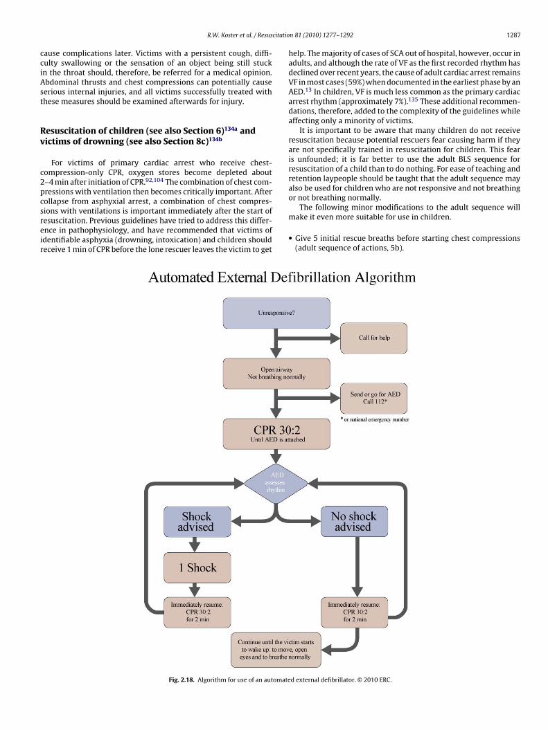
equence for use of an AED
See Fig. 2.18
. Make sure you, the victim, and any bystanders are safe.
. Follow the Adult BLS sequence (steps 1–5).• if the victim is unresponsive and not breathing normally, send
someone for help and to find and bring an AED if available;• if you are on your own, use your mobile telephone to alert
the ambulance service—leave the victim only when there is noother option.
. Start CPR according to the adult BLS sequence. If you are on yourown and the AED is in your immediate vicinity, start by applyingthe AED.
. As soon as the AED arrives• switch on the AED and attach the electrode pads on the victim’s
bare chest (Fig. 2.19);• if more than one rescuer is present, CPR should be continued
while electrode pads are being attached to the chest;• follow the spoken/visual directions immediately;• ensure that nobody is touching the victim while the AED is
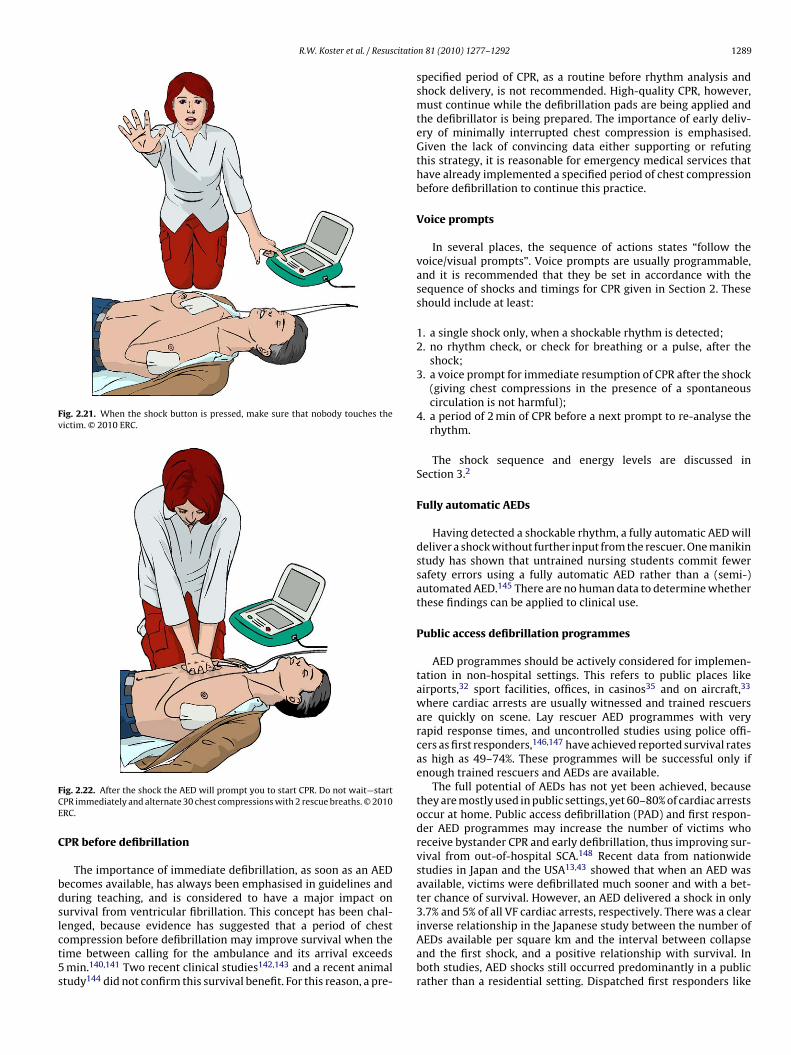
analysing the rhythm (Fig. 2.20).a. If a shock is indicated
• ensure that nobody is touching the victim (Fig. 2.21);• push shock button as directed (fully automatic AEDs will
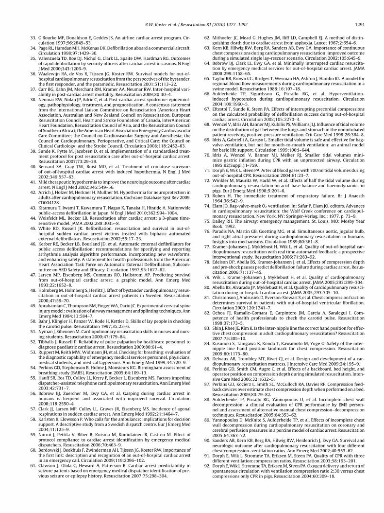
deliver the shock automatically);• immediately restart CPR 30:2 (Fig. 2.22);• continue as directed by the voice/visual prompts.
Fig. 2.19. Attaching the electrode pads. Place the first electrode pad in the mid-axillary line just below the armpit. Place the second electrode just below the rightcollarbone (clavicle). © 2010 ERC.
b. If no shock is indicated• immediately resume CPR, using a ratio of 30 compressions to
2 rescue breaths;• continue as directed by the voice/visual prompts.
6. Continue to follow the AED prompts until• professional help arrives and takes over;
Fig. 2.20. While the AED analyses the heart rhythm, nobody should touch the victim.© 2010 ERC.
R.W. Koster et al. / Resuscitatio
Fig. 2.21. When the shock button is pressed, make sure that nobody touches thevictim. © 2010 ERC.
FCE
C
bdslct5s
ig. 2.22. After the shock the AED will prompt you to start CPR. Do not wait—startPR immediately and alternate 30 chest compressions with 2 rescue breaths. © 2010RC.
PR before defibrillation
The importance of immediate defibrillation, as soon as an AEDecomes available, has always been emphasised in guidelines anduring teaching, and is considered to have a major impact onurvival from ventricular fibrillation. This concept has been chal-
enged, because evidence has suggested that a period of chestompression before defibrillation may improve survival when theime between calling for the ambulance and its arrival exceedsmin.140,141 Two recent clinical studies142,143 and a recent animaltudy144 did not confirm this survival benefit. For this reason, a pre-
n 81 (2010) 1277–1292 1289
specified period of CPR, as a routine before rhythm analysis andshock delivery, is not recommended. High-quality CPR, however,must continue while the defibrillation pads are being applied andthe defibrillator is being prepared. The importance of early deliv-ery of minimally interrupted chest compression is emphasised.Given the lack of convincing data either supporting or refutingthis strategy, it is reasonable for emergency medical services thathave already implemented a specified period of chest compressionbefore defibrillation to continue this practice.
Voice prompts
In several places, the sequence of actions states “follow thevoice/visual prompts”. Voice prompts are usually programmable,and it is recommended that they be set in accordance with thesequence of shocks and timings for CPR given in Section 2. Theseshould include at least:
1. a single shock only, when a shockable rhythm is detected;2. no rhythm check, or check for breathing or a pulse, after the
shock;3. a voice prompt for immediate resumption of CPR after the shock
(giving chest compressions in the presence of a spontaneouscirculation is not harmful);
4. a period of 2 min of CPR before a next prompt to re-analyse therhythm.
The shock sequence and energy levels are discussed inSection 3.2
Fully automatic AEDs
Having detected a shockable rhythm, a fully automatic AED willdeliver a shock without further input from the rescuer. One manikinstudy has shown that untrained nursing students commit fewersafety errors using a fully automatic AED rather than a (semi-)automated AED.145 There are no human data to determine whetherthese findings can be applied to clinical use.
Public access defibrillation programmes
AED programmes should be actively considered for implemen-tation in non-hospital settings. This refers to public places likeairports,32 sport facilities, offices, in casinos35 and on aircraft,33
where cardiac arrests are usually witnessed and trained rescuersare quickly on scene. Lay rescuer AED programmes with veryrapid response times, and uncontrolled studies using police offi-cers as first responders,146,147 have achieved reported survival ratesas high as 49–74%. These programmes will be successful only ifenough trained rescuers and AEDs are available.
The full potential of AEDs has not yet been achieved, becausethey are mostly used in public settings, yet 60–80% of cardiac arrestsoccur at home. Public access defibrillation (PAD) and first respon-der AED programmes may increase the number of victims whoreceive bystander CPR and early defibrillation, thus improving sur-vival from out-of-hospital SCA.148 Recent data from nationwidestudies in Japan and the USA13,43 showed that when an AED wasavailable, victims were defibrillated much sooner and with a bet-ter chance of survival. However, an AED delivered a shock in only3.7% and 5% of all VF cardiac arrests, respectively. There was a clear
inverse relationship in the Japanese study between the number ofAEDs available per square km and the interval between collapseand the first shock, and a positive relationship with survival. Inboth studies, AED shocks still occurred predominantly in a publicrather than a residential setting. Dispatched first responders like
1 citatio
pb
glmpit
rldaaralimrh
ais
U
ptrtai
Fc
R
290 R.W. Koster et al. / Resus
olice and fire fighters will, in general, have longer response times,ut have the potential to reach the whole population.
When implementing an AED programme, community and pro-ramme leaders should consider factors such as the strategicocation of AEDs, development of a team with responsibility for
onitoring and maintaining the devices, training and retrainingrogrammes for the individuals who are likely to use the AED, and
dentification of a group of volunteer individuals who are commit-ed to using the AED for victims of cardiac arrest.149
The logistic problem for first responder programmes is that theescuer needs to arrive, not just earlier than the traditional ambu-ance, but within 5–6 min of the initial call, to enable attemptedefibrillation in the electrical or circulatory phase of cardiacrrest.44 With longer delays, the survival benefits decrease:36,47
few minutes’ gain in time will have little impact when a firstesponder arrives more than 10 min after the call,14,150 or when
first responder does not improve on an already short ambu-ance response time.151 However, small reductions in responsentervals achieved by first-responder programmes that impact on
any residential victims may be more cost-effective than the largereductions in response interval achieved by PAD programmes thatave an impact on fewer cardiac arrest victims.152,153
Programmes that make AEDs publicly available in residentialreas have not yet been evaluated. The acquisition of an AED forndividual use at home, even for those considered at high risk ofudden cardiac arrest, has proved not to be effective.154
niversal AED signage
When a collapse occurs, and an AED must be found rapidly, sim-le and clear signage indicating the location of, and the fastest wayo an AED is important. ILCOR has designed an AED sign that may beecognised worldwide and is recommended for indicating the loca-ion of an AED (Fig. 2.23). More detailed information on design andpplication of this AED sign can be found at: https://www.erc.edu/ndex.php/newsItem/en/nid=204/
ig. 2.23. Universal ILCOR signage to indicate presence of an AED. This sign can beombined with arrows to indicate the direction of the nearest AED.
eferences
1. Recommended guidelines for uniform reporting of data from out-of-hospitalcardiac arrest: the ‘Utstein style’. Prepared by a Task Force of Representativesfrom the European Resuscitation Council, American Heart Association, Heart
and Stroke Foundation of Canada, Australian Resuscitation Council. Resuscita-tion 1991;22:1–26.2. Deakin CD, Nolan JP, Sunde K, Koster RW. European Resuscitation CouncilGuidelines for Resuscitation 2010. Section 3. Electrical therapies: automatedexternal defibrillators, defibrillation, cardioversion and pacing. Resuscitation2010;81:1293–304.
n 81 (2010) 1277–1292
3. Deakin CD, Nolan JP, Soar J, et al. European Resuscitation Council Guidelinesfor Resuscitation 2010. Section 4. Adult advanced life support. Resuscitation2010;81:1305–52.
4. Sans S, Kesteloot H, Kromhout D. The burden of cardiovascular diseasesmortality in Europe. Task Force of the European Society of Cardiology onCardiovascular Mortality and Morbidity Statistics in Europe. Eur Heart J1997;18:1231–48.
5. Atwood C, Eisenberg MS, Herlitz J, Rea TD. Incidence of EMS-treated out-of-hospital cardiac arrest in Europe. Resuscitation 2005;67:75–80.
6. Cobb LA, Fahrenbruch CE, Olsufka M, Copass MK. Changing incidence of out-of-hospital ventricular fibrillation, 1980–2000. JAMA 2002;288:3008–13.
7. Rea TD, Pearce RM, Raghunathan TE, et al. Incidence of out-of-hospital cardiacarrest. Am J Cardiol 2004;93:1455–60.
8. Vaillancourt C, Verma A, Trickett J, et al. Evaluating the effectiveness ofdispatch-assisted cardiopulmonary resuscitation instructions. Acad EmergMed 2007;14:877–83.
9. Agarwal DA, Hess EP, Atkinson EJ, White RD. Ventricular fibrilla-tion in Rochester, Minnesota: experience over 18 years. Resuscitation2009;80:1253–8.
10. Ringh M, Herlitz J, Hollenberg J, Rosenqvist M, Svensson L. Out of hospital car-diac arrest outside home in Sweden, change in characteristics, outcome andavailability for public access defibrillation. Scand J Trauma Resusc Emerg Med2009;17:18.
11. Cummins R, Thies W. Automated external defibrillators and the AdvancedCardiac Life Support Program: a new initiative from the American Heart Asso-ciation. Am J Emerg Med 1991;9:91–3.
12. Waalewijn RA, Nijpels MA, Tijssen JG, Koster RW. Prevention of deteriorationof ventricular fibrillation by basic life support during out-of-hospital cardiacarrest. Resuscitation 2002;54:31–6.
13. Weisfeldt ML, Sitlani CM, Ornato JP, et al. Survival after application of auto-matic external defibrillators before arrival of the emergency medical system:evaluation in the resuscitation outcomes consortium population of 21 million.J Am Coll Cardiol 2010;55:1713–20.
14. van Alem AP, Vrenken RH, de Vos R, Tijssen JG, Koster RW. Use of auto-mated external defibrillator by first responders in out of hospital cardiac arrest:prospective controlled trial. BMJ 2003;327:1312.
15. Nolan J, Soar J, Eikeland H. The chain of survival. Resuscitation 2006;71:270–1.16. Muller D, Agrawal R, Arntz HR. How sudden is sudden cardiac death? Circula-
tion 2006;114:1146–50.17. Lowel H, Lewis M, Hormann A. Prognostic significance of prehospital phase in
acute myocardial infarct. Results of the Augsburg Myocardial Infarct Registry,1985–1988. Dtsch Med Wochenschr 1991;116:729–33.
18. Waalewijn RA, Tijssen JG, Koster RW. Bystander initiated actions in out-of-hospital cardiopulmonary resuscitation: results from the AmsterdamResuscitation Study (ARREST). Resuscitation 2001;50:273–9.
19. Valenzuela TD, Roe DJ, Cretin S, Spaite DW, Larsen MP. Estimating effectivenessof cardiac arrest interventions: a logistic regression survival model. Circulation1997;96:3308–13.
20. Holmberg M, Holmberg S, Herlitz J. Factors modifying the effect of bystandercardiopulmonary resuscitation on survival in out-of-hospital cardiac arrestpatients in Sweden. Eur Heart J 2001;22:511–9.
21. Holmberg M, Holmberg S, Herlitz J, Gardelov B. Survival after cardiac arrestoutside hospital in Sweden. Swedish Cardiac Arrest Registry. Resuscitation1998;36:29–36.
22. SOS-KANTO Study Group. Cardiopulmonary resuscitation by bystanderswith chest compression only (SOS-KANTO): an observational study. Lancet2007;369:920–6.
23. Iwami T, Kawamura T, Hiraide A, et al. Effectiveness of bystander-initiatedcardiac-only resuscitation for patients with out-of-hospital cardiac arrest. Cir-culation 2007;116:2900–7.
24. Rea TD, Eisenberg MS, Culley LL, Becker L. Dispatcher-assisted car-diopulmonary resuscitation and survival in cardiac arrest. Circulation2001;104:2513–6.
25. Kuisma M, Boyd J, Vayrynen T, Repo J, Nousila-Wiik M, Holmstrom P. Emer-gency call processing and survival from out-of-hospital ventricular fibrillation.Resuscitation 2005;67:89–93.
26. Rea TD, Fahrenbruch C, Culley L, et al. CPR with chest compresssions alone orwith rescue breathing. N Engl J Med 2010;363:423–33.
27. Svensson L, Bohm K, Castren M, et al. Compression-only CPR or standard CPRin out-of-hospital cardiac arrest. N Engl J Med 2010;363:434–42.
28. Weaver WD, Hill D, Fahrenbruch CE, et al. Use of the automatic external defib-rillator in the management of out-of-hospital cardiac arrest. N Engl J Med1988;319:661–6.
29. Auble TE, Menegazzi JJ, Paris PM. Effect of out-of-hospital defibrillation by basiclife support providers on cardiac arrest mortality: a metaanalysis. Ann EmergMed 1995;25:642–58.
30. Stiell IG, Wells GA, Field BJ, et al. Improved out-of-hospital cardiac arrestsurvival through the inexpensive optimization of an existing defibrillation pro-gram: OPALS study phase II. Ontario prehospital advanced life support. JAMA1999;281:1175–81.
31. Stiell IG, Wells GA, DeMaio VJ, et al. Modifiable factors associatedwith improved cardiac arrest survival in a multicenter basic life sup-port/defibrillation system: OPALS Study Phase I results. Ontario prehospitaladvanced life support. Ann Emerg Med 1999;33:44–50.
32. Caffrey S. Feasibility of public access to defibrillation. Curr Opin Crit Care2002;8:195–8.
citatio
R.W. Koster et al. / Resus33. O’Rourke MF, Donaldson E, Geddes JS. An airline cardiac arrest program. Cir-culation 1997;96:2849–53.
34. Page RL, Hamdan MH, McKenas DK. Defibrillation aboard a commercial aircraft.Circulation 1998;97:1429–30.
35. Valenzuela TD, Roe DJ, Nichol G, Clark LL, Spaite DW, Hardman RG. Outcomesof rapid defibrillation by security officers after cardiac arrest in casinos. N EnglJ Med 2000;343:1206–9.
36. Waalewijn RA, de Vos R, Tijssen JG, Koster RW. Survival models for out-of-hospital cardiopulmonary resuscitation from the perspectives of the bystander,the first responder, and the paramedic. Resuscitation 2001;51:113–22.
37. Carr BG, Kahn JM, Merchant RM, Kramer AA, Neumar RW. Inter-hospital vari-ability in post-cardiac arrest mortality. Resuscitation 2009;80:30–4.
38. Neumar RW, Nolan JP, Adrie C, et al. Post-cardiac arrest syndrome: epidemiol-ogy, pathophysiology, treatment, and prognostication. A consensus statementfrom the International Liaison Committee on Resuscitation (American HeartAssociation, Australian and New Zealand Council on Resuscitation, EuropeanResuscitation Council, Heart and Stroke Foundation of Canada, InterAmericanHeart Foundation, Resuscitation Council of Asia, and the Resuscitation Councilof Southern Africa); the American Heart Association Emergency CardiovascularCare Committee; the Council on Cardiovascular Surgery and Anesthesia; theCouncil on Cardiopulmonary, Perioperative, and Critical Care; the Council onClinical Cardiology; and the Stroke Council. Circulation 2008;118:2452–83.
39. Sunde K, Pytte M, Jacobsen D, et al. Implementation of a standardised treat-ment protocol for post resuscitation care after out-of-hospital cardiac arrest.Resuscitation 2007;73:29–39.
40. Bernard SA, Gray TW, Buist MD, et al. Treatment of comatose survivorsof out-of-hospital cardiac arrest with induced hypothermia. N Engl J Med2002;346:557–63.
41. Mild therapeutic hypothermia to improve the neurologic outcome after cardiacarrest. N Engl J Med 2002;346:549–56.
42. Arrich J, Holzer M, Herkner H, Mullner M. Hypothermia for neuroprotection inadults after cardiopulmonary resuscitation. Cochrane Database Syst Rev 2009.CD004128.
43. Kitamura T, Iwami T, Kawamura T, Nagao K, Tanaka H, Hiraide A. Nationwidepublic-access defibrillation in Japan. N Engl J Med 2010;362:994–1004.
44. Weisfeldt ML, Becker LB. Resuscitation after cardiac arrest: a 3-phase time-sensitive model. JAMA 2002;288:3035–8.
45. White RD, Russell JK. Refibrillation, resuscitation and survival in out-of-hospital sudden cardiac arrest victims treated with biphasic automatedexternal defibrillators. Resuscitation 2002;55:17–23.
46. Kerber RE, Becker LB, Bourland JD, et al. Automatic external defibrillators forpublic access defibrillation: recommendations for specifying and reportingarrhythmia analysis algorithm performance, incorporating new waveforms,and enhancing safety. A statement for health professionals from the AmericanHeart Association Task Force on Automatic External Defibrillation, Subcom-mittee on AED Safety and Efficacy. Circulation 1997;95:1677–82.
47. Larsen MP, Eisenberg MS, Cummins RO, Hallstrom AP. Predicting survivalfrom out-of-hospital cardiac arrest: a graphic model. Ann Emerg Med1993;22:1652–8.
48. Holmberg M, Holmberg S, Herlitz J. Effect of bystander cardiopulmonary resus-citation in out-of-hospital cardiac arrest patients in Sweden. Resuscitation2000;47:59–70.
49. Aprahamian C, Thompson BM, Finger WA, Darin JC. Experimental cervical spineinjury model: evaluation of airway management and splinting techniques. AnnEmerg Med 1984;13:584–7.
50. Bahr J, Klingler H, Panzer W, Rode H, Kettler D. Skills of lay people in checkingthe carotid pulse. Resuscitation 1997;35:23–6.
51. Nyman J, Sihvonen M. Cardiopulmonary resuscitation skills in nurses and nurs-ing students. Resuscitation 2000;47:179–84.
52. Tibballs J, Russell P. Reliability of pulse palpation by healthcare personnel todiagnose paediatric cardiac arrest. Resuscitation 2009;80:61–4.
53. Ruppert M, Reith MW, Widmann JH, et al. Checking for breathing: evaluation ofthe diagnostic capability of emergency medical services personnel, physicians,medical students, and medical laypersons. Ann Emerg Med 1999;34:720–9.
54. Perkins GD, Stephenson B, Hulme J, Monsieurs KG. Birmingham assessment ofbreathing study (BABS). Resuscitation 2005;64:109–13.
55. Hauff SR, Rea TD, Culley LL, Kerry F, Becker L, Eisenberg MS. Factors impedingdispatcher-assisted telephone cardiopulmonary resuscitation. Ann Emerg Med2003;42:731–7.
56. Bobrow BJ, Zuercher M, Ewy GA, et al. Gasping during cardiac arrest inhumans is frequent and associated with improved survival. Circulation2008;118:2550–4.
57. Clark JJ, Larsen MP, Culley LL, Graves JR, Eisenberg MS. Incidence of agonalrespirations in sudden cardiac arrest. Ann Emerg Med 1992;21:1464–7.
58. Karlsten R, Elowsson P. Who calls for the ambulance: implications for decisionsupport. A descriptive study from a Swedish dispatch centre. Eur J Emerg Med2004;11:125–9.
59. Nurmi J, Pettila V, Biber B, Kuisma M, Komulainen R, Castren M. Effect ofprotocol compliance to cardiac arrest identification by emergency medicaldispatchers. Resuscitation 2006;70:463–9.
60. Berdowski J, Beekhuis F, Zwinderman AH, Tijssen JG, Koster RW. Importance ofthe first link: description and recognition of an out-of-hospital cardiac arrestin an emergency call. Circulation 2009;119:2096–102.
61. Clawson J, Olola C, Heward A, Patterson B. Cardiac arrest predictability inseizure patients based on emergency medical dispatcher identification of pre-vious seizure or epilepsy history. Resuscitation 2007;75:298–304.
n 81 (2010) 1277–1292 1291
62. Mithoefer JC, Mead G, Hughes JM, Iliff LD, Campbell EJ. A method of distin-guishing death due to cardiac arrest from asphyxia. Lancet 1967;2:654–6.
63. Kern KB, Hilwig RW, Berg RA, Sanders AB, Ewy GA. Importance of continuouschest compressions during cardiopulmonary resuscitation: improved outcomeduring a simulated single lay-rescuer scenario. Circulation 2002;105:645–9.
64. Bobrow BJ, Clark LL, Ewy GA, et al. Minimally interrupted cardiac resuscita-tion by emergency medical services for out-of-hospital cardiac arrest. JAMA2008;299:1158–65.
65. Taylor RB, Brown CG, Bridges T, Werman HA, Ashton J, Hamlin RL. A model forregional blood flow measurements during cardiopulmonary resuscitation in aswine model. Resuscitation 1988;16:107–18.
66. Aufderheide TP, Sigurdsson G, Pirrallo RG, et al. Hyperventilation-induced hypotension during cardiopulmonary resuscitation. Circulation2004;109:1960–5.
67. Eftestol T, Sunde K, Steen PA. Effects of interrupting precordial compressionson the calculated probability of defibrillation success during out-of-hospitalcardiac arrest. Circulation 2002;105:2270–3.
68. Wenzel V, Idris AH, Banner MJ, Kubilis PS, Williams JLJ. Influence of tidal volumeon the distribution of gas between the lungs and stomach in the nonintubatedpatient receiving positive-pressure ventilation. Crit Care Med 1998;26:364–8.
69. Idris A, Gabrielli A, Caruso L. Smaller tidal volume is safe and effective for bag-valve-ventilation, but not for mouth-to-mouth ventilation: an animal modelfor basic life support. Circulation 1999;100:I–644.
70. Idris A, Wenzel V, Banner MJ, Melker RJ. Smaller tidal volumes mini-mize gastric inflation during CPR with an unprotected airway. Circulation1995;92(Suppl.):I–759.
71. Dorph E, Wik L, Steen PA. Arterial blood gases with 700 ml tidal volumes duringout-of-hospital CPR. Resuscitation 2004;61:23–7.
72. Winkler M, Mauritz W, Hackl W, et al. Effects of half the tidal volume duringcardiopulmonary resuscitation on acid–base balance and haemodynamics inpigs. Eur J Emerg Med 1998;5:201–6.
73. Ruben H. The immediate treatment of respiratory failure. Br J Anaesth1964;36:542–9.
74. Elam JO. Bag-valve-mask O2 ventilation. In: Safar P, Elam JO, editors. Advancesin cardiopulmonary resuscitation: the Wolf Creek conference on cardiopul-monary resuscitation. New York, NY: Springer-Verlag, Inc.; 1977. p. 73–9.
75. Dailey RH. The airway: emergency management. St. Louis, MO: Mosby YearBook; 1992.
76. Paradis NA, Martin GB, Goetting MG, et al. Simultaneous aortic, jugular bulb,and right atrial pressures during cardiopulmonary resuscitation in humans.Insights into mechanisms. Circulation 1989;80:361–8.
77. Kramer-Johansen J, Myklebust H, Wik L, et al. Quality of out-of-hospital car-diopulmonary resuscitation with real time automated feedback: a prospectiveinterventional study. Resuscitation 2006;71:283–92.
78. Edelson DP, Abella BS, Kramer-Johansen J, et al. Effects of compression depthand pre-shock pauses predict defibrillation failure during cardiac arrest. Resus-citation 2006;71:137–45.
79. Wik L, Kramer-Johansen J, Myklebust H, et al. Quality of cardiopulmonaryresuscitation during out-of-hospital cardiac arrest. JAMA 2005;293:299–304.
80. Abella BS, Alvarado JP, Myklebust H, et al. Quality of cardiopulmonary resusci-tation during in-hospital cardiac arrest. JAMA 2005;293:305–10.
81. Christenson J, Andrusiek D, Everson-Stewart S, et al. Chest compression fractiondetermines survival in patients with out-of-hospital ventricular fibrillation.Circulation 2009;120:1241–7.
82. Ochoa FJ, Ramalle-Gomara E, Carpintero JM, Garcia A, Saralegui I. Com-petence of health professionals to check the carotid pulse. Resuscitation1998;37:173–5.
83. Shin J, Rhee JE, Kim K. Is the inter-nipple line the correct hand position for effec-tive chest compression in adult cardiopulmonary resuscitation? Resuscitation2007;75:305–10.
84. Kusunoki S, Tanigawa K, Kondo T, Kawamoto M, Yuge O. Safety of the inter-nipple line hand position landmark for chest compression. Resuscitation2009;80:1175–80.
85. Delvaux AB, Trombley MT, Rivet CJ, et al. Design and development of a car-diopulmonary resuscitation mattress. J Intensive Care Med 2009;24:195–9.
86. Perkins GD, Smith CM, Augre C, et al. Effects of a backboard, bed height, andoperator position on compression depth during simulated resuscitation. Inten-sive Care Med 2006;32:1632–5.
87. Perkins GD, Kocierz L, Smith SC, McCulloch RA, Davies RP. Compression feed-back devices over estimate chest compression depth when performed on a bed.Resuscitation 2009;80:79–82.
88. Aufderheide TP, Pirrallo RG, Yannopoulos D, et al. Incomplete chest walldecompression: a clinical evaluation of CPR performance by EMS person-nel and assessment of alternative manual chest compression–decompressiontechniques. Resuscitation 2005;64:353–62.
89. Yannopoulos D, McKnite S, Aufderheide TP, et al. Effects of incomplete chestwall decompression during cardiopulmonary resuscitation on coronary andcerebral perfusion pressures in a porcine model of cardiac arrest. Resuscitation2005;64:363–72.
90. Sanders AB, Kern KB, Berg RA, Hilwig RW, Heidenrich J, Ewy GA. Survival andneurologic outcome after cardiopulmonary resuscitation with four different
chest compression–ventilation ratios. Ann Emerg Med 2002;40:553–62.91. Dorph E, Wik L, Stromme TA, Eriksen M, Steen PA. Quality of CPR with threedifferent ventilation:compression ratios. Resuscitation 2003;58:193–201.
92. Dorph E, Wik L, Stromme TA, Eriksen M, Steen PA. Oxygen delivery and return ofspontaneous circulation with ventilation:compression ratio 2:30 versus chestcompressions only CPR in pigs. Resuscitation 2004;60:309–18.
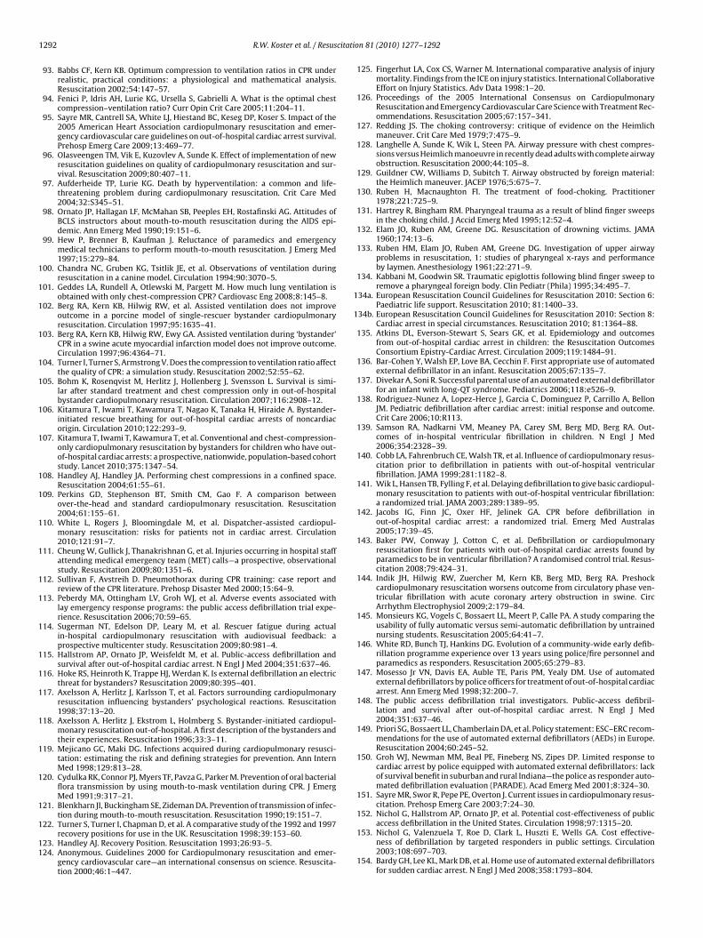
1 citatio
1
1
1
1
1
1
1
1
1
1
1
1
1
1
1
1
1
1
1
1
1
1
1
11
1
1
1
1
1
1
1
1
1
1
1
1
1
1
1
1
1
1
1
1
1
1
1
1
1
1
1
1
1
1access defibrillation in the United States. Circulation 1998;97:1315–20.
153. Nichol G, Valenzuela T, Roe D, Clark L, Huszti E, Wells GA. Cost effective-
292 R.W. Koster et al. / Resus
93. Babbs CF, Kern KB. Optimum compression to ventilation ratios in CPR underrealistic, practical conditions: a physiological and mathematical analysis.Resuscitation 2002;54:147–57.
94. Fenici P, Idris AH, Lurie KG, Ursella S, Gabrielli A. What is the optimal chestcompression–ventilation ratio? Curr Opin Crit Care 2005;11:204–11.
95. Sayre MR, Cantrell SA, White LJ, Hiestand BC, Keseg DP, Koser S. Impact of the2005 American Heart Association cardiopulmonary resuscitation and emer-gency cardiovascular care guidelines on out-of-hospital cardiac arrest survival.Prehosp Emerg Care 2009;13:469–77.
96. Olasveengen TM, Vik E, Kuzovlev A, Sunde K. Effect of implementation of newresuscitation guidelines on quality of cardiopulmonary resuscitation and sur-vival. Resuscitation 2009;80:407–11.
97. Aufderheide TP, Lurie KG. Death by hyperventilation: a common and life-threatening problem during cardiopulmonary resuscitation. Crit Care Med2004;32:S345–51.
98. Ornato JP, Hallagan LF, McMahan SB, Peeples EH, Rostafinski AG. Attitudes ofBCLS instructors about mouth-to-mouth resuscitation during the AIDS epi-demic. Ann Emerg Med 1990;19:151–6.
99. Hew P, Brenner B, Kaufman J. Reluctance of paramedics and emergencymedical technicians to perform mouth-to-mouth resuscitation. J Emerg Med1997;15:279–84.
00. Chandra NC, Gruben KG, Tsitlik JE, et al. Observations of ventilation duringresuscitation in a canine model. Circulation 1994;90:3070–5.
01. Geddes LA, Rundell A, Otlewski M, Pargett M. How much lung ventilation isobtained with only chest-compression CPR? Cardiovasc Eng 2008;8:145–8.
02. Berg RA, Kern KB, Hilwig RW, et al. Assisted ventilation does not improveoutcome in a porcine model of single-rescuer bystander cardiopulmonaryresuscitation. Circulation 1997;95:1635–41.
03. Berg RA, Kern KB, Hilwig RW, Ewy GA. Assisted ventilation during ‘bystander’CPR in a swine acute myocardial infarction model does not improve outcome.Circulation 1997;96:4364–71.
04. Turner I, Turner S, Armstrong V. Does the compression to ventilation ratio affectthe quality of CPR: a simulation study. Resuscitation 2002;52:55–62.
05. Bohm K, Rosenqvist M, Herlitz J, Hollenberg J, Svensson L. Survival is simi-lar after standard treatment and chest compression only in out-of-hospitalbystander cardiopulmonary resuscitation. Circulation 2007;116:2908–12.
06. Kitamura T, Iwami T, Kawamura T, Nagao K, Tanaka H, Hiraide A. Bystander-initiated rescue breathing for out-of-hospital cardiac arrests of noncardiacorigin. Circulation 2010;122:293–9.
07. Kitamura T, Iwami T, Kawamura T, et al. Conventional and chest-compression-only cardiopulmonary resuscitation by bystanders for children who have out-of-hospital cardiac arrests: a prospective, nationwide, population-based cohortstudy. Lancet 2010;375:1347–54.
08. Handley AJ, Handley JA. Performing chest compressions in a confined space.Resuscitation 2004;61:55–61.
09. Perkins GD, Stephenson BT, Smith CM, Gao F. A comparison betweenover-the-head and standard cardiopulmonary resuscitation. Resuscitation2004;61:155–61.
10. White L, Rogers J, Bloomingdale M, et al. Dispatcher-assisted cardiopul-monary resuscitation: risks for patients not in cardiac arrest. Circulation2010;121:91–7.
11. Cheung W, Gullick J, Thanakrishnan G, et al. Injuries occurring in hospital staffattending medical emergency team (MET) calls—a prospective, observationalstudy. Resuscitation 2009;80:1351–6.
12. Sullivan F, Avstreih D. Pneumothorax during CPR training: case report andreview of the CPR literature. Prehosp Disaster Med 2000;15:64–9.
13. Peberdy MA, Ottingham LV, Groh WJ, et al. Adverse events associated withlay emergency response programs: the public access defibrillation trial expe-rience. Resuscitation 2006;70:59–65.
14. Sugerman NT, Edelson DP, Leary M, et al. Rescuer fatigue during actualin-hospital cardiopulmonary resuscitation with audiovisual feedback: aprospective multicenter study. Resuscitation 2009;80:981–4.
15. Hallstrom AP, Ornato JP, Weisfeldt M, et al. Public-access defibrillation andsurvival after out-of-hospital cardiac arrest. N Engl J Med 2004;351:637–46.
16. Hoke RS, Heinroth K, Trappe HJ, Werdan K. Is external defibrillation an electricthreat for bystanders? Resuscitation 2009;80:395–401.
17. Axelsson A, Herlitz J, Karlsson T, et al. Factors surrounding cardiopulmonaryresuscitation influencing bystanders’ psychological reactions. Resuscitation1998;37:13–20.
18. Axelsson A, Herlitz J, Ekstrom L, Holmberg S. Bystander-initiated cardiopul-monary resuscitation out-of-hospital. A first description of the bystanders andtheir experiences. Resuscitation 1996;33:3–11.
19. Mejicano GC, Maki DG. Infections acquired during cardiopulmonary resusci-tation: estimating the risk and defining strategies for prevention. Ann InternMed 1998;129:813–28.
20. Cydulka RK, Connor PJ, Myers TF, Pavza G, Parker M. Prevention of oral bacterialflora transmission by using mouth-to-mask ventilation during CPR. J EmergMed 1991;9:317–21.
21. Blenkharn JI, Buckingham SE, Zideman DA. Prevention of transmission of infec-tion during mouth-to-mouth resuscitation. Resuscitation 1990;19:151–7.
22. Turner S, Turner I, Chapman D, et al. A comparative study of the 1992 and 1997recovery positions for use in the UK. Resuscitation 1998;39:153–60.
23. Handley AJ. Recovery Position. Resuscitation 1993;26:93–5.24. Anonymous. Guidelines 2000 for Cardiopulmonary resuscitation and emer-
gency cardiovascular care—an international consensus on science. Resuscita-tion 2000;46:1–447.
1
n 81 (2010) 1277–1292
25. Fingerhut LA, Cox CS, Warner M. International comparative analysis of injurymortality. Findings from the ICE on injury statistics. International CollaborativeEffort on Injury Statistics. Adv Data 1998:1–20.
26. Proceedings of the 2005 International Consensus on CardiopulmonaryResuscitation and Emergency Cardiovascular Care Science with Treatment Rec-ommendations. Resuscitation 2005;67:157–341.
27. Redding JS. The choking controversy: critique of evidence on the Heimlichmaneuver. Crit Care Med 1979;7:475–9.
28. Langhelle A, Sunde K, Wik L, Steen PA. Airway pressure with chest compres-sions versus Heimlich manoeuvre in recently dead adults with complete airwayobstruction. Resuscitation 2000;44:105–8.
29. Guildner CW, Williams D, Subitch T. Airway obstructed by foreign material:the Heimlich maneuver. JACEP 1976;5:675–7.
30. Ruben H, Macnaughton FI. The treatment of food-choking. Practitioner1978;221:725–9.
31. Hartrey R, Bingham RM. Pharyngeal trauma as a result of blind finger sweepsin the choking child. J Accid Emerg Med 1995;12:52–4.
32. Elam JO, Ruben AM, Greene DG. Resuscitation of drowning victims. JAMA1960;174:13–6.
33. Ruben HM, Elam JO, Ruben AM, Greene DG. Investigation of upper airwayproblems in resuscitation, 1: studies of pharyngeal x-rays and performanceby laymen. Anesthesiology 1961;22:271–9.
34. Kabbani M, Goodwin SR. Traumatic epiglottis following blind finger sweep toremove a pharyngeal foreign body. Clin Pediatr (Phila) 1995;34:495–7.
34a. European Resuscitation Council Guidelines for Resuscitation 2010: Section 6:Paediatric life support. Resuscitation 2010; 81:1400–33.
34b. European Resuscitation Council Guidelines for Resuscitation 2010: Section 8:Cardiac arrest in special circumstances. Resuscitation 2010; 81:1364–88.
35. Atkins DL, Everson-Stewart S, Sears GK, et al. Epidemiology and outcomesfrom out-of-hospital cardiac arrest in children: the Resuscitation OutcomesConsortium Epistry-Cardiac Arrest. Circulation 2009;119:1484–91.
36. Bar-Cohen Y, Walsh EP, Love BA, Cecchin F. First appropriate use of automatedexternal defibrillator in an infant. Resuscitation 2005;67:135–7.
37. Divekar A, Soni R. Successful parental use of an automated external defibrillatorfor an infant with long-QT syndrome. Pediatrics 2006;118:e526–9.
38. Rodriguez-Nunez A, Lopez-Herce J, Garcia C, Dominguez P, Carrillo A, BellonJM. Pediatric defibrillation after cardiac arrest: initial response and outcome.Crit Care 2006;10:R113.
39. Samson RA, Nadkarni VM, Meaney PA, Carey SM, Berg MD, Berg RA. Out-comes of in-hospital ventricular fibrillation in children. N Engl J Med2006;354:2328–39.
40. Cobb LA, Fahrenbruch CE, Walsh TR, et al. Influence of cardiopulmonary resus-citation prior to defibrillation in patients with out-of-hospital ventricularfibrillation. JAMA 1999;281:1182–8.
41. Wik L, Hansen TB, Fylling F, et al. Delaying defibrillation to give basic cardiopul-monary resuscitation to patients with out-of-hospital ventricular fibrillation:a randomized trial. JAMA 2003;289:1389–95.
42. Jacobs IG, Finn JC, Oxer HF, Jelinek GA. CPR before defibrillation inout-of-hospital cardiac arrest: a randomized trial. Emerg Med Australas2005;17:39–45.
43. Baker PW, Conway J, Cotton C, et al. Defibrillation or cardiopulmonaryresuscitation first for patients with out-of-hospital cardiac arrests found byparamedics to be in ventricular fibrillation? A randomised control trial. Resus-citation 2008;79:424–31.
44. Indik JH, Hilwig RW, Zuercher M, Kern KB, Berg MD, Berg RA. Preshockcardiopulmonary resuscitation worsens outcome from circulatory phase ven-tricular fibrillation with acute coronary artery obstruction in swine. CircArrhythm Electrophysiol 2009;2:179–84.
45. Monsieurs KG, Vogels C, Bossaert LL, Meert P, Calle PA. A study comparing theusability of fully automatic versus semi-automatic defibrillation by untrainednursing students. Resuscitation 2005;64:41–7.
46. White RD, Bunch TJ, Hankins DG. Evolution of a community-wide early defib-rillation programme experience over 13 years using police/fire personnel andparamedics as responders. Resuscitation 2005;65:279–83.
47. Mosesso Jr VN, Davis EA, Auble TE, Paris PM, Yealy DM. Use of automatedexternal defibrillators by police officers for treatment of out-of-hospital cardiacarrest. Ann Emerg Med 1998;32:200–7.
48. The public access defibrillation trial investigators. Public-access defibril-lation and survival after out-of-hospital cardiac arrest. N Engl J Med2004;351:637–46.
49. Priori SG, Bossaert LL, Chamberlain DA, et al. Policy statement: ESC–ERC recom-mendations for the use of automated external defibrillators (AEDs) in Europe.Resuscitation 2004;60:245–52.
50. Groh WJ, Newman MM, Beal PE, Fineberg NS, Zipes DP. Limited response tocardiac arrest by police equipped with automated external defibrillators: lackof survival benefit in suburban and rural Indiana—the police as responder auto-mated defibrillation evaluation (PARADE). Acad Emerg Med 2001;8:324–30.
51. Sayre MR, Swor R, Pepe PE, Overton J. Current issues in cardiopulmonary resus-citation. Prehosp Emerg Care 2003;7:24–30.
52. Nichol G, Hallstrom AP, Ornato JP, et al. Potential cost-effectiveness of public
ness of defibrillation by targeted responders in public settings. Circulation2003;108:697–703.
54. Bardy GH, Lee KL, Mark DB, et al. Home use of automated external defibrillatorsfor sudden cardiac arrest. N Engl J Med 2008;358:1793–804.