Eukaryotic Genomes & Chromatin Structure Bio 4342 Genomics SCR Elgin Copyright 2014 Washington...
34
Eukaryotic Genomes & Chromatin Structure Bio 4342 Genomics SCR Elgin Copyright 2014 Washington University
-
Upload
annis-wells -
Category
Documents
-
view
219 -
download
0
Transcript of Eukaryotic Genomes & Chromatin Structure Bio 4342 Genomics SCR Elgin Copyright 2014 Washington...

Eukaryotic Genomes& Chromatin Structure
Bio 4342 Genomics
SCR Elgin
Copyright 2014 Washington University

Minimum haploid DNA content - the C value paradox
R Britten and E Davidson, Science, 1970

Lack of relationship between amount of DNA and organism complexity
Alberts et. al., Molec Biol Cell, 3rd ed

Melting and reassociation of DNA is informative; follow by ultraviolet absorption spectra of DNA
Saenger, (1984)
Mononucleotides
Denatured random coil DNA
Native double helical DNA
Dependence of melting temperature (Tm)
on G+C content of DNA
Saenger, (1984)

Ideal C0t curve
Britten and Kohne, Science, 161, 529 (1968)
Am
ou
nt s
ing
le s
tra
nde
d
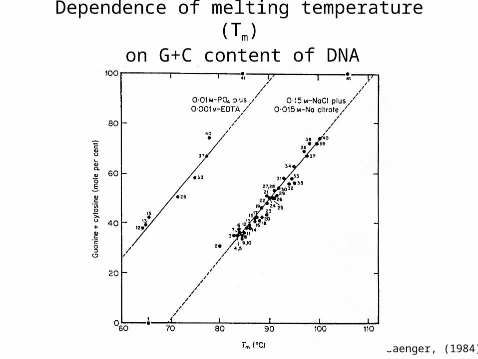
C0t curves of different DNA samples
Britten and Davidson, Carnegie Report

Davidson and Hough, PNAS 63, 342. (1969)
C0t curve of a eukaryotic genome

Composition of the mouse genome:frequency distribution
after Britten and Kohne, Science 161, 529 (1969)

Multigene families
Campbell et. al. Ann Rev Genet (1975)

Satellite DNAs of Drosophila melanogaster
Gall et. al, CSHSQB, 38, 417 (1973)

Satellite DNAs of Drosophila species
Gall et. al, CSHSQB, 38, 417 (1973)
Drosophila virilis
Drosophila americana
Drosophila virilis x Drosophila americana
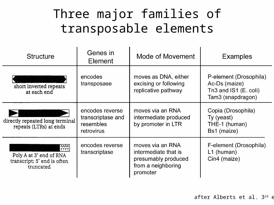
Three major families of transposable elements
after Alberts et al. 3rd ed.

As genomes get larger, an increasing proportion of the DNA is non-coding.
Allis et al: Epigenetics 2007

Repeat distribution is not uniform. Drosophila dot chromosomes are 25% - 30% repetitious DNA
(typical – but up to 80% in D. ananassae)
D mel D vir D mel D vir D vir D mel D vir D vir D vir
Leung et al 2010 Genetics 185: 1519

Considerations for genome sequencing1. Satellite DNA is very difficult to sequence, as there are few
markers to help order reads or subclones; hence centromeric regions of the chromosomes are usually left unsequenced.
2. Middle repetitious DNA also causes difficulties; because one finds nearly identical sequences located in different regions of the genome, mistakes can be made in assembling sequence data. High quality discrepancies can identify different copies of a repeat.
3. Much of the repetitious DNA is packaged in heterochromatin, which maintains these regions in a compact and transcriptionally silent form.
4. However, in many higher organisms, the protein-coding genes are found embedded in repetitious DNA. Check out your favorite human gene on the UCSC Browser by taking off RepeatMasker!
SCR Elgin
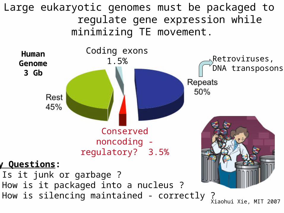
Coding exons1.5%
Conserved noncoding - regulatory? 3.5%
Human Genome
3 Gb
Key Questions: Is it junk or garbage ? How is it packaged into a nucleus ? How is silencing maintained - correctly ?
Large eukaryotic genomes must be packaged to regulate gene expression while minimizing TE movement.
Xiaohui Xie, MIT 2007
Retroviruses,DNA transposons

Felsenfeld et al. Nature 2003, 421: 448
Packaging large genomes:
First step - packaging in a nucleosome array
Second - differential packaging into heterochromatin & euchromatin
DNA
Chromatin
Lodish et.al., Molecular Cell Biology, 4th Edition
Chromosome(metaphase)
Histone protein core

DNA packaging in chromatin & chromosomes
Packing Ratio Model
Naked DNA 20A dia. 10 bp/turn 1 good
Chromatin 100A dia. ~80 bp/turn 6-7 good(100 A fiber)
Chromatin 300A dia. 6 bp/turn ~40 vague
(300 A fiber)
Domains (loops) 20-100 kb/loop ~700 vague
Chromosome 106 - 108 bp ~10,000 definedSCR Elgin

Electron micrograph of chromatin fibers (rat thymus nucleus)
Olins et. al., J. Cell Biol, (1975) 64, 528-537 Chris Woodcock

- Anonymous review of paper submitted by C.F.L. Woodcock, 1973, showing EM pictures of nucleosome arrays
“A eukaryotic chromosome made out of self-assembling 70A units, which could perhaps be made to crystallize, would necessitate rewriting our basic textbooks on cytology and genetics! I have never read such a naïve paper purporting to be of such fundamental significance. Definitely it should not be published anywhere!”
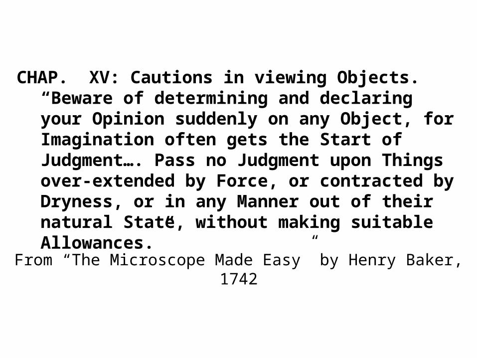
From “The Microscope Made Easy” by Henry Baker, 1742
CHAP. XV: Cautions in viewing Objects.“Beware of determining and declaring your Opinion suddenly on any Object, for Imagination often gets the Start of Judgment…. Pass no Judgment upon Things over-extended by Force, or contracted by Dryness, or in any Manner out of their natural State, without making suitable Allowances.”

Establishing the nucleosome model..- a paradigm shift, 1973-1974
1. Electron microscopy - images
2. Micrococcal nuclease digestion patterns
3. Knowledge of histone:histone interactions
SCR Elgin

Observation of defined lengths of chromatin
Finch et.al., PNAS (1975) 72: 3321
Sucrose gradient fractionation of micrococcal nuclease digestion products
• Top of gradient is on the right• Bottom of gradient is on the left• Fractions collected from shaded areas
Polyacrylamide gel electrophoresis of purified DNA
• Right lane: unfractionated digest
• Left lanes: DNA purified from sucrose gradient peaks
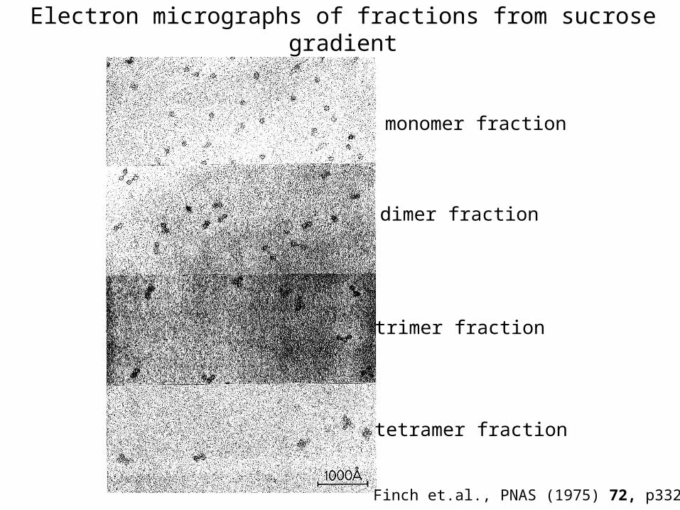
Electron micrographs of fractions from sucrose gradient
monomer fraction
dimer fraction
trimer fraction
tetramer fraction
Finch et.al., PNAS (1975) 72, p3321

Ribbon model of the four histones-stable interactions leading to a histone core
Arents et. al., PNAS (1991) 88, 10148-52
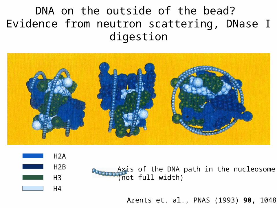
DNA on the outside of the bead? Evidence from neutron scattering, DNase I digestion
Arents et. al., PNAS (1993) 90, 10489-93
H2A
H2B
H3
H4
Axis of the DNA path in the nucleosome(not full width)

The crystal structure of the nucleosome core
Rhodes, Nature (1997) 389, 231-233, after Luger et. al., Nature (1997) 389, 251-260
Resolution: 2.8 Å
Half of the nucleosome structure is shown
One turn of the DNA helix is visible (73 bp)
View is down the superhelix axis
Protein - DNA contacts: white hooks
Seven years effort: required symmetric DNA (inverted repeat)and histones made in E. coli(uniformly free of modifications)
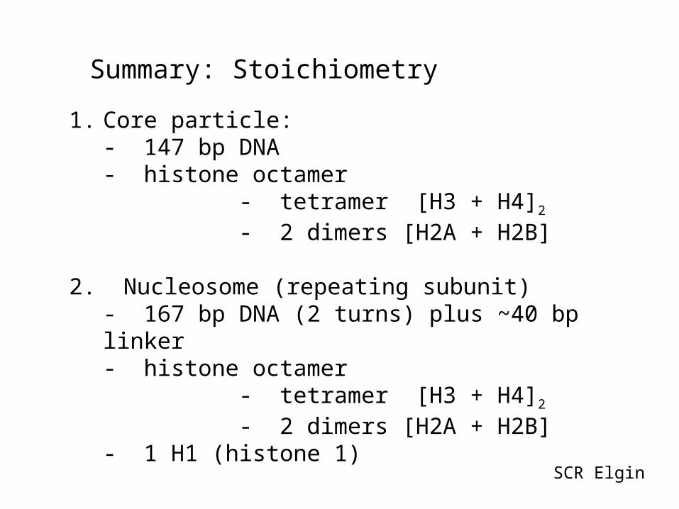
Summary: Stoichiometry
1. Core particle:- 147 bp DNA- histone octamer
- tetramer [H3 + H4]2
- 2 dimers [H2A + H2B]
2. Nucleosome (repeating subunit) - 167 bp DNA (2 turns) plus ~40 bp linker- histone octamer
- tetramer [H3 + H4]2
- 2 dimers [H2A + H2B]- 1 H1 (histone 1)
SCR Elgin

Summary: a model of chromatin structure 10 nm and 30 nm fibers
Griffiths et.al. Introduction to Genetic Analysis, 2000
Histone H1 stabilizes 30 nm fiber.Nearest neighbor pattern?May be a 2-start helix.

Histones are general repressors of template activity.
• Default state of the eukaryotic genome is “off.”
• Nucleosomes block access to promoter in vitro
• Active and inducible genes show 5’ nuclease
accessible hypersensitive sites (HS sites)
• Generation of such sites part of activation
• Remodeling activities are critical for activation
• Histone modification patterns reflect activity state
• Histone variants reflect activity state
SCR Elgin

Carl Wu
Wu et al, 1978 Cell16: 797.
Given recombinant DNA probes, we can analyze the chromatin structure of specific genes

Assessing chromatin structure - genes are packaged in nucleosome arrays with the active TSS
in an open region – a HS site
Analysis: MNase cuts between nucleosomes; DNase I cuts nucleosome-free sites (DH – DNase hypersensitive sites). Results shown for hsp26.

Summary
• Eukaryotic genomes are packaged in a nucleosome
array; an excellent model of the nucleosome based on
EM results, nuclease digestion, and protein-protein
interaction studies is available.
• This chromatin subunit has been crystallized, and the
textbooks have been rewritten!
• Nucleosome packaging negatively impacts gene
expression, keeping the bulk of the genome silent.
• Transcription Start Sites are found in Hypersensitive
Sites, nucleosome-free regions; this selective access
compensates for the large size of the genome.
SCR Elgin