Establishing the “Biological Relevance” of Dipentyl ... et al...Phthalate Reductions in Fetal...
14
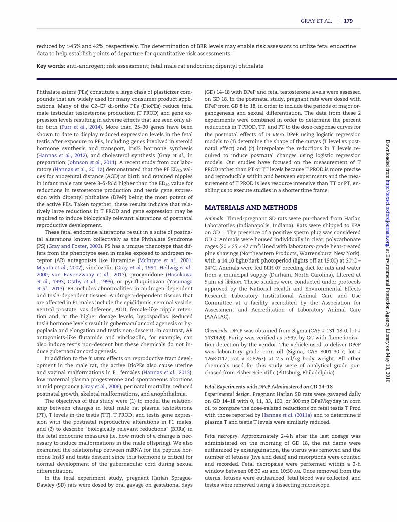
Establishing the “Biological Relevance” of Dipentyl Phthalate Reductions in Fetal Rat Testosterone Production and Plasma and Testis Testosterone Levels Leon Earl Gray Jr,* ,1,2 Johnathan Furr,* ,1 Katoria R. Tatum-Gibbs,* ,1,3 Christy Lambright,* Hunter Sampson, 3 Bethany R. Hannas,* ,4 Vickie S. Wilson,* Andrew Hotchkiss, † and Paul M. D. Foster ‡ *Reproductive Toxicology Branch, Toxicology Assessment Division, National Health and Environmental Effects Laboratory, Office of Research and Development, U.S. Environmental Protection Agency (US EPA), Research Triangle Park, North Carolina 27711; † NCEA, ORD, USEPA, Washington, District of Columbia; and ‡ National Toxicology Program, NIEHS, NIH, DHHS, Research Triangle Park, North Carolina 27709 1 These authors contributed equally to this study. 2 To whom correspondence should be addressed. Reproductive Toxicology Branch, Toxicology Assessment Division, National Health and Environmental Effects Laboratory, Office of Research and Development, U.S. Environmental Protection Agency (US EPA), Research Triangle Park, North Carolina 27711. Fax: 919-541-7750. E-mail: [email protected] 3 Present address: Oak Ridge Institute for Science and Education, Oak Ridge, Tennessee 37831. 4 Present address: The Dow Chemical Company, Toxicology & Environmental Research & Consulting, Midland, Michigan. Disclaimers. This Manuscript has been reviewed in accordance with the policy of the National Health and Environmental Effects Research Laboratory, U.S. Environmental Protection Agency, and approved for publication. Approval does not signify that the contents necessarily reflect the views or policy of the Agency nor does mention of trade names or commercial products constitute endorsement or recommendation for use. This research was supported in part by the Intramural Research Program of the National Institute of Environmental Health Sciences, National Institutes of Health. This article may be the work product of an employee or group of employees of the National Institute of Environmental Health Sciences (NIEHS), National Institutes of Health (NIH), however, the statements, opinions, or conclusions contained therein do not necessarily represent the statements, opinions, or conclusions of NIEHS, NIH, or the United States government. ABSTRACT Phthalate esters (PEs) constitute a large class of compounds that are used for many consumer product applications. Many of the C2–C7 di-ortho PEs reduce fetal testicular hormone and gene expression levels in rats resulting in adverse effects seen later in life but it appears that relatively large reductions in fetal testosterone (T) levels and testis gene expression may be required to adversely affect reproductive development (Hannas, B. R., Lambright, C. S., Furr, J., Evans, N., Foster, P. M., Gray, E. L., and Wilson, V. S. (2012). Genomic biomarkers of phthalate-induced male reproductive developmental toxicity: a targeted RT-PCR array approach for defining relative potency. Toxicol. Sci. 125, 544–557). The objectives of this study were (1) to model the relationships between changes in fetal male rat plasma testosterone (PT), T levels in the testis (TT), T production (PROD), and testis gene expression with the reproductive malformation rates, and (2) to quantify the “biologically relevant reductions” (BRRs) in fetal T necessary to induce adverse effects in the offspring. In the fetal experiment, Harlan Sprague-Dawley rats were dosed with dipentyl phthalate (DPeP) at 0, 11, 33, 100, and 300 mg/kg/day from gestational days (GD) 14–18 and fetal testicular T, PT levels, and T Prod and gene expression were assessed on GD 18. In the postnatal experiment, rats were dosed with DPeP from GD 8–18 and reproductive development was monitored through adulthood. The dose-response curves for TT levels (ED 50 ¼ 53 mg/kg) and T PROD (ED 50 ¼ 45 mg/kg) were similar, whereas PT was reduced at ED 50 ¼ 19 mg/kg. When the reductions in TPROD and Insl3 mRNA were compared with the postnatal effects of in utero DPeP, dose-related reproductive alterations were noted when T PROD and Insl3 mRNA were Published by Oxford University Press on behalf of the Society of Toxicology 2015. This work is written by US Government employees and is in the public domain in the US. 178 TOXICOLOGICAL SCIENCES, 149(1), 2016, 178–191 doi: 10.1093/toxsci/kfv224 Advance Access Publication Date: October 9, 2015 Research Article at Environmental Protection Agency Library on May 18, 2016 http://toxsci.oxfordjournals.org/ Downloaded from
Transcript of Establishing the “Biological Relevance” of Dipentyl ... et al...Phthalate Reductions in Fetal...
Establishing the “Biological Relevance” of Dipentyl
Phthalate Reductions in Fetal Rat Testosterone
Production and Plasma and Testis Testosterone LevelsLeon Earl Gray Jr,*,1,2 Johnathan Furr,*,1 Katoria R. Tatum-Gibbs,*,1,3
Christy Lambright,* Hunter Sampson,3 Bethany R. Hannas,*,4
Vickie S. Wilson,* Andrew Hotchkiss,† and Paul M. D. Foster‡
*Reproductive Toxicology Branch, Toxicology Assessment Division, National Health and EnvironmentalEffects Laboratory, Office of Research and Development, U.S. Environmental Protection Agency (US EPA),Research Triangle Park, North Carolina 27711; †NCEA, ORD, USEPA, Washington, District of Columbia; and‡National Toxicology Program, NIEHS, NIH, DHHS, Research Triangle Park, North Carolina 27709
1These authors contributed equally to this study.2To whom correspondence should be addressed. Reproductive Toxicology Branch, Toxicology Assessment Division, National Health and EnvironmentalEffects Laboratory, Office of Research and Development, U.S. Environmental Protection Agency (US EPA), Research Triangle Park, North Carolina 27711.Fax: 919-541-7750. E-mail: [email protected] address: Oak Ridge Institute for Science and Education, Oak Ridge, Tennessee 37831.4Present address: The Dow Chemical Company, Toxicology & Environmental Research & Consulting, Midland, Michigan.
Disclaimers. This Manuscript has been reviewed in accordance with the policy of the National Health and Environmental Effects Research Laboratory,U.S. Environmental Protection Agency, and approved for publication. Approval does not signify that the contents necessarily reflect the views or policy ofthe Agency nor does mention of trade names or commercial products constitute endorsement or recommendation for use. This research was supportedin part by the Intramural Research Program of the National Institute of Environmental Health Sciences, National Institutes of Health. This article may bethe work product of an employee or group of employees of the National Institute of Environmental Health Sciences (NIEHS), National Institutes of Health(NIH), however, the statements, opinions, or conclusions contained therein do not necessarily represent the statements, opinions, or conclusions ofNIEHS, NIH, or the United States government.
ABSTRACT
Phthalate esters (PEs) constitute a large class of compounds that are used for many consumer product applications. Many ofthe C2–C7 di-ortho PEs reduce fetal testicular hormone and gene expression levels in rats resulting in adverse effects seenlater in life but it appears that relatively large reductions in fetal testosterone (T) levels and testis gene expression may berequired to adversely affect reproductive development (Hannas, B. R., Lambright, C. S., Furr, J., Evans, N., Foster, P. M., Gray,E. L., and Wilson, V. S. (2012). Genomic biomarkers of phthalate-induced male reproductive developmental toxicity: atargeted RT-PCR array approach for defining relative potency. Toxicol. Sci. 125, 544–557). The objectives of this study were (1)to model the relationships between changes in fetal male rat plasma testosterone (PT), T levels in the testis (TT), Tproduction (PROD), and testis gene expression with the reproductive malformation rates, and (2) to quantify the“biologically relevant reductions” (BRRs) in fetal T necessary to induce adverse effects in the offspring. In the fetalexperiment, Harlan Sprague-Dawley rats were dosed with dipentyl phthalate (DPeP) at 0, 11, 33, 100, and 300 mg/kg/dayfrom gestational days (GD) 14–18 and fetal testicular T, PT levels, and T Prod and gene expression were assessed on GD 18.In the postnatal experiment, rats were dosed with DPeP from GD 8–18 and reproductive development was monitoredthrough adulthood. The dose-response curves for TT levels (ED50¼53 mg/kg) and T PROD (ED50¼45 mg/kg) were similar,whereas PT was reduced at ED50 ¼ 19 mg/kg. When the reductions in TPROD and Insl3 mRNA were compared with thepostnatal effects of in utero DPeP, dose-related reproductive alterations were noted when T PROD and Insl3 mRNA were
Published by Oxford University Press on behalf of the Society of Toxicology 2015.This work is written by US Government employees and is in the public domain in the US.
178
TOXICOLOGICAL SCIENCES, 149(1), 2016, 178–191
doi: 10.1093/toxsci/kfv224Advance Access Publication Date: October 9, 2015Research Article
at Environm
ental Protection Agency L
ibrary on May 18, 2016
http://toxsci.oxfordjournals.org/D
ownloaded from
reduced by >45% and 42%, respectively. The determination of BRR levels may enable risk assessors to utilize fetal endocrinedata to help establish points of departure for quantitative risk assessments.
Key words: anti-androgen; risk assessment; fetal male rat endocrine; dipentyl phthalate
Phthalate esters (PEs) constitute a large class of plasticizer com-pounds that are widely used for many consumer product appli-cations. Many of the C2–C7 di-ortho PEs (DioPEs) reduce fetalmale testicular testosterone production (T PROD) and gene ex-pression levels resulting in adverse effects that are seen only af-ter birth (Furr et al., 2014). More than 25–30 genes have beenshown to date to display reduced expression levels in the fetaltestis after exposure to PEs, including genes involved in steroidhormone synthesis and transport, Insl3 hormone synthesis(Hannas et al., 2012), and cholesterol synthesis (Gray et al., inpreparation; Johnson et al., 2011). A recent study from our labo-ratory (Hannas et al., 2011a) demonstrated that the PE ED50 val-ues for anogenital distance (AGD) at birth and retained nipplesin infant male rats were 3–5-fold higher than the ED50 value forreductions in testosterone production and testis gene expres-sion with dipentyl phthalate (DPeP) being the most potent ofthe active PEs. Taken together, these results indicate that rela-tively large reductions in T PROD and gene expression may berequired to induce biologically relevant alterations of postnatalreproductive development.
These fetal endocrine alterations result in a suite of postna-tal alterations known collectively as the Phthalate Syndrome(PS) (Gray and Foster, 2003). PS has a unique phenotype that dif-fers from the phenotype seen in males exposed to androgen re-ceptor (AR) antagonists like flutamide (McIntyre et al., 2001;Miyata et al., 2002), vinclozolin (Gray et al., 1994; Hellwig et al.,2000; van Ravenzwaay et al., 2013), procymidone (Hosokawaet al., 1993; Ostby et al., 1999), or pyrifluquinazon (Yasunagaet al., 2013). PS includes abnormalities in androgen-dependentand Insl3-dependent tissues. Androgen-dependent tissues thatare affected in F1 males include the epididymis, seminal vesicle,ventral prostate, vas deferens, AGD, female-like nipple reten-tion and, at the higher dosage levels, hypospadias. ReducedInsl3 hormone levels result in gubernacular cord agenesis or hy-poplasia and elongation and testis non-descent. In contrast, ARantagonists-like flutamide and vinclozolin, for example, canalso induce testis non-descent but these chemicals do not in-duce gubernacular cord agenesis.
In addition to the in utero effects on reproductive tract devel-opment in the male rat, the active DioPEs also cause uterineand vaginal malformations in F1 females (Hannas et al., 2013),low maternal plasma progesterone and spontaneous abortionsat mid pregnancy (Gray et al., 2006), perinatal mortality, reducedpostnatal growth, skeletal malformations, and anophthalmia.
The objectives of this study were (1) to model the relation-ship between changes in fetal male rat plasma testosterone(PT), T levels in the testis (TT), T PROD, and testis gene expres-sion with the postnatal reproductive alterations in F1 males,and (2) to describe “biologically relevant reductions” (BRRs) inthe fetal endocrine measures (ie, how much of a change is nec-essary to induce malformations in the male offspring). We alsoexamined the relationship between mRNA for the peptide hor-mone Insl3 and testis descent since this hormone is critical fornormal development of the gubernacular cord during sexualdifferentiation.
In the fetal experiment study, pregnant Harlan Sprague-Dawley (SD) rats were dosed by oral gavage on gestational days
(GD) 14–18 with DPeP and fetal testosterone levels were assessedon GD 18. In the postnatal study, pregnant rats were dosed withDPeP from GD 8 to 18, in order to include the periods of major or-ganogenesis and sexual differentiation. The data from these 2experiments were combined in order to determine the percentreductions in T PROD, TT, and PT to the dose-response curves forthe postnatal effects of in utero DPeP using logistic regressionmodels to (1) determine the shape of the curves (T level vs post-natal effect) and (2) interpolate the reductions in T levels re-quired to induce postnatal changes using logistic regressionmodels. Our studies have focused on the measurement of TPROD rather than PT or TT levels because T PROD is more preciseand reproducible within and between experiments and the mea-surement of T PROD is less resource intensive than TT or PT, en-abling us to execute studies in a shorter time frame.
MATERIALS AND METHODS
Animals. Timed-pregnant SD rats were purchased from HarlanLaboratories (Indianapolis, Indiana). Rats were shipped to EPAon GD 1. The presence of a positive sperm plug was consideredGD 0. Animals were housed individually in clear, polycarbonatecages (20� 25� 47 cm3) lined with laboratory-grade heat-treatedpine shavings (Northeastern Products, Warrensburg, New York),with a 14:10 light/dark photoperiod (lights off at 19:00) at 20�C –24�C. Animals were fed NIH 07 breeding diet for rats and waterfrom a municipal supply (Durham, North Carolina), filtered at5 mm ad libitum. These studies were conducted under protocolsapproved by the National Health and Environmental EffectsResearch Laboratory Institutional Animal Care and UseCommittee at a facility accredited by the Association forAssessment and Accreditation of Laboratory Animal Care(AAALAC).
Chemicals. DPeP was obtained from Sigma (CAS # 131-18-0, lot #1431420). Purity was verified as >99% by GC with flame ioniza-tion detection by the vendor. The vehicle used to deliver DPePwas laboratory grade corn oil (Sigma; CAS 8001-30-7; lot #126K0117; cat # C-8267) at 2.5 ml/kg body weight. All otherchemicals used for this study were of analytical grade pur-chased from Fisher Scientific (Pittsburg, Philadelphia).
Fetal Experiments with DPeP Administered on GD 14–18Experimental design. Pregnant Harlan SD rats were gavaged dailyon GD 14–18 with 0, 11, 33, 100, or 300 mg DPeP/kg/day in cornoil to compare the dose-related reductions on fetal testis T Prodwith those reported by Hannas et al. (2011a) and to determine ifplasma T and testis T levels were similarly reduced.
Fetal necropsy. Approximately 2–4 h after the last dosage wasadministered on the morning of GD 18, the rat dams wereeuthanized by exsanguination, the uterus was removed and thenumber of fetuses (live and dead) and resorptions were countedand recorded. Fetal necropsies were performed within a 2-hwindow between 08:30 AM and 10:30 AM. Once removed from theuterus, fetuses were euthanized, fetal blood was collected, andtestes were removed using a dissecting microscope.
GRAY ET AL. | 179
at Environm
ental Protection Agency L
ibrary on May 18, 2016
http://toxsci.oxfordjournals.org/D
ownloaded from

Fetal plasma collection. Fetuses were removed from the uterusand blood was collected from the jugular vein using heparinizedcapillary tubes. Blood was blown from the capillary tube with afine-tip disposable transfer pipet, and transferred into a 0.65-ml(siliconized) microcentrifuge tube. Blood samples from all malesin each litter were pooled and stored on ice until centrifugation.The pooled blood samples were then centrifuged at 3000 rpm at4�C for 10 min. The plasma was removed using a 10-ml pipet,placed in a new tube, and stored at �80�C until assayed for Tby RIA using Coat-a-Count Kits (Siemens, Los Angeles,California) according to the protocol provided by the manufac-turer. Fetal plasma T levels are based upon the 7–9 litters/dosegroup.
Ex vivo testicular T production (T PROD). One testis per male from 3males per litter was used to measure ex vivo T PROD levels(numbers of litters are 16, 11, 10, 12, and 14 in the 0, 11, 33, 100,and 300 mg/kg/day groups, respectively). Paired testes fromother males in some of these litters (up to 3 males per litter)were used for determinations of fetal testis weight (5–6 litters/dose group) and extracted testicular T levels (7–8 litters/dosegroup). Testicular T PROD was measured as previouslydescribed (Furr et al., 2014) in media using a Coat-a-Count radio-immunoassay (RIA) kit for total testosterone according to theprotocol provided by the manufacturer (Siemens HealthcareDiagnostics, Deerfield, Illinois). The intra-assay coefficientof variation was 3.1% (based on variability of the standardcurve). The interassay coefficient of variation was 13.7%. Crossreactivity with DHT was 3.2%. The limit of detection for T was0.2 ng/ml.
Fetal testis gene expression using 96 well QPCR custom arrays. Fetaltestis gene expression was measured using 96 gene custom-designed QPCR arrays (described in detail Hannas et al., 2011b,2012). We have found that phthalates that disrupt testosteronelevels also reduce the expression of 10–12 genes on these arraysincluding those coding for proteins involved in steroid trans-port, and testosterone and Insl3 hormone synthesis (includingCyp11b1, StAR. Cyp11a1, Hsd3b, Cyp17a1, Scarb1, Insl3, Inha,Cyp11b2, Lhcgr, and Dhcr7). Among the DioPEs, several of thesegenes on our arrays typically display ED50 values that are simi-lar to (StAR, Scarb1, and Cyp17a1) or below (Cyp11b1) the ED50
value for T Prod.
Fetal testicular T extraction. Testosterone was extracted fromthe testes collected on GD 18. Paired testes were placed inglass tubes (12� 75 mm2) (n¼ 3 per litter), the testes in the glasstubes were flash frozen with dry ice and stored at �80�C.Detailed extraction methods are provided in the Supplementaryfiles.
Postnatal Experiment with DPeP administered on GD 8–18Experimental design. Pregnant Harlan SD rats were gavaged dailyon GD 8–18 with 0, 11, 33, 100, or 300 mg/kg/day DPeP in corn oilwith 5 dams/dose group. The maternal data and F1 male off-spring up to weaning on postnatal day (PND) 24 were describedby Hannas et al. (2011a). The exposures started on GD 8 ratherthan GD 14, when dosing started in the fetal study, so we couldcompare the dose responses for effects on F1 male reproductivedevelopment induced during the masculinizing window withother adverse effects that are induced earlier in gestation thanis disruption of male sexual differentiation; effects that mightturn out to be relevant to a hazard assessment of thechemical. For example, we have found that in utero phthalate
administration during organogenesis results in F1 female repro-ductive tract malformations, skeletal malformations, and otheradverse effects, effects not seen if phthalate exposure isrestricted to the period of sexual differentiation (Hannas et al.,2013). In addition, exposure to a phthalates from GD 8 to 13 doesnot affect F1 male reproductive tract development (Carruthersand Foster, 2005; Hannas et al., 2013) which is consistent withthe fact that the fetal testis is not producing measurable testos-terone at this stage of development (Feldman and Bloch, 1978;Gangnerau and Picon, 1987; Majdic et al., 1998; Warren et al.,1973). Furthermore, early gestational exposure to phthalateslike DPeP would not be expected to accumulate in tissuesbecause the majority of the chemical and its metabolites arelikely excreted within 24 h, similar to the better studied phtha-lates DBP and DEHP (Kluwe, 1982).
The continuation of the F1 offspring study from weaningthrough adult life is reported here. The AGD (PND 2) and infantmale nipple retention (PND) 13 data from this study werereported by Hannas et al. (2011a) and are shown here for com-parison to the prenatal T PROD, PT and TT data, and the postna-tal effects seen later in life.
Male and female offspring were weaned on PND 24and housed in groups of 2–3 per cage by sex until necropsy.Male offspring were checked daily from 37 to 55 days of age todetermine the age and weight at “puberty” (preputial separa-tion—PPS) and both male (n¼ 98 total) and female (n¼ 103 total)offspring were necropsied beginning at 120 days of age.
F1 females were examined for gross malformations (includ-ing anophthalmia, absent vaginal opening or vaginal canal, ute-rine agenesis and hydrometrocolpos and hydronephrosis; alltraits seen in the female rat—female PS). Weights of the pitui-tary, uterus, ovaries, kidneys, and liver were recorded from 2females per litter (since organ weights were unaffected by inutero exposure to DPeP these measurements were not takenfrom all F1 females).
In F1 males, body, glans penis, seminal vesicle, ventral pros-tate, testis, epididymal, levator ani-bulbocavernosus muscles,Cowper’s glands, kidney, and liver weights were recorded alongwith any gross malformations. In addition, gubernacular cordlengths were measured with a micrometer. Gubernacularabnormalities were either complete absence or elongation of ahypoplastic cord. We defined “abnormal”—elongated gubernac-ular cords as those longer than 17 mm, a value that exceeds thecontrol mean by several standard deviations. Control gubernac-ular cord in this study averaged �9.5 mm with a standard devia-tion of �1.2 mm.
Testes and epididymal tissues were preserved in Bouin’s fix-ative, placed in 70% ethanol after 24 h and sent to ExperimentalPathology Laboratories, Inc., Durham, North Carolina (EPLProject No. 431-603) where they were embedded in paraffin, sec-tioned at 4–6 lm, stained using hematoxylin and eosin and his-topathological analyses were conducted by a board certifiedpathologist.
Since several of the male and female F1 rats in the high dosegroup developed malocclusion of the incisors due to obviousskull abnormalities, skulls were frozen and shipped to SkullsUnlimited International, INC., Oklahoma City, Oklahoma. At thetaxidermist, skulls were individually tagged for identification,cleaned using flesh-eating dermestid beetles (Dermestes macula-tus) and returned for gross examination and morphometricanalysis of the length and width of the left and right zygomaticarch. Since this structure was obviously asymmetrical in theaffected animals it provided a quantitative descriptor of thegross skeletal malformation.
180 | TOXICOLOGICAL SCIENCES, 2016, Vol. 149, No. 1
at Environm
ental Protection Agency L
ibrary on May 18, 2016
http://toxsci.oxfordjournals.org/D
ownloaded from
Statistics. Data analysis was performed using 1-way ANOVAthrough the General Linear Model procedure (PROC GLM) in theStatistical Analysis System (SAS, SAS Institute, Cary, NorthCarolina). If the overall ANOVA was significant at (P< .05) thesignificant differences between control and treated groups weredetermined by a post hoc 2-tailed t test (LSMEANS on SAS)between litter means. Fetal T PROD data were analyzed usinglitter mean values generated from the individual testis incuba-tions from males within a litter. The fetal T PROD data from thisstudy were pooled with DPeP dose-response data collected ear-lier (Furr et al., 2014) and these pooled results were used tomodel the relationship between T PROD and the postnatal con-sequences of in utero DPeP exposure. The effects of in utero DPePexposure on F1 male rat postnatal reproductive developmentwere also analyzed using litter means rather than individualanimal values.
Dose-response curves from all experiments were analyzedusing untransformed and transformed (to percent of control) datain a non-linear 4 parameter logistic (4PL) regression model (sig-moidal fit with variable slope Prism GraphPad 5.01 software,GraphPad Software, Inc., La Jolla, California). For logistic regressionanalyses, the control dose value was set to 1 mg/kg/day ratherthan 0 mg/kg/day so the control data would be included in theanalysis as Prism converts the dose to log10 values and the top ofthe models were constrained to 100% and the bottom to 0%.
In addition, we used both the 4PL and 5 (5PL) logistic regres-sion models to describe the relationship between the fetal tes-tosterone measures (T Prod on the X-axis rather than the DPePdose) and the postnatal effects of DPeP to estimate by interpola-tion the BRR in fetal T PROD (BRRT); BRRT being the estimatedreductions in T levels resulting in postnatal alterations in the2%, 5%, 10%, 25%, 50%, 75%, and 95% of the male offspring. TheBRRT values were estimated using a 4PL which assumes curvesymmetry on both sides of the ED50 as well as a 5PL parametermodel which includes a parameter for asymmetry of the slopeabove and below the ED50 value. The 4PL logistic regressionmodel was also used to define the BRR in Insl3 mRNA withDPeP-induced increases in gubernacular cord abnormalities andundescended testes in F1 male rats.
RESULTS
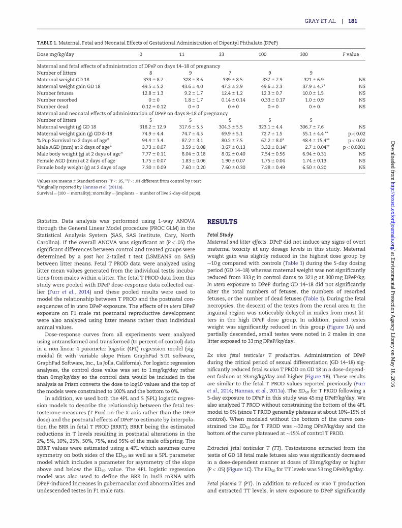
Fetal StudyMaternal and litter effects. DPeP did not induce any signs of overtmaternal toxicity at any dosage levels in this study. Maternalweight gain was slightly reduced in the highest dose group by�10 g compared with controls (Table 1) during the 5-day dosingperiod (GD 14–18) whereas maternal weight was not significantlyreduced from 333 g in control dams to 321 g at 300 mg DPeP/kg.In utero exposure to DPeP during GD 14–18 did not significantlyalter the total numbers of fetuses, the numbers of resorbedfetuses, or the number of dead fetuses (Table 1). During the fetalnecropsies, the descent of the testes from the renal area to theinguinal region was noticeably delayed in males from most lit-ters in the high DPeP dose group. In addition, paired testesweight was significantly reduced in this group (Figure 1A) andpartially descended, small testes were noted in 2 males in onelitter exposed to 33 mg DPeP/kg/day.
Ex vivo fetal testicular T production. Administration of DPePduring the critical period of sexual differentiation (GD 14–18) sig-nificantly reduced fetal ex vivo T PROD on GD 18 in a dose-depend-ent fashion at 33 mg/kg/day and higher (Figure 1B). These resultsare similar to the fetal T PROD values reported previously (Furret al., 2014; Hannas, et al., 2011a). The ED50 for T PROD following a5-day exposure to DPeP in this study was 45 mg DPeP/kg/day. Wealso analyzed T PROD without constraining the bottom of the 4PLmodel to 0% (since T PROD generally plateaus at about 10%–15% ofcontrol). When modeled without the bottom of the curve con-strained the ED50 for T PROD was �32 mg DPeP/kg/day and thebottom of the curve plateaued at �15% of control T PROD.
Extracted fetal testicular T (TT). Testosterone extracted from thetestis of GD 18 fetal male fetuses also was significantly decreasedin a dose-dependent manner at doses of 33 mg/kg/day or higher(P< .05) (Figure 1C). The ED50 for TT levels was 53 mg DPeP/kg/day.
Fetal plasma T (PT). In addition to reduced ex vivo T productionand extracted TT levels, in utero exposure to DPeP significantly
TABLE 1. Maternal, Fetal and Neonatal Effects of Gestational Administration of Dipentyl Phthalate (DPeP)
Dose mg/kg/day 0 11 33 100 300 F value
Maternal and fetal effects of administration of DPeP on days 14–18 of pregnancyNumber of litters 8 9 7 9 9Maternal weight GD 18 333 6 8.7 328 6 8.6 339 6 8.5 337 6 7.9 321 6 6.9 NSMaternal weight gain GD 18 49.5 6 5.2 43.6 6 4.0 47.3 6 2.9 49.6 6 2.3 37.9 6 4.7* NSNumber fetuses 12.8 6 1.3 9.2 6 1.7 12.4 6 1.2 12.3 6 0.7 10.0 6 1.5 NSNumber resorbed 0 6 0 1.8 6 1.7 0.14 6 0.14 0.33 6 0.17 1.0 6 0.9 NSNumber dead 0.12 6 0.12 0 6 0 0 6 0 0 6 0 0 6 0 NSMaternal and neonatal effects of administration of DPeP on days 8–18 of pregnancyNumber of litters 5 5 5 5 5Maternal weight (g) GD 18 318.2 6 12.9 317.6 6 5.5 304.3 6 5.5 323.1 6 4.4 306.7 6 7.6 NSMaternal weight gain (g) GD 8–18 74.9 6 4.4 74.7 6 4.5 69.9 6 5.1 72.7 6 1.5 55.1 6 4.4 ** p< 0.02% Pup Survival to 2 days of agea 94.4 6 3.4 87.2 6 3.1 80.2 6 7.5 67.2 6 8.0* 48.4 6 15.4** p< 0.02Male AGD (mm) at 2 days of agea 3.73 6 0.07 3.59 6 0.08 3.67 6 0.13 3.32 6 0.14* 2.7 6 0.04** p< 0.0001Male body weight (g) at 2 days of agea 7.77 6 0.11 8.04 6 0.18 8.02 6 0.40 7.54 6 0.56 6.94 6 0.31 NSFemale AGD (mm) at 2 days of age 1.75 6 0.07 1.83 6 0.06 1.90 6 0.07 1.75 6 0.04 1.74 6 0.13 NSFemale body weight (g) at 2 days of age 7.30 6 0.09 7.60 6 0.20 7.60 6 0.30 7.28 6 0.49 6.50 6 0.20 NS
Values are means 6 Standard errors; *P< .05, **P< .01 different from control by t testaOriginally reported by Hannas et al. (2011a).
Survival¼ (100 �mortality); mortality¼ (implants � number of live 2-day-old pups).
GRAY ET AL. | 181
at Environm
ental Protection Agency L
ibrary on May 18, 2016
http://toxsci.oxfordjournals.org/D
ownloaded from
reduced circulating PT at doses of 33 mg/kg/day or higher(P< .05) (Figure 1D) (PT ED50¼ 19 mg DPeP/kg/day).
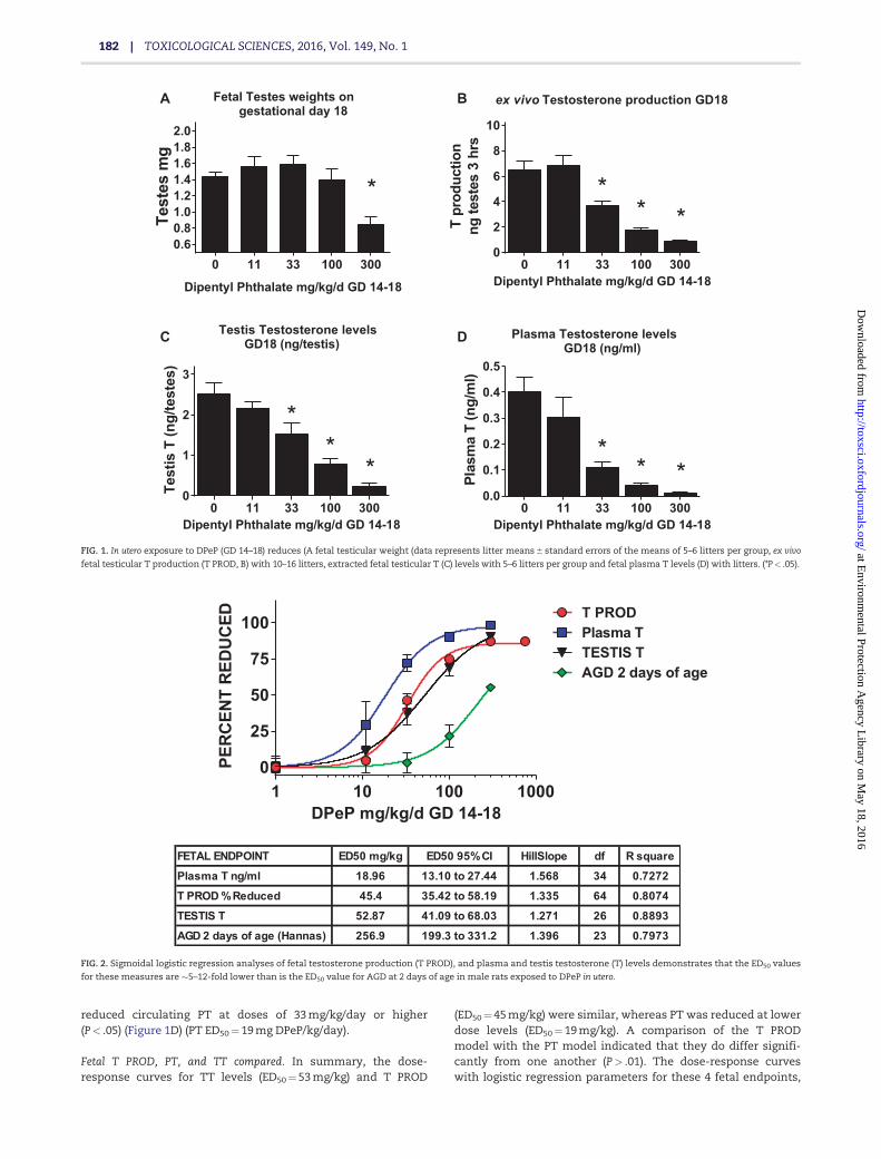
Fetal T PROD, PT, and TT compared. In summary, the dose-response curves for TT levels (ED50¼ 53 mg/kg) and T PROD
(ED50¼ 45 mg/kg) were similar, whereas PT was reduced at lowerdose levels (ED50¼ 19 mg/kg). A comparison of the T PRODmodel with the PT model indicated that they do differ signifi-cantly from one another (P> .01). The dose-response curveswith logistic regression parameters for these 4 fetal endpoints,
Fetal Testes weights ongestational day 18
0 11 33 100 3000.60.81.01.21.41.61.82.0
Dipentyl Phthalate mg/kg/d GD 14-18
Test
esm
g
*
BA
DC
ex vivo Testosterone production GD18
0 11 33 100 3000
2
4
6
8
10
Dipentyl Phthalate mg/kg/d GD 14-18
Tpr
oduc
tion
ngte
stes
3hr
s
***
Testis Testosterone levelsGD18 (ng/testis)
0 11 33 100 3000
1
2
3
Dipentyl Phthalate mg/kg/d GD 14-18
Test
isT
(ng/
test
es)
**
*
Plasma Testosterone levelsGD18 (ng/ml)
0 11 33 100 3000.0
0.1
0.2
0.3
0.4
0.5
Dipentyl Phthalate mg/kg/d GD 14-18
Plas
ma
T(n
g/m
l)**
*
FIG. 1. In utero exposure to DPeP (GD 14–18) reduces (A fetal testicular weight (data represents litter means 6 standard errors of the means of 5–6 litters per group, ex vivo
fetal testicular T production (T PROD, B) with 10–16 litters, extracted fetal testicular T (C) levels with 5–6 litters per group and fetal plasma T levels (D) with litters. (*P< .05).
1 10 100 10000
25
50
75
100
DPeP mg/kg/d GD 14-18
PER
CEN
TR
EDU
CED T PROD
Plasma TTESTIS TAGD 2 days of age
FETAL ENDPOINT ED50 mg/kg ED50 95% CI HillSlope df R square
Plasma T ng/ml 18.96 13.10 to 27.44 1.568 34 0.7272
T PROD % Reduced 45.4 35.42 to 58.19 1.335 64 0.8074
TESTIS T 52.87 41.09 to 68.03 1.271 26 0.8893
AGD 2 days of age (Hannas) 256.9 199.3 to 331.2 1.396 23 0.7973
FIG. 2. Sigmoidal logistic regression analyses of fetal testosterone production (T PROD), and plasma and testis testosterone (T) levels demonstrates that the ED50 values
for these measures are �5–12-fold lower than is the ED50 value for AGD at 2 days of age in male rats exposed to DPeP in utero.
182 | TOXICOLOGICAL SCIENCES, 2016, Vol. 149, No. 1
at Environm
ental Protection Agency L
ibrary on May 18, 2016
http://toxsci.oxfordjournals.org/D
ownloaded from
along with AGD at 2 days of age from the postnatal study, areshown in Figure 2.
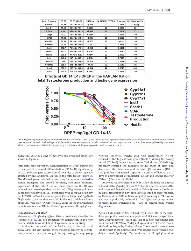
Fetal testis gene expression. Administration of DPeP during thecritical period of sexual differentiation (GD 14–18) significantly(P< .01) reduced gene expression of the suite of genes typicallyaffected by anti-androgen DioPES in the fetal testes (Figure 3).The affected genes included those coding for proteins involved insteroid transport, and steroid hormone, and Insl3 hormone.Expression of the mRNA for all these genes on GD 18 wasreduced in a dose-dependent fashion with ED50 values as low as28 mg DPeP/kg/day (Cyp11b1) compared with 45 mg DPeP/kg/dayfor T PROD. mRNA for several genes (Scarb, Lhcgr, and Cyp17a1)displayed ED50 values that were within the 95% confidence limitsof the ED50 value for T PROD. The ED50 value for the DPeP inducedreduction in testis mRNA for the Insl3 gene was �112 mg/kg/day.
Postnatal Study with DPePMaternal and F1 offspring effects. Effects previously described inHannas et al. (2011a) are presented for comparison to the newpostnatal data beyond 14 days of age from the same study.
Similar to the results of the fetal study, in the postnatalstudy DPeP did not induce overt maternal toxicity or signifi-cantly reduce maternal weight during dosing in any group
whereas maternal weight gain was significantly (P< .02)reduced in the highest dose group (Table 1) during the dosingperiod (GD 8–18). In utero exposure to DPeP during GD 8–18 sig-nificantly reduced the numbers of live pups at birth anddecreased the fetal/neonatal survival [% survival¼ (100 �(100*(number of maternal implants � number of live pups at 2days of age)/number of implants))] at 100 and 300 mg DPeP/kg(Table 1) (Hannas et al., 2011a).
AGD was reduced significantly in 2-day-old male rat pups at100 and 300 mg/kg/day (Figure 2; Table 1) whereas female AGDand male and female body weights (Table 1) were not reducedby DPeP treatment at any dose level at this age (also reportedby Hannas et al., 2011a). Body weight at weaning at 24 days ofage was significantly reduced in the high-dose group. A fewof these males weighed only �60% of control body weight(Figure 4a).
Age and body weight at PPS (PPS-puberty) in male rats. In the high-dose group, the onset and completion of PPS was delayed by inutero DPeP treatment (Figure 4b). Ten of 13 high-dose males hadnot attained full PPS by 55 days of age and in 8 of the 10 males,PPS had not begun by 55 days of age. These results were due tothe fact that these animals had hypospadias rather than a truedelay in male “puberty”. Two males in the 11 mg/kg/day dose
1 10 1000.00
0.25
0.50
0.75
1.00
Effects of GD 14 to18 DPEP in the HARLAN Rat onfetal Testosterone production and testis gene expression
DPEP mg/kg/d GD 14-18
Fold
vers
usC
ontro
l
Cyp11a1Cyp11b1Cyp17a1Insl3Scarb1StARTestosteroneProduction
0Hsd3b
Fetal Endpoint ED 50 ED 50 95% CL HillSlope NUMBER LITTERS R square LEL FROM ANOVA
Cyp11b1 27.95 14.43 to 54.16 -1.242 22 0.8076 33 mg/kg
Scarb1 41.53 23.06 to 74.81 -0.9287 22 0.8363 33T Prod 45.4 35.42 to 58.19 -1.335 66 0.8074 33Lhcgr 55.3 13.51 to 226.4 -0.6606 22 0.4612 100StAR 71.46 45.57 to 112.0 -1.291 22 0.8589 100
Cyp17a1 81.06 53.26 to 123.4 -2.077 22 0.835 100Cyp11a1 97.57 70.04 to 135.9 -1.34 22 0.9024 100
Insl3 112.7 73.98 to 171.6 -1.234 22 0.8431 100Hsd3b 143.7 92.49 to 223.3 -0.9397 22 0.8245 100Wnt7a 236.8 44.76 to 1253 -0.4811 22 0.3652 300Inha 243.4 137.1 to 432.0 -0.7104 22 0.7497 100
Rhox10 266 205.1 to 344.9 -3.314 22 0.8111 300Cyp11b2 305.7 288.5 to 323.9 -44.01 22 0.5689 325
Dhcr7 390.2 257.7 to 590.6 -1.434 22 0.6746 300Nrob1 541.6 106.0 to 2766 -0.3981 22 0.4802 100
FIG. 3. Logistic regression analyses of fetal testosterone production (T PROD) and the mRNA for 13 genes with reduced expression levels as a consequence of in utero
DPeP exposure. Values in the headings are all derived from the 4PL regression model (constrained to 0 and 1.0) except the LEL from the ANOVA (followed by LSMEANS)
which is the lowest dose of DPeP that significantly (P< .05) reduced the gene expression level versus the control.
GRAY ET AL. | 183
at Environm
ental Protection Agency L
ibrary on May 18, 2016
http://toxsci.oxfordjournals.org/D
ownloaded from

group also had not completed PPS by 55 days of age; however,these males did not display penile malformations. Body weightat full PPS was not affected in animals that attained this land-mark by 55 days of age.
Male rat necropsy data collected at 6–7 months of age. In this phaseof the study, we necropsied 30 (5), 23 (5), 18 (5), 14 (4), and 13 (4)
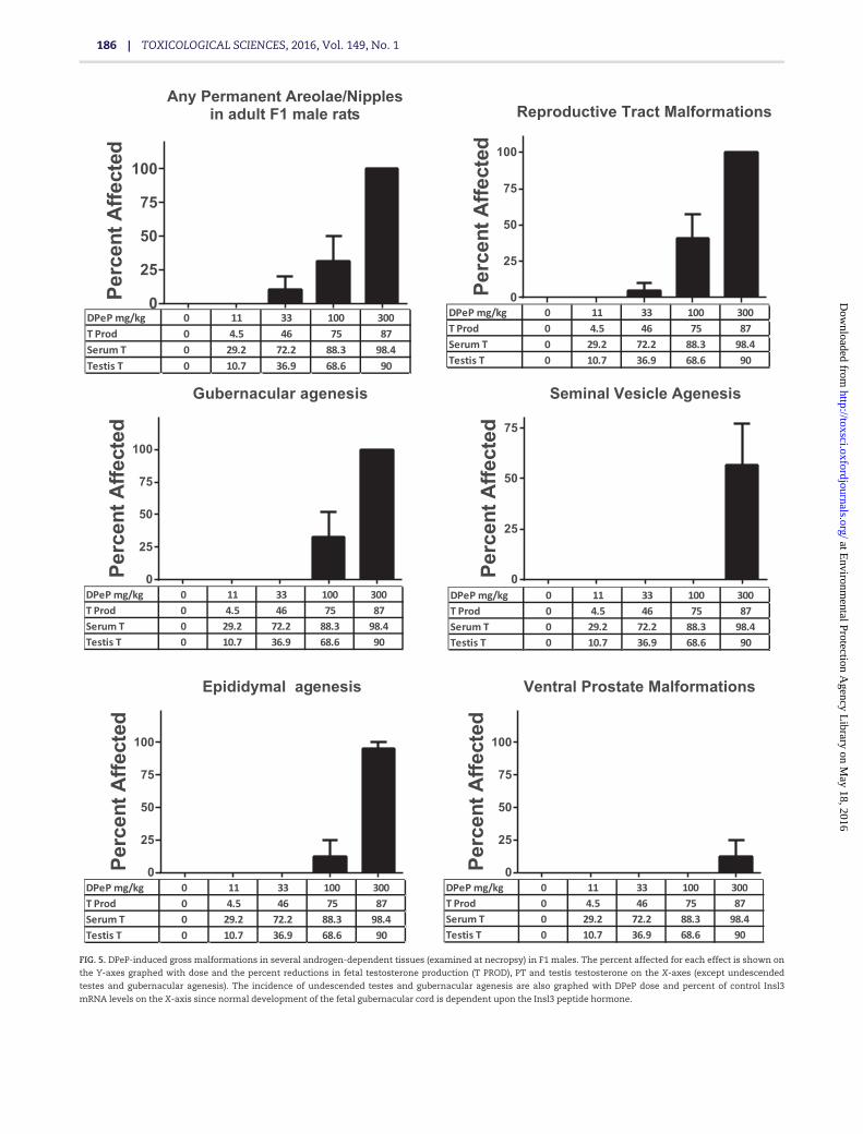
F1 males (litters) in the 0, 11, 33, 100, and 300 mg DPeP/kg/day,respectively. All of the F1 males in the 300 mg/kg/day dosegroup were characterized as displaying combinations of malfor-mations or lesions associated with PS (Table 2); over 90% dis-played gubernacular cord agenesis, agenesis of the vasdeferens, and agenesis of all of a segment of the epididymis and50% displayed penile hypospadias (Table 2; Figure 5).
In the 100 mg/kg/day dose group, F1 males displayed mildlyto severely elongated gubernacular cords or gubernacular agen-esis (36% total of which one displayed complete agenesis),retained female-like nipples (albeit faint, 14%), and epididymalagenesis and testicular abnormalities (7%). In addition, 5.6%(one male) of F1 males exposed to 33 mg DPeP/kg in utero dis-played severe bilateral testicular atrophy. In summary, 0%, 0%,5.6%, 42.8%, and 100% of the F1 males displayed some PS lesionin the 0, 11, 33, 100, and 300 mg DPeP mg/kg/day dose groups,respectively (Figure 5).
The androgen-dependent tissue malformations seen at33 mg DPeP/kg/day were associated with reductions in fetal TPROD, plasma T levels, and testis T of 46%, 72%, and 37%,respectively (Figure 5).
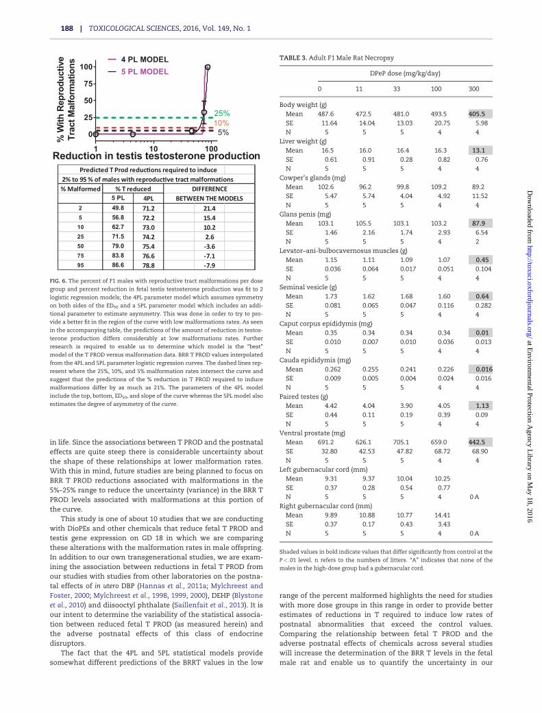
Although the 4PL and 5PL model fits do not differ signifi-cantly from one another, the estimated reductions in T PRODrequired to produce malformations do differ. For example, a 5%malformation rate was predicted to result from a 57% reductionin T PROD with the 5PL model and 72% reduction with the 4PLmodel (Figure 6). While both models have relatively high R2 val-ues of �89%, the slopes are “ambiguous” because they are quitesteep and there are few points on the linear portion of thecurves. The 4PL logistic regression models of the relationshipbetween Insl3 mRNA expression and gubernacular develop-ment and testis non-descent also displayed a R2 values >85%with steep, ambiguous slopes.
F1 male reproductive organ weights and body and liverweight all were reduced by in utero DPeP exposure; however, theeffects were generally significant only in the high-dose group(Table 3). In addition, three of 13 males (23%) exposed in utero to300 mg DPeP/day displayed malocclusion as a result of the skullmalformations (Figs. 7A and B).
Female rat necropsy data collected at 3–4 months of age. Three of 10females exposed in utero to 300 mg DPeP/day also displayed mal-occlusions and skull malformations (Figure 7A) and 1 of 10females in this dose group displayed reproductive tract malfor-mations (uterus unicornis with ipsilateral hydrometrocolpos).In addition, 300 mg DPeP/day permanently reduced F1 femalebody and liver weights but had no effect on kidney, pituitary, orovarian weights (data not shown).
DISCUSSION
Results of this study and other studies (Hannas et al., 2011a,2012), taken together, demonstrate that in utero exposure toDPeP disrupts fetal testicular endocrine function and induces PSreproductive tract malformations in F1 male rats at doses lowerthan most other DioPEs. The ED50s for the postnatal effects arelower than that seen with DEHP (Blystone et al., 2010; Gray et al.,2009) or DBP (Hotchkiss et al., 2010; Mylchreest et al., 1998), orother DioPEs that disrupt fetal male rat endocrine function andpostnatal reproductive development.
Testis T PROD, extracted testis T, and plasma T all were allsignificantly reduced at 33 mg DPeP/kg/day with plasma T
0 11 33 100 300
20
40
60
80
DPeP F1 male Body Weight at Weaning
DPeP mg/kg/d
WEI
GH
T(g
)
0 11 33 100 35
40
45
50
55
60
DPeP male age at Preputial Seperation
DPeP mg/kg/d
Age
atfu
llPP
S
0 11 33 100 300 -5.0×10⁷
0
5.0×10⁷
1.0×10⁸
1.5×10⁸
2.0×10⁸
DPeP male epididymal sperm counts
DPeP mg/kg/d
Mill
ions
FIG. 4. In utero exposure to DPeP on GD 8–18 reduced weaning weight in F1 male
rat offspring at 300 mg/kg/day (A). The age at PPS (puberty) was not delayed in
males without penile malformations (B) Epididymal sperm numbers in males
with an intact epididymis were reduced in a few males in the 33, 100, and
300 mg/kg/day dose groups (C). Data are individual animal values from 4 to 5 lit-
ters/ dose group. Points on the graph represent the values from individual males
in each dose group.
184 | TOXICOLOGICAL SCIENCES, 2016, Vol. 149, No. 1
at Environm
ental Protection Agency L
ibrary on May 18, 2016
http://toxsci.oxfordjournals.org/D
ownloaded from
having a slightly lower ED50 than T PROD or extracted testis T.In addition to reducing T levels in the fetus, DPeP also reducedthe mRNA expression levels for genes involved in steroid trans-port and steroid and Insl3 hormone synthesis; these are thesame genes that are affected by other “active” DioPEs (Hannaset al., 2011b). Furthermore, the relative sensitivity of the mRNAlevels reduced by in utero DPeP exposure is very similar tothat reported for other phthalates. In contrast to the endocrinealterations in the fetal testis, fetal testis weight on GD 18was only reduced at 300 mg/kg/day, the highest dose grouptested.
In the postnatal study, the most sensitive effects to in uteroDPeP were the percent of F1 male rats displaying PS reproduc-tive tract malformations with 5.6% of the males (1 male) dis-playing abnormal testis morphology at 33 mg/kg/day, retained-female-like areolae/nipples (2 of 14 F1 males), and hydroneph-rosis (1 male each exposed to 11 or 33 mg/kg/day). The incidenceand severity of the PS increases with DPeP dose with effects at33 mg DPeP/kg/day being associated with 46%, 37%, and 72%reductions in testis T PROD, extracted testis T and plasma T lev-els, respectively. In addition, pup survival was reduced from94% in controls to 82% in the 33 DPeP mg/kg/day dose group.While none of these effects by themselves were statistically sig-nificant, taken together they could be considered biologicallyrelevant since they are dose-related and typical outcomes afterin utero exposure to other PEs like DBP and DEHP. Statisticallysignificant increases in postnatal abnormalities were notedwhen T Prod, PT, and TT were reduced by 65%, 86%, and 69%,respectively, and all F1-treated males displayed PS malforma-tions when these 3 measures of T were reduced by 85% orhigher.
When the reductions in Insl3 mRNA were compared withthe rates of gubernacular abnormalities and undescendedtestes, dose-related reproductive alterations were notedwhen Insl3 mRNA was reduced by >42%. We did detectpartially undescended, small testes during the fetal necropsiesin males from 1 litter exposed to 33 mg DPeP/kg/day (a dose thatreduces Insl3 mRNA by �20%); however, this effect could havebeen a transient effect and no gubernacular cord alterationswere noted in the postnatal study in this dose group.
The fact that such large reductions in fetal T levels arerequired to induce postnatal reproductive alterations is similar
to the seminiferous tubular fluid (STF) T dose-response requiredto maintain spermatogenesis in the adult rat testis (Zirkin et al.,1989). Zirkin et al. (1989) reported that “Complete spermatogene-sis was maintained despite an 80% reduction in the STF T con-centration from control values to �13 ng/ml. The ability of thetestis to maintain complete spermatogenesis was extremelysensitive to further decreases in STF T concentration. Thus,reduction of the STF T concentration from �13 to 9 ng/mlresulted in a reduction in the number of advanced spermatidsthat were maintained in the testis from �275� 106 to 150� 106.Reduction of the STF T concentration to �4 ng/ml resulted in afurther reduction in the number of advanced spermatids pertestis to about 45� 106. Taken together, these data support thecontention that there is far more T present within the seminif-erous tubules of intact rat testes than is required to maintainquantitatively normal spermatogenesis”.
It is also possible that our estimate of the reductions in T lev-els overestimates the degree to which T is reduced throughoutthe dosing period. We are taking a single measure of these Tlevels on GD 18, the last day of dosing, at a time point when TPROD is maximally affected (about 4 h after the last dose).Therefore, the actual area under the curve for T PROD over the5-day dosing period may not be as dramatically reduced as it isat GD 18 shortly after dosing since the level of T Prod is not con-stant during this developmental stage. T PROD increases by �3-fold from GD 16 to 18 (Hannas et al., 2011a) and, in addition, wewere unable to detect any testis T PROD on GDs 14 and 15 usingthis method.
In addition to the reproductive effects in F1 male rats from inutero DPeP treatment, male and female pup survival also wasreduced in a dose-related manner (P< .05 at 100 and 300 mg/kg/day; Table 1) and some F1 male and female rats in the high-dose group displayed skull defects, malocclusion of the incisors(Figure 7) and some females displayed uterine malformations.These results suggest that toxicity assessments for chemicals inthis class should not focus solely on disruption of endocrine-mediated alterations of sexual differentiation in the male ratreproductive tract.
Our studies to date on PEs have focused on using a broaddose range of each chemical in order to obtain accurate ED50
values for the in utero effects on fetal T PROD, fetal testis geneexpression, and alterations of the male reproductive tract later
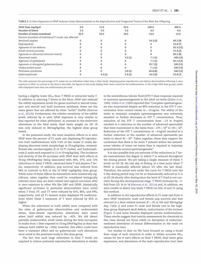
TABLE 2. In Utero Exposure to DPeP Induces Gross Abnormalities in the Reproductive and Urogenital Tracts of the Male Rat Offspring
DPeP dose mg/kg/d 0.0 11.0 33.0 100.0 300.0Number of litters 5.0 5.0 5.0 4.0 4.0Number of males examined 30.0 23.0 18.0 14.0 13.0Percent (number) of individual F1 male rats affectedRetained nipples 0 0 0 14.3 (2) 69.2 (9)Hypospadias 0 0 0 0 53.8 (7)Agenesis of vas deferns 0 0 0 0 92.3 (12)Small ventral prostate 0 0 0 0 15.4 (2)Agenesis or abnormal seminal vesicle 0 0 0 0 69.2 (9)Abnormal testis 0 0 5.6 (1) 7.1 (1) 100 (13)Agenesis of epididymis 0 0 0 7.1 (1) 92.3 (12)Agenesis or elongated gubernaculums 0 0 0 35.7 (5) 100 (13)Undescended testis 0 0 0 7.1 (1) 100 (13)Phthalate syndrome 0 0 5.6 (1) 42.8 (6) 100 (13)Hydronephrosis 0 4.3 (1) 5.6 (1) 14.3 (2) 53.8 (7)
The table presents the percentage of F1 males (on an individual rather than a litter basis), displaying gross reproductive and kidney abnormalities following in utero
exposure to DPeP. In contrast to the data in this table, the figures in this study display litter mean values for the malformations. In the 2 high DPeP dose groups, males
often displayed more than one malformation per male.
GRAY ET AL. | 185
at Environm
ental Protection Agency L
ibrary on May 18, 2016
http://toxsci.oxfordjournals.org/D
ownloaded from
0
25
50
75
100
Any Permanent Areolae/Nipplesin adult F1 male rats
Perc
entA
ffect
ed
DPeP mg/kg 0 11 33 100 300T Prod 0 4.5 46 75 87Serum T 0 29.2 72.2 88.3 98.4Testis T 0 10.7 36.9 68.6 90
0
25
50
75
100
Reproductive Tract Malformations
Perc
entA
ffect
ed
DPeP mg/kg 0 11 33 100 300T Prod 0 4.5 46 75 87Serum T 0 29.2 72.2 88.3 98.4Testis T 0 10.7 36.9 68.6 90
0
25
50
75
100
Gubernacular agenesis
Perc
entA
ffect
ed
DPeP mg/kg 0 11 33 100 300T Prod 0 4.5 46 75 87Serum T 0 29.2 72.2 88.3 98.4Testis T 0 10.7 36.9 68.6 90
0
25
50
75
Seminal Vesicle Agenesis
Perc
entA
ffect
ed
DPeP mg/kg 0 11 33 100 300T Prod 0 4.5 46 75 87Serum T 0 29.2 72.2 88.3 98.4Testis T 0 10.7 36.9 68.6 90
0
25
50
75
100
Epididymal agenesis
Perc
entA
ffect
ed
DPeP mg/kg 0 11 33 100 300T Prod 0 4.5 46 75 87Serum T 0 29.2 72.2 88.3 98.4Testis T 0 10.7 36.9 68.6 90
0
25
50
75
100
Ventral Prostate Malformations
Perc
entA
ffect
ed
DPeP mg/kg 0 11 33 100 300T Prod 0 4.5 46 75 87Serum T 0 29.2 72.2 88.3 98.4Testis T 0 10.7 36.9 68.6 90
FIG. 5. DPeP-induced gross malformations in several androgen-dependent tissues (examined at necropsy) in F1 males. The percent affected for each effect is shown on
the Y-axes graphed with dose and the percent reductions in fetal testosterone production (T PROD), PT and testis testosterone on the X-axes (except undescended
testes and gubernacular agenesis). The incidence of undescended testes and gubernacular agenesis are also graphed with DPeP dose and percent of control Insl3
mRNA levels on the X-axis since normal development of the fetal gubernacular cord is dependent upon the Insl3 peptide hormone.
186 | TOXICOLOGICAL SCIENCES, 2016, Vol. 149, No. 1
at Environm
ental Protection Agency L
ibrary on May 18, 2016
http://toxsci.oxfordjournals.org/D
ownloaded from

0
20
40
60
80
100
HypospadiasPe
rcen
tAffe
cted
DPeP mg/kg 0 11 33 100 300T Prod 0 4.5 46 75 87Serum T 0 29.2 72.2 88.3 98.4Testis T 0 10.7 36.9 68.6 90
0
20
40
60
80
100
Testicular Malformations
Perc
entA
ffect
ed
DPeP mg/kg 0 11 33 100 300T Prod 0 4.5 46 75 87Serum T 0 29.2 72.2 88.3 98.4Testis T 0 10.7 36.9 68.6 90
0
20
40
60
80
100
Vas deferens Agenesis
Perc
entA
ffect
ed
DPeP mg/kg 0 11 33 100 300T Prod 0 4.5 46 75 87Serum T 0 29.2 72.2 88.3 98.4Testis T 0 10.7 36.9 68.6 90
0
20
40
60
80
100
Hydronephrosis
Perc
entA
ffect
ed
DPeP mg/kg 0 11 33 100 300T Prod 0 4.5 46 75 87Serum T 0 29.2 72.2 88.3 98.4Testis T 0 10.7 36.9 68.6 90
0
20
40
60
80
100
Undescended testes
Perc
entA
ffect
ed
DPeP mg/kg 0 11 33 100 300T Prod 0 4.5 46 75 87Serum T 0 29.2 72.2 88.3 98.4Testis T 0 10.7 36.9 68.6 90
020406080
100120
Insl3 mRNA levels vs gubernacularagenesis and testis nondescent
Perc
entA
ffect
ed Gubernacular agenesisUndescended testes
DPeP mg/kg/d 0 11 33 100 300
% Reduc�on in Insl3 mRNA 0 6.94 20.95 42.28 82.14
FIG. 5. Continued
GRAY ET AL. | 187
at Environm
ental Protection Agency L
ibrary on May 18, 2016
http://toxsci.oxfordjournals.org/D
ownloaded from
in life. Since the associations between T PROD and the postnataleffects are quite steep there is considerable uncertainty aboutthe shape of these relationships at lower malformation rates.With this in mind, future studies are being planned to focus onBRR T PROD reductions associated with malformations in the5%–25% range to reduce the uncertainty (variance) in the BRR TPROD levels associated with malformations at this portion ofthe curve.
This study is one of about 10 studies that we are conductingwith DioPEs and other chemicals that reduce fetal T PROD andtestis gene expression on GD 18 in which we are comparingthese alterations with the malformation rates in male offspring.In addition to our own transgenerational studies, we are exam-ining the association between reductions in fetal T PROD fromour studies with studies from other laboratories on the postna-tal effects of in utero DBP (Hannas et al., 2011a; Mylchreest andFoster, 2000; Mylchreest et al., 1998, 1999, 2000), DEHP (Blystoneet al., 2010) and diisooctyl phthalate (Saillenfait et al., 2013). It isour intent to determine the variability of the statistical associa-tion between reduced fetal T PROD (as measured herein) andthe adverse postnatal effects of this class of endocrinedisruptors.
The fact that the 4PL and 5PL statistical models providesomewhat different predictions of the BRRT values in the low
range of the percent malformed highlights the need for studieswith more dose groups in this range in order to provide betterestimates of reductions in T required to induce low rates ofpostnatal abnormalities that exceed the control values.Comparing the relationship between fetal T PROD and theadverse postnatal effects of chemicals across several studieswill increase the determination of the BRR T levels in the fetalmale rat and enable us to quantify the uncertainty in our
1 10 100
0
25
50
75
100
Reduction in testis testosterone production
%W
ithRe
prod
uctiv
eTr
actM
alfo
rmat
ions
25%10%5%
4 PL MODEL5 PL MODEL
Predicted T Prod reduc�ons required to induce2% to 95 % of males with reproduc�ve tract malformations % Malforme Dd IFFERENCE
5 PL 4PL BETWEEN THE MODELS2 49.8 71.2 21.45 56.8 72.2 15.410 62.7 73.0 10.225 71.5 74.2 2.650 79.0 75.4 -3.675 83.8 76.6 -7.195 86.6 78.8 -7.9
% T reduced
FIG. 6. The percent of F1 males with reproductive tract malformations per dose
group and percent reduction in fetal testis testosterone production was fit to 2
logistic regression models; the 4PL parameter model which assumes symmetry
on both sides of the ED50 and a 5PL parameter model which includes an addi-
tional parameter to estimate asymmetry. This was done in order to try to pro-
vide a better fit in the region of the curve with low malformations rates. As seen
in the accompanying table, the predictions of the amount of reduction in testos-
terone production differs considerably at low malformations rates. Further
research is required to enable us to determine which model is the “best”
model of the T PROD versus malformation data. BRR T PROD values interpolated
from the 4PL and 5PL parameter logistic regression curves. The dashed lines rep-
resent where the 25%, 10%, and 5% malformation rates intersect the curve and
suggest that the predictions of the % reduction in T PROD required to induce
malformations differ by as much as 21%. The parameters of the 4PL model
include the top, bottom, ED50, and slope of the curve whereas the 5PL model also
estimates the degree of asymmetry of the curve.
TABLE 3. Adult F1 Male Rat Necropsy
DPeP dose (mg/kg/day)
0 11 33 100 300
Body weight (g)Mean 487.6 472.5 481.0 493.5 405.5SE 11.64 14.04 13.03 20.75 5.98N 5 5 5 4 4
Liver weight (g)Mean 16.5 16.0 16.4 16.3 13.1SE 0.61 0.91 0.28 0.82 0.76N 5 5 5 4 4
Cowper’s glands (mg)Mean 102.6 96.2 99.8 109.2 89.2SE 5.47 5.74 4.04 4.92 11.52N 5 5 5 4 4
Glans penis (mg)Mean 103.1 105.5 103.1 103.2 87.9SE 1.46 2.16 1.74 2.93 6.54N 5 5 5 4 2
Levator–ani-bulbocavernosus muscles (g)Mean 1.15 1.11 1.09 1.07 0.45SE 0.036 0.064 0.017 0.051 0.104N 5 5 5 4 4
Seminal vesicle (g)Mean 1.73 1.62 1.68 1.60 0.64SE 0.081 0.065 0.047 0.116 0.282N 5 5 5 4 4
Caput corpus epididymis (mg)Mean 0.35 0.34 0.34 0.34 0.01SE 0.010 0.007 0.010 0.036 0.013N 5 5 5 4 4
Cauda epididymis (mg)Mean 0.262 0.255 0.241 0.226 0.016SE 0.009 0.005 0.004 0.024 0.016N 5 5 5 4 4
Paired testes (g)Mean 4.42 4.04 3.90 4.05 1.13SE 0.44 0.11 0.19 0.39 0.09N 5 5 5 4 4
Ventral prostate (mg)Mean 691.2 626.1 705.1 659.0 442.5SE 32.80 42.53 47.82 68.72 68.90N 5 5 5 4 4
Left gubernacular cord (mm)Mean 9.31 9.37 10.04 10.25SE 0.37 0.28 0.54 0.77N 5 5 5 4 0 A
Right gubernacular cord (mm)Mean 9.89 10.88 10.77 14.41SE 0.37 0.17 0.43 3.43N 5 5 5 4 0 A
Shaded values in bold indicate values that differ significantly from control at the
P< .01 level. n refers to the numbers of litters. “A” indicates that none of the
males in the high-dose group had a gubernacular cord.
188 | TOXICOLOGICAL SCIENCES, 2016, Vol. 149, No. 1
at Environm
ental Protection Agency L
ibrary on May 18, 2016
http://toxsci.oxfordjournals.org/D
ownloaded from

DPeP SKULL ZYGOMATIC ARCH LENGTHLEFT TO RIGHT VARIATION (mm)
0 11 33 100
300
0
2
4
6
8
10
DEL
TAZY
GO
MA
TIC
AR
CH
LEN
GTH
S(m
m)
FIG. 7. Examples of the malformed skulls of F1 males and females exposed to 300 mg DPeP/kg are shown in the photographs with a control male and female skull and a
treated male and female skull. The graph presents one skull measure that quantifies this lesion—“Delta zygomatic arch lengths (mm)” which represents the absolute
value of the length of one arch subtracted from the other.
GRAY ET AL. | 189
at Environm
ental Protection Agency L
ibrary on May 18, 2016
http://toxsci.oxfordjournals.org/D
ownloaded from
predictions of the levels of malformations that result from spe-cific reductions in fetal T PROD.
FUNDING
Supported in part by NIH NTP/NIEHS IA RW7592285501-1.
ACKNOWLEDGMENTS
We thank Brandy Riffle, Nicola Evans, Mary Cardon, andPhillip Hartig for the excellent technical assistance with sev-eral phases of this research project. We also thank Drs AllenDavis and Vicki Sutherland for their thorough and construc-tive reviews of the manuscript.
SUPPLEMENTARY DATA
Supplementary data are available online at http://toxsci.oxfordjournals.org/.
REFERENCESBlystone, C. R., Kissling, G. E., Bishop, J. B., Chapin, R. E., Wolfe, G.
W., and Foster, P. M. (2010). Determination of the di-(2-ethylhexyl) phthalate NOAEL for reproductive developmentin the rat: importance of the retention of extra animals toadulthood. Toxicol. Sci. 116, 640–646.
Carruthers, C. M., and Foster, P. M. (2005). Critical window ofmale reproductive tract development in rats following gesta-tional exposure to di-n-butyl phthalate. Birth Defects Res. 74,277–285.
Feldman, S. C., and Bloch, E. (1978). Developmental pattern oftestosterone synthesis by fetal rat testes in response to lutei-nizing hormone. Endocrinology 102, 999–1007.
Furr, J. R., Lambright, C. S., Wilson, V. S., Foster, P. M., and Gray,L. E., Jr (2014). A short-term in vivo screen using fetal testos-terone production, a key event in the phthalate adverse out-come pathway, to predict disruption of sexualdifferentiation. Toxicol. Sci. 140, 403–424.
Gangnerau, M. N., and Picon, R. (1987). Onset of steroidogenesisand differentiation of functional LH receptors in rat fetal tes-ticular cultures. Arch. Androl. 18, 215–224.
Gray, L. E., and Foster, P. M. D. (2003). Significance of experimen-tal studies for assessing adverse effects of endocrine-disrupting chemicals. Pure Appl. Chem. 75, 2125–2141.
Gray, L. E., Jr, Barlow, N. J., Howdeshell, K. L., Ostby, J. S., Furr,J. R., and Gray, C. L. (2009). Transgenerational effects of Di (2-ethylhexyl) phthalate in the male CRL:CD(SD) rat: addedvalue of assessing multiple offspring per litter. Toxicol. Sci.110, 411–425.
Gray, L. E., Jr, Laskey, J., and Ostby, J. (2006). Chronic di-n-butylphthalate exposure in rats reduces fertility and alters ovar-ian function during pregnancy in female Long Evans hoodedrats. Toxicol. Sci. 93, 189–195.
Gray, L. E., Jr, Ostby, J. S., and Kelce, W. R. (1994). Developmentaleffects of an environmental antiandrogen: the fungicide vin-clozolin alters sex differentiation of the male rat. Toxicol.Appl. Pharmacol. 129, 46–52.
Hannas, B. R., Furr, J., Lambright, C. S., Wilson, V. S., Foster, P. M.,and Gray, L. E., Jr (2011a). Dipentyl phthalate dosing duringsexual differentiation disrupts fetal testis function and post-natal development of the male Sprague-Dawley rat withgreater relative potency than other phthalates. Toxicol. Sci.120, 184–193.
Hannas, B. R., Howdeshell, K. L., and Furr, J. (2013). In uterophthalate effects in the female rat: a model for MRKH syn-drome. Toxicol. Lett. 223, 315–321.
Hannas, B. R., Lambright, C. S., Furr, J., Evans, N., Foster, P. M.,Gray, E. L., and Wilson, V. S. (2012). Genomic biomarkers ofphthalate-induced male reproductive developmental toxic-ity: a targeted RT-PCR array approach for defining relativepotency. Toxicol. Sci. 125, 544–557.
Hannas, B. R., Lambright, C. S., Furr, J., Howdeshell, K. L., Wilson,V. S., and Gray, L. E., Jr (2011b). Dose-response assessment offetal testosterone production and gene expression levels inrat testes following in utero exposure to diethylhexyl phthal-ate, diisobutyl phthalate, diisoheptyl phthalate, and diiso-nonyl phthalate. Toxicol. Sci. 123, 206–216.
Hellwig, J., van Ravenzwaay, B., Mayer, M., and Gembardt, C.(2000). Pre- and postnatal oral toxicity of vinclozolin in Wistarand Long-Evans rats. Regul. Toxicol. Pharmacol. 32, 42–50.
Hosokawa, S., Murakami, M., Ineyama, M., Yamada, T., Koyama,Y., Okuno, Y., Yoshitake, A., Yamada, H., and Miyamoto, J.(1993). Effects of procymidone on reproductive organs andserum gonadotropins in male rats. J. Toxicol. Sci. 18, 111–124.
Hotchkiss, A. K., Rider, C. V., Furr, J., Howdeshell, K. L., Blystone,C. R., Wilson, V. S., and Gray, L. E., Jr (2010). In utero exposureto an AR antagonist plus an inhibitor of fetal testosteronesynthesis induces cumulative effects on F1 male rats. Reprod.Toxicol. (Elmsford, N.Y) 30, 261–270.
Johnson, K. J., McDowell, E. N., Viereck, M. P., and Xia, J. Q. (2011).Species-specific dibutyl phthalate fetal testis endocrine dis-ruption correlates with inhibition of SREBP2-dependent geneexpression pathways. Toxicol. Sci. 120, 460–474.
Kluwe, W. M. (1982). Overview of phthalate ester pharmacoki-netics in mammalian species. Environ. Health Perspect. 45, 3–9.
Majdic, G., Saunders, P. T., and Teerds, K. J. (1998).Immunoexpression of the steroidogenic enzymes 3-betahydroxysteroid dehydrogenase and 17 alpha-hydroxylase,C17,20 lyase and the receptor for luteinizing hormone (LH) inthe fetal rat testis suggests that the onset of Leydig cell ste-roid production is independent of LH action. Biol. Reprod. 58,520–525.
McIntyre, B. S., Barlow, N. J., and Foster, P. M. (2001). Androgen-mediated development in male rat offspring exposed toflutamide in utero: permanence and correlation of earlypostnatal changes in anogenital distance and nipple reten-tion with malformations in androgen-dependent tissues.Toxicol. Sci. 62, 236–249.
Miyata, K., Yabushita, S., Sukata, T., Sano, M., Yoshino, H.,Nakanishi, T., Okuno, Y., and Matsuo, M. (2002). Effects ofperinatal exposure to flutamide on sex hormones andandrogen-dependent organs in F1 male rats. J. Toxicol. Sci. 27,19–33.
Mylchreest, E., Cattley, R. C., and Foster, P. M. D. (1998). Male re-productive tract malformations in rats following gestationaland lactational exposure to di(n-butyl) phthalate: an antian-drogenic mechanism? Toxicol. Sci. 43, 47–60.
Mylchreest, E., and Foster, P. M. (2000). DBP exerts its antiandro-genic activity by indirectly interfering with androgen signal-ing pathways. Toxicol. Appl. Pharmacol. 168, 174–175.
Mylchreest, E., Sar, M., Cattley, R. C., and Foster, P. M. (1999).Disruption of androgen-regulated male reproductive devel-opment by di(n-butyl) phthalate during late gestation inrats is different from flutamide. Toxicol. Appl. Pharmacol. 156,81–95.
Mylchreest, E., Wallace, D. G., Cattley, R. C., and Foster, P. M.(2000). Dose-dependent alterations in androgen-regulated
190 | TOXICOLOGICAL SCIENCES, 2016, Vol. 149, No. 1
at Environm
ental Protection Agency L
ibrary on May 18, 2016
http://toxsci.oxfordjournals.org/D
ownloaded from
male reproductive development in rats exposed to Di(n-bu-tyl) phthalate during late gestation. Toxicol. Sci. 55, 143–151.
Ostby, J., Kelce, W. R., Lambright, C., Wolf, C. J., Mann, P., andGray, L. E., Jr (1999). The fungicide procymidone alters sexualdifferentiation in the male rat by acting as an androgen-recep-tor antagonist in vivo and in vitro. Toxicol. Ind. Health 15, 80–93.
Saillenfait, A. M., Sabate, J. P., Robert, A., Cossec, B., Roudot, A. C.,Denis, F., and Burgart, M. (2013). Adverse effects of diisooctylphthalate on the male rat reproductive development followingprenatal exposure. Reprod. Toxicol. (Elmsford, N.Y.) 42, 192–202.
van Ravenzwaay, B., Kolle, S. N., Ramirez, T., and Kamp, H. G.(2013). Vinclozolin: a case study on the identification of
endocrine active substances in the past and a future perspec-tive. Toxicol. Lett. 223, 271–279.
Warren, D. W., Haltmeyer, G. C., and Eik-Nes, K. B. (1973).Testosterone in the fetal rat testis. Biol. Reprod. 8, 560–565.
Yasunaga, R., Ikuta, J., Murata, Y., Inoue, K., Koga, H., Masaki, T.,and Tamura, H. (2013). Ligand-independent androgen recep-tor antagonism caused by the newly developed pesticide pyr-ifluquinazon (PFQ). Reprod. Toxicol. (Elmsford, N.Y.) 35, 1–6.
Zirkin, B. R., Santulli, R., Awoniyi, C. A., and Ewing, L. L. (1989).Maintenance of advanced spermatogenic cells in the adultrat testis: quantitative relationship to testosterone concen-tration within the testis. Endocrinology 124, 3043–3049.
GRAY ET AL. | 191
at Environm
ental Protection Agency L
ibrary on May 18, 2016
http://toxsci.oxfordjournals.org/D
ownloaded from