Essentials of Human · 2018-02-03 · Essentials series The Essentials are an international,...
30
Transcript of Essentials of Human · 2018-02-03 · Essentials series The Essentials are an international,...


Essentials of Human Disease in Dentistry
Essentials series
The Essentials are an international, best‐selling series of textbooks, all of which are designed to support lecture series or themes on core topics within the health sciences.
Roitt’s Essential Immunology, 13th Editionby Peter J. Delves, Seamus J. Martin, Dennis R. Burton, Ivan M. RoittNovember 2016
Essential Practical Prescribingby Georgia Woodfield, Benedict Lyle Phillips, Victoria Taylor, Amy Hawkins, Andrew StantonApril 2016
Essential Primary Careby Andrew Blythe (Editor), Jessica Buchan (Editor)March 2016
Hoffbrand’s Essential Haematology, 7th Editionby A. Victor Hoffbrand, Paul A. H. MossSeptember 2015
Essentials of Human Disease
in DentistrySecond Edition
Mark Greenwood BDS, MDS, PhD, FDSRCS, MB ChB, FRCS (Eng. and Ed.),
FRCS (OMFS), FHEANHS Consultant, Oral and Maxillofacial Surgery
Honorary Clinical Professor of Medical EducationDentistry at the School of Dental Sciences
Newcastle UniversityNewcastle upon Tyne, UK

This edition first published 2018© 2018 John Wiley & Sons Ltd
Edition HistoryJohn Wiley & Sons (1e, 2009)
All rights reserved. No part of this publication may be reproduced, stored in a retrieval system, or transmitted, in any form or by any means, electronic, mechanical, photocopying, recording or otherwise, except as permitted by law. Advice on how to obtain permission to reuse material from this title is available at http://www.wiley.com/go/permissions.
The right of Mark Greenwood to be identified as the author of this work has been asserted in accordance with law.
Registered OfficesJohn Wiley & Sons, Inc., 111 River Street, Hoboken, NJ 07030, USAJohn Wiley & Sons Ltd, The Atrium, Southern Gate, Chichester, West Sussex, PO19 8SQ, UK
Editorial Office9600 Garsington Road, Oxford, OX4 2DQ, UK
For details of our global editorial offices, customer services, and more information about Wiley products visit us at www.wiley.com.
Wiley also publishes its books in a variety of electronic formats and by print‐on‐demand. Some content that appears in standard print versions of this book may not be available in other formats.
Limit of Liability/Disclaimer of Warranty
The contents of this work are intended to further general scientific research, understanding, and discussion only and are not intended and should not be relied upon as recommending or promoting scientific method, diagnosis, or treatment by physicians for any particular patient. In view of ongoing research, equipment modifications, changes in governmental regulations, and the constant flow of information relating to the use of medicines, equipment, and devices, the reader is urged to review and evaluate the information provided in the package insert or instructions for each medicine, equipment, or device for, among other things, any changes in the instructions or indication of usage and for added warnings and precautions. While the publisher and authors have used their best efforts in preparing this work, they make no representations or warranties with respect to the accuracy or completeness of the contents of this work and specifically disclaim all warranties, including without limitation any implied warranties of merchantability or fitness for a particular purpose. No warranty may be created or extended by sales representatives, written sales materials or promotional statements for this work. The fact that an organization, website, or product is referred to in this work as a citation and/or potential source of further information does not mean that the publisher and authors endorse the information or services the organization, website, or product may provide or recommendations it may make. This work is sold with the understanding that the publisher is not engaged in rendering professional services. The advice and strategies contained herein may not be suitable for your situation. You should consult with a specialist where appropriate. Further, readers should be aware that websites listed in this work may have changed or disappeared between when this work was written and when it is read. Neither the publisher nor authors shall be liable for any loss of profit or any other commercial damages, including but not limited to special, incidental, consequential, or other damages.
Library of Congress Cataloging‐in‐Publication Data
Names: Greenwood, M. (Mark), author.Title: Essentials of human disease in dentistry / by Mark Greenwood.Other titles: Textbook of human disease in dentistryDescription: Second edition. | Hoboken, NJ : Wiley, 2018. | Preceded by Textbook of human
disease in dentistry / Mark Greenwood, Robin A. Seymour, and John G. Meechan. 2009. | Includes bibliographical references and index. |
Identifiers: LCCN 2017040601 (print) | LCCN 2017041146 (ebook) | ISBN 9781119251828 (pdf ) | ISBN 9781119251859 (epub) | ISBN 9781119251842 (pbk.)
Subjects: | MESH: Dental Care–methods | Clinical Medicine–methods | Disease | Comprehensive Dental Care | Handbooks
Classification: LCC RK305 (ebook) | LCC RK305 (print) | NLM WU 49 | DDC 617.6/3–dc23LC record available at https://lccn.loc.gov/2017040601
Cover Design: WileyCover Image: © MedicalRF.com/Gettyimages
Set in 10/12pt Adobe Garamond by SPi Global, Pondicherry, India
10 9 8 7 6 5 4 3 2 1
Contributors viiiPreface to the first edition xiPreface to the second edition xiiAcknowledgements xiiiAbout the companion website xiv
1 Clinical examination and history taking 1M Greenwood
2 Inflammation and anti‑inflammatory drugs 11CM Robinson and RA Seymour
3 Principles of infection and infection control 21S Waugh, E Ong, S Hogg, JG Meechan, RA Seymour, M Greenwood, C Taylor and G Toms
3A Sterilisation, disinfection and antiseptics 22
3B Principles of infection and infection control, diagnosis and treatment of bacterial infections 27
3C Viruses and antiviral agents relevant to dentistry 38
3D Infection with immunodeficiency virus and implications for the oral cavity: Infection with HIV 46
3E Fungi and antifungal agents 53
4 Immunological disease 55C Stroud and H Bourne
5 Cardiovascular disorders 67RH Jay, G Stansby, T Barakat, RA Seymour, JG Meechan, CM Robinson, M Greenwood, S Hogg and A Balakrishnan
5A Introduction to cardiovascular disease (CVD) 68
5B Heart failure 78
5C Cardiac arrhythmias 81
5D Valvular heart disease 84
5E Hypertension 87
5F Anticoagulants, drugs affecting blood clotting 92
5G Dental implications of CVD 95
5H Peripheral vascular and cardiac surgical disorders 97
Contents
vi / Contents
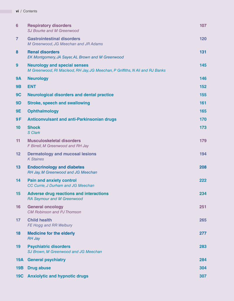
6 Respiratory disorders 107SJ Bourke and M Greenwood
7 Gastrointestinal disorders 120M Greenwood, JG Meechan and JR Adams
8 Renal disorders 131EK Montgomery, JA Sayer, AL Brown and M Greenwood
9 Neurology and special senses 145M Greenwood, RI Macleod, RH Jay, JG Meechan, P Griffiths, N Ali and RJ Banks
9A Neurology 146
9B ENT 152
9C Neurological disorders and dental practice 155
9D Stroke, speech and swallowing 161
9E Ophthalmology 165
9 F Anticonvulsant and anti‐Parkinsonian drugs 170
10 Shock 173S Clark
11 Musculoskeletal disorders 179F Birrell, M Greenwood and RH Jay
12 Dermatology and mucosal lesions 194K Staines
13 Endocrinology and diabetes 208RH Jay, M Greenwood and JG Meechan
14 Pain and anxiety control 222CC Currie, J Durham and JG Meechan
15 Adverse drug reactions and interactions 234RA Seymour and M Greenwood
16 General oncology 251CM Robinson and PJ Thomson
17 Child health 265FE Hogg and RR Welbury
18 Medicine for the elderly 277RH Jay
19 Psychiatric disorders 283SJ Brown, M Greenwood and JG Meechan
19A General psychiatry 284
19B Drug abuse 304
19C Anxiolytic and hypnotic drugs 307

Contents / vii
20 Haematology 309J Hanley, A Lennard and S Mathia
20A General haematology, haemato‐oncology 310
20B Haemostasis 322
20C Transfusion medicine 329
21 Medical emergencies 334M Greenwood
Appendix: normal reference ranges 346Index 347
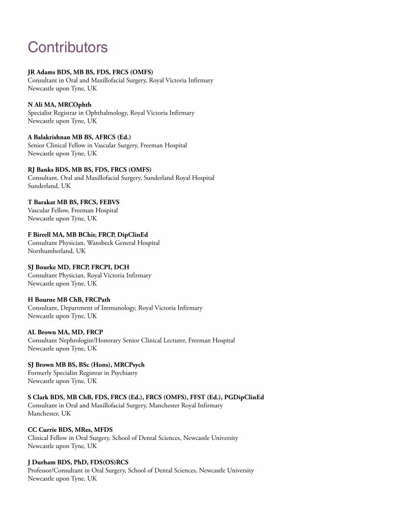
JR Adams BDS, MB BS, FDS, FRCS (OMFS)Consultant in Oral and Maxillofacial Surgery, Royal Victoria InfirmaryNewcastle upon Tyne, UK
N Ali MA, MRCOphthSpecialist Registrar in Ophthalmology, Royal Victoria InfirmaryNewcastle upon Tyne, UK
A Balakrishnan MB BS, AFRCS (Ed.)Senior Clinical Fellow in Vascular Surgery, Freeman HospitalNewcastle upon Tyne, UK
RJ Banks BDS, MB BS, FDS, FRCS (OMFS)Consultant, Oral and Maxillofacial Surgery, Sunderland Royal HospitalSunderland, UK
T Barakat MB BS, FRCS, FEBVSVascular Fellow, Freeman HospitalNewcastle upon Tyne, UK
F Birrell MA, MB BChir, FRCP, DipClinEdConsultant Physician, Wansbeck General HospitalNorthumberland, UK
SJ Bourke MD, FRCP, FRCPI, DCHConsultant Physician, Royal Victoria InfirmaryNewcastle upon Tyne, UK
H Bourne MB ChB, FRCPathConsultant, Department of Immunology, Royal Victoria InfirmaryNewcastle upon Tyne, UK
AL Brown MA, MD, FRCPConsultant Nephrologist/Honorary Senior Clinical Lecturer, Freeman HospitalNewcastle upon Tyne, UK
SJ Brown MB BS, BSc (Hons), MRCPsychFormerly Specialist Registrar in PsychiatryNewcastle upon Tyne, UK
S Clark BDS, MB ChB, FDS, FRCS (Ed.), FRCS (OMFS), FFST (Ed.), PGDipClinEdConsultant in Oral and Maxillofacial Surgery, Manchester Royal InfirmaryManchester, UK
CC Currie BDS, MRes, MFDSClinical Fellow in Oral Surgery, School of Dental Sciences, Newcastle UniversityNewcastle upon Tyne, UK
J Durham BDS, PhD, FDS(OS)RCSProfessor/Consultant in Oral Surgery, School of Dental Sciences, Newcastle UniversityNewcastle upon Tyne, UK
Contributors

Contributors / ix
M Greenwood BDS, MDS, PhD, FDSRCS, MB ChB, FRCS (Eng. and Ed.), FRCS (OMFS), FHEAConsultant/Clinical Professor, Oral and Maxillofacial Surgery UnitNewcastle Dental Hospital/Royal Victoria InfirmaryNewcastle upon Tyne, UK
P Griffiths FRCS, FRCOphthConsultant/Senior Lecturer in Ophthalmology, Royal Victoria InfirmaryNewcastle upon Tyne, UK
J Hanley MB ChB, MD, FRCP, FRCPathConsultant Haematologist, Royal Victoria InfirmaryNewcastle upon Tyne, UK
FE Hogg BDS, MFDS, MPaedDent, Med, FHEASpecialist Registrar in Paediatric DentistryGlasgow Dental Hospital/Royal Hospital for ChildrenGlasgow, UK
S Hogg BSc, PhD, FHEAFormerly Senior Lecturer, School of Dental Sciences, Newcastle UniversityNewcastle upon Tyne, UK
RH Jay MA, MD, FRCP, DipClinEdConsultant Physician, Royal Victoria InfirmaryNewcastle upon Tyne, UK
A Lennard MB BS (Hons), FRCP, FRCPathConsultant Haematologist/Honorary Senior Lecturer, Royal Victoria InfirmaryNewcastle upon Tyne, UK
RI Macleod BDS, PhD, FDSRCSE, DDRRCRConsultant in Oral and Maxillofacial Radiology (retired), School of Dental SciencesNewcastle UniversityNewcastle upon Tyne, UK
S Mathia MB BS, MRCP (UK)Specialist Registrar in Haematology, Royal Victoria InfirmaryNewcastle upon Tyne, UK
JG Meechan BDS, BSc, PhD, FDSRCPS, FDSRCS (Ed.)Honorary Consultant/Senior Lecturer (retired), School of Dental SciencesNewcastle UniversityNewcastle upon Tyne, UK
EK Montgomery MB BS, MRCP, NephrolConsultant Nephrologist, Newcastle Hospitals NHS Foundation TrustNewcastle upon Tyne, UK
E Ong MB BS, MSc, FRCP, FRCPI, DTM&HConsultant Physician/Honorary Senior Lecturer in MedicineDepartment of Infectious Diseases and Tropical Medicine, Royal Victoria InfirmaryNewcastle upon Tyne, UK
x / Contributors
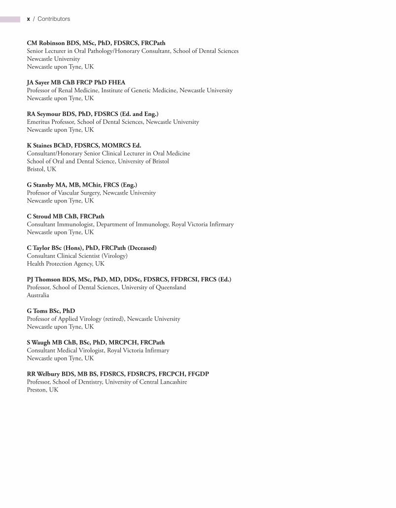
CM Robinson BDS, MSc, PhD, FDSRCS, FRCPathSenior Lecturer in Oral Pathology/Honorary Consultant, School of Dental SciencesNewcastle UniversityNewcastle upon Tyne, UK
JA Sayer MB ChB FRCP PhD FHEAProfessor of Renal Medicine, Institute of Genetic Medicine, Newcastle UniversityNewcastle upon Tyne, UK
RA Seymour BDS, PhD, FDSRCS (Ed. and Eng.)Emeritus Professor, School of Dental Sciences, Newcastle UniversityNewcastle upon Tyne, UK
K Staines BChD, FDSRCS, MOMRCS Ed.Consultant/Honorary Senior Clinical Lecturer in Oral MedicineSchool of Oral and Dental Science, University of BristolBristol, UK
G Stansby MA, MB, MChir, FRCS (Eng.)Professor of Vascular Surgery, Newcastle UniversityNewcastle upon Tyne, UK
C Stroud MB ChB, FRCPathConsultant Immunologist, Department of Immunology, Royal Victoria InfirmaryNewcastle upon Tyne, UK
C Taylor BSc (Hons), PhD, FRCPath (Deceased)Consultant Clinical Scientist (Virology)Health Protection Agency, UK
PJ Thomson BDS, MSc, PhD, MD, DDSc, FDSRCS, FFDRCSI, FRCS (Ed.)Professor, School of Dental Sciences, University of QueenslandAustralia
G Toms BSc, PhDProfessor of Applied Virology (retired), Newcastle UniversityNewcastle upon Tyne, UK
S Waugh MB ChB, BSc, PhD, MRCPCH, FRCPathConsultant Medical Virologist, Royal Victoria InfirmaryNewcastle upon Tyne, UK
RR Welbury BDS, MB BS, FDSRCS, FDSRCPS, FRCPCH, FFGDPProfessor, School of Dentistry, University of Central LancashirePreston, UK

The concept of human disease teaching in dentistry arose out of General Dental Council recommendations in their report ‘The First Five Years’, published in 2002 and currently under revision. This suggested an integrated approach to the teaching of general medicine and surgery, and the related disciplines. There are slight variations in the subjects incorporated into human disease teaching across UK dental schools, but general medicine and surgery teaching is common to all. Pharmacology, pathology and microbiology are also included in the course at Newcastle University, and the material in this book forms the basis of the theoretical elements of that course.
A dentist should have a firm grounding in all these disciplines to facilitate a broader knowledge and understanding of human disease, which can then be focused on issues of direct relevance to dentistry. This approach enables informed dialogue with colleagues in other healthcare professions, including general medical practitioners.
It is hoped that this book will prove useful to dental undergraduates studying human disease, and that it will also be of use to candidates preparing for the membership examinations of the Royal Colleges of Surgeons.
M GreenwoodRA SeymourJG Meechan
Newcastle upon Tyne, UKJanuary 2009
Preface to the first edition

This second edition of Textbook of Human Disease in Dentistry, now called Essentials of Human Disease in Dentistry, aims to build on the strengths of the first edition, but has been updated in several key areas. The underlying principle remains the same – to provide the dental student/dental practitioner with a firm back-ground knowledge of the relevant aspects of general medicine and surgery, pharmacology, pathology and microbiology. New additions to this edition include multiple choice–style questions at the end of each chapter, together with relevant references.
It is hoped that this edition will continue to be of value to undergraduate students of dentistry and those preparing for their postgraduate dental examinations.
M GreenwoodNewcastle upon Tyne, UK
August 2017
Preface to the second edition

I would like to acknowledge the contributing authors of this book, both for this text and the first edition, and for the invaluable help they give to the smooth running of the undergraduate course in Newcastle.
Acknowledgement is also due to Iain Macleod for his help with some of the illustrations, David Sales for many of the line drawings, and Beryl Leggatt and S Wilkinson for the secretarial help.
Where illustrations have been imported from other publications, due acknowledgement has been given.
Acknowledgements

About the companion website
Don’t forget to visit the companion website for this book:
www.wiley.com/go/greenwood/human‐disease‐in‐dentistry
The companion website provides all the figures from the book in PowerPoint for download.
Scan this QR code to visit the companion website:

Essentials of Human Disease in Dentistry, Second Edition. Mark Greenwood. © 2018 John Wiley & Sons Ltd. Published 2018 by John Wiley & Sons Ltd.Companion website: www.wiley.com/go/greenwood/human-disease-in-dentistry
1
Key topics
◾ Essential components of a medical history
◾ Key issues that may arise from the medical history
Learning objectives
◾ To be familiar with the main components of a medical history.
◾ To be aware of the medical terms used in taking a medical history, and their meaning.
◾ To be aware of the normal vital signs.
Clinical examination and history takingM Greenwood
CHAPTER 1

2 / Chapter 1: Clinical examination and history taking
Components of a medical history
The medical history aims to: ▪ Enable the formulation of a differential diagnosis or
diagnosis ▪ Put the patient’s disease process into the correct medical and
social context. ▪ Establish a rapport with the patient.
Clinicians engaged in obtaining medical histories should introduce themselves to the patients and give their designa-tions. The taking of the history may then commence and should follow a scheme similar to that shown in Table 1.1.
Presenting complaint
The presenting complaint can be recorded in medical terms, but often is better expressed in the patient’s own words. When recording the history in writing, quotation marks should be placed around the patient’s words. In a verbal case presenta-tion, it should be stated that the patient’s own words are being used. It is important to avoid presumptive diagnoses in the pre-senting complaint. For example, patients do not present with iron deficiency anaemia; they may present with symptoms that arise from it. It should be remembered that symptoms are the features of the illness that the patient describes; signs are physi-cal findings obtained by the clinician.
History of the presenting complaint
The history of the presenting complaint should be a chrono-logical but succinct account of the patient’s problem. It is important to start at the onset of the problem and describe its progression. Symptoms should be similarly described.
Points to include when asking patients about pain are as follows: ▪ Site ▪ Character – for example, tight/band-like (in the chest,
suggestive of cardiac origin) ▪ Does the pain radiate anywhere? ▪ Onset – sudden or gradual
▪ Severity (ask the patient to rate on a scale of 1–10, with 10 being the most severe)
▪ Duration ▪ Exacerbating/relieving factors (including the use and
efficacy of medication) ▪ Preceding events or associated features ▪ Has the pain occurred before? / Is it getting better or
worse?
Past medical history
It is worth asking a generic set of opening questions – for exam-ple, ‘Do you have any heart or chest problems?’ Questioning should then focus on specific disorders – for example, asthma, diabetes, epilepsy, hypertension, hepatitis, jaundice or tubercu-losis. It is also worth specifically asking about any previous problems with the arrest of haemorrhage. Past problems with intravenous sedation or general anaesthesia should be noted. It is worth asking about any previous history of rheumatic fever, which may have led to cardiac valve damage. In 2008, the National Institute for Health and Care Excellence (NICE) discontinued the regular use of antibiotic prophylaxis for bacteraemia-producing dental procedures in patients with car-diac damage. There were some concerns about this, however, such as the lack of an evidence base for prophylaxis and the fact that Europe and the USA differed in their practices.
Before 2008, a consistent upward trend was apparent in the population-corrected incidence of infective endocarditis in England. Soon after the implementation of the NICE guide-lines, the slope of the trend line increased further, although there is no direct evidence that this was due to the discontinua-tion of antibiotic prophylaxis in dentistry. In 2016, NICE mod-ified the guidance slightly to state that: ‘Antibiotic prophylaxis against infective endocarditis is not recommended routinely [my emphasis] for people undergoing dental procedures’. This addition emphasises NICE’s standard advice on healthcare professionals’ responsibilities. Doctors and dentists should offer the most appropriate treatment options, in consultation with their patients and/or their carers or guardians. In doing so, they should take into account the recommendations of NICE guid-ance and the values and preferences of patients, and also apply their clinical judgement.
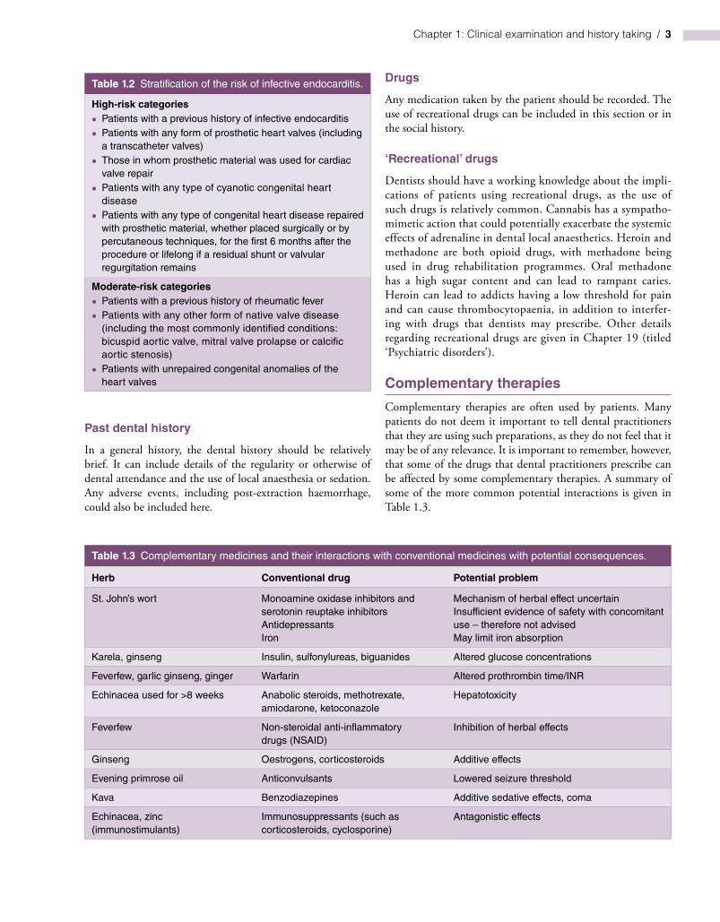
To guide decision-making, NICE has provided infor-mation regarding which might be considered high-risk and moderate-risk groups for the development of infective endocarditis – see Table 1.2.
It is clearly important that positive findings be recorded. Some important negative findings too are worth recording.
Allergies
Any known allergies should be recorded. This is one aspect of the medical history that should be recorded even if there are no known allergies. Any allergies that are identified should be highlighted in the clinical record.
Table 1.1 Areas to be covered in a medical history.
Presenting complaint
History of presenting complaint
Past medical history
Allergies
Past dental history
Drugs
Social history
Family history
Psychiatric history
Chapter 1: Clinical examination and history taking / 3
Past dental history
In a general history, the dental history should be relatively brief. It can include details of the regularity or otherwise of dental attendance and the use of local anaesthesia or sedation. Any adverse events, including post-extraction haemorrhage, could also be included here.
Drugs
Any medication taken by the patient should be recorded. The use of recreational drugs can be included in this section or in the social history.
‘Recreational’ drugs
Dentists should have a working knowledge about the impli-cations of patients using recreational drugs, as the use of such drugs is relatively common. Cannabis has a sympatho-mimetic action that could potentially exacerbate the systemic effects of adrenaline in dental local anaesthetics. Heroin and methadone are both opioid drugs, with methadone being used in drug rehabilitation programmes. Oral methadone has a high sugar content and can lead to rampant caries. Heroin can lead to addicts having a low threshold for pain and can cause thrombocytopaenia, in addition to interfer-ing with drugs that dentists may prescribe. Other details regarding recreational drugs are given in Chapter 19 (titled ‘Psychiatric disorders’).
Complementary therapies
Complementary therapies are often used by patients. Many patients do not deem it important to tell dental practitioners that they are using such preparations, as they do not feel that it may be of any relevance. It is important to remember, however, that some of the drugs that dental practitioners prescribe can be affected by some complementary therapies. A summary of some of the more common potential interactions is given in Table 1.3.
Table 1.3 Complementary medicines and their interactions with conventional medicines with potential consequences.
Herb Conventional drug Potential problem
St. John’s wort Monoamine oxidase inhibitors and serotonin reuptake inhibitorsAntidepressantsIron
Mechanism of herbal effect uncertainInsufficient evidence of safety with concomitant use – therefore not advisedMay limit iron absorption
Karela, ginseng Insulin, sulfonylureas, biguanides Altered glucose concentrations
Feverfew, garlic ginseng, ginger Warfarin Altered prothrombin time/INR
Echinacea used for >8 weeks Anabolic steroids, methotrexate, amiodarone, ketoconazole
Hepatotoxicity
Feverfew Non-steroidal anti-inflammatory drugs (NSAID)
Inhibition of herbal effects
Ginseng Oestrogens, corticosteroids Additive effects
Evening primrose oil Anticonvulsants Lowered seizure threshold
Kava Benzodiazepines Additive sedative effects, coma
Echinacea, zinc (immunostimulants)
Immunosuppressants (such as corticosteroids, cyclosporine)
Antagonistic effects
Table 1.2 Stratification of the risk of infective endocarditis.
High‐risk categories• Patients with a previous history of infective endocarditis• Patients with any form of prosthetic heart valves (including
a transcatheter valves)• Those in whom prosthetic material was used for cardiac
valve repair• Patients with any type of cyanotic congenital heart
disease• Patients with any type of congenital heart disease repaired
with prosthetic material, whether placed surgically or by percutaneous techniques, for the first 6 months after the procedure or lifelong if a residual shunt or valvular regurgitation remains
Moderate‐risk categories• Patients with a previous history of rheumatic fever• Patients with any other form of native valve disease
(including the most commonly identified conditions: bicuspid aortic valve, mitral valve prolapse or calcific aortic stenosis)
• Patients with unrepaired congenital anomalies of the heart valves

4 / Chapter 1: Clinical examination and history taking
Implanted cardiac devices
Some ultrasonic scalers and ultrasonic baths produce electro-magnetic interference and may therefore be a risk to patients with implanted cardiac devices such as pacemakers and implanted defibrillators. Other such devices include electronic apex locators and electrocautery devices. There is a degree of confusion in the current literature regarding what devices are and are not considered safe to use, and consultation with the appropriate authorities is therefore important.
Social history
This should be a succinct but comprehensive assessment of the patient’s social circumstances. It should include the following details: ▪ Smoking behaviour ▪ Alcohol consumption – type and quantity – recommended
not to exceed 14 units per week (female) and 21 units per week (male)
▪ Occupation (or previous occupation if retired) ▪ Home circumstances – a brief description of the residence –
for example, a house, flat or sheltered accommodation. Who else lives in the household?
Family history
Any disorders with a genetic origin should be recorded.
Psychiatric history
This will only need to be included in specific cases. More detail is given in Chapter 18 (titled ‘Medicine for the elderly’).
In hospital practice, after the history comes the systems review. Specific questions are asked to further refine the avail-able knowledge on the patient’s overall medical condition. Many schemes are described, and the following scheme has been adapted for the dental clinician.
General questions
As with the history, a series of general questions can help to encompass the wide-ranging possibilities in terms of the underlying medical problems. Questions cover the following topics: ▪ Appetite ▪ Weight loss ▪ Fevers ▪ The presence of lumps or bumps ▪ Any rashes or itchy rashes ▪ Lethargy or fatigue
Cardiovascular system
▪ Chest pain (a differential diagnosis is given in Chapter 21, titled ‘Medical emergencies’)
▪ Dyspnoea – difficult or disordered breathing (beware of co-existing/alternative respiratory causes)
▪ If dyspnoea on exertion, try and quantify in terms of metres walked or number of stairs climbed before dyspnoea occurs
▪ Paroxysmal nocturnal dyspnoea (waking up in the night feeling breathless – see Chapter 5, titled ‘Cardiovascular disorders’)
▪ Orthopnoea (breathlessness on lying flat – see Chapter 5) ▪ Ankle oedema – beware of other possible causes of lower
limb swelling ▪ Palpitations (awareness of the beating of the heart) ▪ Calf claudication (distance walked until pain occurs in the
‘calf ’ muscles of the leg, referred to as the ‘claudication distance’)
Respiratory system
▪ The presence of cough, and its duration ▪ Whether the cough produces sputum ▪ Haemoptysis (coughing up blood) ▪ Wheezing
Gastrointestinal system
▪ Indigestion ▪ Nausea or vomiting ▪ Dysphagia (difficulty swallowing) ▪ Odynophagia (pain on swallowing) ▪ Haematemesis (vomiting of blood), described as looking
like ‘coffee grounds’ ▪ Change in bowel habits ▪ Change in bowel motion – for example, pale stool and dark
urine is virtually pathognomonic of obstructive jaundice (see Chapter 7, titled ‘Gastrointestinal disorders’)
▪ Melaena is the production of black stool containing blood altered by gastric acid; fresh blood indicates bleeding from further down the gastrointestinal tract
Neurological system
A brief overview is required, in particular: ▪ Any history of fits or faints ▪ Disturbance in sensation – particularly in the orofacial region ▪ Headache or facial pain
Musculoskeletal system
▪ Gait (overlaps with neurological system) ▪ Pain/swelling/stiffness of joints ▪ Impairment of function
Genitourinary system
This is usually of little relevance to the dental practitioner. Repeated urinary tract infections may be relevant insofar as the patient may be undergoing antibiotic treatment – of which the dental practitioner should be aware. For dental patients in gen-eral hospital settings, enquiry is useful regarding symptoms of prostatism. Some patients who require significant surgical
Chapter 1: Clinical examination and history taking / 5
procedures may require catheterisation, and an enlarged pros-tate gland can lead to difficulties with catheter insertion. ‘Hesitancy’ is the term that is used to describe difficulty in ini-tiating the urine stream, and ‘terminal dribbling’ is difficulty in stopping. Frequency of urination and nocturia (passing urine at night) should all be included.
Clinical observations in the clothed patient
While it is evident that clinical examination is important, much of the background to a patient’s medical condition is gained from the history. Physical examination often serves to confirm what is suspected from the history.
Overall view of the patient
Does the patient look generally well? Is the patient of normal weight, or is he or she cachectic or obese? It is important to note whether the patient is alert or appears to be confused (Table 1.4 lists potential causes of confusion in a patient). As soon as the patient enters the surgery, note should be taken of the gait. Is the patient pale or flushed or of normal complexion? Is he or she breathless?
Not all the preceding observations are necessarily diagnos-tic of the precise nature of disease. However, if something does not look normal, then it probably is not, and an explanation needs to be found.
In hospitalised patients, it is important that the vital signs be recorded (Table 1.5). This is discussed further in a following section (titled ‘Vital signs’).
Examination of the hands
In the hands, there are several observable signs that can be of interest to a dental practitioner. The overall appearance of the
hands should be noted, together with any abnormalities of the nails, skin and muscles.
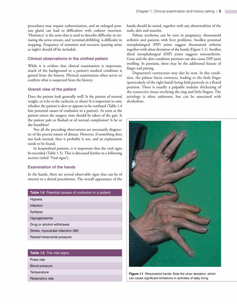
Palmar erythema can be seen in pregnancy, rheumatoid arthritis and patients with liver problems. Swollen proximal interphalangeal (PIP) joints suggest rheumatoid arthritis together with ulnar deviation of the hands (Figure 1.1). Swollen distal interphalangeal (DIP) joints suggests osteoarthritis. Gout and the skin condition psoriasis can also cause DIP joint swelling. In psoriasis, there may be the additional feature of finger nail pitting.
Dupuytren’s contracture may also be seen. In this condi-tion, the palmar fascia contracts, leading to the little finger (particularly of the right hand) being held passively in a flexed position. There is usually a palpable nodular thickening of the connective tissue overlying the ring and little fingers. The aetiology is often unknown, but can be associated with alcoholism.
Table 1.4 Potential causes of confusion in a patient.
Hypoxia
Infection
Epilepsy
Hypoglycaemia
Drug or alcohol withdrawal
Stroke, myocardial infarction (MI)
Raised intracranial pressure
Table 1.5 The vital signs.
Pulse rate
Blood pressure
Temperature
Respiratory rateFigure 1.1 Rheumatoid hands. Note the ulnar deviation, which can cause significant limitations in activities of daily living.

6 / Chapter 1: Clinical examination and history taking
Clubbing of the fingers should always be looked for and can represent disease processes in diverse systems (Figure 1.2). There is a loss of the angle between the nail and nail bed, and the fingernail has an exaggerated curvature in the longitu-dinal plane. The area around the nail fold feels boggy to palpation. Potential causes of finger clubbing are given in Table 1.6.
The fingernails may also show splinter haemorrhages that can result from mild trauma, but these may also be a sign of endocarditis (see Chapter 5, titled ‘Cardiovascular disorders’). Leukonychia (white fingernails) may be seen in patients with liver disease. Koilonychia (spoon-shaped fingernails) can be seen in patients with chronic iron deficiency anaemia.
The face
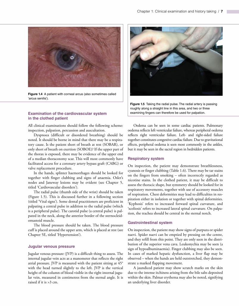
If the patient’s complexion is examined, it may display evidence of jaundice. This is rather subjective and unreliable. The best area to look for jaundice is the sclera of the eyes. The clinical and metabolic syndrome seen in chronic kidney dis-ease known as ‘uraemia’ may also impart a yellowish tinge to the skin. The eyelids may exhibit xanthelasma – deposits in the eyelids, signifying hyperlipidaemia (Figure 1.3). Corneal arcus (Figure 1.4) can be seen in some patients. It is sometimes associated with an increased risk of coronary artery disease. There may also be the malar flush of mitral stenosis or the butterfly rash seen in systemic lupus erythematosus (SLE; see Chapter 11, titled ‘Musculoskeletal disorders’).
Central cyanosis may be seen by asking the patient to pro-trude the tongue – a bluish hue is indicative of this. Peripheral cyanosis (seen in the nail beds) is caused by peripheral vasocon-striction, which may be normal, seen in cold conditions or in shock, but may also signify peripheral vascular insufficiency. Figure 1.3 Xanthelasma.
Figure 1.2 Finger clubbing. There is a loss of angle between the nail surface and the skin of the finger, and the nail bed is ‘boggy’ to pressure.
Table 1.6 Causes of finger clubbing.
Cardiothoracic causes• Infective endocarditis• Cyanotic congenital cardiac disease• Intrathoracic pus – for example, lung abscess, bronchiectasis• Bronchial carcinoma• Fibrosing alveolitis
Gastrointestinal causes• Inflammatory bowel disease• Cirrhosis of the liver
Other causes• Familial• Secondary to thyrotoxicosis• Idiopathic
Chapter 1: Clinical examination and history taking / 7
Examination of the cardiovascular system in the clothed patient
All clinical examinations should follow the following scheme: inspection, palpation, percussion and auscultation.
Dyspnoea (difficult or disordered breathing) should be noted. It should be borne in mind that there may be a respira-tory cause. Is the patient short of breath at rest (SOBAR), or only short of breath on exertion (SOBOE)? If the upper part of the thorax is exposed, there may be evidence of the upper end of a median thoracotomy scar. This will most commonly have facilitated access for a coronary artery bypass graft (CABG) or valve replacement procedure.
In the hands, splinter haemorrhages should be looked for together with finger clubbing and signs of anaemia. Osler’s nodes and Janeway lesions may be evident (see Chapter 5, titled ‘Cardiovascular disorders’).
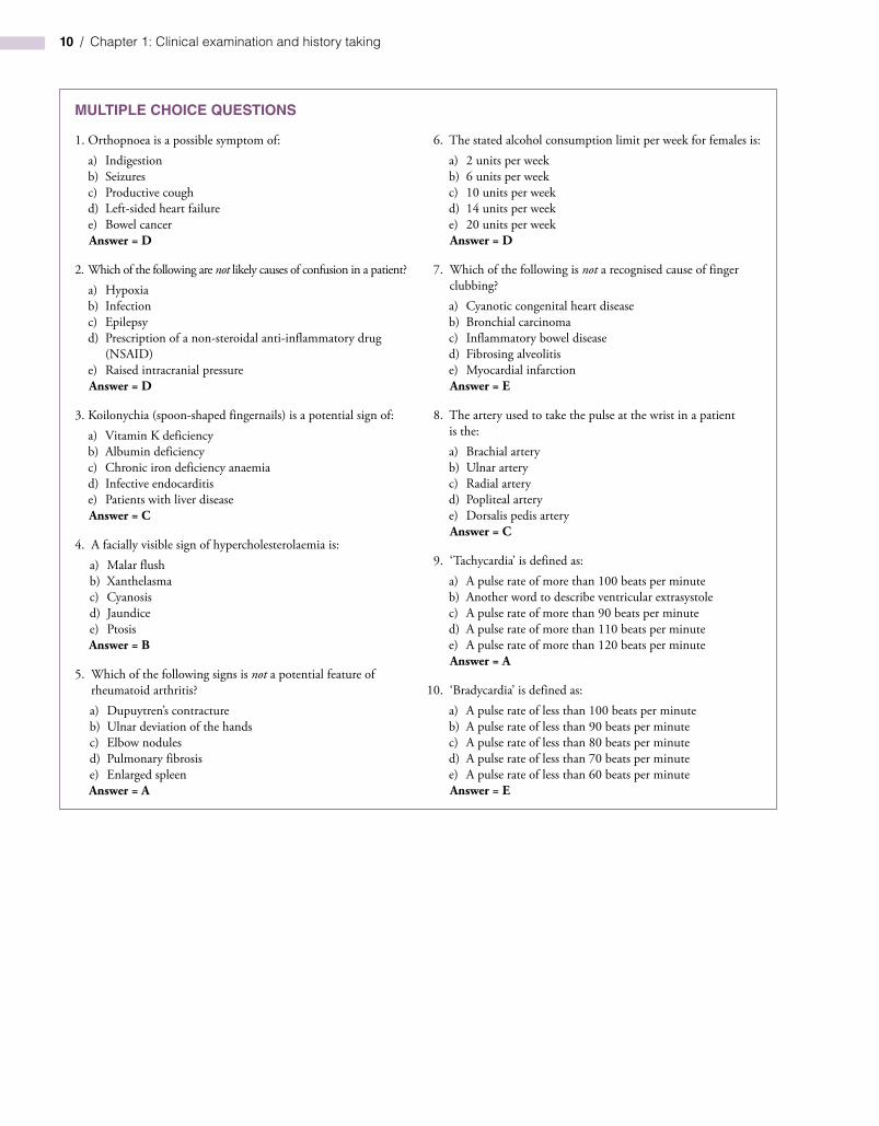
The radial pulse (thumb side of the wrist) should be taken (Figure 1.5). This is discussed further in a following section (titled ‘Vital signs’). Some dental practitioners are proficient in palpating a central pulse in addition to the radial pulse (which is a peripheral pulse). The carotid pulse (a central pulse) is pal-pated in the neck, along the anterior border of the sternocleid-omastoid muscle.
The blood pressure should be taken. The blood pressure cuff is placed around the upper arm, which is placed at rest (see Chapter 5E, titled ‘Hypertension’).
Jugular venous pressure
Jugular venous pressure (JVP) is a difficult thing to assess. The internal jugular vein acts as a manometer that reflects the right atrial pressure. JVP is measured with the patient sitting at 45° with the head turned slightly to the left. JVP is the vertical height of the column of blood visible in the right internal jugu-lar vein, measured in centimetres from the sternal angle. It is raised if it is >3 cm.
Oedema can be seen in some cardiac patients. Pulmonary oedema reflects left ventricular failure, whereas peripheral oedema reflects right ventricular failure. Left- and right-sided failure together constitutes congestive cardiac failure. Due to gravitational effects, peripheral oedema is seen most commonly in the ankles, but it may be seen in the sacral region in bedridden patients.
Respiratory system
On inspection, the patient may demonstrate breathlessness, cyanosis or finger clubbing (Table 1.6). There may be tar stains on the fingers from smoking – often incorrectly regarded as nicotine stains. In the clothed patient, it may be difficult to assess the thoracic shape, but symmetry should be looked for in respiratory movements, together with use of accessory muscles of respiration. Chest deformities may lead to difficulties in res-piration either in isolation or together with spinal deformities. ‘Kyphosis’ refers to increased forward spinal curvature, and ‘scoliosis’ refers to increased lateral spinal curvature. On palpa-tion, the trachea should be central in the sternal notch.
Gastrointestinal system
On inspection, the patient may show signs of purpura or spider naevi. Spider naevi can be emptied by pressing on the centre, and they refill from this point. They are only seen in the distri-bution of the superior vena cava. Leukonychia may be seen (a sign of hypoalbuminaemia). Finger clubbing may also be seen. In cases of marked hepatic dysfunction, a liver flap may be observed – when the hands are held outstretched, they demon-strate a marked flapping movement.
A jaundiced patient may show scratch marks on the skin due to the intense itchiness arising from the bile salts deposited within the skin. Palmar erythema may also be noted, signifying an underlying liver disorder.
Figure 1.5 Taking the radial pulse. The radial artery is passing roughly along a straight line in this area, and two or three examining fingers can therefore be used for palpation.
Figure 1.4 A patient with corneal arcus (also sometimes called ‘arcus senilis’).

8 / Chapter 1: Clinical examination and history taking
It is unusual for a dental practitioner to be called upon to examine other systems. A diagram of the abdomen is shown in Figure 1.6.
Vital signs
All hospital patients should have their vital signs measured. Vital signs are summarised in Table 1.5.
In contemporary hospital practice, vital signs are reviewed as part of the National Early Warning Score (NEWS). Any changes (normal score being zero) should prompt a review of the patient.
Pulse
The pulse is usually taken from the radial artery. In very small children and babies, the brachial pulse may be palpated in the antecubital fossa. The pulse should be assessed for its rate (in beats per minute), rhythm and volume. The rhythm of the pulse may be regular or irregular. If the pulse is irregular, this may be in a predictable pattern, in which case it is described as ‘regularly irregular’. If the pulse is completely disordered, it is described as ‘irregularly irregular’. The most common example of the latter is in patients with atrial fibrillation (see Chapter 5, titled ‘Cardiovascular disorders’). It should be ascertained whether the pulse is strong, or weak and ‘thready’. A bounding pulse can be a sign of carbon dioxide retention in patients with chronic obstructive pulmonary disease (COPD).
A pulse rate of >100 beats/min is described as ‘tachycardia’, and a pulse rate of <60 beats/min is described as ‘bradycardia’ (causes given in Table 1.7). Other abnormalities of the pulse are listed in Table 1.8.
Blood pressure
The method for measuring blood pressure is given in Chapter 5E (titled ‘Hypertension’). The figures quoted are given in millimetres of mercury. The upper figure is the systolic blood pressure (120–140 mmHg), and the lower figure the diastolic blood pressure (60–90 mmHg). Pathological changes in blood pressure are discussed in Chapter 5 (titled ‘Cardiovascular disorders’).
Temperature
The normal body temperature, measured orally, is 35.5–37.5°C. Many automated digital devices are now available for measuring body temperature, often in the form of a probe inserted into the external auditory meatus. In infants, the ther-mometer may be inserted into the armpit (axilla).
Respiratory rate
Several disease processes may be manifest by alterations in the respiratory rate and are discussed in the relevant chapters. The normal respiratory rate in a resting adult who is fit and well is 12–18 breaths/min.
Specific lesions
It is useful to have a standard set of parameters to be used in the assessment of lumps and ulcers. These can be applied to any clinical situation with minor modifications if required. These are summarised in Table 1.9 (lumps) and Table 1.10 (ulcers).
Right
Liver
Bladder
Left
Spleen
Left kidney
Umbilicus
Figure 1.6 A schematic diagram of the abdomen (not to scale).
Table 1.7 Causes of tachycardia and bradycardia.
Tachycardia (pulse rate >100 beats/min)• Physiological – for example, exercise, emotion• Related to fever• Secondary to drugs – for example, adrenaline, atropine• Hyperthyroidism• Smoking• Excess caffeine
Bradycardia (pulse rate <60 beats/min)• Physiological – for example, in athletes• Immediately post-vaso-vagal attack• Sick sinus syndrome• Hypothyroidism
Table 1.8 Commonly seen abnormalities of the radial pulse.
Sinus tachycardia – pulse >100 beats/min
Sinus bradycardia – pulse <60 beats/min
Atrial fibrillation – irregularly irregular pulse
Ventricular extrasystole – ‘missed beats’

Chapter 1: Clinical examination and history taking / 9
Table 1.9 Generic features to be considered in the assessment of lumps.
History
When/how was the lump first noticed?
Are there any symptoms?
Has the lump changed since it was noticed?
Does the lump ever disappear?
Are there any other lumps?
Examination
Site
Size
Shape
Surface – smooth or not – fixed to skin/deep structures
Colour of overlying skin/mucosa
Is it tender?
Edge – indistinct or well defined
Consistency – soft, fluctuant, rubbery or hard
Is it compressible?
Is it pulsatile?
Does it transilluminate when the light from a torch is shone through it?
Enlargement of local lymph nodes?
Consider blood and nerve supply to surrounding area
Is this a localised lump, or part of an associated generalised condition?
Table 1.10 Features to be considered in the assessment of ulcers.
History
Where/how was it noticed?
Symptoms
Changes since noticed
Any previous history of similar ulcers?
Examination
Site
Size
Shape
Base – slough, granulation tissue, deeper anatomy visible
Edge – sloping, suggesting healing
Punched out (square edge)
Undermined edge – for example, TB
Rolled – basal cell cancer (see Chapter 11, titled ‘Musculoskeletal disorders’)
Everted – squamous cell cancer (Chapter 11)
Depth
Discharge – swab for microbiological analysis
Enlargement of local lymph nodes?
Consider blood and nerve supply to surrounding area
Is this a localised ulcer, or part of an associated generalised condition?
Most of the assessments of any patient’s medical condition is made on the basis of a thorough history.
Examination findings usually serve to confirm suspicions and refine findings.
SU
MM
AR
Y
FURTHER READING
Oxford Handbook of Clinical Medicine. Oxford: Oxford University Press; 2014.
Scully’s Medical Problems in Dentistry. London: Churchill Livingstone; 2014.
Antibiotic prophylaxis. Available from: https://www.nice.org/guidance/cg64 (last update 2016).
General Dental Council. Maintaining Standards: Guidance to Dentists on Professional and Personal Conduct. Available from: http://www.gdc-uk.org/Newsandpublications//Publications/ Publications/MaintainingStandards.
Resuscitation Council (UK). Available from: http://www.resusc.org.uk/pages/medental.htm.
10 / Chapter 1: Clinical examination and history taking
MULTIPLE CHOICE QUESTIONS
1. Orthopnoea is a possible symptom of:a) Indigestionb) Seizuresc) Productive coughd) Left-sided heart failuree) Bowel cancerAnswer = D
2. Which of the following are not likely causes of confusion in a patient?a) Hypoxiab) Infectionc) Epilepsyd) Prescription of a non-steroidal anti-inflammatory drug
(NSAID)e) Raised intracranial pressureAnswer = D
3. Koilonychia (spoon-shaped fingernails) is a potential sign of:a) Vitamin K deficiencyb) Albumin deficiencyc) Chronic iron deficiency anaemiad) Infective endocarditise) Patients with liver diseaseAnswer = C
4. A facially visible sign of hypercholesterolaemia is:a) Malar flushb) Xanthelasmac) Cyanosisd) Jaundicee) PtosisAnswer = B
5. Which of the following signs is not a potential feature of rheumatoid arthritis?a) Dupuytren’s contractureb) Ulnar deviation of the handsc) Elbow nodulesd) Pulmonary fibrosise) Enlarged spleenAnswer = A
6. The stated alcohol consumption limit per week for females is:a) 2 units per weekb) 6 units per weekc) 10 units per weekd) 14 units per weeke) 20 units per weekAnswer = D
7. Which of the following is not a recognised cause of finger clubbing?a) Cyanotic congenital heart diseaseb) Bronchial carcinomac) Inflammatory bowel diseased) Fibrosing alveolitise) Myocardial infarctionAnswer = E
8. The artery used to take the pulse at the wrist in a patient is the:a) Brachial arteryb) Ulnar arteryc) Radial arteryd) Popliteal arterye) Dorsalis pedis arteryAnswer = C
9. ‘Tachycardia’ is defined as:a) A pulse rate of more than 100 beats per minuteb) Another word to describe ventricular extrasystolec) A pulse rate of more than 90 beats per minuted) A pulse rate of more than 110 beats per minutee) A pulse rate of more than 120 beats per minuteAnswer = A
10. ‘Bradycardia’ is defined as:a) A pulse rate of less than 100 beats per minuteb) A pulse rate of less than 90 beats per minutec) A pulse rate of less than 80 beats per minuted) A pulse rate of less than 70 beats per minutee) A pulse rate of less than 60 beats per minuteAnswer = E
11
Essentials of Human Disease in Dentistry, Second Edition. Mark Greenwood. © 2018 John Wiley & Sons Ltd. Published 2018 by John Wiley & Sons Ltd.Companion website: www.wiley.com/go/greenwood/human-disease-in-dentistry
Key topics
◾ Overview of the pathology and clinical features of wound healing and inflammation
◾ Descriptive terms used in the clinicopathology of inflammatory disorders
Learning objectives
◾ To be familiar with the main pathological processes involved in wound healing and inflammation.
◾ To be familiar with some of the drugs commonly used in the treatment of inflammatory conditions.
Inflammation and anti‑inflammatory drugsCM Robinson and RA Seymour
CHAPTER 2

12 / Chapter 2: Inflammation and anti‑inflammatory drugs
Cell and tissue injury – Introduction
Cells are under constant threat of injury as a consequence of changes in their local environment. There are numerous poten-tially injurious agents that a cell may encounter. The type of mediator depends on the location of the cell within the body. There are agents that cause physical damage to cell integ-rity – for example, mechanical trauma, thermal injury and chemical damage. Cellular viability is affected by the deleteri-ous effects of microorganisms. Cells are also prone to damage following reductions in oxygen and nutrient supply. Cell injury may be induced by DNA‐damaging agents, such as ionising radiation.
Cell injury may be reversible or irreversible. This depends on the type of injurious agent, the duration of the adverse con-ditions and the ability of the cell to adapt to the changes. If the cell is unable to adapt and survive, there are two distinct path-ways leading to cell death – necrosis and apoptosis (pro-grammed cell death). Consequences of tissue injury are shown in Table 2.1 and described in the following text.
Necrosis
Necrosis is the death of groups of cells. It has profound conse-quences on tissue integrity and function. In some circum-stances, for example, in the heart, ischaemic necrosis of the myocardium can result in complete organ failure and death of the individual. Various types of necrosis are described, depend-ing on the microscopic appearances observed within tissues when stained with haematoxylin and eosin (H&E). Haematoxylin stains cell nuclei dark blue, whereas eosin stains the cytoplasm and the connective tissue proteins a reddish pink colour.
Coagulative necrosis
Coagulative necrosis is the most common type of necrosis and can occur in most organs and tissues. Immediately following cell death, the architecture of the tissue is retained. The cell outlines are discernible along with tissue architecture. There is progressive loss of nuclear staining, and there is a loss of cyto-plasmic detail, with condensation and breakdown of intracel-lular proteins. Eventually, all that remains are the ‘ghost’
outlines of cells embedded in the extracellular matrix that comprises the tissue. Accompanying necrosis is an ensuing inflammatory reaction, which results in progressive dissolution of the damaged tissue with variable attempts at regeneration and repair, depending on the affected tissue.
Colliquative necrosis
Colliquative necrosis is observed in the brain following cerebral infarction. The brain has little in terms of robust collagenous supporting tissues, and the necrotic neurons and glial tissue break down to form a liquid material (liquefactive degeneration). The latter is progressively removed by microglia (macrophages/histiocytes), leaving a fluid‐filled cavity.
Caseous necrosis
Caseous necrosis is characterised by structureless necrotic tis-sue, which has the textural quality of crumbly cheese. It is typi-cally seen in the context of granulomatous inflammation caused by Mycobacterium tuberculosis.
Fibrinoid necrosis
Fibrinoid necrosis is seen in malignant hypertension and dis-eases characterised by vasculitis. Necrosis of smooth muscle cells in the arterial wall causes plasma to leak into the tunica media with deposition of fibrin. Fibrin takes on a distinct bright red colour when stained with H&E.
Fat necrosis
Damage to adipose tissue and the release of stored intracellular fat elicits an intense acute inflammatory response, which is typically followed by a dense fibrotic repair reaction.
Gangrene
Gangrene is necrosis with accompanying putrefaction (breakdown) of the tissues. Gangrene is typically seen in the lower extremities (legs and toes), and is usually the result of supervening infection of the necrotic tissue with bacteria, particularly Clostridium.
Apoptosis
Apoptosis is the process whereby cells can be removed in a con-trolled manner, without disrupting the integrity of the organ or tissue. Apoptosis is also called ‘programmed cell death’, which emphasises that it is a cellular process induced and executed by specific biochemical pathways. Apoptosis has a pivotal role in development, and controls organ size and tissue morphogene-sis. In the adult, apoptosis counterbalances cellular prolifera-tion, helping to maintain optimal cell numbers and cell types within a particular tissue. Apoptosis is also essential for the establishment of a functional immune system, through dele-tion of unwanted lymphocyte clones. Apoptosis is considered to play an important role in ageing, and deregulated apoptosis is involved in the pathogenesis of neoplasia.
Table 2.1 Potential consequences of tissue injury.
Inflammation
Coagulative necrosis
Colliquative necrosis
Caseous necrosis
Fibrinoid necrosis
Fat necrosis
Gangrene
Chapter 2: Inflammation and anti‑inflammatory drugs / 13
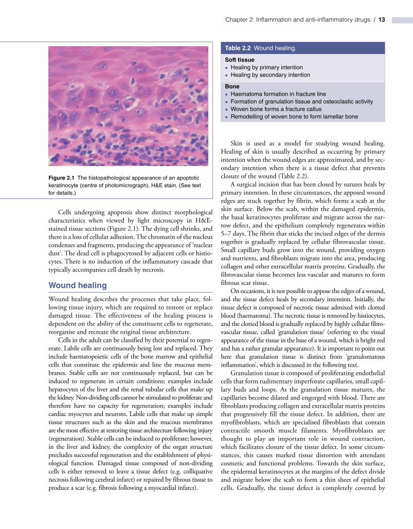
Cells undergoing apoptosis show distinct morphological characteristics when viewed by light microscopy in H&E‐stained tissue sections (Figure 2.1). The dying cell shrinks, and there is a loss of cellular adhesion. The chromatin of the nucleus condenses and fragments, producing the appearance of ‘nuclear dust’. The dead cell is phagocytosed by adjacent cells or histio-cytes. There is no induction of the inflammatory cascade that typically accompanies cell death by necrosis.
Wound healing
Wound healing describes the processes that take place, fol-lowing tissue injury, which are required to restore or replace damaged tissue. The effectiveness of the healing process is dependent on the ability of the constituent cells to regenerate, reorganise and recreate the original tissue architecture.
Cells in the adult can be classified by their potential to regen-erate. Labile cells are continuously being lost and replaced. They include haematopoietic cells of the bone marrow and epithelial cells that constitute the epidermis and line the mucous mem-branes. Stable cells are not continuously replaced, but can be induced to regenerate in certain conditions; examples include hepatocytes of the liver and the renal tubular cells that make up the kidney. Non‐dividing cells cannot be stimulated to proliferate and therefore have no capacity for regeneration; examples include cardiac myocytes and neurons. Labile cells that make up simple tissue structures such as the skin and the mucous membranes are the most effective at restoring tissue architecture following injury (regeneration). Stable cells can be induced to proliferate; however, in the liver and kidney, the complexity of the organ structure precludes successful regeneration and the establishment of physi-ological function. Damaged tissue composed of non‐dividing cells is either removed to leave a tissue defect (e.g. colliquative necrosis following cerebral infarct) or repaired by fibrous tissue to produce a scar (e.g. fibrosis following a myocardial infarct).
Skin is used as a model for studying wound healing. Healing of skin is usually described as occurring by primary intention when the wound edges are approximated, and by sec-ondary intention when there is a tissue defect that prevents closure of the wound (Table 2.2).
A surgical incision that has been closed by sutures heals by primary intention. In these circumstances, the apposed wound edges are stuck together by fibrin, which forms a scab at the skin surface. Below the scab, within the damaged epidermis, the basal keratinocytes proliferate and migrate across the nar-row defect, and the epithelium completely regenerates within 5–7 days. The fibrin that sticks the incised edges of the dermis together is gradually replaced by cellular fibrovascular tissue. Small capillary buds grow into the wound, providing oxygen and nutrients, and fibroblasts migrate into the area, producing collagen and other extracellular matrix proteins. Gradually, the fibrovascular tissue becomes less vascular and matures to form fibrous scar tissue.
On occasions, it is not possible to appose the edges of a wound, and the tissue defect heals by secondary intention. Initially, the tissue defect is composed of necrotic tissue admixed with clotted blood (haematoma). The necrotic tissue is removed by histiocytes, and the clotted blood is gradually replaced by highly cellular fibro-vascular tissue, called ‘granulation tissue’ (referring to the visual appearance of the tissue in the base of a wound, which is bright red and has a rather granular appearance). It is important to point out here that granulation tissue is distinct from ‘granulomatous inflammation’, which is discussed in the following text.
Granulation tissue is composed of proliferating endothelial cells that form rudimentary imperforate capillaries, small capil-lary buds and loops. As the granulation tissue matures, the capillaries become dilated and engorged with blood. There are fibroblasts producing collagen and extracellular matrix proteins that progressively fill the tissue defect. In addition, there are myofibroblasts, which are specialised fibroblasts that contain contractile smooth muscle filaments. Myofibroblasts are thought to play an important role in wound contraction, which facilitates closure of the tissue defect. In some circum-stances, this causes marked tissue distortion with attendant cosmetic and functional problems. Towards the skin surface, the epidermal keratinocytes at the margins of the defect divide and migrate below the scab to form a thin sheet of epithelial cells. Gradually, the tissue defect is completely covered by
Table 2.2 Wound healing.
Soft tissue• Healing by primary intention• Healing by secondary intention
Bone• Haematoma formation in fracture line• Formation of granulation tissue and osteoclastic activity• Woven bone forms a fracture callus• Remodelling of woven bone to form lamellar bone
Figure 2.1 The histopathological appearance of an apoptotic keratinocyte (centre of photomicrograph). H&E stain. (See text for details.)

14 / Chapter 2: Inflammation and anti‑inflammatory drugs
full‐thickness epidermis, and the scab is exfoliated. Sometimes, the production of fibrous repair tissue is excessive, and the der-mis becomes bulky and lumpy – this is referred to as a ‘hyper-trophic’ or ‘keloid’ scar (Figure 2.2).
Wound healing proceeds in a similar manner following bone injury (Table 2.2). For example, following the fracture of a limb bone, a haematoma forms at the site of injury and invests dead pieces of soft tissue and devitalised fragments of bone. The soft tissue is removed by histiocytes, and the bone is removed by osteoclasts. Granulation tissue gradually replaces the haematoma, and woven bone is laid down to form a frac-ture callus. The woven bone of the callus is eventually remod-elled to lamellar bone and, consequently, within 6–8 weeks, there is very little evidence of the injury; this is known as ‘regeneration’. Healing of long bones is dependent, however, on close apposition of the broken ends of the bone and immo-bilisation of the fracture site. Failure to ensure the latter may result in failure of bone regeneration and the production of a fibrous union (pseudarthrosis; false joint).
The liver has a complex architecture, with hepatocytes arranged in a lobular configuration around hepatic vessels and bile ducts. Hepatocytes are stable cells that can be induced to proliferate, and small groups of cells are capable of regenerating and recreating the lobular architecture following injury. However, following extensive damage to the liver – for example, owing to long‐standing alcohol abuse – there is disorganised
nodular regeneration of hepatocytes, as opposed to lobular regeneration, and there is also extensive fibrosis, which results in liver cirrhosis. The kidney is similar, in that the tubular cells are capable of regeneration, but the complex microanatomy of the organ is difficult to recreate, and organ damage usually heals by fibrosis.
Inflammation – Introduction
Inflammation is a protective response, the goal of which is to eliminate the injurious agent and the consequences of tissue damage. Ideally, the inflammatory process facilitates repair of damaged tissue; however, in some circumstances, inflamma-tion can have harmful effects on the tissues and compromise the well‐being of the individual.
Inflammation is a complex reaction that occurs in vascular-ised tissue and comprises changes in blood vessels, which lead to the accumulation of fluid that contains leukocytes, the inflammatory exudate. Inflammation is classified into acute and chronic phases. Acute inflammation is usually of relatively short duration, lasting minutes, hours or a few days. It is char-acterised by the accumulation of tissue fluid, plasma proteins and the emigration of leukocytes, principally neutrophils.
Chronic inflammation is of longer duration, and is character-ised by the accumulation of specialised immune cells – for exam-ple, lymphocytes, plasma cells and macrophages. Long‐standing inflammation may cause further tissue damage, which is termed ‘bystander damage’. After neutralisation/elimination of the cause of inflammation, there follows attempts at repair. The process of repair is characterised by the proliferation of blood vessels pro-ducing richly vascular granulation tissue, and there is usually attendant fibrosis. The inflammatory cascade is orchestrated by chemical mediators triggered by the inflammatory stimulus. The chemical mediators are derived from the plasma, and are released by a variety of cells involved in inflammation.
Conventionally, the process of inflammation is described by the suffix ‘‐itis’, preceded by the organ and/or tissues affected (Table 2.3).
Inflammation is caused by: ▪ Microorganisms: bacteria, viruses, fungi, parasites. ▪ Physical agents: mechanical trauma, thermal injury, chemi-
cal damage, exposure to radiation. ▪ Tissue necrosis. ▪ Hypersensitivity reactions.
The clinical signs of inflammation, called the ‘cardinal signs’, are redness, heat, swelling, pain and loss of function (Table 2.4). Redness is a consequence of increased blood flow to the inflamed tissue, and is called ‘hyperaemia’. In peripheral tissues, for exam-ple, the skin, the increased blood flow causes the skin surface to feel warmer. In addition, inflammatory chemical mediators contribute to a rise in body temperature, called ‘pyrexia’. The development of swelling is mainly a consequence of accumulat-ing tissue fluid, called ‘oedema’. Progression of the inflammatory response produces symptoms of pain; there is distortion and stretching of the tissues, and inflammatory chemical mediators
Figure 2.2 A hypertrophic scar. Keloid scars extend beyond the wound margins.