Esophagus, Stomach & Duodenum, Part II - American...
71
® Vol 40 | 7 | 2014 SELECTED READINGS in GENERAL SURGERY Esophagus, Stomach & Duodenum, Part II AMERICAN COLLEGE OF SURGEONS | DIVISION OF EDUCATION Blended Surgical Education and Training for Life Gastroparesis in the diabetic patient page 7 Peptic ulcer disease page 20 Gastric cancer page 27 Bariatric surgery page 32
Transcript of Esophagus, Stomach & Duodenum, Part II - American...

® Vol 40 | 7 | 2014
SE L EC T E D R E A DI NGS in GENER A L SU RGERY
Esophagus, Stomach & Duodenum, Part II
AMERICAN COLLEGE OF SURGEONS | DIVISION OF EDUCATIONBlended Surgical Education and Training for Life
Gastroparesis in the diabetic patientpage 7
Peptic ulcer diseasepage 20
Gastric cancerpage 27
Bariatric surgerypage 32
Cover: Printed on paper manufactured from 10% post-consumer waste and Green-e certified renewable energy.
Interior: Printed on paper manufactured from 100% post-consumer waste, Green Seal certified and processed chlorine free.
American College of SurgeonsDivision of Education633 N. Saint Clair St.Chicago, IL 60611-3211
[email protected]/publications/srgs
®
SE L E C T E D R E A DI NG S in G E N E R A L SU RG E RY
®Vo
l 40 | 7 | 2014A
ME
RIC
AN
CO
LL
EG
E O
F S
UR
GE
ON
SEsophagus, Stom
ach & D
uodenum, Part II

iAmerican College of Surgeons www.facs.org/publications/srgs SRGS Vol 40 | 7 | 2014
| ESOPHAGUS, STOMACH & DUODENUM, PART II
Editor-in chief Lewis Flint, MD, FACS
ACS steering committeeL. D. Britt, MD, MPH, FACS, chair
Ajit K. Sachdeva, MD, FACS, FRCSC
Patrice Gabler Blair, MPH
Editorial board Nita Ahuja, MD, FACS, The Johns Hopkins Medical Institutions, Baltimore, MD
L. D. Britt, MD, MPH, FACS, Eastern Virginia Medical School, Norfolk, VA
Ara Darzi, FRCS (Eng.), KBE, FmedSci, FACS, Imperial College of London, London, UK
Karen Deveney, MD, FACS, Oregon Health and Science University, Portland, OR
Michael B. Edye, MD, FACS, University of Western Sydney, Seven Hills, Australia
Jean C. Emond, MD, FACS, Columbia University Medical Center/New York-Presbyterian Hospital, New York, NY
John Ferrara, MD, FACS, Virginia Tech Carilion School of Medicine, Roanoke, VA
Donald E. Fry, MD, FACS, Michael Pine & Associates, Chicago, IL
Amy L. Halverson, MD, FACS, Northwestern Memorial Hospital, Chicago, IL
Tyler G. Hughes, MD, FACS, Memorial Hospital, McPherson, KS
Roger Keith, MD, FACS, University of Saskatchewan, Saskatoon, Canada
Solly Mizrahi, MD, FACS, Soroka Medical Center, Beer Sheva, Israel
Chandrajit Premanand Raut, MD, MSc, FACS, Brigham and Women’s Hospital, Boston, MA
Raul J. Rosenthal, MD, FACS, Cleveland Clinic Florida-Weston, Fort Lauderdale, FL
Ajit K. Sachdeva, MD, FACS, FRCSC, American College of Surgeons, Chicago, IL
Eduardo de Santibañes, MD, PhD, FACS, Instituto Universitario del Hospital Italiano de Buenos Aires, Buenos Aires, Argentina
Murray Shames, MD, FACS, University of South Florida, Tampa, FL
Nathaniel J. Soper, MD, FACS, Northwestern Memorial Hospital, Chicago, IL
Steven Steinberg, MD, FACS, The Ohio State University Hospitals, Columbus, OH
Christopher B. Weldon, MD, PhD, FACS, Children’s Hospital Boston, Boston, MA
Editorial and business officesACS-SRGS 633 N. Saint Clair St. Chicago, IL 60611-3211P 800-631-0033 or 312-202-5227 F 312-202-5009 [email protected] | www.facs.org/publications/srgs
Managing editor: Lynanne Feilen, [email protected]
The American College of Surgeons is a scientific and educational organization of surgeons that was founded in 1913 to raise the standards of surgical practice and improve the quality of care for surgical patients. The College is dedicated to the ethical and competent practice of surgery. Its achievements have significantly influenced the course of scientific surgery in America and have established it as an important advocate for all surgical patients. The College has more than 79,000 members and is the largest organization of surgeons in the world.
ACS disclosure policyIn accordance with ACCME accreditation criteria, ACS must ensure that anyone in a position to control the content of SRGS has disclosed all relevant financial relationships with any commercial interest. Members of the Editorial Board and those providing editorial assistance are required to disclose all financial relationships. All reported conflicts are managed by a designated official to ensure bias-free content. However, if you perceive a bias, please contact us at [email protected]. The following relationships were disclosed in 2014:
Donald E. Fry, MD, FACS, has disclosed a commercial interest in Ethicon and Merck; Chandrajit P. Raut, MD, MSc, FACS, has disclosed a commercial interest in Novartis; Raul J. Rosenthal, MD, FACS, has disclosed a commercial interest in Baxter, Covidien, Ethicon, Gore, and Storz; Nathaniel J. Soper, MD, FACS, has disclosed a commercial interest in Miret Surgical, Inc., and TransEnterix.
Unless specifically stated otherwise, the opinions expressed and statements made in this publication reflect the authors’ personal observations and do not imply endorsement by nor official policy of the American College of Surgeons.
Subscription informationVisit www.facs.org/publications/srgs for order information. Prepayment in U.S. dollars is required to activate a subscription.
Back issues:Current subscribers can purchase back issues (print only) for $35/issue; nonsubscribers, $70/issue.
Payment should be sent to:ACS-SRGS 633 N. Saint Clair St. Chicago, IL 60611-3211
Renew online at www.facs.org/ publications/srgs/subscriptions/renew.
To place an order over the telephone:
Call 800-631-0033 or 312-202-5227. Please have your ACS-SRGS ID number and credit card information available.
Address changes:Please notify us of any address changes six weeks prior to a move.
Missing issues:Lost or missing issues must be reported within eight weeks after the issue has been mailed. Consult www.facs.org/publications/srgs for mailing dates. Two missing issues per year per subscription can be replaced.
To change your address or to report a missing issue: Call 800-631-0033 or 312-202-5227 Fax 312-202-5009 E-mail [email protected]
Postmaster:Send address changes to: ACS-SRGS 633 N. Saint Clair St. Chicago, IL 60611-3211

ii American College of Surgeons www.facs.org/publications/srgs SRGS Vol 40 | 7 | 2014
| ESOPHAGUS, STOMACH & DUODENUM, PART II
Continuing medical educationAccreditation The American College of Surgeons is accredited by the Accreditation Council for Continuing Medical Education (ACCME) to provide continuing medical education for physicians.
CME credit The American College of Surgeons designates this enduring material for a maximum of 10 AMA PRA Category 1 Credits.™* Physicians should claim only the credit commensurate with the extent of their participation in the activity.
*Of the AMA PRA Category 1 Credits™ listed above, a maximum of 10 credits meet the requirements for Self-Assessment.
Learning objectives This activity is designed for general surgeons, surgical residents, and allied professionals. Regular reading of SRGS should enable learners to:
• Maintain an excellent knowledge base in all areas of general surgery
• Develop comparative and critical literature reading skills
• Apply newly acquired knowledge to surgical practice
• Prepare effectively for recertification exams
Additional information at www.facs.org/publications/srgs/cme
Maintenance of certification The American Board of Surgery (ABS) recognizes SRGS as a resource for surgeons enrolled in its Maintenance of Certification (MOC) program. Successful completion of the SRGS program fulfills MOC Part II requirements that focus on lifelong learning and self-assessment.
ACS in cooperation with ABS has created a process wherein ACS members can directly submittheir ACS CME transcript to the ABS for MOC purposes. For more information, go to www.facs.org, click Member Login and enter your ACS user name and password. Then, go to My Profile, My CME, and click on “Send Credit to ABS.”
For information on ABS’s MOC requirements, go to http://absurgery.org and click on “Maintenance of Certification (MOC)” or e-mail [email protected].
Questions about ACS CME can be e-mailed to [email protected] or call 866-918-4799.
Statement of purposeSelected Readings in General Surgery (SRGS) is a topic oriented, in-depth review of the field of general surgery presented eight times annually as an educational offering of the Division of Education of the American College of Surgeons. The mission of the Division of Education is to improve the quality of surgical care through lifelong learning, based on educational programs and products designed to enhance the competence or performance of practicing surgeons, surgery residents, and members of the surgical team. The intent of the publication is to analyze relevant medical literature to give the surgeon the knowledge necessary to practice state-of-the-art surgery. To accomplish this goal, the editor selects 100–125 pertinent articles from the literature for each issue. Each article is reviewed and an overview is written that places the content of these articles in the perspective of the best, day-to-day, clinical practice. In addition to the overview, 12–18 full-text articles are reprinted in each issue.
The overview is compiled with the assistance of an 18-member, international board of editors who are experts in the various focus areas that comprise the specialty of surgery. In addition, the editorial board has representation and expertise in such important fields as
medical evidence evaluation, surgical education, outcomes research, standard setting, and performance improvement. SRGS is a unique resource because the overview and selected full-text articles provide the reader with the most valuable and pertinent content illuminated with informed opinion and critique. Unnecessary material is eliminated. SRGS does not present itself as infallible and the editor-in-chief takes responsibility for the content that appears in each issue. The editor-in-chief and the editorial board recognize that there is no such thing as the “average” surgical patient, and that the information in the literature must be interpreted in the light of the clinical presentation of each individual patient.
CopyrightMaterial printed in SRGS is covered by copyright law. The overview and CME tests are copyrights of the American College of Surgeons. Permission has been obtained from individual journal publishers to reprint articles that appear in SRGS. Copying all or portions of this journal for distribution to a group practice, residency program, university, hospital, or colleague is strictly prohibited.
© 2014 American College of Surgeons All rights reserved
Title Volume/Issue Publication Date
General Oncology, Part I V40N1 Published
General Oncology, Part II V40N2 Published
Ethics, Palliative Care, Patient Safety & Business Aspects of Surgical Practice
V40N3 Published
Pediatric Surgery V40N4 Published
Breast Diseases V40N5 Published
Esophagus, Stomach & Duodenum, Part I
V40N6 Published
Esophagus, Stomach & Duodenum, Part II
V40N7 Published
Biliary Tract & Pancreas, Part I V40N8 December
2014 SRGS Publishing Schedule
Visit http://www.facs.org/publications/srgs/issues/upcoming for previously published issues and next year’s topics.

iiiAmerican College of Surgeons www.facs.org/publications/srgs SRGS Vol 40 | 7 | 2014
CME pretest ................................................. iv
Bibliography of articles selectedfor further study .......................................vii
Introduction ..................................................1 Endoscopy training for surgeons .........2
Functional disorders of the stomach and duodenum ..........................5Gastroparesis
Gastroparesis in the diabetic patient
Treatment of gastroparesis
Management of postoperative ileus
Postoperative gastric atony
Belching, hiccups, and aerophagia
Gastrostomy: indications and techniques ..........................................14Percutaneous gastrostomy
Open and minimally invasive gastrostomy
Gastroduodenal ulcer disease ............19Peptic ulcer disease
Management of peptic ulcer bleeding
Management of peptic ulcer perforation
Benign gastric outlet obstruction
Stress gastrointestinal bleeding
Gastric cancer ........................................... 27Endoscopic detection and therapy for early gastric cancer
Adjuvant therapies and surgical approaches for gastric cancer
Minimally invasive gastrectomy for early gastric cancer
Management of advanced gastric cancer
Malignant gastric outlet obstruction
Bariatric surgery ...................................... 32Principles of management of bariatric surgery patients
Effectiveness of bariatric surgical procedures
Factors contributing to the safety of bariatric surgical procedures
Determination of performance metrics for bariatric centers
Indications, techniques, and outcomes for specific bariatric procedures
Management of complications of bariatric operations
Venous thromboembolism
Anastomotic and suture line leak
Complications specific to laparoscopic adjustable gastric band (LAGB) placement
Management of biliary complications following bariatric surgery
Marginal ulcer
Intestinal obstruction following bariatric surgery
Management of failed weight loss after bariatric procedures
References ...................................................51
CME posttest ............................................ 58
Literature Overview Editor: Lewis Flint, MD, FACS
Associate editors for this issue: Robert Yates, MD, and Carlos Pellegrini, MD, FACS
Table of Contents
VOLUME 40 | 7 | 2014
ESOPHAGUS, STOMACH & DUODENUM, PART II

iv American College of Surgeons www.facs.org/publications/srgs SRGS Vol 40 | 7 | 2014
CME Pretest
VOLUME 40 | 7 | 2014
ESOPHAGUS, STOMACH & DUODENUM, PART II
1. The minimum number of colonoscopies required to satisfy the surgery resident training requirements of the Accreditation Council for Graduate Medical Education is which of the following?
a) 200
b) 160
c) 50
d) 10
e) 35
2. In patients with gastroparesis, abnormalities are found in which of the following cell populations?
a) Mucosal cells
b) Smooth muscle cells
c) Endothelial cells
d) Interstitial cells of Cajal
e) Adventitial cells
3. All of the following are symptoms of diabetic gastroparesis except which one?
a) Abdominal pain
b) Nausea
c) Early satiety
d) Vomiting
e) Flatulence
4. Which of the following statements is true about prokinetic therapy for patients with gastroparesis?
a) Prokinetic drug therapy is the only available long-term treatment for gastroparesis
b) Oral erythromycin is more effective than intravenous erythromycin
c) Tardive dyskinesia is a side effect of metoclopramide therapy
d) Lack of response; the prokinetic drug therapy predicts failure of gastric electric stimulation
e) Prokinetic drug therapy should not be used until the patient requires “venting” with a nasogastric tube or gastrostomy tube
5. Which of the following prokinetic agents has significant adverse cardiac side effects?
a) Metoclopramide
b) Cisapride
c) Clarithromycin
d) Erythromycin
e) Domperidone
6. Which of the following is a contributing factor to postoperative ileus?
a) Use of alvimopan
b) Gum chewing
c) Opiate analgesics
d) Nonopioid epidural analgesia
e) Minimally invasive surgical techniques
A pretest is mandatory to earn CME credit on the posttest. The pretest should be completed BEFORE reading the overview. Both tests must be completed online. See new log-in procedures on page viii.
vAmerican College of Surgeons www.facs.org/publications/srgs SRGS Vol 40 | 7 | 2014
Pretest | ESOPHAGUS, STOMACH & DUODENUM, PART II
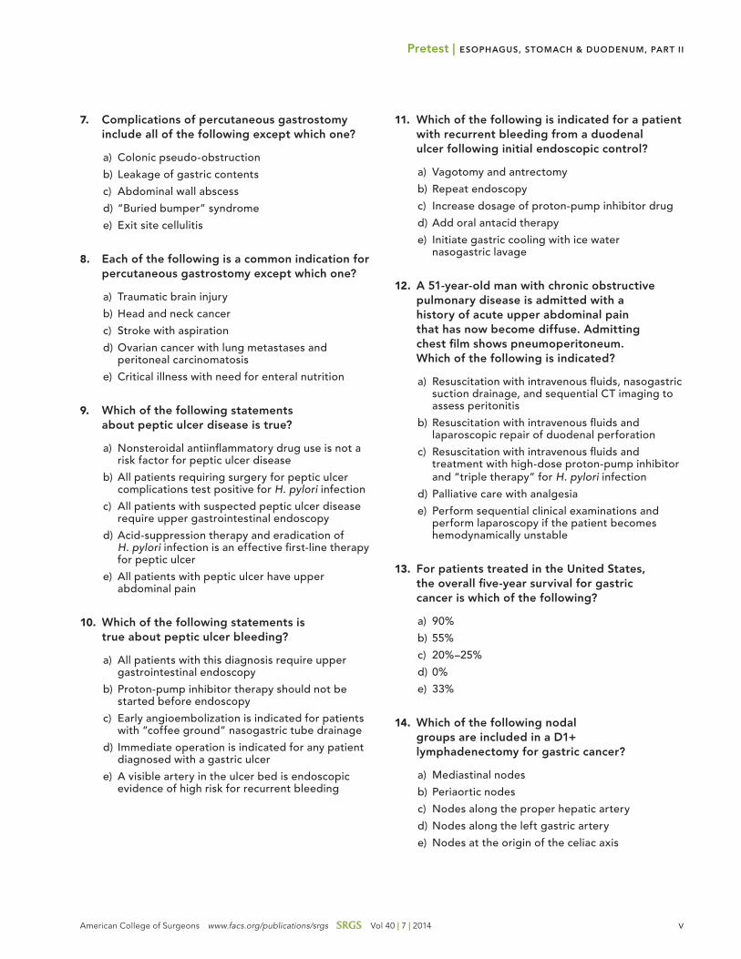
7. Complications of percutaneous gastrostomy include all of the following except which one?
a) Colonic pseudo-obstruction
b) Leakage of gastric contents
c) Abdominal wall abscess
d) “Buried bumper” syndrome
e) Exit site cellulitis
8. Each of the following is a common indication for percutaneous gastrostomy except which one?
a) Traumatic brain injury
b) Head and neck cancer
c) Stroke with aspiration
d) Ovarian cancer with lung metastases and peritoneal carcinomatosis
e) Critical illness with need for enteral nutrition
9. Which of the following statements about peptic ulcer disease is true?
a) Nonsteroidal antiinflammatory drug use is not a risk factor for peptic ulcer disease
b) All patients requiring surgery for peptic ulcer complications test positive for H. pylori infection
c) All patients with suspected peptic ulcer disease require upper gastrointestinal endoscopy
d) Acid-suppression therapy and eradication of H. pylori infection is an effective first-line therapy for peptic ulcer
e) All patients with peptic ulcer have upper abdominal pain
10. Which of the following statements is true about peptic ulcer bleeding?
a) All patients with this diagnosis require upper gastrointestinal endoscopy
b) Proton-pump inhibitor therapy should not be started before endoscopy
c) Early angioembolization is indicated for patients with “coffee ground” nasogastric tube drainage
d) Immediate operation is indicated for any patient diagnosed with a gastric ulcer
e) A visible artery in the ulcer bed is endoscopic evidence of high risk for recurrent bleeding
11. Which of the following is indicated for a patient with recurrent bleeding from a duodenal ulcer following initial endoscopic control?
a) Vagotomy and antrectomy
b) Repeat endoscopy
c) Increase dosage of proton-pump inhibitor drug
d) Add oral antacid therapy
e) Initiate gastric cooling with ice water nasogastric lavage
12. A 51-year-old man with chronic obstructive pulmonary disease is admitted with a history of acute upper abdominal pain that has now become diffuse. Admitting chest film shows pneumoperitoneum. Which of the following is indicated?
a) Resuscitation with intravenous fluids, nasogastric suction drainage, and sequential CT imaging to assess peritonitis
b) Resuscitation with intravenous fluids and laparoscopic repair of duodenal perforation
c) Resuscitation with intravenous fluids and treatment with high-dose proton-pump inhibitor and “triple therapy” for H. pylori infection
d) Palliative care with analgesia
e) Perform sequential clinical examinations and perform laparoscopy if the patient becomes hemodynamically unstable
13. For patients treated in the United States, the overall five-year survival for gastric cancer is which of the following?
a) 90%
b) 55%
c) 20%–25%
d) 0%
e) 33%
14. Which of the following nodal groups are included in a D1+ lymphadenectomy for gastric cancer?
a) Mediastinal nodes
b) Periaortic nodes
c) Nodes along the proper hepatic artery
d) Nodes along the left gastric artery
e) Nodes at the origin of the celiac axis

vi American College of Surgeons www.facs.org/publications/srgs SRGS Vol 40 | 7 | 2014
Pretest | ESOPHAGUS, STOMACH & DUODENUM, PART II
15. A 56-year-old woman is found to have unresectable pancreatic cancer. Which of the following can be used to prevent postoperative gastric outlet obstruction?
a) Permanent gastrostomy
b) Gastrojejunostomy
c) Permanent transpyloric stent placement
d) Total gastrectomy
e) Palliative total pancreatectomy
16. Obesity is defined as which of the following?
a) BMI > 30
b) BMI > 45
c) BMI > 23
d) BMI > 35
e) BMI > 40
17. Each of the following is a bariatric procedure that alters nutrient absorption except which one?
a) Biliary-pancreatic bypass
b) Jejunoileal bypass
c) Roux-en-Y gastric bypass with 150 cm Roux limb
d) Laparoscopic sleeve gastrectomy with duodenal switch
e) Laparoscopic adjustable gastric band
18. Which of the following is a common comorbid condition in morbidly obese patients?
a) Dermatitis
b) Halitosis
c) Chronic sore throat
d) Type 2 diabetes
e) Mitral insufficiency
19. Recommended indications for bariatric surgery in practice guidelines include all of the following except which one?
a) Patient must qualify for Medicare
b) Patient should participate in a preoperative weight loss program
c) Patient should be psychologically stable
d) Patient BMI of 40 or more or 35 or more with comorbid conditions
e) Patient must be acceptable operative risk
20. Which of the following procedures includes removal of 80% of the stomach?
a) Vertical banded gastroplasty
b) Jejunoileal bypass
c) Laparoscopic sleeve gastrectomy
d) Roux-en-Y gastric bypass
e) Biliary pancreatic diversion
© 2014 American College of Surgeons
viiAmerican College of Surgeons www.facs.org/publications/srgs SRGS Vol 40 | 7 | 2014
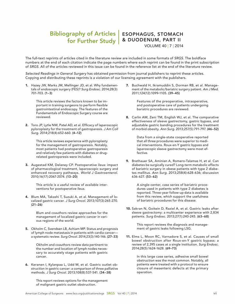
1. Hazey JW, Marks JM, Mellinger JD, et al. Why fundamen-tals of endoscopic surgery (FES)? Surg Endosc. 2014;28(3): 701-703. (1–3)
This article reviews the factors known to be im-portant in training surgeons to perform flexible gastrointestinal endoscopy. The features of the Fundamentals of Endoscopic Surgery course are reviewed.
2. Toro JP, Lytle NW, Patel AD, et al. Efficacy of laparoscopic pyloroplasty for the treatment of gastroparesis. J Am Coll Surg. 2014;218(4):652-660. (4–12)
This article reviews experience with pyloroplasty for the management of gastroparesis. Notably, most patients had postoperative gastroparesis and relatively few patients with diabetes or drug-related gastroparesis were included.
3. Augestad KM, Delaney CP. Postoperative ileus: impact of pharmacological treatment, laparoscopic surgery and enhanced recovery pathways. World J Gastroenterol. 2010;16(17):2067-2074. (13–20)
This article is a useful review of available inter-ventions for postoperative ileus.
4. Blum MA, Takashi T, Suzuki A, et al. Management of lo-calized gastric cancer. J Surg Oncol. 2013;107(3):265-270. (21–26)
Blum and coauthors review approaches for the management of localized gastric cancer in vari-ous regions of the world.
5. Okholm C, Svendsen LB, Achiam MP. Status and prognosis of lymph node metastasis in patients with cardia cancer— a systematic review. Surg Oncol. 2014;23(3):140-146. (27–33)
Okholm and coauthors review data pertinent to the number and location of lymph nodes neces-sary to accurately stage patients with gastric cancer.
6. Keranen I, Kylanpaa L, Udd M, et al. Gastric outlet ob-struction in gastric cancer: a comparison of three palliative methods. J Surg Oncol. 2013;108(8):537-541. (34–38)
This report reviews options for the management of malignant gastric outlet obstruction.
7. Buchwald H, Ikramuddin S, Dorman RB, et al. Manage-ment of the metabolic/bariatric surgery patient. Am J Med. 2011;124(12):1099-1105. (39–45)
Features of the preoperative, intraoperative, and postoperative care of patients undergoing bariatric procedures are reviewed.
8. Carlin AM, Zeni TM, English WJ, et al. The comparative effectiveness of sleeve gastrectomy, gastric bypass, and adjustable gastric banding procedures for the treatment of morbid obesity. Ann Surg. 2013;257(5):791-797. (46–52)
Data from a single-state cooperative reported that all three procedures were superior to medi-cal interventions. Roux-en-Y gastric bypass and laparoscopic sleeve gastrectomy were most ef-fective.
9. Brethauer SA, Aminian A, Romero-Talamas H, et al. Can diabetes be surgically cured? Long-term metabolic effects of bariatric surgery in obese patients with type 2 diabe-tes mellitus. Ann Surg. 2013;258(4):628-636; discussion 636-627. (53–62)
A single-center, case series of bariatric proce-dures used in patients with type 2 diabetes is reported. Three-year follow-up data is available from this review, which support the usefulness of bariatric procedures for this disease.
10. Sakran N, Goitein D, Raziel A, et al. Gastric leaks after sleeve gastrectomy: a multicenter experience with 2,834 patients. Surg Endosc. 2013;27(1):240-245. (63–68)
This report reviews the diagnosis and manage-ment of gastric leaks following LSG.
11. Elms L, Moon RC, Varnadore S, et al. Causes of small bowel obstruction after Roux-en-Y gastric bypass: a review of 2,395 cases at a single institution. Surg Endosc. 2014;28(5):1624-1628. (69–73)
In this large case series, adhesive small bowel obstruction was the most common. Notably, all patients were treated with a protocol to ensure closure of mesenteric defects at the primary operation.
The full-text reprints of articles cited in the literature review are included in some formats of SRGS. The boldface numbers at the end of each citation indicate the page numbers where each reprint can be found in the print subscription of SRGS. All of the articles reviewed in this issue can be found in the reference list at the end of the literature review.
Selected Readings in General Surgery has obtained permission from journal publishers to reprint these articles. Copying and distributing these reprints is a violation of our licensing agreement with the publishers.
Bibliography of Articles for Further Study
VOLUME 40 | 7 | 2014
ESOPHAGUS, STOMACH & DUODENUM, PART II

viii American College of Surgeons www.facs.org/publications/srgs SRGS Vol 40 | 7 | 2014
New SRGS Log-in Procedures
On August 5, 2014, the American College of Surgeons (ACS) launched its new public website, facs.org, designed for easy access from desktops, tablets, and smartphones. ACS retired its mem-bers-only Web portal, efacs.org, migrating some features to the new public website, and replacing others with a new online community platform.
SRGS print subscribers who participate in CME program:
1. Go to www.facs.org
2. Log in with your ACS username and password at “Member Login” (even if you are not an ACS member)
3. Click on Publications, under Selected Readings in General Surgery, click on Continuing Medical Education
4. You can log in seamless to your CME index page by clicking on “CME Test Login” from the left-hand side
of the SRGS page.
SRGS Connect subscribers:
1. Go to www.facs.org
2. Log in with your ACS username and password at “Member Login” (even if you are not an ACS member)
3. Click on Publications, under Selected Readings in General Surgery, click on SRGS Connect Login
4. Once logged in to SRGS Connect, click on “Test Your Knowledge” to access your CME test index page.
If you are having trouble logging in to SRGS content, please power off and relaunch your browser. Do not use any saved bookmarks or passwords; follow the new procedures outlined above.
The new ACS website is optimized for Microsoft Windows IE 8+, Apple Mac Safari 6+, Mozilla Firefox 14+, Google Chrome 26+, Apple Mobile Safari 6+, Google Android Internet Browser 4+, Google Android Chrome 20+.
®
SE L EC T E D R E A DI NGS in GE N E R A L SU RGE RY

1American College of Surgeons www.facs.org/publications/srgs SRGS Vol 40 | 7 | 2014
This issue of SRGS is the second of a two-part offering on the management of surgical patients with diseases of the esophagus, stomach, and duodenum. The issue surveys disorders of the stomach and duodenum. I am grateful for
the able editorial assistance of Robert Yates MD, and Carlos Pellegrini, MD, FACS, who assisted me in the selection of articles for this series. In this second overview, we focus on topics of interest to general surgeons who might encounter patients with upper gastrointestinal symptoms. The topics surveyed have been chosen based on current relevance and because the subjects are important components of the practice of general surgery. As is our practice at SRGS, new articles are included and articles previously discussed in the 2010 edition that continue to be relevant and useful are retained.
Introduction
VOLUME 40 | 7 | 2014
ESOPHAGUS, STOMACH & DUODENUM, PART II
2 American College of Surgeons www.facs.org/publications/srgs SRGS Vol 40 | 7 | 2014
Endoscopy training for surgeons | ESOPHAGUS, STOMACH & DUODENUM, PART II
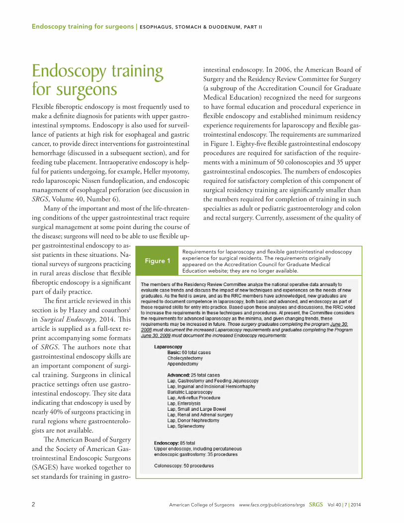
Endoscopy training for surgeonsFlexible fiberoptic endoscopy is most frequently used to make a definite diagnosis for patients with upper gastro-intestinal symptoms. Endoscopy is also used for surveil-lance of patients at high risk for esophageal and gastric cancer, to provide direct interventions for gastrointestinal hemorrhage (discussed in a subsequent section), and for feeding tube placement. Intraoperative endoscopy is help-ful for patients undergoing, for example, Heller myotomy, redo laparoscopic Nissen fundoplication, and endoscopic management of esophageal perforation (see discussion in SRGS, Volume 40, Number 6).
Many of the important and most of the life-threaten-ing conditions of the upper gastrointestinal tract require surgical management at some point during the course of the disease; surgeons will need to be able to use flexible up-per gastrointestinal endoscopy to as-sist patients in these situations. Na-tional surveys of surgeons practicing in rural areas disclose that flexible fiberoptic endoscopy is a significant part of daily practice.
The first article reviewed in this section is by Hazey and coauthors1 in Surgical Endoscopy, 2014. This article is supplied as a full-text re-print accompanying some formats of SRGS. The authors note that gastrointestinal endoscopy skills are an important component of surgi-cal training. Surgeons in clinical practice settings often use gastro-intestinal endoscopy. They site data indicating that endoscopy is used by nearly 40% of surgeons practicing in rural regions where gastroenterolo-gists are not available.
The American Board of Surgery and the Society of American Gas-trointestinal Endoscopic Surgeons (SAGES) have worked together to set standards for training in gastro-
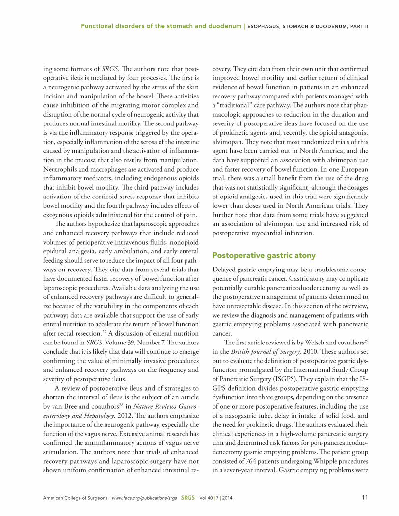
intestinal endoscopy. In 2006, the American Board of Surgery and the Residency Review Committee for Surgery (a subgroup of the Accreditation Council for Graduate Medical Education) recognized the need for surgeons to have formal education and procedural experience in flexible endoscopy and established minimum residency experience requirements for laparoscopy and flexible gas-trointestinal endoscopy. The requirements are summarized in Figure 1. Eighty-five flexible gastrointestinal endoscopy procedures are required for satisfaction of the require-ments with a minimum of 50 colonoscopies and 35 upper gastrointestinal endoscopies. The numbers of endoscopies required for satisfactory completion of this component of surgical residency training are significantly smaller than the numbers required for completion of training in such specialties as adult or pediatric gastroenterology and colon and rectal surgery. Currently, assessment of the quality of
Requirements for laparoscopy and flexible gastrointestinal endoscopy experience for surgical residents. The requirements originally appeared on the Accreditation Council for Graduate Medical Education website; they are no longer available.
Figure 1

3American College of Surgeons www.facs.org/publications/srgs SRGS Vol 40 | 7 | 2014
Endoscopy training for surgeons | ESOPHAGUS, STOMACH & DUODENUM, PART II
training and the process of granting endoscopy privileges are based primarily on numbers of procedures performed during training.
Hazey and coauthors1 note that learning curves for endoscopy skills may differ in surgeons versus gastro-enterologists and determining requirements for all en-doscopists based on a single set of numbers may not be feasible. Available evidence suggests that surgeons can become competent at the case number levels suggested by the American Board of Surgery. The authors cite data indicating that upper endoscopies performed by surgeons showed acceptable completion rates; similar data was available for colonoscopies. Other data did suggest that colonoscopies performed by surgeons who did at least 100 colonoscopies annually had completion rates and missed lesion rates similar to colon and rectal surgeons and gastroenterologists.
Additional perspective on determining the appro-priate case numbers to achieve competency in flexible endoscopy is provided by Vassiliou and coauthors2 in the American Journal of Surgery, 2010. The authors obtained data to calculate Global Assessment of Gastrointestinal Endoscopic Skills (GAGES) scores for upper endosco-pies and colonoscopies performed by 139 surgeons and gastroenterologists from 11 United States and non-U.S. academic centers. The improvement curves for GAGES scores plateaued at 35 procedures for upper endoscopy and 50 procedures for colonoscopy. The authors stress that the data suggest that case numbers may not be the best way to determine competence in gastrointestinal endoscopy. They also stress that a GAGES score that would define competence has not been finalized. They recommend that competency in endoscopy is probably best guaranteed by training in an environment that has adequate clinical volume, a trained dedicated teaching staff, mentoring capability, and a defined curriculum. Simulation training may be an additional means of facilitating the acquisition of competence.
Despite the fact that data comparing performance metrics for endoscopies done by surgeons and gastro-enterologists indicate acceptable levels of performance by surgeons, there are challenges for surgical residency programs wishing to produce trainees that are competent and meet national caseload requirements. In many teach-ing institutions, endoscopy equipment and facilities might
be controlled by nonsurgeon specialists; surgical faculty with an interest in training residents in endoscopy are required to create an adequate training curriculum and mentor committed trainees but these resources may not be available. Institutional infrastructure is necessary to gather data on endoscopy performance and outcomes. For example, a high-quality screening colonoscopy is necessary to ensure that the patient is free of cancer or precancerous adenomas. Data from studies of screening colonoscopy have suggested that a judgment about the quality of a screening colonoscopy cannot be made with-out certain specific information about the quality of the endoscopist (adenoma detection rate, withdrawal time, frequency of complete inspection of the entire colon) and the quality of the individual examination (completeness of bowel preparation).3
Perspective on current challenges facing surgical resi-dency programs seeking to provide optimum gastrointesti-nal endoscopy training is provided in an article by Subhas and coauthors4 in the American Journal of Surgery, 2010. The authors conducted an internet-based survey of surgical training programs in the U.S. The survey was composed of 10 questions designed to assess the size of the program, the availability of faculty to provide training in endoscopy, the average number of endoscopy procedures performed by residents, and the program director’s perspectives on the success of endoscopic training in the program. Ad-ditional questions assessed features of the educational environment, including dedicated endoscopic training fa-cilities and any negative effects of competition from other specialties. The response to the survey was small (30%). The data showed that 42% of programs had four or fewer residents. Ten percent of programs could not fulfill the requirements set forth by accrediting agencies and 55% of programs did not have dedicated endoscopic faculty or educational facilities. The data showed that fewer than 20% of programs had resident endoscopic experience that consistently exceeded 100 cases during residency. This article was a presentation to the plenary session of the 2009 annual meeting of the Midwest Surgical Associa-tion. The discussion that occurred after the presentation is included with the article. Questions focused on the risk that the sample was not representative of surgical residency programs in the U.S. The authors concluded that the data suggest a need for surgical residency programs

4 American College of Surgeons www.facs.org/publications/srgs SRGS Vol 40 | 7 | 2014
to provide an endoscopic curriculum, dedicated faculty, and endoscopic training facilities to increase confidence that surgical residents are competent in gastrointestinal endoscopy when training is completed.
Hazey and coauthors1 offer one potential solution for the lack of a formal endoscopic training curriculum for surgical residents. They suggest adoption of the Funda-mentals of Endoscopic Surgery (FES) course developed by SAGES. The course is composed of a 12-segment, online, didactic component, a test to document learning of the didactic component, and a five-segment, virtual reality, skills development component. They note that the course is similar to Fundamentals of Laparoscopic Surgery (FLS) course developed by SAGES, and it has been assessed for validity. The course can be used by learners beginning in medical school and extending into a clinical practice career. The authors recommend that the American Board of Surgery adopt the FES as a requirement for residency training similar to the adoption of FLS.
Another article discussing the optimum structure of endoscopy training for surgeons is by Bittner and co-authors5 in the Journal of Surgical Education, 2007. The authors note that avenues available to obtain the neces-sary expertise in flexible gastrointestinal endoscopy in-clude use of simulation, assignment of residents to work with surgeons who perform high volumes of endoscopy procedures, and development of cooperative training ar-rangements with nonsurgeon endoscopists. They further note that reported endoscopy experiences from residents in colon and rectal surgery programs and practicing co-lon and rectal surgeons are 5- to 10-fold higher than the requirements for general surgery residents. Bittner and as-sociates cite data from published reports confirming more rapid endoscopy completion and lower complication rates for surgeons with high versus low endoscopy volumes. Although no reliable data document a firm minimum number of procedures to guarantee competence, available information suggests that the range is large (50–130 upper endoscopies and 50–140 colonoscopies).
Data from analyses of administrative databases indi-cate that surgeon endoscopists are more likely than gastro-enterologists to miss important lesions during endoscopy, particularly colonoscopy.6 These reports should be viewed critically, however, because administrative data are not able to confirm the indication for endoscopy (screening
versus diagnostic), the quality of the bowel preparation, or the completeness of the procedure. Despite this, data suggesting that patients undergoing colonoscopy by sur-geons are at higher risk for subsequent development of colon cancer than patients undergoing colonoscopy by gastroenterologists should stimulate efforts to document the quality of endoscopy performed by surgeons.
Bittner and colleagues provide additional data on the current status of endoscopy training in general surgery programs in the U.S. They cite data indicating that as of 2006, less than two-thirds of programs have formal en-doscopy experience and only one-third of the experiences are led by surgeons with formal training in endoscopy. The challenge of providing adequate endoscopy experience is made more difficult because intraoperative endoscopy by a surgeon, other than the surgeon responsible for the primary operation, currently cannot be counted toward endoscopy experience. The authors conclude with a clear discussion of the use of simulation as a means of facilitat-ing endoscopy training. They stress that the presence of a standard curriculum for endoscopy training that includes simulation improves resident endoscopy performance and reduces cost of training. The reduction in training cost may, in fact, offset the expense of the simulator equip-ment. They conclude that acquisition of high-quality endoscopy training during surgical residency is feasible and desirable but requires commitment on the part of residency program directors, training institutions, and trainees.
Editorial comment: The articles described in the
foregoing section document the importance of a
standardized curriculum, simulation, and adequate
clinical training to produce surgeons who will be
able to integrate flexible fiberoptic intestinal en-
doscopy into their daily practice. Integration of
flexible gastrointestinal endoscopy training into
surgical residencies is still evolving. Forces that are
making competence in flexible endoscopy critical
for general surgeons include the necessity to pre-
pare surgeons for rural practice and the continued
evolution of minimally invasive surgical procedures
that often require intraoperative endoscopy. The
further development of natural orifice, endoscopi-
cally aided, surgical procedures will add challenges
Endoscopy training for surgeons | ESOPHAGUS, STOMACH & DUODENUM, PART II

5American College of Surgeons www.facs.org/publications/srgs SRGS Vol 40 | 7 | 2014
for training programs. Past experience has shown
that residency program directors and residents will
rise to the occasion and produce excellent train-
ing structures leading to satisfactory competence.
Achieving documentation of competence may be
more difficult than structuring the educational
programs necessary to satisfy requirements of
accrediting bodies. It is very important that sur-
geons assume leadership roles in efforts designed
to document effectiveness of education programs,
competence of trainees, and excellent outcomes
in daily practice.
Functional disorders of the stomach and duodenumUpper gastrointestinal symptoms commonly accompany a group of conditions called “functional gastrointestinal disorders.” These conditions are encountered in patients with psychiatric disorders and with other specific dis-eases. Diagnosis and management of these conditions can be challenging because there is significant overlap with symptoms of functional disorders and symptoms experienced by healthy individuals. The relationships be-tween brain function and the gastrointestinal tract are becoming clearer as research in this area progresses. There are data to suggest that one functional bowel disorder, irritable bowel syndrome, has a genetic component, is more common in offspring of parents with depression and anxiety syndromes, and is associated with structural changes in the brain.7,8
The symptoms encountered in patients with func-tional gastrointestinal disorders include dyspepsia, upper abdominal pain, nausea, vomiting, anorectal dysfunction, and bladder dysfunction. The most severe and disabling of these functional conditions is gastroparesis, which is an important complication of diabetes. In this section of the overview, we review several aspects of functional gastrointestinal disorders of importance to surgeons. The
topics reviewed include gastroparesis, postoperative ileus, postoperative gastric atony, and the aerophagia/rumina-tion syndromes.
Gastroparesis
Gastroparesis is characterized by symptoms of absent, delayed, or inadequate gastric emptying without evidence of mechanical obstruction. The medical history will dis-close that patients experience early satiety with inability to finish a normal meal, abdominal pain, nausea, vomiting, and, in some patients, dyspepsia. Although gastric emp-tying assessment using scintigraphy studies is generally required to document the presence of gastroparesis, the relationship between disordered emptying documented on emptying tests and symptom severity is not consistent.
Clinical practice guidelines for diagnosis and man-agement of gastroparesis have been promulgated by the American College of Gastroenterology and appear in an article by Camillieri and coauthors.9 The guidelines document is available free from the American College of Gastroenterology website at www.gi.org. Because the symptoms of gastroparesis overlap with other conditions, including accelerated gastric emptying, confirmation of delayed gastric emptying by scintigraphy and documenta-tion of the absence of mechanical obstruction are recom-mended. The most important finding on radioisotope scintigraphy is delayed emptying of solid food at four hours after intake. Scintigraphic studies are 85% accurate for identifying gastroparesis. Data cited in the guidelines document suggest that adding emptying studies for liq-uids may increase accuracy.
Data from a study of diabetic patients with gastropa-resis suggest that delayed gastric emptying documented by abnormal gastric scintigraphy is related to morbidity. This study by Hyett and coauthors10 is in Gastroenterol-ogy, 2009. The data come from a parallel cohort study of three groups of patients; each group contained 94 patients and the groups were followed for an eight-year interval. Data were collected to document hospitalizations, doc-tor’s office visits, emergency room visits, hemoglobin A1C levels, medications, and new disease diagnoses. Compared with groups of patients with diabetes and symptoms of gastroparesis but normal gastric emptying and patients with diabetes without gastroparesis symptoms, the pa-
Functional disorders of the stomach and duodenum | ESOPHAGUS, STOMACH & DUODENUM, PART II

6 American College of Surgeons www.facs.org/publications/srgs SRGS Vol 40 | 7 | 2014
tients with gastroparesis and abnormal scintigraphy had increased morbidity. These patients experienced more hospitalizations, emergency room visits, and doctor’s office visits. There were more diagnoses of retinopathy, hyper-tension, and renal disease in the group with abnormal scintigraphy. The morbidity differences remained when the data were adjusted for diabetes control, as reflected by hemoglobin A1C levels. Mortality was not different when the groups were compared. The authors concluded that severe gastroparesis with documented abnormal gastric emptying was associated with increased diabetes-related morbidity.
The guidelines9 recommend that laboratory studies to evaluate the patient for diabetes and thyroid dysfunc-tion should be part of the initial evaluation. Because gastroparesis can occur as a side effect of the use of nar-cotic drugs and certain medications used in the manage-ment of diabetes, the guidelines recommend including questions regarding these factors in the medical history. Gastroparesis can occur following a viral infection; the guidelines recommend questioning for symptoms of a viral prodrome.
The clinical diagnosis of gastroparesis is usually based on symptoms such as nausea, vomiting, and early satiety. Clinical experience has shown that surgeons are often called to participate in the care of patients with gastro-paresis when patients present with abdominal pain. An article discussing the frequency of abdominal pain as a symptom of gastroparesis is by Cherian and coauthors11 in Clinics in Gastroenterology and Hepatology, 2010. In this study, the authors evaluated responses to three question-naires designed to evaluate abdominal symptoms, pain, and overall quality of life. The data disclosed that 90% of patients with gastroparesis experienced abdominal pain and this pain was severe enough to reduce quality of life. Pain was epigastric in nearly half the patients and was frequently induced by eating, but also was nocturnal in al-most three-quarters of patients with resultant disturbance of sleep. The authors conclude that therapies designed to target the pain component of gastroparesis are important factors in treatment strategies.
Gastroparesis is one component of a group of disor-ders of gastric motor and sensory function. These con-ditions are discussed in an article by Tack12 in Current
Opinion in Gastroenterology, 2009. The author reviews data suggesting that the pathogenesis of gastroparesis has a genetic component. There have also been documented abnormalities of the interstitial cells of Cajal within the stomach. Abnormal neural function has been documented in animal models of gastroparesis characterized by loss of nitric oxide synthase within gastric nerves. This finding is similar to findings in humans with achalasia (see discus-sion in SRGS, Volume 40, Number 6). Other studies have suggested contributions of infection and inflammation, allergic reactions to foods, and autonomic dysfunction as contributors to gastroparesis, but the results have not been consistent over a number of studies. The author concludes by reviewing therapeutic options including prokinetic drugs, antidepressants, and anxiolytic agents. In refractory cases, gastric electrical stimulation is recommended. All of these interventions have been used with varying levels of success in achieving reductions in symptom severity and improved gastric emptying.
The epidemiology of gastroparesis is the topic of an article by Jung and coauthors13 in Gastroenterology, 2009. The authors followed patients with definite gastroparesis (symptoms plus abnormal scintigraphy), probable gastro-paresis (symptoms plus retained food on barium gastric imaging or endoscopy), or possible gastroparesis (symp-toms only or abnormal scintigraphy without symptoms) in Olmsted County, Minnesota. The data disclosed that the age-adjusted prevalence of gastroparesis was 9.6/100,000 persons. There was a definite preponderance of women in the gastroparesis group. When all individuals with gastroparesis were considered, women had a higher than predicted mortality. Individuals in the definite gastropare-sis group had a lower than predicted mortality compared with other patients with gastroparesis. The authors note that the definite gastroparesis group contained signifi-cantly more patients with “idiopathic” gastroparesis com-pared with the other groups where comorbidities such as diabetes, Parkinson’s disease, connective tissue disorders, and central nervous system degenerative disease were com-mon. They suggest that the increased frequency of these diseases in the probable and possible gastroparesis groups is one explanation for the observed increased mortality. These population-based data contrast with the findings
Functional disorders of the stomach and duodenum | ESOPHAGUS, STOMACH & DUODENUM, PART II

7American College of Surgeons www.facs.org/publications/srgs SRGS Vol 40 | 7 | 2014
of Hyett and coauthors10 (discussed earlier) and the dif-ference is probably the result of the larger sample sizes in the Jung study.
The health burden of gastroparesis is the topic of two articles discussed here. The first is by Wang and coau-thors14 in the American Journal of Gastroenterology, 2008. These authors queried the National Inpatient Sample database from 1995-2004 to determine trends in hospi-talizations for gastroparesis in the U.S. Hospitalizations for gastroparesis as the primary diagnosis increased from 3,977 in 1995 to 10,252 in 2004. There was a similar increase in hospitalizations for gastroparesis as a second-ary diagnosis. The authors note that there were increases in total hospitalizations and hospitalizations for diabetes during the same interval, but these were smaller than the increase due to gastroparesis. Compared with other upper gastrointestinal diagnoses, gastroparesis had the largest increase in hospitalizations and the longest lengths of stay. The authors suggest that the increase in hospitaliza-tions could result from improved diagnostic accuracy, an increased appreciation for the diagnosis of gastroparesis among clinicians, increases in the prevalence of diabetes, and increased longevity of diabetic patients.
Factors contributing to the need for hospitalization in patients with gastroparesis are the focus of an article by Uppalapati and coauthors.15 The authors reviewed the medical records of 63 patients admitted on 103 occasions for exacerbations of gastroparesis symptoms. Gastroparesis related to diabetes and idiopathic gastroparesis accounted for an equal number of admissions and these two forms of gastroparesis accounted for more than 80% of the total admissions. Poor diabetic control and intolerance of medications were common causes leading to hospital admission. Evidence of inflammation was frequently de-tected (increased erythrocyte sedimentation rate and/or elevated C-reactive protein) but documented bacterial infection was not frequently diagnosed. Of interest is the observation that adrenal insufficiency was diagnosed in 9% of patients. The authors recommend searching for a source of infection in patients with elevated inflammatory markers and exclusion of adrenal insufficiency in patients hospitalized with gastroparesis. Another study of gastric antral, full-thickness biopsies in patients with gastropa-resis16 documented histologic evidence of inflammation in more than 50% of patients with diabetic gastroparesis.
This additional evidence suggests that inflammatory con-ditions, including infections, may precipitate exacerba-tions of gastroparesis.
Gastroparesis in the diabetic patient
Gastroparesis is one of the most troubling gastrointestinal complications of diabetes. An article reviewing the patho-physiology and management of diabetic gastroparesis is by Alam and coauthors17 in Diabetes Therapy, 2010. The authors cite data suggesting that evidence of delayed gas-tric emptying can be discovered in 30%-40% of patients with Type 1 and Type 2 diabetes although only a small proportion of diabetics have severe gastroparesis symp-toms. Of interest are data indicating that in the early stages of Type 2 diabetes gastric emptying may actually be accelerated. The underlying pathophysiology of dia-betic gastroparesis is a combination of vagal neuropathy and disordered function of the gastric pacemaker. The consequences of these changes include decreased antral contractions and dysfunction of the gastric response to hypoglycemia. Absence of normal gastric emptying in response to low blood sugar levels may lead to symptoms of hypoglycemia that termed the “gastric hypoglycemia” syndrome.
The full spectrum of gastrointestinal manifesta-tions of diabetes is discussed in an article by Sellin and Chang18 in Nature Clinical Practice Gastroenterology and Hepatology, 2008. The authors begin the review by noting that the common gastrointestinal disorders associated with diabetes include gastroesophageal reflux, candida esophagitis, gastroparesis, constipation, and diarrhea. They further note that women with diabetes are more likely to develop gastrointestinal complications than are men. The pathogenesis of the gastrointestinal complica-tions associated with diabetes can result from the effects of hyperglycemia on the various neural signaling pathways within the gastrointestinal tract. Hyperglycemia is notable for its inhibitory effects on vagal nerve function that can lead to symptoms. They emphasize that gastrointestinal manifestations of diabetes do not correlate with the dura-tion of the disease or with systemic evidence of autonomic nerve dysfunction. The intestinal cells of Cajal are targets of neural damage associated with diabetes. Reduced levels of Cajal cell trophic factors have been demonstrated in
Functional disorders of the stomach and duodenum | ESOPHAGUS, STOMACH & DUODENUM, PART II

8 American College of Surgeons www.facs.org/publications/srgs SRGS Vol 40 | 7 | 2014
animal models of diabetes. Increased neural cell apop-tosis as well as hypoxia from microvascular disease and hyperglycemia all predispose these pacemaker cells of the intestine to damage and degeneration.
Alam and coauthors17 note that additional factors that influence gastric emptying in diabetic patients include the degree of insulin resistance and body mass index. Hyperglycemia can also cause “blunting” of nitric oxide function leading to depressed gastric motility. Elevated levels of dopamine also depress gastric emptying and re-ductions of dopamine levels is the main reason for the effectiveness of prokinetic agents such as metoclopramide.
Treatment of gastroparesis
Alam and coauthors17 note that the most important factors in the initial treatment of gastroparesis are normalization of fluid and electrolytes, measures to optimize glycemic control and support of nutrition. The practice guidelines document9 recommends enteral nutritional support. If gastric feedings in reduced volumes, administered fre-quently, are tolerated, this approach can be used, but intermittent exacerbations of obstructive symptoms may interrupt gastric feedings. In this setting, nasoenteric tube, postpyloric feedings are recommended. The guidelines note that parenteral nutrition is not recommended.
The mainstay of pharmacologic treatment of diabetic gastroparesis is prokinetic therapy. The first-line drug is metoclopramide; as mentioned earlier, the main mecha-nism of action is reduction of dopamine levels. Alam and coauthors17 cite evidence confirming improvement of gastric emptying in randomized trials using this agent. Unfortunately, there was not a close correlation of im-proved scintigraphic evidence of gastric emptying and improvement of symptoms. Metoclopramide is effective in improving gastroparesis symptoms in most patients, but its effectiveness may be reduced with prolonged use. Cen-tral nervous system side effects such as anxiety, movement disorders, and tardive dyskinesia become more frequent with prolonged use of metoclopramide. Domperidone is another antidopamine agent that has been used in Europe for treatment of diabetic gastroparesis. This agent is not currently approved for use in the U.S. Motilin agonists, such as erythromycin, have been shown to be effective in the treatment of diabetic gastroparesis. Concerns over the
emergence of resistant microorganisms as a result of use of erythromycin to treat noninfectious diseases have limited the use of this agent. Azithromycin and clarithromycin are additional motilin agonist antibiotic agents that may be effective in treating diabetic gastroparesis. Currently available data are not sufficient to support a recommenda-tion for the use of these agents except for patients with gastroparesis symptoms refractory to other drugs. Of note is that a recommendation for use of these drugs, even in refractory cases, is not included in the practice guidelines document.9
Nonantibiotic motilin agonist drugs, such as metimci-nal and ABT-229, have been used in exploratory clinical studies. Gastric emptying was improved, but frequently without significant improvement in symptoms in most patients. Alam and coauthors17 note that cisapride has been effective in treating gastroparesis symptoms, but this drug and similar agents that act by increasing cholinergic responses by actions on the 5-HT receptor have been limited by frequent cardiac side effects (rhythm distur-bances). A new group of agents with potential value in the treatment of diabetic gastroparesis are ghrelin and ghrelin agonists. Ghrelin acts through the GHRS-1 re-ceptor that increases gastric emptying by enhancing the migratory motor complex. The authors cite data indicat-ing that ghrelin has potential value for improving gastric emptying but, as with other drugs, symptom improvement was inconsistent. Studies in nondiabetic gastroparesis have not supported the use of ghrelin. The authors note that one positive aspect of ghrelin and ghrelin agonists is the fact that these agents have a very good safety and side ef-fect profile compared to other prokinetic agents. For this reason, further research and development are indicated.
Additional perspective on the various options for management of gastroparesis is found in an article by Hejazi and McCallum19 in Gastrointestinal Endoscopy Clin-ics of North America, 2009. The authors begin by noting that significant gastroparesis symptoms are found in up to 7% of patients with diabetes, and this fact makes the disorder a major concern in the management of patients with diabetes. As noted previously, the symptoms oc-cur predominantly in women and the mean age of onset is the mid-30s. Estimates indicate that there may be as many as five million patients with gastroparesis in the U.S. The authors note that prokinetic agents represent the
Functional disorders of the stomach and duodenum | ESOPHAGUS, STOMACH & DUODENUM, PART II

9American College of Surgeons www.facs.org/publications/srgs SRGS Vol 40 | 7 | 2014
first line of therapy for gastroparesis (along with dietary modifications and optimization of diabetic control as noted previously). The dopamine D2 receptor antagonist metoclopramide and the motilin receptor agonists eryth-romycin and azithromycin represent the most commonly used pharmacologic agents. These drugs are frequently administered in combination with antiemetic medications (particularly the serotonin 5-HT3 antagonist, ondan-setron), antidepressants, antianxiety drugs, and others.
A study comparing erythromycin and azithromycin for treatment of gastroparesis is by Moshiree and coau-thors20 in Digestive Disease and Sciences, 2010. Gastric antral function was measured in 30 patients during clini-cal evaluation for gastroparesis. The effect of the drugs on antral function was equivalent at a dose of 250 mg intravenously. At a dose of 500 mg intravenously, azithro-mycin produced superior antral muscle function. The authors note that there are fewer drug-drug interactions with azithromycin and this may offer a therapeutic ad-vantage. Unfortunately, no data about improvement of patient symptoms were reported. The practice guidelines document9 recommends the use of adjunctive antiemetic agents and tricyclic antidepressant drugs. These agents may improve symptoms when used in conjunction with prokinetic agents.
Other modalities suggested for treatment of diabetic gastroparesis are endoscopic injection of botulinum toxin into the pyloric muscle, gastric electrical stimulation, and the use of Chinese medicine approaches, such as acupuncture. Alam and colleagues note that botulinum toxin injection has been useful in achalasia. Available high-quality data from randomized trials have failed to confirm benefit from botulinum toxin injection except in patients where pylorospasm can be documented. Clinical practice guidelines9 do not recommend the use of botu-linum toxin injections.
Gastric electrical stimulation using two electrodes implanted in the serosa along the greater curvature of the stomach at points 9 and 10 centimeters from the pylorus with stimulation from a pulse generator placed on the anterior abdomen has demonstrated improved gastric emptying. Alam and coauthors17 note that this approach is applicable in relatively few patients (those with severe refractory symptoms leading to multiple hospitalizations). Clinical practice guidelines9 recommend the compas-
sionate use of this approach for severe refractory diabetic gastroparesis. Hejazi and McCallum19 report two stud-ies from their institution using the Enterra™ stimulator (Medtronic, Minneapolis, MN). The first study followed 37 patients for a mean of 45 months. Significant symptom improvement and reduced medication use were observed at one year and persisted at three years. Fifteen of the 37 patients required nutritional support at the beginning of the study, but only five still required support at three years. In a second study of 46 patients with up to 10 years of followup, standard gastrointestinal symptoms scores improved from a mean of 20 to a mean of 7. Complica-tions were seen in 13% of patients and the device failed to improve symptoms in 13 patients.
A review of the application of traditional Chinese medicines (herbal mixtures) and acupuncture for the treatment of diabetic gastroparesis is from Pang and co-authors21 in the World Journal of Gastroenterology, 2014. The authors note that Chinese herbal remedies with or without acupuncture are employed frequently in Chi-nese health care facilities for patients with diabetic and nondiabetic gastroparesis. Anecdotal data indicate that these approaches are frequently associated with symp-tomatic improvement. Data cited by the authors from one retrospective trial conducted by a single practitioner documented a significant improvement in symptoms when patients treated with herbal medicines and acupuncture were compared with patients treated without these ap-proaches. Data with sufficient quality to support a recom-mendation are not currently available.
A review of surgical approaches to the management of gastroparesis is by Jones and Maganti22 in the American Journal of Gastroenterology, 2003. The authors conducted a systematic review of the literature and identified 17 acceptable articles. All available studies were unblinded, included relatively few patients, and focused primarily on patients with postsurgical gastroparesis. From the avail-able data, the authors concluded that venting gastrostomy provides symptom relief in patients with severe refractory gastroparesis of all types. Jejunostomy was associated with improved nutritional status in most patients but complica-tions, such as tube malfunction and tube displacement, caused significant morbidity. Pyloroplasty was useful in selected patients with postsurgical gastroparesis and completion gastrectomy was beneficial for patients with long-standing severe postsurgical gastroparesis.
Functional disorders of the stomach and duodenum | ESOPHAGUS, STOMACH & DUODENUM, PART II

10 American College of Surgeons www.facs.org/publications/srgs SRGS Vol 40 | 7 | 2014
A recent article presenting a retrospective analysis of a clinical case series of pyloroplasty as treatment for gastroparesis is by Toro and coauthors23 in the Journal of the American College of Surgeons, 2014. This article is supplied as a full-text reprint accompanying some for-mats of SRGS. The authors reviewed outcomes in 50 patients who underwent laparoscopic pyloroplasty for gastroparesis. Ten patients were known to be diabetic and five patients had the diagnosis of diabetic gastroparesis. Symptoms improved in all of these patients at one month postoperatively. Most patients had postsurgical gastropa-resis and symptom improvement occurred in 82% of the total series. Gastric emptying studies showed improved emptying in all patients. Subsequent interventions were necessary in five patients and consisted of gastrectomy, duodenojejunostomy, and gastric stimulator placement. These data were presented at plenary session of the 2013 annual meeting of the Southern Surgical Association. The discussion that followed the presentation, included with the article, notes that the clinical features of patients operated on in this series indicated a highly selected group of patients mostly with postsurgical gastroparesis. The use of laparoscopic pyloroplasty is unlikely to be a suitable option for most patients with gastroparesis in whom the condition is associated with diabetes or narcotic drug use.
The use of gastric bypass for treatment of morbidly obese patients with diabetic or idiopathic gastroparesis is described in an article by Papasavas and coauthors24 in Surgery of Obesity and Related Diseases, 2014. The au-thors present outcomes data on a group of seven patients who underwent laparoscopic Roux-en-Y gastric bypass. A follow-up period up to two years was available. All patients had significant improvement in gastroparesis symptoms and four patients who were taking prokinetic agents preoperatively were able to discontinue these. There were no major perioperative complications. Three patients required readmission for non-gastroparesis conditions. The authors concluded that gastric bypass has potential value for the management of morbidly obese patients with gastroparesis.
Hejazi and McCallum25 note that gastric resection has been used to treat patients with refractory gastroparesis after a prior gastric resection and in a small number of patients with diabetic gastroparesis. They report a study from their institution of total gastrectomy used in nine
patients as a last resort treatment. Six patients were avail-able for followup at an average of 3.5 years. All patients had symptom improvement averaging more than 50%. Hospitalizations and emergency department visits were reduced. Quality of life had improved. Additional dis-cussion of gastrectomy for management of postoperative gastric atony is found in a later section of the overview.
Management of postoperative ileus
Postoperative ileus is a frequent cause of delayed return to oral feedings and delayed hospital discharge after ab-dominal surgery. Ileus can also complicate burn injury and nonsurgical critical illness. Postoperative ileus has gastric, small bowel, and colonic components. Postopera-tive nausea and vomiting can be due to gastric empty-ing delays after operation and is also a complication of using certain anesthetic agents and narcotic analgesics. The consistent observation that small bowel feeding can resume within the first few hours after a major operation is clear evidence that small intestinal ileus is transient and frequently may not be clinically significant. Traditionally, surgeons are taught to withhold oral feedings until bowel sounds are audible on abdominal auscultation and the patient has passed flatus. The origin of these sounds is thought to be the small intestine, although the success of feeding after bowel sounds return and flatus is passed may indicate that the sounds have a colonic origin because the colonic component of ileus is thought to be the last component to resolve.
As clearer understanding of the pathogenesis of ileus has evolved, strategies have been developed to shorten times to feeding, reduce the use of nasogastric decompres-sion, and hasten hospital discharge. The more severe forms of postoperative ileus occur in elderly high-risk patients (colonic pseudo-obstruction) and after pancreaticoduo-denectomy (post-Whipple gastric atony). The problem of gastric atony following pancreaticoduodenectomy is discussed later this section.
Increasing use of minimally invasive approaches and enhanced recovery pathways has been associated with changes in the frequency and severity of postoperative ileus. This topic is reviewed in an article by Augestad and Delaney26 in the World Journal of Gastroenterology, 2010. This article is supplied as a full-text reprint accompany-
Functional disorders of the stomach and duodenum | ESOPHAGUS, STOMACH & DUODENUM, PART II
11American College of Surgeons www.facs.org/publications/srgs SRGS Vol 40 | 7 | 2014
ing some formats of SRGS. The authors note that post-operative ileus is mediated by four processes. The first is a neurogenic pathway activated by the stress of the skin incision and manipulation of the bowel. These activities cause inhibition of the migrating motor complex and disruption of the normal cycle of neurogenic activity that produces normal intestinal motility. The second pathway is via the inflammatory response triggered by the opera-tion, especially inflammation of the serosa of the intestine caused by manipulation and the activation of inflamma-tion in the mucosa that also results from manipulation. Neutrophils and macrophages are activated and produce inflammatory mediators, including endogenous opioids that inhibit bowel motility. The third pathway includes activation of the corticoid stress response that inhibits bowel motility and the fourth pathway includes effects of exogenous opioids administered for the control of pain.
The authors hypothesize that laparoscopic approaches and enhanced recovery pathways that include reduced volumes of perioperative intravenous fluids, nonopioid epidural analgesia, early ambulation, and early enteral feeding should serve to reduce the impact of all four path-ways on recovery. They cite data from several trials that have documented faster recovery of bowel function after laparoscopic procedures. Available data analyzing the use of enhanced recovery pathways are difficult to general-ize because of the variability in the components of each pathway; data are available that support the use of early enteral nutrition to accelerate the return of bowel function after rectal resection.27 A discussion of enteral nutrition can be found in SRGS, Volume 39, Number 7. The authors conclude that it is likely that data will continue to emerge confirming the value of minimally invasive procedures and enhanced recovery pathways on the frequency and severity of postoperative ileus.
A review of postoperative ileus and of strategies to shorten the interval of ileus is the subject of an article by van Bree and coauthors28 in Nature Reviews Gastro-enterology and Hepatology, 2012. The authors emphasize the importance of the neurogenic pathway, especially the function of the vagus nerve. Extensive animal research has confirmed the antiinflammatory actions of vagus nerve stimulation. The authors note that trials of enhanced recovery pathways and laparoscopic surgery have not shown uniform confirmation of enhanced intestinal re-
covery. They cite data from their own unit that confirmed improved bowel motility and earlier return of clinical evidence of bowel function in patients in an enhanced recovery pathway compared with patients managed with a “traditional” care pathway. The authors note that phar-macologic approaches to reduction in the duration and severity of postoperative ileus have focused on the use of prokinetic agents and, recently, the opioid antagonist alvimopan. They note that most randomized trials of this agent have been carried out in North America, and the data have supported an association with alvimopan use and faster recovery of bowel function. In one European trial, there was a small benefit from the use of the drug that was not statistically significant, although the dosages of opioid analgesics used in this trial were significantly lower than doses used in North American trials. They further note that data from some trials have suggested an association of alvimopan use and increased risk of postoperative myocardial infarction.
Postoperative gastric atony
Delayed gastric emptying may be a troublesome conse-quence of pancreatic cancer. Gastric atony may complicate potentially curable pancreaticoduodenectomy as well as the postoperative management of patients determined to have unresectable disease. In this section of the overview, we review the diagnosis and management of patients with gastric emptying problems associated with pancreatic cancer.
The first article reviewed is by Welsch and coauthors29 in the British Journal of Surgery, 2010. These authors set out to evaluate the definition of postoperative gastric dys-function promulgated by the International Study Group of Pancreatic Surgery (ISGPS). They explain that the IS-GPS definition divides postoperative gastric emptying dysfunction into three groups, depending on the presence of one or more postoperative features, including the use of a nasogastric tube, delay in intake of solid food, and the need for prokinetic drugs. The authors evaluated their clinical experiences in a high-volume pancreatic surgery unit and determined risk factors for post-pancreaticoduo-denectomy gastric emptying problems. The patient group consisted of 764 patients undergoing Whipple procedures in a seven-year interval. Gastric emptying problems were
Functional disorders of the stomach and duodenum | ESOPHAGUS, STOMACH & DUODENUM, PART II

12 American College of Surgeons www.facs.org/publications/srgs SRGS Vol 40 | 7 | 2014
identified in 344 patients (44.5%). Their data disclose that the main risk factors identified on multivariate analysis were female gender, New York Heart Association grade > 1, and the presence of a major postoperative complication, such as infection and complex pancreatic fistula. More severe postoperative complications were directly related to advanced grades of delayed gastric emptying. The authors conclude that delayed gastric emptying is present to some degree in nearly half of the patients undergoing Whipple resection. Most of these will not require intervention. More severe grades of delayed gastric emptying are associ-ated with the presence of postoperative complications and these advanced grades require treatment with nasogastric suction, measures to implement enteral nutrition, and prokinetic agents.
The next article reviewed is by Leung and Silverman30 in Digestive Diseases and Sciences, 2009. These authors present a proposed algorithm for the efficient diagnosis and management of patients with gastroparesis associ-ated with pancreatic cancer. The algorithm is based on a systematic review of available literature. The algorithm proposes a water-soluble gastric imaging study to deter-mine the presence of mechanical obstruction. The au-thors stress that water-soluble contrast does not coat the internal surface of the stomach and, thus, an early upper gastrointestinal endoscopy examination, the second com-ponent of the diagnostic phase of the algorithm, can be accomplished. The purpose of the endoscopic examination is to confirm the absence of mechanical obstruction and exclude gastroduodenal mucosal disease. Patients identi-fied with pancreatic-cancer-associated gastroparesis are treated initially with prokinetic agents. If the gastroparesis is refractory, percutaneous endoscopic gastrostomy with jejunal extension for enteral feeding is the final step. The algorithm suggested by these authors is reproduced with permission as Figure 2.
Patients who have postoperative gastroparesis and who are acceptable operative risks might be effectively treated with completion gastrectomy. An article describ-ing a single institution’s clinical experience with this ap-proach is by Speicher and coauthors31 in the Journal of Gastrointestinal Surgery, 2009. These authors describe their experience with 44 patients seen over an extended 19-year interval. Each patient had refractory gastric atony and underwent near total or total gastrectomy for man-
agement of this condition. Disabling symptoms present preoperatively included abdominal pain and vomiting (present in more than 98% of patients). Symptoms present in more than three-quarters of patients included nausea, diet limitation, heartburn, and weight loss. There was one perioperative death and complications, including anasto-motic stricture and anastomotic leak, occurred in 36% of patients. Twenty-nine patients were available for followup at intervals ranging from 0.5–15 years. Symptoms had improved in all patients available for followup and nearly 80% of patients believed their overall health status had improved. Postoperative weight loss averaged 10% and symptoms such as early satiety were common. Osteopo-rosis was frequently diagnosed in this patient group. The authors stress the need for postoperative supplementation of vitamin B12, calcium, folate, iron, and vitamin D. In conclusion, they recommend that patients who undergo completion gastrectomy for postoperative gastroparesis have feeding jejunostomies inserted to facilitate postop-erative nutritional support.
Belching, hiccups, and aerophagia
Belching, hiccups, and aerophagia are increasingly en-countered in children and adults. Surgeons may be called on to participate in the care of these patients because patients with aerophagia may present with significant ab-dominal distention and some of the symptoms of the con-ditions are also associated with gastroesophageal reflux.
A review article by Bredenoord32 in Clinics in Gas-troenterology and Hepatology, 2013, summarizes current knowledge relevant to the diagnosis and management of these disorders. The author notes that belching and hiccups occur in healthy individuals and that diagnostic testing is necessary only when symptoms become exces-sive or chronic. He emphasizes the fact that belching is a different entity than aerophagia. Excessive belching may be due to gastric belching wherein swallowed air is evacuated from the stomach because of transient relax-ation of the lower esophageal sphincter. More commonly, excessive belching is due to supragastric belching that is produced by relaxation of the upper esophageal sphincter and contraction of the diaphragm. This action creates negative pressure in the thoracic cavity and esophagus;
Functional disorders of the stomach and duodenum | ESOPHAGUS, STOMACH & DUODENUM, PART II

13American College of Surgeons www.facs.org/publications/srgs SRGS Vol 40 | 7 | 2014
air is suctioned into the esophagus and then evacuated as a belch. Excessive belching may be a symptom of GERD and clinical testing to exclude GERD is indicated in the evaluation of the patient with excessive belching.
Aerophagia, in contrast, is a disorder where air is swallowed producing abdominal distention, bloating, flatulence, and abdominal pain. Symptoms may mimic ileus or intestinal obstruction and surgeons might be con-sulted to assist in management of this condition. Excessive supragastric belching can be treated with a combination of speech therapy and psychotherapy. Aerophagia is also treated with speech therapy and psychotherapy. Excessive
distention and bloating may require nasogastric intuba-tion. Gas dispersal agents, such as simethicone, might be helpful in selected patients.
Inability to belch is a potential cause of troublesome abdominal distention and bloating. This symptom is en-countered occasionally as a postoperative complication of antireflux procedures for GERD (gas-bloat syndrome). In most patients, the symptoms are transient and respond to simple measures such as simethicone therapy. Occa-sionally, inability to belch is an indication to take down the fundoplication. Restoring lower esophageal sphincter function with a partial fundoplication has not consistently reduced the frequency of gas-bloat syndrome.
Functional disorders of the stomach and duodenum | ESOPHAGUS, STOMACH & DUODENUM, PART II
Algorithm for the management of pancreatic cancer associated gastroparesis. Reproduced with permission from Leung and Silverman.30 Figure 2

14 American College of Surgeons www.facs.org/publications/srgs SRGS Vol 40 | 7 | 2014
Hiccups may be a normal phenomenon or they may indicate the presence of a serious underlying disease, such as cerebral ischemia, azotemia, GERD, peptic ulcer dis-ease, or myocardial infarction. Hiccups that require evalu-ation and management by a health care professional are usually termed persistent (lasting for 48 hours or more) or refractory (lasting for more than one month). A thorough history, physical examination, and selected laboratory studies are necessary to exclude life-threatening conditions in patients with persistent or refractory hiccups. Treatment of persistent or refractory hiccups includes management of any underlying condition discovered and pharmacologic therapy with chlorpromazine.
Gastrostomy: indications and techniquesGastrostomy tube placement is indicated for gastric de-compression in patients with disorders of gastric empty-ing or gastric outlet obstruction who might be at risk for vomiting and aspiration, and as access for enteral nutrition and drug administration. Patient groups likely to need gastrostomy tube placement include injured and critically ill patients requiring enteral nutrition, patients with head and neck cancer, patients with vomiting from gastropare-sis or malignant gastroduodenal obstruction, and patients after stroke who are unable to swallow normally. Gastros-tomy placement can be accomplished using percutane-ous endoscopic and interventional radiology techniques. Laparoscopic or open gastrostomy procedures can be used as adjunctive procedures added to another intraabdominal procedure or as an independent intervention.
Percutaneous gastrostomy
Percutaneous endoscopic gastrostomy is performed most often using the technique pioneered by Gauderer and Ponsky.33,34 The procedure can be performed at the bed-side with analgesia and sedation. Commercially avail-able (for example, Bard Access Systems) kits provide all
needed equipment, including gastrostomy tubes in sizes 20 and 28 French. A complete upper gastrointestinal flex-ible endoscopic examination is conducted. A site for the gastrostomy is selected by using the endoscope to place the gastric wall against the anterior abdominal wall at a point 3–4 cm below the left costal margin. The endoscope light transilluminates the gastric wall, the operator then presses on the external surface of the anterior abdominal wall until a site for insertion of the needle catheter is se-lected by the endoscopist, observing an indentation in the gastric wall. The anterior abdominal wall is prepped as a sterile field, a 1-cm vertical incision is made, and a plastic needle-catheter combination is inserted through the an-terior abdominal wall and into the gastric lumen. A snare placed into the stomach through the biopsy channel of the endoscope is used to grasp the catheter before the needle is removed. The needle is removed and a wire or suture is passed through the catheter into the stomach lumen. The snare is used to grasp the wire or suture and the wire/suture is pulled up the esophagus and out of the mouth. The external end of the gastrostomy tube is secured to the wire/suture and the wire/suture is pulled back through the stomach; the external end of the gastrostomy tube acts as a dilator to enlarge the tract through the abdominal wall. An internal “bumper” assists in holding the gastric wall against the internal surface of the abdominal wall and an external bar assists in securing the external portion of the gastrostomy tube. The internal surface of the stomach is inspected endoscopically to make certain that the gastric mucosa at the tube exit point is not “blanched” from ex-cessive pressure from the internal bundle. Similarly, the skin of the external abdominal wall is inspected to make sure that the external fixation is not too tight.
While it is important to make certain there is ad-equate apposition of the stomach and abdominal wall, it is also important to make sure that the tube fixation is not excessively tight because this can produce necrosis, and leakage or migration of the “bumper” through the gastric wall and into the abdominal wall (“buried bumper syndrome”). Migration of the bumper into the gastric and/or abdominal wall has often resulted in the need for an open operation for excision of the buried bumper and revision of the gastrostomy. A less invasive technique for dealing with “buried bumper syndrome,” where the bumper is encased in the soft tissue layers of the gastric
Gastrostomy: indications and techniques | ESOPHAGUS, STOMACH & DUODENUM, PART II

15American College of Surgeons www.facs.org/publications/srgs SRGS Vol 40 | 7 | 2014
and anterior abdominal wall and no peritoneal leakage has occurred, is found in an article by Turner and Dea-kin35 in Surgical Endoscopy, 2009. These authors describe a technique for retrieving a buried bumper that they have used successfully in 22 patients. The technique is briefly described in an excerpt from their article.
“The existing PEG is divided 5 cm from the skin.
A pair of stent-grasping forceps is inserted via the
tube. A snare then is passed via the gastroscope,
caught in the stent-grasping forceps, and brought
out via the PEG tube. Next, the tube is split as
deeply as possible into the PEG exit site, and the
snare is closed around the tube. Gentle traction is
applied along the endoscope, allowing the internal
bumper to concertina and pop through the mu-
cosa. Another PEG can now be placed at a separate
site, although the authors have successfully used
the same tract.”
They did not observe any technical failures with this technique and all patients were able to retain a function-ing gastrostomy.
Outcomes for percutaneous endoscopic gastrostomy, according to the underlying primary diagnosis, is the focus of an article by Poulose36 and coauthors in Sur-gical Endoscopy, 2013. The authors used a prospectively maintained database that incorporated administrative and medical record data to determine disease-specific mortal-ity for patients who underwent percutaneous endoscopic gastrostomy. The database information was linked to the Social Security Death Index to help determine mortality rates. Time of death was recorded out to one year fol-lowing gastrostomy tube placement. Not surprisingly, the most common cause of early and late mortality was malignant disease at a nongastric site (mostly pancreatic cancer), followed by gastric malignancy. Mortality rates for stroke, neuromuscular disease, and head and neck cancer were similar at 32%. Cerebral palsy patients and patients with gastroparesis had the lowest mortality rate (< 7% at one year) while the 12-month mortality rate for trauma patients was 13%. Because the 60-day mortality rate for patients with gynecologic, gastrointestinal, and thoracic malignancy was 50% and the one-year mortal-ity for this group was 72%, the authors recommend a
careful review by a multidisciplinary team of caregivers before a decision for gastrostomy is made in a patient with advanced malignancy. They note that palliative gas-trostomy can be useful to reduce abdominal discomfort and vomiting in patients with malignant disease, but a palliative intent could be documented in only 29% of the gastrostomies placed in patients with malignant disease.
Additional data on rates of mortality and morbidity in patients undergoing percutaneous endoscopic gastros-tomy appear in a paper by Lee and coauthors37 in Surgical Endoscopy, 2013. The authors reviewed outcomes in more than 1,600 patients who had percutaneous endoscopic gastrostomy procedures in seven tertiary care medical centers in Korea over a seven-year interval. The patients studied were older (median age 66 years) and the proce-dures were primarily done for malignant disease, stroke, and neuromuscular disorders. Overall 30-day mortality was 14%. A Kaplan-Meyer mortality graph showed that overall mortality was 35% at one year and 40% at two years, with a six-year mortality rate of 60%. The authors found that hypoalbuminemia, as an index of nutritional status, and CRP level, as an indicator of acute inflamma-tion, were both risk factors for early and late mortality. The overall complication rate was 14.2%. Wound infection, peristomal infection, and bleeding were the most frequent, significant complications. Of interest was that antibiotic prophylaxis reduced the frequency of fever without docu-mented infection, but had no apparent effect on the rate of peristomal infection. The authors note that compliance with clinical practice guidelines on the use of prophylactic antibiotics and discontinuation of anticoagulant drugs was suboptimal, with approximately an 80% compliance rate. This might partially explain the lack of effect of antibiotic prophylaxis and the significant rate of bleeding complications. The authors concluded that percutaneous endoscopic gastrostomy is a relatively safe procedure, but avenues for improvement in care processes exist.
In “real world” practice, percutaneous gastrostomy, with or without the assistance of upper gastrointestinal endoscopy, is performed by surgeons, critical care spe-cialists, gastroenterologists, and radiologists. Because of the wide variety of specialists performing the procedure, an analysis of a community experience is useful to gain insight into the practice patterns and outcomes of this intervention. Data from such a community assessment
Gastrostomy: indications and techniques | ESOPHAGUS, STOMACH & DUODENUM, PART II

16 American College of Surgeons www.facs.org/publications/srgs SRGS Vol 40 | 7 | 2014
is found in an article by Pruthi and coauthors38 in the American Journal of Gastroenterology, 2010. In this report, the authors describe a retrospective medical record review of percutaneous endoscopic gastrostomy performed in six hospitals in the city of Winnipeg, Manitoba, Canada, over a two-year interval. From a total sample of 418 patients, the authors found that 376 patients underwent percutane-ous endoscopic gastrostomy and 42 patients underwent radiologic gastrostomy. The most common conditions associated with the need for percutaneous endoscopy were stroke, head and neck cancer, and traumatic brain injury. The procedure could not be completed in 4% of patients because of extreme obesity, enlargement of the left lobe of the liver, and unexpected ascites. Complications were encountered in 24% of patients. In 6% of patients, the complications were termed major. Peritonitis from leakage at the gastrostomy site was encountered in 1% of patients; there was one death in this group of four patients.
Complications were associated with older patient age and the presence of comorbid conditions. Pruthi and colleagues provide data suggesting that the most common minor complication following percutaneous gastrostomy was cellulitis in the tissue surrounding the tube exit site. One episode of abscess at the tube exit site occurred. Cellulitis and/or abscess occurred in 8.5% of patients. The authors note that perioperative antibiotics were administered less often in the group of patients who underwent radiologic gastrostomy. They suggest that this omission may have contributed to a higher frequency of peritubular cellulitis in the radiologic gastrostomy group. Examination of the tabulated data in the article discloses, however, that the frequency of cellulitis after radiologic gastrostomy was the same (13%–15%) whether or not the patient received antibiotics. A higher rate of cellulitis (23% vs 6%) was observed in patients undergoing percutaneous endoscopic gastrostomy if perioperative antibiotics were not given. No information is provided about drug choice and timing of dose for perioperative antibiotics.
Data comparing outcomes of percutaneous endoscop-ic gastrostomy with outcomes from radiologic gastrostomy are presented in an article by Galaski and coauthors39 in the Canadian Journal of Gastroenterology, 2009. The data presented were gathered in a retrospective analysis of a single institution in Canada. The most common indica-tion for gastrostomy tube insertion by either technique
was dysphagia/aspiration. The reported case series is small, consisting of 30 percutaneous endoscopic gastrostomies and 44 radiologic gastrostomies. The authors report that the patients undergoing radiologic gastrostomies were more likely to be older, and to have ascites. Patients un-dergoing percutaneous endoscopic gastrostomy were more seriously ill and were more likely to be admitted to the intensive care unit. The authors observed no procedure-related deaths and overall rates of complications were similar between the two groups. The authors conclude that both methods of gastrostomy placement are safe. Radiologic gastrostomy placement was associated with a lower procedural cost (less than $200/procedure).
Most patients who receive percutaneous endoscopic gastrostomy are at increased risk of complications because of associated malignant disease, central nervous system disease, critical illness, and advanced age. Separation of the gastric wall from the internal peritoneal surface of the abdominal wall with attendant leakage of gastric con-tent and peritonitis is the most common life-threatening complication of percutaneous endoscopic gastrostomy. This and other complications of percutaneous endoscopic gastrostomy, are discussed in an article by Shah and coau-thors40 in Surgical Endoscopy, 2009. This article provides data from a retrospective review of 322 patients who un-derwent percutaneous endoscopic gastrostomy during an admission to a single intensive care unit over a three-year interval. The objective of the study was to identify risk factors for complications after percutaneous endoscopic gastrostomy in critically ill patients. They found 16 of 322 patients who underwent laparotomy within a mean inter-val of 11 days following gastrostomy placement. Overall hospital mortality for the entire group of 322 patients was 23% reflective of critical illness. If peritonitis developed, requiring laparotomy, the mortality rate was 56%.
The authors found statistically significant associations between high body mass index and low serum albumin levels and the need for laparotomy. They stress that pneu-moperitoneum was observed in 8.4% of the entire patient group. They note that pneumoperitoneum was observed in more than 50% of patients requiring laparotomy. The authors stress that they did not obtain radiographs on all patients so the frequency of benign pneumoperito-neum may be underestimated. While observed pneumo-peritoneum after gastrostomy placement is of concern,
Gastrostomy: indications and techniques | ESOPHAGUS, STOMACH & DUODENUM, PART II

17American College of Surgeons www.facs.org/publications/srgs SRGS Vol 40 | 7 | 2014
these authors stress that most patients with documented pneumoperitoneum will not develop peritonitis. In 14 of the 16 patients who underwent laparotomy, they found separation of the gastric wall from the internal surface of the abdominal wall with gastric content leakage. Shah and colleagues recommend that all patients be followed care-fully after gastrostomy placement for the development of signs of peritonitis and hemodynamic instability. A high index of suspicion for gastric leakage is necessary to mini-mize delay in laparotomy in this high-risk patient group.
A large retrospective clinical experience seeking to determine the frequency and clinical importance of pneu-moperitoneum following percutaneous endoscopic gas-trostomy is found in an article by Blum and coauthors41 in the American Surgeon, 2009. These authors reviewed the findings from the medical records of 322 patients who underwent chest and/or abdominal radiographs within five days of percutaneous endoscopic gastrostomy place-ment. The authors note that most of the radiographs were obtained for evaluation of respiratory symptoms. From the 322 patients, 39 were identified with pneumoperitoneum. Six of the 39 patients had gastrostomy complications re-quiring operation. Five of the six had local or systemic
signs of peritonitis and underwent exploratory laparotomy. One of the patients had no abnormality discovered on exploration of the abdomen. This patient was sedated and ventilated, and did not have a dependable abdominal examination. Significant complications were found in the remaining five patients including gastrostomy leakage, duodenal perforation, and injury to the left lobe of the liver. The discussion section of the article stresses that patients with clinical suspicion for peritonitis who under-went CT scanning had intraabdominal fluid discovered in all patients with significant complications. The authors recommend, therefore, that clinicians consider obtaining abdominal CT scans in patients with clinical suspicion for peritonitis and free air. The presence of intraabdominal fluid would then assist in selecting patients for laparotomy. A clinical algorithm suggested by the authors is repro-duced with permission as Figure 3).
Additional data on postgastrostomy complications re-quiring emergency intervention are presented in an article by Schulenberg and coauthors42 in Endoscopy, 2010. This article presents data from a single institution’s experience with 38 patients who required emergency operative in-tervention after placement of a percutaneous endoscopic
gastrostomy over a three-year in-terval. Over the course of the same interval, 1,260 patients underwent percutaneous endoscopic gastros-tomy. The frequency of emergency intervention for complications was 2.1%. Most patients required inter-vention for separation of the gastric wall from the internal abdominal wall with leakage of gastric content. Four patients developed “buried bumper syndrome” with leakage and peritonitis. One patient de-veloped a gastrocutaneous fistula as a late complication. The article stresses that most of the complica-tions requiring operation were from technical complications of the pro-cedure.
The most common complica-tions in the published literature are gastric leakage, bleeding from
Gastrostomy: indications and techniques | ESOPHAGUS, STOMACH & DUODENUM, PART II
Algorithm for management of pneumoperitoneum following percutaneous endoscopic gastrostomy. Reproduced with permission from Blum.41
Figure 3

18 American College of Surgeons www.facs.org/publications/srgs SRGS Vol 40 | 7 | 2014
puncture of major blood vessels, infection and peritonitis from passage of the gastrostomy tube through adjacent hollow viscera such as the transverse colon, and buried bumper syndrome. Although the overall risk of serious complications is small (averaging 3.5% in the reported adult literature), the authors note that all of the complica-tions were potentially preventable and urge special care to ensure that there is sufficient, but not excess, tension on the gastrostomy tube so that a firm, permanent seal at the site of tube exit from the stomach occurs. The impor-tance of bumper height in producing pressure-associated complications at the gastrostomy site is confirmed in an article by Glazer and coauthors43 in the Journal of Trauma and Acute Care Surgery, 2013.
Head and neck cancer patients comprise a large pro-portion of the patients requiring gastrostomy for feeding or to prevent aspiration. Development of a protocol to facilitate a selective approach to gastrostomy placement rather than using gastrostomy tubes for all patients un-dergoing chemotherapy and surgical resection is the focus of an article by Habib and coauthors44 in Oral Surgery, Oral Medicine, Oral Pathology, Oral Radiology, 2014. The authors conducted a single-center retrospective study of all patients undergoing treatment for head and neck malignancy with curative intent. A total group of 947 patients were treated over an interval of four years. The analysis showed that patient age, tumor site and clinical stage were the main drivers of need for gastrostomy. Age 70 and over was associated with a reduced need for gas-trostomy for oral cancer patients but not for pharyngeal cancer, laryngeal cancer, or other site lesions. Clinical tumor stage 2 or higher was associated with at least a 25% risk of needing gastrostomy placement for pharyngeal, laryngeal, and other site lesions in all age groups. A 25% need for gastrostomy in oral cancer patients was noted for clinical stage 3 or higher. The authors conclude that a protocol taking into consideration the patient’s age, tumor site, and tumor stage can accurately select patients for gastrostomy tube placement.
Patients with ventriculoperitoneal shunts in place comprise a group that may be at high risk for complica-tions following percutaneous endoscopic gastrostomy. Clinical experience with this type of patient is described in an article by Chong and coauthors45 in the Medical Journal of Malaysia, 2013. The authors describe experience
with 12 patients. Two of the patients had ventriculoperi-toneal shunt placed after gastrostomy placement and the remainder had the shunt in place prior to gastrostomy placement. Eight patients received 48 hours of antibiotic prophylaxis and one minor tube site infection occurred in this group. One tube site infection and occlusion of the shunt occurred in one patient who did not receive antibiotic prophylaxis. The authors conclude that gas-trostomy tube placement before or after placement of a ventriculoperitoneal shunt is feasible and relatively safe. Their analysis supported the use of 48 hours of antibiotic prophylaxis in this patient group.
Open and minimally invasive gastrostomy
Percutaneous endoscopic gastrostomy or radiologic gas-trostomy can be chosen for patients deemed at high risk for general anesthesia and open gastrostomy. For select patients requiring gastrostomy, the open approach using the Stamm or Witzel techniques might be applicable.
An article comparing outcomes of percutaneous en-doscopic gastrostomy with open and laparoscopic gastros-tomy is by Bankhead and coauthors46 in Nutrition Clinical Practice, 2005. The authors conducted a retrospective case series review involving more than 90 patients. Laparo-scopic gastrostomy was used in 39 patients, with open and percutaneous endoscopic gastrostomy used in 29 and 23 patients, respectively. Outcomes analysis showed that overall morbidity was higher for laparoscopic gastrostomy, but this involved self-limited stoma complications (bleed-ing, infection) in three patients. The authors concluded that percutaneous endoscopic gastrostomy, if feasible, is the preferred approach.
An article describing a technique using local anes-thesia for open gastrostomy is by Sharma and coauthors47 in the American Surgeon, 2009. The article describes a retrospective medical record analysis of 54 patients who underwent modified Stamm gastrostomy using local an-esthesia. The article focuses on the use of this procedure in patients with central nervous system diseases such as amyotrophic lateral sclerosis. Sharma and coauthors note that such patients might not tolerate general anesthesia and they might also be intolerant of oral intubation nec-
Gastrostomy: indications and techniques | ESOPHAGUS, STOMACH & DUODENUM, PART II

19American College of Surgeons www.facs.org/publications/srgs SRGS Vol 40 | 7 | 2014
essary for the completion of percutaneous endoscopic gastrostomy. The technique preferred by Sharma and col-leagues is described in a direct quote from their paper.
“The technique is a modification of Stamm’s origi-
nal description in 1894 and uses a vertical midline
incision in the upper abdomen under local anes-
thesia to locate the stomach. The latter is grasped
with a Babcock clamp and eviscerated through the
incision. A single elliptical purse string suture of 2-0
silk is placed along the anterior greater curvature
of the stomach; a stab wound with a No. 10 Bard
Parker scalpel blade permits insertion of a 24-Fr
Malecot catheter and the purse string suture is
tied securely. The catheter port is cut to a point
and passed through the omentum. The point is
then passed through an abdominal stab wound,
left lateral to the skin incision, and made with a
No. 15 blade. The catheter is tested for water tight-
ness and pulled to near the abdominal wall. It is
not sutured to the skin because the stab wound
is small, hence tight about the catheter, which is
taped securely to the abdominal wall after wound
closure. A water-soluble contrast study, through the
tube, is performed in the immediate postoperative
period and if ‘‘no extravasation’’ is reported, tube
feeding, as tolerated, can begin before discharge
the day of surgery.”
There were no perioperative deaths and no short-term complications observed. All patients were discharged from the hospital, and feeding was successful in all patients. The article mentions that long-term complications consisted mainly of unintentional removal of the gastrostomy tube. Given the fact that the initial tube is placed through the omentum into the stomach, and the exit point through the skin of the abdomen is small and tight, leakage is unusual. If the extubation occurs more than two weeks following insertion, reintubation with a Foley catheter is usually possible. If difficulty in reintubation is encoun-tered, placement of an angiographic guide wire under fluoroscopic control may be successful. If gastric leakage and peritonitis are suspected, careful examination and inpatient followup are indicated.
Gastrostomy tubes can also be placed using mini-mally invasive surgical techniques. An article describing this approach is by Kandil and coauthors48 in the Journal of the Society of Laparoendoscopic Surgeons, 2010. The data provided are retrospective and came from a chart review of 14 patients. The patients required gastrostomy because of head and neck cancer or brain injury. One of the pa-tients was morbidly obese. Each of the patients had been rejected for placement of percutaneous endoscopic gas-trostomy. Two of the patients had undergone prior open abdominal surgical procedures. The technique used by Kandil and colleagues is clearly described and illustrated in the article. There were no deaths or major complica-tions. Two unintentional tube removals were managed with fluoroscopically guided reinsertion. Although the followup for this group of patients was relatively short, successful feeding was achieved, and the technique is a useful addition to the armamentarium of surgeons dealing with patients needing gastrostomy access.
Editorial comment: From the content of the ar-
ticles reviewed above, it is obvious that surgeons
encountering patients requiring gastrostomy access
for feeding and/or enteral nutrition will need to
have several alternatives available to make certain
that successful gastrostomy access is achieved. For-
tunately, percutaneous endoscopic gastrostomy,
open Stamm gastrostomy, and minimally invasive
gastrostomy are useful options for the surgeon.
Gastroduodenal ulcer diseaseGastroduodenal ulceration results from acid-induced injury in most patients; acid injury is facilitated by H. pylori infection. Benign gastric ulcers, especially those located in the distal stomach, are also caused by hyper-acidity. Histamine-2 receptor blockers and proton-pump inhibitors are efficient and effective treatments for acid-related ulcer disease. Over the last decade, much has been learned about the contribution of H. pylori infection to the pathogenesis of gastroduodenal ulcer disease. Eradi-
Gastroduodenal ulcer disease | ESOPHAGUS, STOMACH & DUODENUM, PART II
20 American College of Surgeons www.facs.org/publications/srgs SRGS Vol 40 | 7 | 2014
cation of H. pylori infection is now an integral part of the management strategy for acid-related ulcer disease. Emerging data suggest that complications of ulcer disease (hemorrhage, perforation, gastroduodenal obstruction) that require urgent or emergent surgical intervention may not have diminished in frequency despite the im-provements in primary treatment of ulcer disease. This seeming paradox is thought to be to the result of a form of gastroduodenal ulcer disease that is not caused by hy-peracidity and H. pylori infection. Contributing to this form of ulcer disease are factors such as increased use of nonsteroidal antiinflammatory drugs, anticoagulants, and corticosteroids. Also contributing are increases in associated factors that predispose to ulceration, such as chronic obstructive pulmonary disease, smoking, and alcohol consumption. In this section, the role of surgery in contemporary management of gastroduodenal ulcer disease is discussed.
Peptic ulcer disease
The first two articles discussed provide perspective on the characteristics of the subsets of ulcer disease that require surgical intervention. A report by Zelickson and coau-thors49 in the American Surgeon, 2011, focuses on the fact that ulcers requiring operation are rarely associated with H. pylori infection. The authors retrospectively reviewed data from a prospectively maintained, acute care surgery registry. Over a five-year interval, 128 patients underwent urgent or emergent operation for gastroduodenal ulcer disease. Bleeding and perforation were the predominant clinical presentations necessitating operation. Gastric out-let obstruction and intractability necessitated operation in 9% and 8.6% of patients, respectively. Testing for H. pylori infection was done in 82 patients; 40% of the tests were positive. The authors estimate that H. pylori infec-tion probably was a causative factor in at least 26% of patients requiring operation. Other factors that may have contributed to ulcer disease were tobacco use, alcohol use, intake of nonsteroidal antiinflammatory drugs, use of corticosteroids, and chronic obstructive pulmonary dis-ease. Vagotomy with pyloroplasty, vagotomy and gastric resection, and gastric resection without vagotomy were
performed in 47% of patients, and simple closure of a perforation was done in 48% of patients. Perioperative mortality was 12.5%.
The authors hypothesize that treatment of perforated, bleeding, obstructing, or intractable gastroduodenal ulcer disease should include vagotomy with or without gastric resection. Because only a minority of patients have H. pylori infection, a simple closure of a perforated ulcer or suture control of a bleeding ulcer followed by triple therapy (proton-pump inhibitor, clarithromycin, and ei-ther amoxacillin or metronidazole) will not be an opti-mal remedy for the condition if most of the patients are not infected. The need for a flexible approach to surgical treatment of peptic ulcer complications is emphasized in a second report by Sarosi and coauthors50 in the American Journal of Surgery, 2005. The authors reviewed outcomes of all patients undergoing gastroduodenal procedures in a single center over a 5½-year interval. Of 128 gastro-duodenal procedures, 48 were performed for peptic ulcer complications. Patients were elderly (median age 60 years) and ASA class 3 and 4 were common. The indications for operation were mostly perforation and bleeding. A posi-tive test for H. pylori was documented in 47% of patients and 53% of patients were using nonsteroidal antiinflam-matory drugs. Most patients (67%) were treated with perforation closure or suture control of a bleeding ulcer. Perioperative mortality was 23%. In 33 patients followed for a median interval of 18 months, there was only one ulcer recurrence. The authors conclude that conserva-tive procedures (perforation closure and suture control of bleeding) combined with triple therapy and cessation of nonsteroidal antiinflammatory drug use is adequate treatment in elderly, high-risk patients with complications of peptic ulcer disease necessitating operation.
A population-based study of the health burden, risk factors, and general clinical presentation of gastroduo-denal ulcer disease is presented in an article by Aro and coauthors51 in the American Journal of Epidemiology, 2006. The authors conducted a questionnaire-based assessment of symptoms in 3,000 randomly selected citizens from two communities in northern Sweden. There was a 74% response rate to the questionnaire. A subsample of patients (n=1,001) was invited to undergo upper gastrointestinal endoscopy. Seventy-three percent of the subsample elected to have endoscopy. Forty-one ulcers were diagnosed and
Gastroduodenal ulcer disease | ESOPHAGUS, STOMACH & DUODENUM, PART II

21American College of Surgeons www.facs.org/publications/srgs SRGS Vol 40 | 7 | 2014
these were equally divided between gastric and duodenal ulcers. Nausea and symptoms of gastroesophageal reflux were predictors of ulcer disease, but upper abdominal pain was not. Eight of the patients with ulcer disease were asymptomatic. Nine of the patients with endoscopically diagnosed ulcer disease had no symptoms, no history of nonsteroidal antiinflammatory agent use, and no evidence of H. pylori infection. These ulcers were deemed idiopath-ic. Risk factors for gastric ulcer were obesity, smoking, and aspirin use. Risk factors for duodenal ulcer were smok-ing, low-dose aspirin use, and H. pylori infection. One patient without any “alarm symptoms” (dysphagia, early satiety, blood in stool) had malignant change in a gastric ulcer diagnosed by endoscopy. The authors concluded that gastroduodenal ulcer disease is more common in the general population than previously thought (incidence = 4.1%). A significant portion of patients is asymptomatic and idiopathic ulcers are more common than anticipated.
Use of aspirin and nonsteroidal antiinflammatory drugs are factors known to increase the risk of gastroduo-denal ulcer disease. Patients using these drugs also have an increased risk of upper gastrointestinal complications such as bleeding and perforation. Data evaluating the risk of upper gastrointestinal complications with these drugs and with the newer selective inhibitors of cyclooxygen-ase-2 (COX2) are scarce. A study evaluating these risks is by Garcia Rodriguez and Tolosa52 in Gastroenterology, 2007. The authors conducted a nested-case control study using patients enrolled in a regional health care database in England; 1,561 patients with documented upper gas-trointestinal complications (UGIC) were identified. The author found that the adjusted risk of UGIC in users of nonsteroidal antiinflammatory agents was increased 3- to 5-fold compared with nonusers. The adjusted relative risk was 3.7. A lower risk was associated with COX2 inhibi-tor use. For both classes of users, the risk of complica-tions was dose related. Long-acting drugs were associated with higher risk, suggesting that the duration of exposure of the gastroduodenal mucosa to elevated plasma levels of the drugs was one mechanism of injury. The authors found a strong interaction between aspirin and COX2 inhibitors. The relative reduction in risk for use of COX2 inhibitors was eliminated by concomitant aspirin use.
Acid-suppressing drugs were effective reducing the risk of UGIC in patients taking nonsteroidal anti-inflammatory drugs or COX2 inhibitors.
A review of the clinical entity of gastroduodenal ulcer disease is found in a report by Napolitano53 in Gastroen-terology Clinics of North America, 2009. The author begins the review by noting that population estimates suggest that up to 10% of Americans may have gastroduodenal ulcer disease. She further notes that treatment of H. py-lori infection is an important first-line therapy for ulcer disease. Because H. pylori infection is found in 70% of patients with gastric ulcer and 95% of patients with duo-denal ulcer (readers should note that these figures contrast and are somewhat higher than the population estimates from the report by Aro and coauthors51 discussed above), eradication of infection should reduce the health burden of ulcer disease. In fact, available data reviewed by Na-politano suggest improved ulcer healing and lower rates of ulcer recurrence when triple therapy (proton-pump inhibitor, clarithromycin and either amoxicillin or met-ronidazole) is used in H. pylori positive patients. Addi-tional data cited by Napolitano suggest that prolonged acid-suppression therapy is not necessary after successful eradication of H. pylori infection.
Despite the effectiveness of therapy using acid-sup-pression and antibiotics, eradication of H. pylori might not protect against hospitalizations for serious ulcer com-plications. Data supporting this contention are found in an article by Manuel and coauthors54 in Alimentary Pharmacology and Therapeutics, 2007. The authors queried discharge diagnoses for five large tertiary care centers during the interval 1996–2005. Peptic ulcer disease as a primary or secondary diagnosis was present in more than 17,000 discharges during the study interval. The data did not disclose any downward trend in the frequency of pep-tic ulcer disease as a primary or secondary diagnosis. The authors note that a concomitant increase in risk factors for ulcer, such as use of nonsteroidal antiinflammatory agents, may help explain the persistent levels of peptic ulcer disease discharge diagnoses. They further suggest that complications of peptic ulcer disease may develop independent of the population prevalence of peptic ulcer disease. They recommend that additional studies be done to help clarify this issue.
Gastroduodenal ulcer disease | ESOPHAGUS, STOMACH & DUODENUM, PART II

22 American College of Surgeons www.facs.org/publications/srgs SRGS Vol 40 | 7 | 2014
A final risk factor that influences outcomes from pep-tic ulcer complications is chronic obstructive pulmonary disease. This topic is addressed in an article by Christensen and coauthors55 in Chest, 2008. These authors used a combination of administrative databases to examine the frequency and impact of chronic obstructive pulmonary disease in patients hospitalized for complications of peptic ulcer disease in northern Denmark during the interval 1991–2004. The data disclosed that more than 10% of patients hospitalized with UGIC had been previously hos-pitalized for COPD. Furthermore, a diagnosis of COPD was associated with a significant increase in mortality from peptic ulcer perforation. The increased mortality risk for peptic ulcer bleeding was smaller but still significant. The largest mortality risk was associated with COPD treated with corticosteroids.
Editorial comment: The data in the articles dis-
cussed above confirm the frequency of peptic ulcer
disease in the general population and effective-
ness of therapy with acid-suppressing drugs and
eradication of H. pylori infection as a means of
improving rates of ulcer healing and reducing rates
of ulcer recurrence. Because of the influences of
other patient-related factors, such as advancing
population age, increased use of drugs that raise
the risk of ulcer disease, and increasing comorbid-
ity rates, the ulcer treatments have not reduced
the rates of serious ulcer complications. Surgical
management is required for a significant propor-
tion of these patients.
Management of peptic ulcer bleeding
Bleeding proximal to the ligament of Treitz is a prevalent and important diagnosis among patients hospitalized for complications of peptic ulcer disease. The diagnosis and management of upper gastrointestinal tract hemorrhage from peptic ulcer are reviewed in an article by Gralnek and coauthors56 in the New England Journal of Medicine, 2008. The authors begin by noting that upper gastroin-testinal bleeding accounts for more than 400,000 hospital admissions in the U.S. each year. Whether the rate of hospitalization is decreasing or remaining stable is un-clear based on available data (previous articles discussed
would suggest a stable rate of hospitalization). Data cited in the article indicate that patients hospitalized for pep-tic ulcer bleeding are increasingly older with more than two-thirds of patients aged more than 65 years. Initial management of peptic ulcer bleeding includes routine resuscitative measures. An early decision is required about the use of endoscopy.
Gralnek and colleagues note that the decision process can be supported with clinical scoring systems such as the Blatchford score and the Rockall score. These scor-ing systems use clinical variables such as the presence of hypotension and tachycardia, serious coexistent cardio-vascular, renal, or pulmonary disease, hemoglobin levels, and patient age to determine the need for immediate endoscopy. The clinical Rockall score is obtained before endoscopy; the complete Rockall score includes endo-scopic findings (reproduced with permission as Figure 4 and Figure 5, respectively). Of the two scoring systems, the Rockall score is the most detailed and likely to be the most useful. A clinical Rockall score of zero and a full Rockall score of 2 or less define a patient at low risk for continued bleeding, justifying the use of pharmacologic therapy as a primary therapeutic strategy with endoscopy performed if there is evidence of ongoing bleeding. Other clinical findings such as bright red blood in nasogastric tube drainage have not been helpful in defining patients who require early endoscopy.
Rebleeding risk based on endoscopic findings can be predicted using the Forrest classification. This classifica-tion system is more than 40 years old. A reassessment of its usefulness is the focus of an article by de Groot and coauthors57 in Endoscopy, 2014. The authors note that the Forrest classification divides bleeding ulcers based on the presence of a spurting arterial hemorrhage (class 1a), an ulcer with an oozing base (1b), and ulcer with a visible vessel (2a), an ulcer with an adherent clot (2b), hematin in the ulcer base (2c), and an ulcer with a clean base (3). Risk of rebleeding gradually declines from a rate of 90% for Forrest class 1a to 5% for Forrest class 3. To reevaluate the value of the Forrest classification system the authors reviewed outcomes in 397 patients undergoing endos-copy for suspected peptic ulcer bleeding. All gastric and duodenal ulcers were evaluated using the Forrest classifi-cation. Rebleeding risk for Forrest 1a patients was 59%. Rebleeding rates were 25%–30% for Forrest 1b, 2a, and
Gastroduodenal ulcer disease | ESOPHAGUS, STOMACH & DUODENUM, PART II

23American College of Surgeons www.facs.org/publications/srgs SRGS Vol 40 | 7 | 2014
2b patients and 5.6% for Forrest class 3. Forrest classification predicted rebleed-ing more accurately for gastric ulcers than duodenal ulcers and the classifi-cation was not useful for prediction of mortality. The authors concluded that the Forrest classification is useful for prediction of rebleeding especially in patients with gastric ulcers.
Napolitano53 cites data suggesting that early therapy with proton-pump inhibitor intravenous infusion is effec-tive in reducing the mortality of upper gastrointestinal tract bleeding. Therapy is implemented prior to endoscopy and continued after endoscopy in patients with endoscopic findings that are con-sidered “high risk.” Gralnek and col-leagues56 note that the goals of early endoscopy are to define the source of bleeding, establish prognosis, and apply endoscopic therapies to stop bleeding. They note that available data support the use of vasoconstrictor injection combined with thermal therapy to the bleeding site if high-risk endoscopic evidence is noted (actively bleeding vessel, visible vessel without active bleeding). For bleeding gastric ulcers that are successfully treated with endo-scopic therapy, follow-up endoscopy is necessary and cost-effective to exclude the presence of gastric cancer.58
Risk factors for failure of endo-scopic management of peptic ulcer bleeding are examined in a report by Bratanic and coauthors59 in Hepato-gastroenterology, 2013. The authors re-viewed outcomes in 251 patients who had endoscopic hemostasis for peptic ulcer bleeding. Rebleeding occurred in 13.5% of patients and mortality was 7.2%. Most patients who failed initial endoscopic therapy were managed with repeat endoscopic hemostasis but 14
Gastroduodenal ulcer disease | ESOPHAGUS, STOMACH & DUODENUM, PART II
The Blatchford upper gastrointestinal bleeding score. Reproduced with permission from Gralnek.56
Figure 4
The Rockall upper gastrointestinal bleeding score. Reproduced with permission from Gralnek.56
Figure 5

24 American College of Surgeons www.facs.org/publications/srgs SRGS Vol 40 | 7 | 2014
patients required surgical control. Risk of rebleeding was accurately predicted by the Forrest classification. Risk fac-tors for mortality were low Rockall score, severe anemia, hypotension, and prolonged time from onset of bleeding to endoscopic control. Gralnek and coauthors56 note that factors associated with failure of endoscopic therapy in-clude a history of peptic ulcer disease (especially if there is a history of bleeding), shock on admission, active bleeding at the time of endoscopy, ulcer diameter > 2 cm, large vis-ible vessel ( > 2 mm diameter), ulcer location on the lesser curve of the stomach, and ulcer location in the posterior or superior duodenal bulb. Confirmation of these risk factors is found in a report by Elmunzer and coauthors60 in the American Journal of Gastroenterology, 2008.
Gralnek and coauthors56 confirm the value of the pharmacologic approach described in the review by Na-politano.53 Gralnek and associates note that therapy with other agents, such as octreotide and somatostatin, has not been shown to be effective.
Patients who rebleed after endoscopic therapy can be managed with repeat endoscopy, transcatheter embo-lization, or operation. The standard operation for peptic ulcer hemorrhage includes visualization of the ulcer bed and suture ligation of the suspected feeding vessels at the 12-, 3-, 6-, and 9-o’clock positions of the ulcer bed using nonabsorbable suture. With the availability of effective pharmacologic therapy, acid-reducing operations are used less frequently for acute bleeding. A national database analysis of the frequency of vagotomy for acute upper gastrointestinal bleeding is found in a report by Reuben and coauthors61 in the Journal of Gastrointestinal Surgery, 2007. This report supplies national data indicating that vagotomy is performed in less than half the surgical pro-cedures done for acute upper gastrointestinal bleeding. Napolitano53 and Gralnek and coauthors56 cite compara-tive studies of angioembolization and these confirm the effectiveness of both approaches for controlling the risk of recurrent bleeding. Both reviews cite a sensible algo-rithm consisting of two attempts at endoscopic therapy with angioembolization or operation, depending on risk assessment after failure of the second endoscopic attempt, in the patient without massive bleeding. Massive rebleed-ing following attempted endoscopic control is best treated
(if expertise is available) with angioembolization to sta-bilize the patient, followed by operation for permanent hemostasis.
Management of peptic ulcer perforation
Napolitano53 presents a discussion of perforation as a pep-tic ulcer complication. The general approaches to peptic ulcer perforation have evolved along with continued prog-ress in the pharmacologic management of peptic ulcer disease. Perforation may complicate peptic ulcer disease in up to 10% of patients. A select group of stable patients with no evidence of ongoing leakage of gastroduodenal content as confirmed by CT imaging might be candidates for nonoperative treatment. In most patients, the diagnosis is based on a history of acute onset of abdominal pain, abdominal tenderness with signs of peritoneal irritation, and evidence of leakage on abdominal imaging (free air and/or leakage of CT contrast). For patients who are able to be resuscitated and stabilized, immediate operation to close the perforation is indicated. Suture closure of duodenal perforations with or without omental patching is effective therapy. Perforations of gastric ulcers are man-aged with suture closure for distal ulcers located near the pylorus. Ulcer excision with histologic evaluation is nec-essary for proximal gastric ulcer perforation. Napolitano notes that H. pylori infection may be present in up to 70% of patients with peptic ulcer perforation. Documentation of the presence of H. pylori infection is a necessary com-ponent of the management of these patients. Resectional therapy is indicated in patients with giant duodenal ulcer with perforation, perforations that extend into adjacent organs, and perforations associated with destruction of large amounts of the duodenal wall.
Minimally invasive procedures to achieve closure of peptic ulcer perforations are used with increasing fre-quency. The most common minimally invasive approach is laparoscopy with suture closure of the ulcer with or without an omental patch. If an omental patch is used, it is frequently anchored in place using the ends of two of the sutures used to close the ulcer. An evaluation of a sutureless omental patch closure is the focus of an article by Wang and coauthors62 in the World Journal of Surgery, 2014. The authors report a retrospective medical record review involving 107 patients. All patients were favor-
Gastroduodenal ulcer disease | ESOPHAGUS, STOMACH & DUODENUM, PART II

25American College of Surgeons www.facs.org/publications/srgs SRGS Vol 40 | 7 | 2014
able operative risks. Forty-three patients had an omental patch placed without suture anchoring after mobilizing the greater omentum to provide sufficient length of the omental patch that it could be applied without tension. The remaining patients had conventional ulcer closures with a suture-anchored omental patch. The omental patch used in the sutureless patch group was marked with a hemoclip so that its location could be assessed postopera-tively. There were no postoperative deaths and no leaks were documented. Radiologic examination confirmed that the sutureless patches remained in place. The au-thors reported significantly shorter operating times for the sutureless patch technique. The authors acknowledge that this finding needs to be interpreted with caution since the surgeons performing the sutured and sutureless closures differed.
A report comparing outcomes of simple ulcer clo-sure with omental patch closure is by Abd Ellatif and coauthors63 in the International Journal of Surgery, 2013. The authors report a retrospective medical record review to document outcomes in 179 patients operated on for perforated peptic ulcer in a single center over a five-year interval. Omental patch closure was used in 108 patients and suture-only closure was used in 79 patients. There were two deaths in the omental patch group and one in the suture-only group. Complication rates were similar in both groups. Leak occurred in 4% of both groups. Operating time was shorter (12 minutes on average) in the suture-only group. The authors concluded that a suture-only closure was not associated with adverse outcomes and could be accomplished with shorter operating times.
Outcomes of operation for peptic ulcer perforation vary depending on the risk status of patients studied. Data reported in an article by Lui and Davis64 suggest that mortality following peptic ulcer perforation is observed in up to 40% of patients depending on risk. Risk factors for mortality include advanced age, major medical co-morbid conditions, hemodynamic instability, and a long interval between perforation and operation. The article by Bhogal and coauthors65 reports operating on patients in their 70s and 80s, but the proportion of patients in these age groups is not given. Similarly, other risk factor information is missing. Bertleff and coauthors66 report a randomized prospective trial of laparoscopic versus open repair of perforated ulcer. Their data confirm that recovery
times are shorter and pain levels less in patients repaired laparoscopically. Major complications were observed more often in patients undergoing open operation.
Interest in natural orifice minimally invasive abdomi-nal operations (NOTES) is increasing. A prospective, pilot safety study of the use of NOTES for closure of perforated peptic ulcers is by Benginer and coauthors67 in Surgical Endoscopy, 2013. The authors report results from three patients. In all patients, an umbilical laparoscopy port was placed to create the pneumoperitoneum and document the procedure. A flexible upper gastrointestinal endoscope was passed into the duodenum and maneuvered through the perforation. After lavage of the peritoneal cavity, the omentum was grasped and drawn through the perfora-tion and secured intraluminally with a clip. All patients recovered without any evidence of leak on postoperative contrast imaging. The authors concluded that the NOTES technique shows promise and is worth further study.
Editorial comment: Available data support the
use of laparoscopic suture closure with or without
omental patch in stable, good-risk patients with
perforated peptic ulcer. Results in terms of mor-
tality and morbidity are equivalent with open and
laparoscopic repair. The use of open repair will
depend on patient risk, the need to minimize time
under anesthesia, and local availability of resources.
These factors are confirmed in a systematic review
of the available literature conducted by Lunevicius
and Morkevicius68 in the British Journal of Surgery,
2005.
Complicated perforations may require gastrec-
tomy. Large duodenal perforations may be treated
with controlled tube drainage as described by Lal
and coauthors69 in the American Journal of Surgery,
2009. In the unusual circumstance where gastric
resection is required, reconstruction with a gas-
troduodenostomy (Billroth I) is preferred. Where
Billroth I reconstruction is not possible there has
been abiding concern about postoperative symp-
toms associated with gastrojejunostomy (Billroth
II) or Roux-en-Y reconstruction. Billroth II recon-
struction is associated with bile reflux with alkaline
gastritis. The Roux-en-Y reconstruction has been
associated with abdominal pain (Roux-en-Y syn-
Gastroduodenal ulcer disease | ESOPHAGUS, STOMACH & DUODENUM, PART II

26 American College of Surgeons www.facs.org/publications/srgs SRGS Vol 40 | 7 | 2014
drome). A recent prospective randomized study70
evaluated Billroth II reconstruction compared with
Roux-en-Y reconstruction. With followup averaging
more than 15 years, these authors found Roux-en-Y
reconstruction to be associated with higher patient
satisfaction and a lower incidence of endoscopic
findings of gastritis.
Benign gastric outlet obstruction
Gastric outlet obstruction due to peptic ulcer disease is a rare entity in contemporary surgical practice. When gastric outlet obstruction from peptic stricture is en-countered the condition can be treated with endoscopic techniques that are discussed in detail in a later section of the overview dealing with malignant obstruction of the gastric outlet and duodenum. Open or minimally invasive pyloroplasty or gastrojejunostomy can be used in patients who are acceptable operative risks.
A specific variant of benign gastric outlet obstruc-tion is Bouveret syndrome. This condition is the result of migration of a gallstone through a cholecystenteric fistula into the duodenum with obstruction of the pylorus. This condition is discussed in an article by Fancellu71 and coau-thors in the Journal of Gastrointestinal Surgery, 2010. The authors begin by noting that this condition is associated with older patients who often may have serious comorbid conditions that make their risk for operation high. Data cited by the authors indicate reported mortality risks of 10%–12% for Bouveret syndrome. They stress that large stones (> 2.5 cm in diameter) are required to produce py-loric obstruction. Computerized tomography (CT) imag-ing is the most useful adjunctive measure to confirm the diagnosis. CT images will disclose the obstructing stone as well as the inflamed and scarred gall bladder. Ideally a one-stage operation, that removes the stone, achieves safe cholecystectomy, and repairs the cholecystenteric fistula, is the most desirable option. The one-stage opera-tion relieves the obstruction and minimizes the risk of subsequent obstruction or hemorrhage from the scarred residual gall bladder and cholecystenteric fistula. Patients who may be unacceptable operative risks can be treated
with endoscopic fragmentation of the stone. Extracor-poreal shock wave lithotripsy has also been reported as a successful approach.
Stress gastrointestinal bleeding
With improved resuscitation, management of risk factors, and early implementation of enteral nutrition, stress gas-trointestinal bleeding has become an unusual complica-tion in critically ill patients. A systematic review of the literature relevant to the contemporary management of prophylaxis for stress gastrointestinal bleeding in critically ill patients is by Marik and coauthors72 in Critical Care Medicine, 2010. The authors identified 17 studies that met the requirements of randomized, prospective trials comparing histamine-2 receptor blockers with placebo. Three of the trials identified patients who had successful implementation of enteral nutrition. The data disclose that histamine-2 blockers reduce the risk of stress gastro-intestinal bleeding. This effect is not seen in patients who have effective enteral nutrition implemented. Histamine-2 blockers do not increase the risk of pneumonia except in patients with effective enteral nutrition. The authors conclude that patients who are successfully fed enterally may not require histamine-2 receptor blockade, and that use of these drugs may increase the risk of pneumonia in the enterally fed patients. Another systematic review by Chin and coauthors73 evaluated available studies compar-ing proton-pump inhibitors to histamine-2 blockers for prophylaxis of stress ulcer bleeding. Seven randomized, controlled trials were identified for review. The review dis-closed that results for stress ulcer prophylaxis were equiva-lent for proton-pump inhibitors and histamine-2 blockers. Thus, the decision to use one or the other class of drugs probably can be made based on patient tolerance and cost.
Gastroduodenal ulcer disease | ESOPHAGUS, STOMACH & DUODENUM, PART II

27American College of Surgeons www.facs.org/publications/srgs SRGS Vol 40 | 7 | 2014
Gastric cancerGastric cancer continues to be a deadly disease with overall five-year survivals in the 20% range. Survival re-mains low despite the fact that distal gastric cancer is less common than previously believed, and proximal gastric cancers behave more like cancers of the esophagus.74 Pa-tients who develop gastric cancer often have other foregut disorders such as gastroesophageal reflux. Patients with “alarm symptoms” defined as dysphagia, weight loss, early satiety, and occult blood present on stool examination have been deemed candidates for endoscopic diagnosis. Ef-forts to improve outcomes have focused on identification of patients at risk for development of gastric cancer, use of endoscopic surveillance to identify early stage disease, employment of neoadjuvant and adjuvant radiation and chemotherapy, and the use of extended lymph node dis-section for patients who are candidates for gastrectomy. In this section of the overview, we review several articles that supply useful information on the problem of gastric cancer. Clinical experience with large case series of gas-tric cancer, particularly early gastric cancer, is located mainly in large centers in Asia. Published results from these centers make up many of the articles reviewed in this section of the overview.
Endoscopic detection and therapy for early gastric cancer
The use of enhanced endoscopy techniques for detection of esophageal carcinoma was discussed in SRGS, Volume 40, Number 6. A review of endoscopic approaches for the management of gastric cancer is by Uedo and coau-thors75 in Annals of Gastroenterology, 2012. The authors note that gastric cancer is the fourth most common cancer worldwide and the second most common cause of cancer death. The prevalence of this malignancy is highest in Asia. Early gastric cancer (EGC) was defined in 1962 as gastric cancer confined to the mucosa and submucosa of the stomach. Data cited by the authors confirms that the risk of lymph node metastasis is < 3% in mucosal tumors and this rises to 15%–20% in submucosal tumors. With this knowledge, it becomes obvious that mucosal tumors might not require gastrectomy and lymph node dissec-tion but could be successfully treated with endoscopic
approaches. The five-year survival rate of 95% for EGC provides further evidence that endoscopic resection might be useful in this patient group.
Prediction of lymph node metastasis in EGC can be facilitated by evaluating tumor appearance. Higher risk for lymph node metastasis is present in tumors that are ulcerated, large, scarred, or macroscopically depressed in appearance. Depth of invasion can be assessed us-ing chromoendoscopy, narrow band endoscopic imaging with magnification, and/or endoscopic ultrasound. If the risk of lymph node metastasis is minimal based on these studies, techniques are available currently that permit the en bloc removal of even large lesions. Histologic evalua-tion of the excised lesion can be used to determine lesion size, presence of ulceration, margin status, and depth of penetration. Lymph node metastasis risk is minimal with tumors less than 3 cm in diameter, absence of ulceration, negative margins, and no penetration into the submucosa or involvement of venous or lymphatic channels. The au-thors cite data from the literature and from their own center that confirm a curative resection rate of 74%–90% for endoscopic submucosal dissection, which is their cur-rent procedure of choice. Risk of complications, such as perforation and bleeding, are 3.1% and 7%, respectively. Long-term survival exceeds 90%. The authors recommend annual surveillance endoscopy and eradication of any H. pylori infection after endoscopic submucosal dissec-tion. Because of the small but significant risk of missed lesions at the primary endoscopic resection, Nishida and coauthors76 recommend surveillance endoscopy by an experienced endoscopist every six months for the first year after endoscopic resection of EGC.
A report by Manner and coauthors77 in the Ameri-can Journal of Gastroenterology, 2009, supplies long-term follow-up data on patients treated with endoscopic resec-tion. The authors evaluated 179 patients for endoscopic therapy for gastric cancer. Extensive endoscopic evalu-ation with videoendoscopy and chromoendoscopy was used for diagnosis. Sixty-five percent of patients were H. pylori positive and were treated. Forty-three patients met inclusion criteria consisting of intramucosal tumor with diameter of 30 mm or less, well or moderately well dif-ferentiated tumor, and no lymphatic or vascular invasion. Endoscopic resection using the “suck and cut” technique was performed. Three patients were excluded because
Gastric cancer | ESOPHAGUS, STOMACH & DUODENUM, PART II

28 American College of Surgeons www.facs.org/publications/srgs SRGS Vol 40 | 7 | 2014
long-term follow-up data were not available. Of the re-maining 39 patients, 38 achieved complete remission. There were five patients who had significant bleeding after the procedure and one perforation occurred that did not require intervention. During long-term followup, averag-ing nearly five years, recurrence of metachronous tumor was diagnosed in 29% of patients. No tumor-related deaths occurred. The authors conclude that endoscopic resection is a useful approach for patients with localized intramucosal gastric cancer. Complications occur but these are usually managed without the need for opera-tion. Because of the significant risk of recurrence and/or metachronous lesions, careful followup is necessary.
Adjuvant therapies and surgical approaches for gastric cancer
There is significant regional variability in approaches to the management of gastric cancer. Experience with gastric cancer in Western countries, especially North America, contrasts sharply with experience from Asia. In the U.S., most patients are diagnosed late in the course of the dis-ease, making endoscopic therapies impossible. For most patients with localized lesions confirmed by endoscopy and imaging, gastrectomy and lymphadenectomy have been the surgical procedures used. There is increasing understanding of the potential value of preoperative or postoperative radiation therapy and chemotherapy, and these approaches are being used with increasing frequency.
A review of approaches to management of localized gastric cancer is by Blum and coauthors78 in the Journal of Surgical Oncology, 2013. This article is supplied as a full-text reprint accompanying some formats of SRGS. The authors note that gastrectomy is used by most centers in the U.S. for patients with localized gastric cancer and no imaging evidence of lymph node metastasis, while neoadjuvant chemotherapy, chemoradiation therapy, postoperative adjuvant chemotherapy, or chemoradiation therapy is considered for patients with more advanced lesions (evidence of lesions > 2 cm in diameter and/or ulceration or deep penetration into the submucosa and/or evidence or suspicion of lymph node metastasis). The authors cite data confirming that neoadjuvant chemo-therapy with platinum-based drugs and 5-fluorouracil was associated with optimum (R0) resection status in
70%–84% of treated patients. The authors review data for postoperative chemotherapy and note that Asian and some European studies have shown benefit for postopera-tive chemotherapy.
A metaanalysis of available evidence on the use of postoperative chemoradiation therapy is by Soon and co-authors79 in the Journal of Medical Imaging and Radiation Oncology, 2014. The data analysis showed a small but significant improvement in overall survival (20% improve-ment) for patients receiving postoperative chemoradiation therapy for treatment of gastric cancer. Of note is that of the six strongest studies included, five were conducted in Asia.
A review of data from trials of multimodal therapy for gastric cancer conducted in Europe and North America is presented in an article by Proserpio and coauthors80 in the World Journal of Gastrointestinal Surgery, 2014. The authors note that gastric cancers treated by gastrectomy and extended lymphadenectomy (D2 lymph node dis-section) is the traditional treatment for gastric cancers in Western countries, but the approach produces five-year survivals of only 20%–25%; these suboptimal outcomes have stimulated trials of chemotherapy, radiation therapy, and combined therapy approaches. The authors cite data from several trials from Western countries that have not shown significant benefit for postoperative chemoradia-tion therapy. Data from several trials have confirmed a survival benefit from postoperative chemotherapy, but data to support a single superior combination of drugs is not currently available. Several combinations of platinum-based therapy with and without fluorouracil drugs are currently used. The authors note that data from several trials have confirmed that neoadjuvant chemotherapy leads to an increase in rates of complete (R0) resection of gastric cancer without concomitant increases in peri-operative mortality or morbidity. Available data suggest that neoadjuvant chemotherapy improves overall survival, but the data are not sufficiently strong to support an un-equivocal recommendation.
A final article by Knight and coauthors81 in Gas-tric Cancer, 2013, presents a systematic literature review of multimodal therapy with the goal to create a North American practice guideline for the management of gas-tric cancer. The practice guideline that resulted from the systematic review recommends a multidisciplinary pre-
Gastric cancer | ESOPHAGUS, STOMACH & DUODENUM, PART II

29American College of Surgeons www.facs.org/publications/srgs SRGS Vol 40 | 7 | 2014
operative evaluation. If neoadjuvant therapy or adjuvant chemoradiation therapy is contemplated, preoperative laparoscopy may be used to exclude peritoneal metastasis. The guidelines indicate that postoperative chemoradiation therapy and neoadjuvant chemotherapy are both accept-able approaches. Adjuvant chemotherapy may be chosen if the approaches described above are contraindicated. Of note is that these recommendations are based on data from a postoperative chemoradiation therapy trial conducted in the 1990s by the Southwest Oncology Group (SWOG). The article by Proserpio and coauthors80 reviewed previ-ously emphasizes that the SWOG trial data have been criticized and later trials of postoperative chemoradiation therapy have not shown benefit.
The traditional approach to the surgical management of gastric cancer has been performance of total gastrec-tomy with lymph node dissection. In Asia, the D2 lymph node dissection (all perigastric nodes adjacent to the gas-tric arteries are included along with nodes along the celiac axis, the common hepatic artery, the splenic artery, and the proper hepatic artery) is recommended. In most West-ern countries and in North America a D1+ dissection is recommended (all perigastric nodes and nodes along the celiac axis and common hepatic artery). Removal of a minimum of 23 nodes is recommended. Lymphadenec-tomy has been recommended because of data confirming that survival is closely related to the location of lymph node metastasis. It seems likely, therefore, that lymph node dissection serves to improve the accuracy of staging and may be associated with improved survival.
An article by Okholm and coauthors82 in Surgical Oncology, 2014, conducted a systematic review of the lit-erature. This article is supplied as a full-text reprint ac-companying some formats of SRGS. The analysis showed that survival was best when lymph node metastasis was confined to nodes in proximity to the primary tumor and that survival progressively decreased in metastases located in nodes more distant from the primary tumor. Additional data from an analysis of outcomes in 300 Asian patients who underwent total gastrectomy and D2 lymphadenectomy is by Song and coauthors83 in Gastro-enterology Research and Practice, 2014. The authors re-viewed long-term survival outcomes in patients who were lymph node negative to determine whether there was any association of the number of excised lymph nodes and
overall survival. The data analysis showed that survival was significantly associated with the depth of tumor in-vasion and the number of lymph nodes excised. The best outcomes for each tumor grade occurred when 25-30 nodes were removed. The authors concluded that lymph node dissection facilitates accurate tumor staging and influences survival.
Another article examining the influence of nodal dis-section on outcomes is by Smith and coauthors84 in the Journal of Clinical Oncology, 2005. The authors queried the SEER database to determine the optimum number of nodes associated with long-term survival after gas-trectomy for gastric cancer. The data support an impact of lymph node number on survival. Improved long-term survival was noted for all groups of increasing lymph node numbers above 10. The authors conclude that their data support the use of the D2 gastrectomy for treatment of gastric cancer.
Two studies have evaluated extended lymphadenec-tomy beyond the D2 level for patients with gastric can-cer.85, 86 Both studies concluded that extended para-aortic lymphadenectomy had no survival benefit beyond the D2 gastrectomy.
A randomized prospective study of D2 versus D1 gas-trectomy is by Songun and coauthors87 in Lancet Oncol-ogy, 2010. This report describes a randomized prospective analysis of more than 1,000 patients treated with either D1 or D2 gastrectomy for gastric cancer in a randomized prospective trial involving multiple institutions. Fifteen years of followup were available. The data disclose that five-year survival for D1 gastrectomy was 21% compared with 29% for D2 gastrectomy; this was statistically signifi-cant. The authors note that perioperative morbidity was higher for the D2 gastrectomy. They conclude that there is now randomized prospective data supporting the D2 gastrectomy. Remaining open is the question of whether the increased rate of morbidity justifies the small increase in five-year survival.
Minimally invasive gastrectomy for early gastric cancer
Minimally invasive total or partial gastrectomy is feasible especially for patients who have early gastric cancer with a low risk for lymph node metastasis. Although the mini-
Gastric cancer | ESOPHAGUS, STOMACH & DUODENUM, PART II

30 American College of Surgeons www.facs.org/publications/srgs SRGS Vol 40 | 7 | 2014
mally invasive techniques are associated with an extended learning curve, experienced surgeons can perform the resections with adequate lymph node harvest. Operative times in centers with the largest experience with mini-mally invasive gastrectomy and lymphadenectomy are not longer than for open gastrectomy and the techniques are associated with reduced perioperative morbidity and faster recovery times. A review of minimally invasive approaches to gastrectomy for early gastric cancer is by El-Sedfy and coauthors88 in the World Journal of Gastroenterology, 2014. The authors conducted a systematic review of available literature. The analysis showed that minimally invasive gastrectomy was associated with reduced volumes of blood loss and faster recovery times for patients with early gastric cancer. The addition of robotic technology was associated with favorable impressions by surgeons that the ability to achieve an adequate lymphadenectomy was improved with robotic technology. Data are not available to document improved oncologic outcomes or survival with robotic gastrectomy. Trial data is becoming available for the use of minimally invasive gastrectomy for patients with more advanced tumors. Available data support the feasibility and safety of minimally invasive approaches for patients with all stages of disease. In addition, pylorus-preserving gastrectomy and partial gastrectomy are safe and effective using minimally invasive techniques.
A metaanalysis of available data comparing open and laparoscopic distal gastrectomy is by Ohtani and coauthors89 in the Journal of Surgical Research, 2011. The authors collected data from five randomized trials compar-ing open gastrectomy with laparoscopic gastrectomy in 326 patients with gastric cancer. The data analysis showed that the minimally invasive approach was associated with less blood loss, less pain, and faster recovery. Perioperative and long-term mortality was equivalent for the two ap-proaches. Operative times were longer for the minimally invasive approach and fewer lymph nodes were removed with laparoscopy compared with the open technique. Of note is that data from the laparoscopic procedures showed that the number of nodes removed for all patients was within the recommended range but, on average, more nodes were removed with the open technique. Two meta-analyses90, 91 have shown similar outcomes for laparoscopic total gastrectomy compared with open total gastrectomy in patients with gastric cancer. Although relatively few
randomized trial data are available for laparoscopic total gastrectomy, data involving nearly 3,000 patients from observational studies document the short-term safety and effectiveness of this approach.
Management of advanced gastric cancer
Up to 35% of patients with gastric cancer will have meta-static disease at the time of diagnosis. The role of gastrec-tomy is debatable in this patient group. A metaanalysis of available data on this topic is by Sun and coauthors92 in BMC Cancer, 2013. The authors reviewed 14 studies involving more than 3,000 patients. The data analysis showed that palliative gastrectomy was most effective for patients with distant lymph node metastases and perito-neal metastasis. Gastrectomy produced one-year survival rates of 50%-60% in these patients. In highly selected patients limited hepatic resection seemed to improve sur-vival in patients with localized hepatic spread. Survival at two years was less than 10%.
Another article dealing with the need for gastrec-tomy in patients with metastatic disease is by Sarela and coauthors93 in Archives of Surgery, 2007. The authors re-view data on 67 patients found to have metastatic disease during preoperative workup. The metastatic disease was documented with imaging or with laparoscopy. Eighty percent of the metastatic foci were intraperitoneal and 20% were distant metastases. More than two-thirds of patients had good functional status. Median survival was seven months and no patient survived beyond 10 months. During followup, interventions were required for bleeding, obstruction, and perforation, but only six patients required laparotomy and no gastrectomies were performed. Other complications were treated with ra-diotherapy, chemotherapy, and endoscopic procedures. Good functional status was related to success of palliative procedures.
Additional data on the use of radiation therapy for bleeding from unresectable gastric cancer are found in an article by Hashimoto and coauthors94 in the Journal of Cancer Research Clinical Oncology, 2009. These authors report on the use of radiotherapy for bleeding from un-resectable gastric cancer in 19 patients. Therapy was suc-cessful on the primary attempt in nearly 70% of patients. Bleeding was controlled by endoscopy in all patients who
Gastric cancer | ESOPHAGUS, STOMACH & DUODENUM, PART II

31American College of Surgeons www.facs.org/publications/srgs SRGS Vol 40 | 7 | 2014
did not respond to radiation therapy. Median survival was a short 1.5 months. Given the very limited life expectancy for patients with advanced gastric cancer, efforts to avoid formal laparotomy seem warranted. According to the data reviewed, these approaches are safe and associated with reasonable patient comfort.
An article reviewing data relevant to current non-surgical treatment options for patients with advanced gastric cancer is by Cervantes and coauthors95 in Cancer Treatment Reviews, 2013. The authors conducted a system-atic review of the literature to determine if chemotherapy provided benefit to patients with advanced gastric can-cer. The analysis showed that single-agent chemotherapy improved quality of life in most patients for periods of 4–6 months. Multiple agent chemotherapy was beneficial only for patients with good performance status. The au-thors concluded that data were not sufficient to support a recommendation for one chemotherapy approach over another.
Malignant gastric outlet obstruction
Gastric outlet obstruction is a frequent complication of advanced gastric cancer and unresectable pancreatic cancer. An article reviewing options for management of malignant gastric outlet obstruction is by Chopita and coauthors96 in Gastrointestinal Endoscopy Clinics of North America, 2007. The authors note that the main surgical approach to potential or established malignant gastric outlet obstruction is surgical gastrojejunostomy or gas-troduodenostomy. These approaches are successful if the anastomosis is placed near the pylorus on the dorsal wall of the stomach and oriented so that gastric contents as well as duodenal contents drain preferentially into the afferent limb. Early complications include functional obstruction of the afferent limb resulting in preferential drainage of duodenal content into the stomach and inadequate drain-age of the stomach. This is a technical complication that can be avoided with careful attention to constructing a properly oriented anastomosis of adequate diameter. The main late complication is dumping syndrome. The authors note that the availability of endoscopically placed stents has offered another alternative to surgical gastroenteros-
tomy. Several studies they cite show that stent placement is successful more than 90% of the time with satisfactory palliation achieved in more than 80% of patients.
An article comparing three approaches to palliation of advanced gastric cancer is by Keranen and coauthors97 in the Journal of Surgical Oncology, 2013. This article is supplied as a full-text reprint accompanying some formats of SRGS. The authors conducted a retrospective medical record review of 97 patients with gastric cancer and gastric outlet obstruction. Endoscopic stenting was associated with the fastest recovery of oral intake and the shortest length of hospitalization. Palliative resection was associ-ated with the best long-term outcomes, but patients who are potential candidates for palliative gastrectomy need to be acceptable operative risks and have good functional status. Better outcomes were also observed if patients could be successfully treated with chemotherapy after gastrectomy or stenting.
Because gastric outlet obstruction is a complication of advanced gastric cancer that severely limits quality of life in this patient group, and survival times after diagnosis of gastric outlet obstruction are short, averaging two months, approaches to this problem need to be designed with qual-ity of life in mind. An article dealing with this topic is by Schmidt and coauthors98 in the American Journal of Surgery, 2009. This report supplies data on 50 patients enrolled in a prospective protocol where quality of life was assessed with standard validated instruments. Because of death of patients and refusal to participate because of terminal symptoms, only 10 patients completed the entire study. Thirteen patients had open, laparoscopic, or endoscopic procedures for palliation of gastric outlet obstruction that did not involve stent placement. Stent placement was attempted in 34 patients and was successful in 24 patients. Of the remaining 10 patients, open opera-tions were necessary to palliate gastric outlet obstruction or treat stent migration.
The authors conclude that a variety of procedures are indicated to successfully palliate advanced gastric cancer. Stent placement is associated with lower morbidity and a higher quality of life. Stent placement is favored as an initial intervention. Additional data confirm the value of stent placement.99,100 The procedure can be performed with conscious sedation. Immediate ability to resume oral intake is observed in 90%–95% of patients and hospital
Gastric cancer | ESOPHAGUS, STOMACH & DUODENUM, PART II
32 American College of Surgeons www.facs.org/publications/srgs SRGS Vol 40 | 7 | 2014
stays are short. Palliative stenting is a valuable adjunct in the management of patients requiring palliation for advanced gastric cancer.
Gastric venting using palliative percutaneous en-doscopic gastrostomy is a potential means of reducing symptom severity and improving quality of life in patients with malignant gastric outlet obstruction. This approach is the focus of an article by Issaka and coauthors101 in Surgical Endoscopy, 2014. The authors reviewed outcomes in 98 patients with malignant gastric outlet obstruction seen in a single center over a 12-year interval. Patients were treated initially with percutaneous gastrostomy tube insertion and if this was unsuccessful, radiologic place-ment or open gastrostomy was attempted. Placement was ultimately successful in all but one patient. Satisfactory palliation of symptoms was achieved in 91% of patients. Significant complications were observed in nine patients and these were mostly due to tube occlusion or displace-ment. Five patients had infectious complications mostly occurring in patients with ascites. One of these complica-tions was fatal. The authors conclude that percutaneous venting gastrostomy is feasible and safe in patients with malignant gastric outlet obstruction. They suggest that preplacement drainage of ascites may help to reduce in-fectious complications.
Prevention of gastric outlet obstruction is theoreti-cally possible with gastrojejunostomy performed at the time of the operation, confirming unresectability. Success with this intervention might come at the cost of complica-tions related to the operation. This topic is examined in a systematic review of the available literature by Huser and coauthors102 in the British Journal of Surgery, 2009. The studies reported, in total, more than 200 patients. There was a significant reduction in the risk of postoperative gastric outlet obstruction in patients who had preventive gastroenterostomy. Operative complications were similar in both groups. Delayed gastric emptying occurred in a similar number of patients in both groups. Patients with preventive gastroenterostomy had a longer hospital stay (three days longer, on average) than did patients who did not undergo gastroenterostomy. The authors concluded that preventive gastroenterostomy was useful in patients with unresectable pancreatic cancer.
Bariatric surgeryAn article providing an overview of the health burden of obesity and the role of bariatric surgery in the manage-ment of this set of problems is by Buchwald and coau-thors103 in the American Journal of Medicine, 2011. This article is supplied as a full-text reprint accompanying some formats of SRGS. The authors cite data confirming the significant health burden of obesity worldwide. In the U.S., for example, an estimate of the proportion of the population that is overweight is 68%. Morbidly obese patients account for nearly 15% of the population, with 5% having a body mass index in excess of 40. Because few patients with a body mass index over 35 are free of comorbid conditions, the present and future health bur-den of obesity is significant mainly due to the comorbid conditions that complicate obesity, such as diabetes, car-diovascular disease, osteoarthritis, and an increased risk of certain cancers such as breast cancer.
Another article describes an attempt to estimate the future health burden of coronary heart disease as overweight and obese adolescents become adults. Bib-bins-Domingo and coauthors104 (New England Journal of Medicine, 2007) used standard computer estimation software (Coronary Heart Disease Policy Model) to es-timate the number of overweight and obese 35 year olds in the year 2020, based on the number of obese and overweight adolescents in 2000. The computer program allows estimates of the excess incidence and prevalence of coronary heart disease, the excess number of coronary events, and excess deaths from coronary heart disease. The estimate disclosed that the range of obesity in 35-year-old men would be 30%–37% and the range in women would be 34%–44%. The authors estimate that this increase in obesity would result in as much as a 16% increase in the prevalence of coronary heart disease and an excess number of coronary heart disease events exceeding 100,000. The authors conclude that predictive models are subject to a number of errors but that successful, aggressive treatment of adolescent and childhood obesity is likely to result in a decline in prevalence of coronary heart disease.
Bariatric surgery | ESOPHAGUS, STOMACH & DUODENUM, PART II

33American College of Surgeons www.facs.org/publications/srgs SRGS Vol 40 | 7 | 2014
Principles of management of bariatric surgery patients
Buchwald and coauthors103 note that the standard indica-tions for bariatric surgery are a BMI > 40 or a BMI > 35 in the presence of significant comorbid conditions. There is interest among clinicians who care for bariatric patients to lower the BMI for consideration of surgical interven-tion to 30 in patients with significant, life-shortening complications such as type 2 diabetes.
The preoperative evaluation and preparation of the patient are facilitated by the use of a multidisciplinary team. Input by a psychologist and a nutritionist is valuable because of the need for patients to understand the dietary changes that will follow the operation and the behavioral adjustments that will be necessary to accommodate these changes. Participation in a weight loss program is helpful. The authors cite data suggesting that preoperative weight loss is associated with improved postoperative outcomes in terms of weight loss. Most morbidly obese patients are vitamin D deficient; obese young women often are iron deficient and up to a third of patients are vitamin A defi-cient. Many of the patients have obstructive sleep apnea but only a small proportion have been tested to document the diagnosis. The authors note that there is an increased risk of renal stone formation following Roux-en-Y gastric bypass and dietary changes to reduce this risk are indi-cated. Radiologic and endoscopic evaluations of the upper gastrointestinal tract are indicated only if symptoms are present. Contrast imaging of the upper gastrointestinal tract may be helpful to diagnose hiatal hernia or motility defects in patients who are candidates for laparoscopic adjustable gastric band placement (LAGB).
The next article reviewed is by Farrell and coau-thors105 in Surgical Endoscopy, 2009. The article supplies an evidence-based review of the literature supporting the practice guidelines for the surgical management of obesity promulgated by the Society of American Gastrointestinal and Endoscopic Surgeons (SAGES). The guidelines docu-ment is available free from the SAGES website at www.sages.org. The article opens by noting that approximately 5% of the citizens of the U.S. are morbidly obese. Data cited suggest there are 9 million Americans who meet the indications for a bariatric surgical procedure for control of obesity. Data is also cited that confirm the lack of efficacy for nonsurgical interventions for obesity. Long-
term studies indicate that over a 10-year interval of active medical intervention for weight control, body mass index actually increased by 1.6%. Available data (some of the data is discussed later in the overview) confirm that weight loss equivalent to 13%–25% of preoperative body weight can be expected from bariatric procedures. Lower weight loss is experienced with the minimally invasive restrictive procedures (adjustable gastric band) and greater weight loss is observed in patients with combined restrictive and malabsorptive procedures.
Effectiveness of bariatric surgical procedures
Bariatric operations have evolved over time. Initial ef-forts focused on altering nutrient absorption (jejunoileal bypass), but these procedures produced numerous undesir-able side effects, the most significant of which was perma-nent liver damage. The next stage focused on restricting gastric capacity using procedures such as vertical banded gastroplasty, LAGB, and laparoscopic sleeve gastrectomy. Currently, operations are available that combine reduc-tion in gastric capacity with altered nutrient absorption (Roux-en-Y gastric bypass and sleeve gastrectomy with biliopancreatic bypass and/or duodenal switch. Figure 6 is an illustration of these procedures (Cleve Clin J Med 2006;73(11):993-1007).
An article reporting outcomes in terms of weight loss and changes in the presence and severity of comorbid conditions (diabetes, hypertension, dyslipidemia) at three years after operation is by Courcoulas and coauthors106 in JAMA, 2013. The authors report outcomes data from a 10-institution consortium providing bariatric surgery services. The analysis included 2,458 patients who under-went bariatric surgery between 2006–2009. Followup of at least three years was available for all patients in 2012. All patients had preoperative BMI > 35. At three years postoperatively the proportion of baseline weight lost was 42% for patients undergoing Roux-en-Y gastric bypass (RYGB) and 15.9% for patients undergoing LAGB. Re-mission of diabetes and dyslipidemia was 60%-62% for RYGB and 28% for LAGB. Hypertension remission was 38% for RYGB and 18% for LAGB. Most of the weight loss occurred during the first postoperative year.
Bariatric surgery | ESOPHAGUS, STOMACH & DUODENUM, PART II

34 American College of Surgeons www.facs.org/publications/srgs SRGS Vol 40 | 7 | 2014
In an editorial by Ikramuddin and Livingston107 that accompanied the article by Courcoulas and coauthors, they stress the importance of including the health bur-den of reinterventions that are necessary in 10%-20% of patients who undergo bariatric procedures. They em-phasize the importance of additional research to improve our understanding of the variable trajectories of weight loss and remission of comorbid conditions. They refer to an editorial by Field and coauthors108 in JAMA, 2013, that summarizes research into the molecular subtyping of obesity. Field and colleagues note that varying results of medical and surgical interventions for obesity might be explained by differences in secretion patterns of in-
sulin and satiety hormones as well as differing psychological patterns of desire for food, binge eating, physical activity, and other factors. They emphasize that results of in-terventions for obesity cannot be interpreted using one standard. As more data are generated from re-search on patients who have under-gone bariatric procedures, a better understanding of varying outcomes should result. large
Additional data on the effec-tiveness of bariatric procedures are presented in an article by Carlin and coauthors109 in Annals of Sur-gery, 2013. This article is supplied as a full-text reprint accompany-ing some formats of SRGS. These authors used data on patients who underwent RYGB, laparoscopic sleeve gastrectomy (LSG), and LAGB from the Michigan Bar-iatric Surgery Cooperative. They matched patient characteristics to create groups of patients in each op-eration type who had large number of clinical characteristics in com-mon. By doing this, a comparison that was statistically valid could be made. The data analysis showed that both RYGB and LSG were superior to LAGB for weight loss, improved
quality of life, and control of comorbid conditions. Over-all complication rates were highest for LAGB and lowest for LSG. The rates of staple line leak and venous throm-boembolic events were higher for LSG than for the other procedures, but this did not reach statistical significance. The authors concluded that RYGB and LSG are effective and safe procedures. While LAGB had lower levels of weight loss than the other two procedures and reinterven-tions were more frequent after LAGB, it was superior to medical therapy for weight loss.
Bariatric surgery | ESOPHAGUS, STOMACH & DUODENUM, PART II
Illustration of bariatric surgical procedures. Reproduced with permission from Brethauer and coauthors.
Figure 6
35American College of Surgeons www.facs.org/publications/srgs SRGS Vol 40 | 7 | 2014
Data from a randomized clinical trial comparing LSG and RYGB are presented by Peterli and coauthors110 in Annals of Surgery, 2013. The authors present results at one year of followup for 217 patients randomized to receive one or the other procedure. Mean BMI was 44 and the mean age of the group was 48 years. The groups were well matched for clinical and demographic characteristics. The data analysis showed that operating time was shorter for LSG. Weight loss at one year was equivalent for the two procedures. Overall complications were higher for patients with RYGB, but rates of major complications were not different for the two comparison groups. The authors concluded that both procedures were safe and effective options for the surgical management of severe obesity.
Valuable data providing evidence for the effectiveness of bariatric surgical procedures come from two articles by Sjostrom and coauthors.111,112 These articles provide data from a prospective, controlled trial of morbidly obese patients in Sweden. The article in JAMA, 2012, provides long-term followup data in the comparative frequencies of cardiovascular events in patients who underwent bariat-ric surgery compared with patients treated with medical weight loss interventions. Two thousand and ten subjects underwent bariatric surgery and 2,037 were treated with conventional nonoperative weight loss techniques. At a median followup of 15 years, the analysis documented a 53% reduced incidence of major cardiovascular events (stroke and myocardial infarction) in patients who had undergone bariatric surgery. In the 2014 article,111 this same group of investigators report data on rates of re-mission of diabetes as well as rates of microvascular and macrovascular events related to diabetes. A median of 15-year follow-up data were available. The diabetes remission rate for patients undergoing bariatric surgery was 72% at two years and declined to 30% at 10 years compared with remission rates of 6% and 3% for medical therapy. There were significant reductions in microvascular and macrovascular diabetes-related events in patients under-going surgery.
This same group has reported data in 2007 on weight loss and mortality.113 After one year of followup, patients treated nonoperatively had no significant change in BMI. Patients treated with bariatric procedures had weight loss-es ranging from 20%–32%, with lower weight losses ob-served with LAGB procedures (the technique is discussed
later in the overview). At 10 years of followup, weight losses for surgically treated patients had stabilized in the range of 14%–25%. There was a significant reduction in the overall mortality for the surgically treated group be-cause of reduced frequency of myocardial infarction and malignant disease. In a 2004 article in the New England Journal of Medicine, Sjostrom and coauthors114 presented two- and 10-year follow-up data regarding various indi-ces of lifestyle and health, particularly focusing on levels of physical activity. They found statistically significant improvements in levels of physical activity and present evidence for improvements in patients with diabetes and hyperuricemia.
Editorial comment: The data reported from the
large Swedish prospective trial are very valuable.
They need to be interpreted in light of the fact
that the bariatric procedures performed on their
patients (nonadjustable gastric banding, vertical
banded gastroplasty) are no longer performed.
Additional data on mortality rate changes after bar-iatric operations are found in an article by Perry and co-authors115 in Annals of Surgery, 2008. In this article, data was analyzed from the Medicare claims and enrollment database. Outcomes for obese patients who did and did not undergo bariatric procedures were assessed two years after operation. Mortality risk significantly reduced for patients who had bariatric surgery. Significant reductions in diagnoses of hypertension, diabetes, cardiovascular events, and hyperlipidemia were noted when the surgical and nonoperative groups were compared.
One of the most important comorbid conditions ac-companying obesity is type 2 diabetes. An article review-ing the potential mechanisms of improvement of glucose homeostasis and diabetes after weight loss is by Bradley and coauthors116 in Gastroenterology, 2012. Readers are encouraged to review this article. Data on the effects of bariatric procedures on this associated condition are presented in an article by Ikramuddin and coauthors117 in JAMA, 2013. The authors present data from an unblinded, randomized prospective trial comparing outcomes of dia-betes for 120 patients enrolled in a program of intensive medical management and then randomized to RYGB or intensive medical management alone. BMI ranged from
Bariatric surgery | ESOPHAGUS, STOMACH & DUODENUM, PART II

36 American College of Surgeons www.facs.org/publications/srgs SRGS Vol 40 | 7 | 2014
35–39 and all patients had been diagnosed with type 2 diabetes at least six months prior to randomization. Effec-tiveness was judged on the basis of a composite endpoint of HbA1c of < 7, LDL < 100, and systolic blood pressure < 130 mm Hg. After one year, the endpoint was achieved by 49% of the patients in the surgery group compared with 19% of the medical therapy alone group.
Analysis showed that achievement of the endpoint was primarily driven by achievement of the HbA1c goal. The surgery group patients required significantly fewer medications and lost 26.1% of excess weight compared with 6% excess weight loss in the medical therapy group. There were perioperative and late complications in 10 sur-gery patients. One of these, a suture line leak led to serious long-term morbidity. The rate of serious adverse events was higher in the surgery group but this did not reach statistical significance. The authors concluded that their data suggest that bariatric surgery is an effective treat-ment for diabetes in moderate to severely obese patients but long-term followup is needed and the benefits need to be weighed against the occurrence of adverse events and surgical complications.
Another article reporting five-year outcomes of dia-betes in patients undergoing bariatric procedures is by Brethauer and coauthors118 in Annals of Surgery, 2013. This article is supplied as a full-text reprint accompanying some formats of SRGS. Outcomes data was available for 217 patients. Diabetes remission was defined as HbA1c levels of < 6 and fasting blood sugar of < 100 when off diabetic medications. Sustained weight loss during the follow-up interval was best for RYGB compared with LSG and LAGB. At a median followup of 6.5 years, 50% of patients had complete or partial remission and an additional 34% showed improvement. A shorter interval between diag-nosis of diabetes and greater short-term weight loss were predictive of complete and partial remission. Diabetes recurred in 19% of patients and a longer duration between diagnosis of diabetes and weight regain were predictive of recurrence. These data were presented at the plenary ses-sion of the 2013 annual meeting of the American Surgical Association. The discussion that occurred following the presentation is included with the article. The discussion emphasized the importance of documenting the effects of weight loss on diabetic complications, especially renal
disease. The discussants also focused on the importance of matching the bariatric procedure to the needs of the patient.
The final comparison series is by Nguyen and coau-thors119 in Annals of Surgery, 2009. This article describes a randomized prospective comparison of laparoscopic gastric band procedures and laparoscopic Roux-en-Y gastric bypass operations. One-hundred-eleven patients underwent gastric bypass and 86 underwent gastric band application. Four years of follow-up data were available on these patients. There were no perioperative deaths, but one gastric bypass patient died during long-term fol-lowup. Weight loss was greater at all follow-up points for the gastric bypass patients. Quality of life improved in all patients in both groups. Treatment failure occurred, over long-term followup, in 17% of the gastric band patients. Anastomotic stricture occurred in 14% of the gastric by-pass patients. The authors found that gastric band failure occurred more often in men than in women. Despite the fact that operative time and hospital lengths of stay were both longer for gastric bypass patients, the difference in cost for the two procedures was only $1,500; the gastric bypass had a larger overall cost. These cost figures are for the primary procedure only and do not reflect costs of long-term complications and revision procedures.
This study was presented to the plenary session of the 2009 annual meeting of the American Surgical As-sociation, and interesting perspectives were offered during the discussion, which is included with the article. One important factor in judging the quality of comparison series has to do with comparability of study groups. When asked about this issue, the principal author acknowledged difficulty obtaining informed consent for randomization when there is a significant difference in the “safety” of the two operations as perceived by the patients. The authors note that roughly four patients need to be interviewed to obtain one patient willing to be randomized. In response to a question about the superior long-term weight loss after gastric bypass, they stressed that although there is a lower weight loss with the gastric band, the weight loss realized is superior to any nonoperative option. He also emphasizes that poor weight loss in the gastric band group is frequently associated with significant dysphagia follow-ing increases in band pressure. Finally, in response to a question about the frequency of anastomotic stricture and
Bariatric surgery | ESOPHAGUS, STOMACH & DUODENUM, PART II
37American College of Surgeons www.facs.org/publications/srgs SRGS Vol 40 | 7 | 2014
marginal ulceration, the authors note that the stricture rate declined after adoption of the 25-mm anastomotic stapler. The rate of marginal ulcer diagnosed relates, in the authors’ opinions, to the aggressive approach they take to postoperative endoscopy in the gastric bypass patients.
Editorial comment: It is apparent from the data
available that gastric bypass offers superior weight
loss compared with other procedures. Extremely
obese patients may benefit from the more complex
duodenal switch operation. There are important
trade-offs that patients need to understand when
being counseled about bariatric procedures. These
include the superior weight loss with the bypass/
malabsorptive procedures versus the lower risk of
life-threatening complications with the adjustable
gastric band. It is important to remember that the
weight loss achieved with the gastric band is su-
perior to any nonoperative strategy. An important
consideration becomes an estimate of the amount
of weight loss necessary to ameliorate the health
risks associated with obesity. Estimates of the
amount of weight loss necessary to achieve normal
life expectancy ranged from 30%–50% of excess
weight. These levels might be achieved with the
gastric band in many patients. One disappointing
factor is the high cost of the gastric band procedure
that approaches the cost of gastric bypass.
The effects of bariatric surgery on the course of diabe-tes in patients who are not morbidly obese is the focus of an article by Maggard-Gibbons and coauthors120 in JAMA, 2013. The authors conducted a systematic review of avail-able literature and identified three acceptable randomized trials. The analysis of the data suggest that bariatric sur-gery has good short-term effectiveness for the treatment of diabetes in patients with 30–35 BMI. The authors emphasize that long-term data on effectiveness, mortal-ity, and morbidity are not available and current evidence is not sufficiently strong to justify and recommendation.
Factors contributing to the safety of bariatric surgical procedures
Farrell and coauthors105 go on to discuss indications for bariatric surgery. Decisions to offer bariatric surgical pro-cedures are mainly based on the 1991 National Institutes of Health (NIH) criteria. These criteria state that bariatric surgery is indicated for patients who are morbidly obese (BMI > 40 or BMI > 35 with comorbid conditions) where the potential benefit of the operation exceeds the risk of the procedure. In general, patients should be capable of understanding the changes that will occur with the proce-dure and should be psychologically well adjusted enough to understand and cooperate with nutritional therapy postoperatively. The authors go on to cite data confirming a rapid increase in bariatric surgical procedures between 1998 and 2003. More recent data121,122 suggest that the incidence of bariatric surgical procedures has plateaued in the U.S. and that only a small proportion of the patients who might benefit from the procedures are obtaining them. The underlying reasons for this phenomenon are unclear but are likely to include a multifactorial array of reasons, including the availability of services and medical insurance reimbursement problems.
An analysis of perioperative mortality and morbidity following bariatric surgical procedures is found in a multi-institutional analysis of results in 10 centers specializing in bariatric surgery. The article is by Flum and coauthors123 in the New England Journal of Medicine, 2009. This study analyzed data on 3,412 patients who underwent Roux-en-Y gastric bypass (more than 87% were performed lapa-roscopically) and 1,198 patients who underwent LAGB placement. A composite 30-day endpoint was established that included perioperative death, major adverse event, reoperation, venous thromboembolism, and failure to be discharged from the hospital. The overall mortality for the procedures in these specialized centers was 0.3%. Significant adverse events occurred in 4.3% of patients. Risk of the composite endpoint increased with a history of venous thromboembolism, sleep apnea, and impaired functional status. Higher BMI was also associated with the composite endpoint. The authors note that they were not able to relate race, ethnicity, gender, or age to out-comes. This may be because small numbers of nonwhite patients and older patients were included.
Bariatric surgery | ESOPHAGUS, STOMACH & DUODENUM, PART II

38 American College of Surgeons www.facs.org/publications/srgs SRGS Vol 40 | 7 | 2014
An article documenting outcomes of bariatric proce-dures in Medicare beneficiaries provides data on results of bariatric procedures in older patients. The article is by Flum and coauthors124 in JAMA, 2005,and describes an analysis of data from the Medicare Part B claims database. The database had evaluable information on more than 16,000 Medicare beneficiaries who had undergone bar-iatric surgical procedures. Early mortality (death within one year of operation) was the main object of the query. Overall mortality at 30 days, 90 days, and one year was 2%, 2.8%, and 4.6%, respectively. Men were at higher risk for death at all of the intervals analyzed. Analysis of outcomes data for a cohort of patients over 65 disclosed a 4.8% mortality at 30 days, 6.9% at 90 days, and 11.1% at one year. One hundred thirty-six patients aged 75 years and older were analyzed and this group had a five-fold greater risk of death at one year than patients in the 64–75 year old group. The authors concluded that early periop-erative death was associated with male gender, advanced age, and lower surgeon caseload volume.
Additional data examining the relationship of surgeon and institution volume to outcomes for bariatric opera-tions performed in a single state is found in an article by Hollenbeak and coauthors125 in Surgery, 2008. This article describes an analysis of data from a large state administrative database (the Pennsylvania Health Care Cost Containment Council database) for the interval 1999–2003. The authors were able to identify records of nearly 15,000 patients who underwent bariatric op-erations. The endpoints chosen were in-hospital mortal-ity, 30-day mortality, and hospital lengths of stay for the primary operations. Surgeon and hospital caseload volumes were stratified according to high volume (> 100 cases/year), medium volume (50–100 cases/year), and low volume ( < 50 cases/year). Data were adjusted for patient age, gender, ethnicity, payer, and the Medis Admission Severity Group score. The analysis disclosed that only one-third of the patients were operated on in low- and medium-volume institutions. Over the course of the study, hospital volumes generally increased and mortality rates decreased. Nonetheless, there was a clear association between mortality and male gender, illness severity score, and volume. Odds of patient death were significantly higher for low- and medium-volume hospitals and surgeons.
In the discussion section of the article, the authors emphasize that a clear causal relationship between casel-oad volume and outcomes has not been established. It is possible that high-volume surgeons and hospitals “learn by doing.” It is just as likely that high-volume institutions have implemented processes and developed infrastructure that supports improved outcomes. Patient factors identi-fied by several authors as important markers for risk of mortality include patient age, male gender, and a his-tory of venous thromboembolism. Whether high-volume surgeons use selection processes that exclude some of the highest risk patients is not known. Available data disclose that patient readmission rates for high- and low-volume surgeons are equivalent; this observation suggests (but does not confirm) that high-volume surgeons are not turn-ing away high-risk patients.
Editorial comment: Analysis of the data from the
article by Flum and coauthors124 discloses that pro-
cedures were not categorized according to open
or laparoscopic approaches. Of interest is that no
laparoscopic adjustable gastric bands were used.
This may reflect that these devices were not ap-
proved for use in the U.S. until 2002. The data
clearly define the risk for bariatric surgery in very
elderly individuals. The question of risk versus ben-
efit of a bariatric procedure needs to be carefully
considered in this patient group. The association
between higher caseload volumes and outcomes
is particularly striking for older patients undergo-
ing bariatric procedures. The article by Flum and
colleagues (discussed in the section just concluded)
indicates significantly lower mortality rates for pa-
tients in the oldest age groups when procedures
are performed in high-volume centers by high-vol-
ume surgeons. Flum and associates recommended
that the patient selection process, preoperative
preparation protocols, and perioperative care pro-
cesses of high-volume centers be examined and, if
possible, exported to centers with less good results.
The response to research that associates increasing caseload volume and improved outcomes has stimulated several organizations to establish programs for designation of centers of excellence in bariatric surgery. For example,
Bariatric surgery | ESOPHAGUS, STOMACH & DUODENUM, PART II

39American College of Surgeons www.facs.org/publications/srgs SRGS Vol 40 | 7 | 2014
the Leapfrog group has added bariatric surgery to its list of procedures. Several organizations such as the American College of Surgeons and the Association for Bariatric and Metabolic Surgery have developed criteria for identifica-tion of bariatric surgery centers of excellence. Caseload volume is one of several factors considered in the accredi-tation process. Further research is required to determine whether high-volume centers obtain better results through expertise, processes of care, or patient selection. Recently, evidence has emerged suggesting that certain high-volume centers do, in fact, exclude high-risk patients. Other data suggest that institutional rather than surgeon volume drive improved outcomes. Low-volume surgeons in high volume centers may get results equivalent to high-volume surgeons in the same institutions. Additional research to determine the best means for identifying high-quality care is needed. One effort in this area is discussed in the next section.
Determination of performance metrics for bariatric centers
An article that evaluates the best methods for predicting high-quality performance for bariatric surgery is from Dimick and coauthors126 in the Journal of the American College of Surgeons, 2009. In this study the authors re-viewed data relevant to all patients undergoing bariatric surgery in a single state during the years 2003 and 2004. The authors calculated risk-adjusted morbidity using a series of previously validated ICD9 diagnoses that in-cluded such things as hemorrhage, pulmonary embolus, anastomotic leak, and others. They explain that they did not use mortality as an important outcome because of the small number of deaths recorded. They emphasize that the continuous improvement in mortality for bariatric proce-dures has made morbidity a much more valid marker for quality. Once risk-adjusted mortality was established for the first time period, hospital volumes were assessed and categorized into four equal groups, according to volume. Hierarchical modeling techniques were used to determine the extent to which volume and risk-adjusted morbidity would work to identify future institutional performance. The authors found that risk-adjusted morbidity predicted more than 80% of future hospital performance. They note that caseload volumes are attractive as a means of
determining hospital and surgeon quality because of the ease of data acquisition. The disadvantage is that caseload volumes seem not to be an accurate and valid means of predicting quality outcomes. The authors recommend that outcomes measures be used instead of caseload volumes to predict future institutional performance.
Indications, techniques, and outcomes for specific bariatric procedures
Farrell and colleagues105 discuss the development of the various weight loss operations. The jejunoileal bypass was introduced in 1954. This procedure produced weight loss by altering nutrient absorption. Unfortunately, the bacte-rial flora of the intestine was also altered, and the bacterial overgrowth produced liver damage that greatly limited the acceptance of this procedure. The gastric bypass, an operation producing a small gastric pouch combined with altered absorption produced by Roux-en-Y reconstruction of gastrointestinal continuity, was pioneered by Mason in the mid-1960s. It has evolved with variations designed to alter the volume of the gastric pouch and the length of the Roux limb. Contemporary practice suggests that a gastric pouch of 30–40 mL in volume be created with a staple line that excludes the gastric fundus and is constructed mainly along the lesser curve of the stomach, including the area of the gastroesophageal junction. The length of the Roux limb varies between 75 cm and 150 cm depending on patient. Heavier patients (BMI > 50) are candidates for the longer Roux limb. A minimally invasive variant of this procedure was developed and the contemporary open and laparoscopic Roux-en-Y gastric bypass are the most common obesity operations performed in the U.S.
Farrell and colleagues105 supply practice guidelines for Roux-en-Y gastric bypass. Farrell and associates stress that effective weight loss after laparoscopic Roux-en-Y gastric bypass (LRYGP) depends on the proper combina-tion of restrictive and malabsorptive functions. The article cites data to indicate that patients with BMI > 50 may derive improved weight loss benefits with creation of an alimentary limb of 200 or 300 cm. This improvement in weight loss comes at the expense of increased risk for nutritional problems that may require revision operations. They emphasize that most patients will achieve satisfac-tory weight loss with alimentary limbs in the 100–150
Bariatric surgery | ESOPHAGUS, STOMACH & DUODENUM, PART II

40 American College of Surgeons www.facs.org/publications/srgs SRGS Vol 40 | 7 | 2014
cm range. They note that the laparoscopic RYGP is a demanding surgical procedure. Surgeons will require a learning experience consisting of 50–100 operations to reach a satisfactory competence level.
The LAGB is an entirely restrictive procedure notable for its ease of application and low complication rate. The device most commonly used currently is the Lap-Band™ (Allergan Inc., Irvine, CA). Although the adjustable gas-tric band can be inserted laparoscopically with a procedure of short duration; its cost is equivalent to the laparoscopic Roux-en Y gastric bypass.121 The adjustable gastric band is placed via a retrogastric tunnel that begins in the area of the pars flaccida of the lesser omentum and extends to an opening on the greater curvature of the stomach near the left crus of the diaphragm. The tunnel is fashioned to create a gastric pouch of approximately 15 mL. Once the tunnel is complete, the gastric band is placed around the stomach and the upper and lower anterior gastric walls are sutured over the band. The inflation port for the band is placed in the anterior abdominal wall near the site of the left upper quadrant laparoscopic port. Farrell’s article stresses that the pars flaccida lesser omental approach may require a larger diameter gastric band than previ-ous versions of the operation because additional lesser curvature, fat pad tissue will need to be encircled. Farrell and colleagues note that many surgeons insert a calibra-tion balloon into the stomach so that the band can be positioned to produce a 15–25 mL pouch. The adjustable gastric band operation is a simpler procedure with low morbidity compared with the Roux-en-Y gastric bypass. Disadvantages include the need for pouch adjustments and complications such as band slippage, band erosion, and inadequate weight loss.
An article dealing with postoperative mortality fol-lowing placement of the gastric band is the topic of an article by Gagner and coauthors127 in the Journal of the American College of Surgeons, 2008. The authors point out that available data confirm the safety of the LAGB procedure but they stress that there are early and late complications, such as bleeding and band erosion, that can be life threatening. This article seeks to quantify the risk of postoperative death by conducting a systematic review of reported mortalities following insertion of the LAGB. The authors were able to identify 24 articles re-porting data on 9,682 patients. Forty-eight deaths were
reported (0.51%). Half of these occurred within 30 days of operation. The most common cause of early death was pulmonary embolus and the second most common cause was myocardial infarction. Nine deaths occurred that were directly attributable to complications of the gastric band (gastric perforation or hemorrhage). Long-term mortality and the causes of long-term deaths could not be assessed from the available articles.
Additional studies cited by the authors confirm mor-tality rates ranging from 0.1% to 0.5% in large collected case series. The authors conclude with the recommenda-tion that aggressive protocols to prevent venous throm-boembolism (early ambulation, intraoperative pneumatic compression, use of prophylactic low-dose anticoagulant therapy) become routine in bariatric centers. They further recommend that patients are carefully evaluated for evi-dence of coronary artery disease. Careful operative tech-nique and vigilant postoperative followup are necessary to prevent gastric injury, bleeding, and other band-related complications. Management of complications occurring after bariatric procedures, including venous thromboem-bolism, is discussed later in the overview.
Given the public health importance of the increasing frequency of obesity in children and adolescents, there has been interest in application of bariatric procedures in this age group. The safety of the LAGB procedure along with the low rate of complications and the relative ease of reversal of the operation makes this an attractive option for younger patients. An article discussing this subject by O’Brien and coauthors128 in JAMA, 2010. The article describes a prospective, randomized, controlled trial con-ducted in 50 adolescents; half of the group was random-ized to receive the LAGB and the remainder received optimum, nonoperative weight loss treatment. The groups were similar except for a significantly higher mean systolic blood pressure observed in the nonoperative group. At two years after randomization, 24 of 25 patients in the surgical group had completed followup compared with 18 of 25 patients in the nonoperative group. The treat-ment was effective (50% or more of excess weight lost in two years) in 84% of the surgical group and 12% of the nonoperative group. There was a significant improvement in markers of the metabolic syndrome in the surgical patients. Revision procedures were required in 28% of the surgical group over two years.
Bariatric surgery | ESOPHAGUS, STOMACH & DUODENUM, PART II

41American College of Surgeons www.facs.org/publications/srgs SRGS Vol 40 | 7 | 2014
The authors stress the potential benefit of the LAGB in this group of young patients. The operation was safe, with no significant perioperative complications. They em-phasize the need for careful supervision of the patients during followup to ensure the consumption of small meals and to diagnose the need for revision procedures early. The effort required is probably equivalent to the effort invested in the nonoperative program. They recommend consideration of the gastric band procedure in obese adolescent patients but they note that long-term data on effectiveness is required.
The laparoscopic sleeve gastrectomy can be performed as an isolated procedure or as a component of the biliary-pancreatic diversion duodenal switch operation, pioneered by Scopinaro129 in the early 1980s. The full procedure, as it is currently practiced, consists of two stages with the first creating a laparoscopic gastric sleeve over a 44 or 46 French bougie by excising the greater curvature of the stomach with a laparoscopic stapling device. In the second stage, the stomach is separated from the proximal duodenum 3 cm distal to the pylorus, and the stomach is anastomosed to a 150 cm Roux limb of jejunum and ileum that empties into the colon at the ileocecal junction. This limb is termed the “alimentary limb.” The proximal duodenum is closed with staples and the distal jejunal end of the bowel forms the biliary-pancreatic limb. This limb is anastomosed to the distal ileum at a point 100 cm proximal to the ileocecal junction. This operation creates a new alimentary tract with a stomach of restricted size combined with a degree of malabsorption because of the short alimentary limb.
Practice guidelines for the biliary-pancreatic diver-sion operation are included in the article by Farrell and colleagues. Farrell and coauthors note that rapid weight loss is common after biliary-pancreatic diversion duodenal switch and this persists for six months postoperatively when the pace of weight loss decreases. Postoperative di-arrhea is common after the procedure and nutritional deficits, such as iron deficiency anemia, calcium malab-sorption, and vitamin D deficiency, require postoperative monitoring. Secondary hyperparathyroidism can occur and it is not preventable with empirical supplementation of calcium and vitamin D.
Results of biliary-pancreatic diversion duodenal switch are the focus of an article by Gueda and coau-thors130 in Obesity Surgery, 2004. The article describes a single-center case series consisting of 74 patients. The mean followup was 68 months. There were no deaths and suture line leaks were not reported. All of the operations in this series were open procedures and, for this reason, incisional hernia was a problematic outcome in 34% of patients. The proportion of excess weight lost was 67% at two years and weight was maintained at 70% at five years of followup. The authors stress that protein malnutrition, anemia, and vitamin deficiencies are common and close followup of patients is required.
Because of its combined restrictive and malabsorp-tive components, biliary-pancreatic diversion duodenal switch has potential value as a treatment of extremely obese patients (BMI > 50). An article comparing outcomes in this group of patients following treatment with biliary-pancreatic diversion duodenal switch or Roux-en-Y gastric bypass is by Prachand and coauthors131 in the Journal of Gastrointestinal Surgery, 2010. This article describes a prospectively collected database with 198 patients who underwent duodenal switch compared with 152 patients who underwent RYGBP. All patients met criteria for the diagnosis of super obesity. There was one death in the duo-denal switch group. Satisfactory weight loss was achieved in all patients. Resolution of comorbidities was seen more frequently in the duodenal switch patients. The authors conclude that the duodenal switch operation holds out promise for improved resolution of comorbid conditions in patients with BMI > 50.
As noted above, LSG can be used as an isolated op-eration for treatment of obesity and its complications. An article describing clinical results using this approach is by Felberbauer and coauthors132 in Obesity Surgery, 2008. This article describes a case series consisting of 126 lapa-roscopic sleeve gastrectomies performed in three Austrian centers. The operation the authors describe consisted of laparoscopy with complete dissection of the greater curva-ture of the stomach up to the left crus of the diaphragm. Successive application of laparoscopic staples permitted the creation of a gastric tube using an intraluminal 48 French bougie as a guide. The entire fundus of the stom-ach was resected. There were no perioperative deaths. Staple line leaks occurred in three patients; these were
Bariatric surgery | ESOPHAGUS, STOMACH & DUODENUM, PART II

42 American College of Surgeons www.facs.org/publications/srgs SRGS Vol 40 | 7 | 2014
closed surgically in all instances. Gastrectomy was neces-sary in one patient 10 months after the primary operation because of gastric sleeve stenosis. In a follow-up interval averaging 19 months, 64% of patients lost at least 50% of their excess weight. Inadequate weight loss occurred in 7% of patients.
The authors conclude that isolated LSG is associated with satisfactory weight loss in the majority of patients. Postoperative vitamin deficiency and anemia did not oc-cur. The authors postulate that one potential mechanism of weight loss is from removal of the ghrelin-producing cells of the gastric fundus. Additional data regarding the applicability of primary laparoscopic sleeve gastrectomy is found in an article by Tucker and coauthors133 in the Jour-nal of Gastrointestinal Surgery, 2008. The authors report results of primary laparoscopic sleeve gastrectomy in 148 patients. Mean preoperative BMI was 44 kg/M2 and 99% of the procedures were completed laparoscopically. There were no perioperative deaths. Suture line leak was the main perioperative complication. Four patients required readmission to the hospital with non-life-threatening con-ditions. This article confirms the safety of laparoscopic sleeve gastrectomy, but does not report weight loss or results relative to amelioration of obesity-related comorbid conditions.
The final article dealing with results of laparoscopic gastric sleeve resection is by Fuks and coauthors134 in Surgery, 2009. The article presents a single institution case series consisting of 135 consecutive patients. All the procedures were completed laparoscopically, with a mean operating time of less than two hours. Postopera-tive complications were encountered in 5% of patients and the most troubling complication was gastrectomy suture line leakage, which occurred in seven patients. A variety of procedures was necessary to deal with suture line leaks including suture closure, application of fibrin glue, and use of intraluminal stents. The authors found that most of the leaks occurred in patients with preopera-tive BMI of > 50. Most patients required more than one procedure to manage the leaks. The mean proportion of excess weight lost at six months was 39%; at one year this proportion had reached 50%. The authors cite the advantages of the laparoscopic sleeve gastrectomy. These include maintenance of gastrointestinal continuity and
lack of malabsorptive complications. They stress that long-term outcomes of weight loss are not yet available, and suture line leakage remains a troublesome complication.
Management of complications of bariatric operations
An overview of perioperative complications and long-term morbidity of bariatric procedures is by Campanile and coauthors135 in Langenbeck’s Archives of Surgery, 2013. The authors conducted an extensive review of available literature. They note that the most common acute com-plications after bariatric procedures vary according to the procedure performed. The most common complication of LAGB is slippage of the band. Suture line leaks and suture line bleeding are the complications seen acutely after RYGB and LSG. Venous thromboembolic events are unusual, but important, events following bariatric procedures. Complications that emerge during intermedi-ate and long-term followup include marginal ulcerations resulting in bleeding, perforation, or stricture and chole-lithiasis that can develop especially in patients with rapid weight loss. Intestinal obstruction can occur from inter-nal hernia, trocar site hernias, or adhesions. The authors emphasize the need to assess the patient’s hemodynamic status and resuscitate the patient to achieve stability prior to proceeding with other diagnostic studies. Unexplained tachycardia can be a sign of suture line leakage. CT imag-ing may be helpful to identify complications.
Another article providing an overview of acute com-plications of bariatric procedures is by Hussain and El-Hasani136 in the World Journal of Emergency Surgery, 2013. The authors conducted a systematic review of articles deal-ing with management of acute complications of bariatric procedures in the emergency department. They specifi-cally looked for characteristics of care that contributed to good and adverse outcomes. They note that the overall mortality of a postoperative bariatric emergency ranges from 0.4%–2.2%, but increases to 6.5% if operative in-tervention is necessary. Their analysis found that most adverse outcomes were preventable. Factors contribut-ing to adverse outcomes were incomplete evaluation of symptoms and failure to communicate with the primary surgical team.
Bariatric surgery | ESOPHAGUS, STOMACH & DUODENUM, PART II

43American College of Surgeons www.facs.org/publications/srgs SRGS Vol 40 | 7 | 2014
The next article discussed focuses on hospital read-mission rates after bariatric procedures. The article is by Kellogg and coauthors137 in Surgery for Obesity and Related Diseases, 2009. The authors reviewed records from the hospital where the primary operation was performed as well as records from surrounding hospitals. Records of 1,222 patients were included; of these, 173 patients had at least one emergency department visit, hospitalization, or reoperation during the postoperative interval. Fifty-eight percent of patients were seen at the primary hospital and the remainder at outlying facilities. Open Roux-en-Y gastric bypass carried a higher risk of readmission than did the laparoscopic procedure. The risk of emergency department visit, rehospitalization, or reoperation within the first 90 days postoperatively was 21%. Patients who returned to the emergency department or were directly readmitted were most likely to have nausea and vomiting, abdominal pain, or wound complications. Unemployed, disabled, or retired patients had a significantly higher risk of readmission. The authors note that a specific strength of their study is the inclusion of records from hospitals other than the primary hospital. They believe that this inclusion offers some protection against the underestima-tion of readmission risk. They emphasize that payers have concerns about the cost of bariatric procedures. Estimates of financial liability should be based on the best avail-able data. Previous studies of readmission rates reported risks of 5%-11%, but these data came from the primary hospital only. The risk of readmission has likely been underestimated in earlier studies.
A final article dealing with complications from pri-mary and revision laparoscopic sleeve gastrectomy is by Lalor and coauthors138 in Surgery for Obesity and Related Diseases, 2008. The authors report data from a record review of 164 patients. One hundred forty-eight of the op-erations were done as primary procedures and 16 patients had operation for revision of a failed previous operation. For primary operations, the overall major complication rate was 2.9%. The major complications included suture line leak, hemorrhage, intraabdominal abscess, and sleeve stricture. For revision procedures, major complications occurred in 25% of patients and included suture line leak, conversion to open operation because of adhesions, and aborted procedure.
Venous thromboembolism
Venous thromboembolic events (VTE) are unusual, but important, complications that occur after bariatric pro-cedures; pulmonary embolus is an important and all too often lethal complication of bariatric procedures. Preven-tive measures such as intraoperative pneumatic compres-sion, early ambulation, and prophylactic anticoagulant use have decreased, but not eliminated, this complication.
A systematic review of the literature pertinent to the management of VTE following bariatric operations is by Brotman and coauthors139 in JAMA-Surgery, 2013. The analysis of the literature showed that evidence supporting interventions for prevention and management of VTE was not strong. Available evidence supports the fact that low-molecular-weight heparin (LMWH) prophylaxis at standard dosing is more effective than unfractionated heparin in this patient group. Evidence suggested that vena cava filters were not effective for prevention of pul-monary embolus and were associated with increased risk for deep vein thrombosis. A single study provided evidence that suggests benefit from extending LMWH therapy for 30 days postoperatively. There was no strong evidence supporting the use of augmented dosing of LMWH.
An article detailing a protocol-driven approach to venous thromboembolism prevention based on risk as-sessment and modification of the protocol based on risk is by Caruana and coauthors140 in Surgery, 2009. The authors used a before and after design for this study. The historical control group was 1,341 patients. This group had a 1% incidence of pulmonary embolus, diagnosed by computed tomography or autopsy in each instance. Risk factors for pulmonary embolus were developed based on characteristics of the patients with pulmonary embolus in the historical control group. From these characteristics, a pulmonary embolism risk score was developed. The table from this article detailing the score is reproduced with permission as Figure 7. Patients with a score of less than 4 were managed with routine venous thromboembolism prophylaxis including intermittent pneumatic compres-sion, early ambulation, and low-dose unfractionated heparin. Patients judged to be at intermittent risk had scores equal to 4. These patients had continuation of the low-dose unfractionated heparin until three weeks after
Bariatric surgery | ESOPHAGUS, STOMACH & DUODENUM, PART II

44 American College of Surgeons www.facs.org/publications/srgs SRGS Vol 40 | 7 | 2014
operation. Patients at high risk were defined as scores > 4 and this group had in-hospital measures, postoperative low-dose unfractionated heparin, and a preoperative vena cava filter placed. The frequency of bleeding requiring one or more units of transfused blood was 2% in the historical control group and 1.2% in the protocol group. No data regarding complications or retrieval rates for vena cava filters are presented. The authors note that the incidence of pulmonary embolus in the protocol group was 0.36%, representing a significant reduction compared with the historical control group.
Anastomotic and suture line leak
With the exception of LAGB insertion, all open and laparoscopic bariatric procedures involve suture lines and anastomoses. Open and laparoscopic gastric bypass operations require partitioning the stomach, a gastrojeju-nal anastomosis, and a distal anastomosis for reestablish-ment of gastrointestinal continuity. Laparoscopic sleeve gastrectomy requires creation of a long staple line along the long axis of the stomach. Suture line and anastomotic leakage are potentially life-threatening complications of bariatric procedures.
An article providing data from a multicenter retro-spective record review of leaks diagnosed after LSG is by Sakran and coauthors141 in Surgical Endoscopy, 2013. This article is supplied as a full-text reprint accompanying some formats of SRGS. The authors collected outcomes data on 2,834 patients who had LSG between 2006 and 2010. Leaks were diagnosed in 44 patients and leaks led to mortality in four patients. Intraoperative leak tests were performed in 33 of the 44 patients and were nega-tive in 32 patients. Leaks were diagnosed in the 3–7 day postoperative interval in 73% of patients. A high index of suspicion was the most sensitive means of diagnos-ing leaks. CT imaging was the most helpful adjunctive diagnostic procedure. Operation was required for man-agement of the leak in 61% of patients. The remaining patients could be managed with percutaneous drainage; endoscopic management including stent placement was helpful for patients with persistent leakage.
Another article presenting data on the diagnosis and clinical management of leaks after laparoscopic sleeve gastrectomy are the focus of an article by Burgos and coauthors142 in Obesity Surgery, 2009. These authors con-firm the infrequent occurrence of this complication (leak occurred in 3% of their patients) but they stress that presentation with signs of peritonitis is an indication for immediate reoperation. Four of the seven patients in this series required immediate re-exploration. There were no deaths observed in this series. Many leaks can be man-aged laparoscopically.
Leaks are clinically manifest in a variety of ways and can become clinically evident within days to weeks fol-lowing the primary procedure. An article reviewing the clinical presentations and diagnostic strategies for post-bariatric procedure leaks is by Gonzalez and coauthors143 in the Journal of the American College of Surgeons, 2007. The article describes a review of medical record data from a prospectively collected database from four tertiary care centers. More than 3,000 patients underwent open or laparoscopic gastric bypass. Anastomotic leaks developed in 2.1% of patients, equally divided between open and laparoscopic procedures. The clinical diagnosis was made intraoperatively and up to four weeks after operation. The most frequent clinical symptoms were unexplained tachycardia, fever, and abdominal pain. Leucocytosis was present in only one-third of patients. Adjunctive imag-
Bariatric surgery | ESOPHAGUS, STOMACH & DUODENUM, PART II
Figure 7 Pulmonary embolism risk score. Reproduced with permission from Caruana.140

45American College of Surgeons www.facs.org/publications/srgs SRGS Vol 40 | 7 | 2014
ing studies were positive in 30% of patients undergoing contrast, upper gastrointestinal imaging and in 54% of patients undergoing CT imaging. Both studies were nega-tive in 30% of patients with leakage.
Because the diagnostic tests are so often negative or misleading, the authors stress the importance of including re-exploration of the abdomen as a diagnostic as well as therapeutic intervention. Patients presenting with acute abdominal symptoms are candidates for immediate re-exploration and most of these patients can be managed with suture closure of the leak, anastomotic revision, and, if indicated, drainage. Stable patients, especially those presenting days to weeks after operation are candidates for percutaneous drainage. Mortality for patients developing leaks was 10% and deaths occurred only in patients who underwent re-exploration. Risk factors for leakage includ-ed male gender, multiple comorbid conditions, previous abdominal procedures, and revision bariatric procedures. The authors stress the importance of vigilance and a high sensitivity for early signs of clinical deterioration.
Another article reviewing means of diagnosing and managing anastomotic and suture line leakage is by Durak and coauthors144 in Surgery for Obesity and Related Dis-eases, 2008. The article reviews data from 1,133 patients who underwent open or laparoscopic gastric bypass. Anas-tomotic leak was diagnosed in 1.5% of patients. In 10 of the 17 patients who developed leaks, previous abdominal operations had occurred. Re-exploration was the treat-ment used in most patients. Re-exploration and repair was possible laparoscopically in most patients. Leaks occurred at several points along the continuum of experience for the surgical team and, thus, were not related to experience. One of the patients died (mortality 6%). The authors rec-ommend early re-exploration for patients who have signs of peritonitis and sepsis. Patients who are stable and who have controlled leaks may be managed nonoperatively. Similar distributions of clinical presentations of leaks are observed after LSG.
An article describing nonoperative management for patients presenting without signs of peritonitis and sepsis after laparoscopic sleeve gastrectomy is by Casella and coauthors145 in Obesity Surgery, 2009. Medical records of 200 patients who underwent laparoscopic sleeve gastrec-tomy as a primary therapeutic measure for obesity were reviewed. Leaks were diagnosed in six patients. All pa-
tients were clinically stable and had abdominal collections drained percutaneously. Persistent fistula drainage was managed with placement of internal stents or with fibrin glue injection. No patient died and the mean hospital stay was 41 days. These authors stress the importance of careful evaluation of patients suspected of having a leak. Clinical findings of peritonitis should prompt immedi-ate re-exploration. They had no patients with peritonitis in their series. The report supports the use of indwelling stents for patients with persistent fistula.
An article describing experience with endoscopic stent placement in patients with persistent leak following bar-iatric procedures is by El-Mourad and coauthors146 in Surgical Endoscopy, 2013. The authors report outcomes data on 47 patients with leaks treated by stent placement. Stents were placed after resuscitation and successful per-cutaneous drainage. Stent placement was successful in all patients and fistula healing occurred after stent placement in 41 patients. Five of the six remaining patients were successfully treated with laparoscopic repair of the leak and only one patient required an open operation. There was no mortality in this group of patients.
Additional data describing the use of indwelling stents are found in an article by Eubanks and coauthors147 in the Journal of the American College of Surgeons, 2008. The article is a retrospective review of 19 patients who were treated for anastomotic leak, gastric fistula, or stricture following bariatric operations. Stents designed for intra-esophageal placement were used primarily. There was no procedure-related mortality and 90% of patients had immediate symptomatic improvement. Oral intake was possible within 12–24 hours in nearly three-quarters of patients. Stent migration requiring intervention occurred in 58% of patients, including three patients who required operation for stent migration into the distal small bowel. The authors conclude that stents can be used success-fully in the management of complications after bariatric procedures. Stent migration is a persistent problem with this approach.
Bariatric surgery | ESOPHAGUS, STOMACH & DUODENUM, PART II
46 American College of Surgeons www.facs.org/publications/srgs SRGS Vol 40 | 7 | 2014
Complications specific to laparoscopic adjustable gastric band (LAGB) placement
The main complications observed after adjustable gastric band placement include refractory gastroesophageal reflux symptoms, pouch dilation, anterior prolapse of the band (slippage), and posterior prolapse of the band. Gastric pouch dilation leads to unsatisfactory weight loss. Band erosion is a major complication. Management of inad-equate weight loss after bariatric procedures is discussed in the last section.
Campanile and coauthors135 note that LAGB slippage is most often heralded by acute persistent vomiting. An-terior prolapse of the stomach resulting in malposition of the gastric band is caused by failure of the gastro-gastric suture line placed in the anterior wall of the stomach to hold the band in place. Posterior prolapse is caused by migration of the gastric band posteriorly toward the pylorus in the lesser peritoneal sac. This complication was more common when the band was placed by dis-secting the lesser peritoneal sac posterior to the stomach for placement of the band. With the development of the pars flaccida tunnel technique for band placement, the complication is much less frequent probably because the pars flaccida approach permits band placement cephalad to the lesser peritoneal sac.
The fact that posterior prolapse has been reduced, but not eliminated, is the focus of an article by Sherwinter and coauthors148 in Obesity Surgery, 2006. The article reports three instances of posterior band migration occurring in a case series of more than 1,100 gastric band applications. The authors describe the nonspecific symptoms of prolapse that can include symptoms of gastroesophageal reflux, abdominal pain, nausea, vomiting, and inadequate weight loss. Plain radiographs of the abdomen will frequently dis-close a change in the orientation of the gastric band from a transverse positioning to a vertical positioning. This change is illustrated nicely in the article. The treatment of prolapse is replacement of the gastric band, which the authors were able to accomplish laparoscopically.
Another complication specific to the gastric band is band erosion. This occurs when the gastric wall under the band undergoes necrosis and the band is then in com-munication with the gastric lumen. This complication is the focus of an article by Suter and coauthors149 in Obe-
sity Surgery, 2004. These authors report on a case series of 347 patients operated on between 1995 and 2002. Band erosion occurred in 6.8% of patients. The authors note that many patients are asymptomatic; band erosion is diagnosed at the time of endoscopy for evaluation of nonspecific foregut symptoms, such as dyspepsia. One of their patients developed perforation at the site of the band erosion with generalized peritonitis. Other symptoms included abdominal pain and inadequate weight loss. On discovery of band erosion, band removal is necessary. Options open to the patient for reestablishing weight loss include later replacement with another band, nonoperative diet-induced weight loss, and conversion of the gastric band to another weight loss operation. The authors offered their patients band removal and immediate conversion to Roux-en-Y gastric bypass. They found that patients who developed erosion had higher BMI than the mean for the entire group. They were able to convert to gastric bypass in all patients who accepted this option. Satisfac-tory weight loss was reestablished in all but one of the converted patients. There were no perioperative deaths. One patient developed a suture line leak and required reoperation.
Management of biliary complications following bariatric surgery
A medium- and long-term complication of RYGB is cho-lelithiasis. Occasionally, evaluation and management of complications of cholelithiasis is necessary using ERCP. Previous approaches to this problem have necessitated per-cutaneous gastrostomy of the distal stomach segment to introduce an endoscope and perform ERCP (GERCP). An article comparing an alternative technique, double-balloon enteroscopy (DBERCP), to the traditional approach is by Choi and coauthors150 in Surgical Endoscopy, 2013. The authors compared outcomes in 44 patients who under-went GERCP, with 28 patients undergoing DBERCP. The data analysis showed that the main disadvantages of GERCP were the need to wait for gastrostomy maturation and the fact that most complications of the procedure were related to the gastrostomy. GERCP was associated with a 97% success rate. The main disadvantages of the DBERCP were failure to achieve access to the papilla of Vater. Visualization of the papilla was possible in 78%
Bariatric surgery | ESOPHAGUS, STOMACH & DUODENUM, PART II

47American College of Surgeons www.facs.org/publications/srgs SRGS Vol 40 | 7 | 2014
of patients, but successful cannulation occurred in only 63% and therapeutic intervention was possible in only 53% of patients. The authors conclude that significant improvements are necessary for both interventions.
Marginal ulcer
Marginal ulcer is a recognized complication of bariat-ric procedures that use a gastrojejunal anastomosis. The symptoms of marginal ulcer are nonspecific. An article by Nguyen and coauthors151 in Surgical Endoscopy, 2007, notes that the actual frequency of marginal ulcer depends on the frequency of endoscopy for postoperative symp-toms. Other than hemorrhage and perforation, marginal ulcer symptoms are similar to postoperative symptoms caused by other conditions and include abdominal pain, symptoms of gastroesophageal reflux, and nausea.
An article reviewing the diagnosis and management of postoperative marginal ulcer after gastric bypass is by Gumbs and coauthors152 in Surgery for Obesity and Re-lated Diseases, 2006. The article reports data gathered in a prospectively maintained database. Three hundred forty-seven patients were reviewed and marginal ulcer was diagnosed in 4%. Patients presented with hemorrhage or abdominal pain. Symptoms emerged during the first postoperative year in all patients. In all patients, symptoms were controlled with high-dose proton-pump inhibitors and/or endoscopic interventions. Weight loss rates were sustained in the diagnosed patients. Repeat endoscopy confirmed healing of the ulcers. The authors note that several patients had postoperative abdominal pain but did not undergo endoscopy so this experience may actually underestimate the frequency of postoperative marginal ulcer. They emphasize that the most serious complication of marginal ulcer development is the formation of gastro-gastric fistula. This complication may result in inadequate weight loss. Gastro-gastric fistula developed in 19% of patients with marginal ulcer, but also developed in two patients without marginal ulcer. Gastro-gastric fistula was treated with fibrin glue injection or laparoscopic closure.
Another article on gastro-gastric fistula after gastric bypass procedures is by Carrodeguas and coauthors153 in Surgery for Obesity and Related Diseases, 2005. These authors present a retrospective medical records review of 1,292 patients who underwent laparoscopic gastric by-
pass; 1.5% percent of the patients developed gastro-gastric fistula. Patients presented with symptoms of abdominal pain, nausea, vomiting, and inadequate weight loss. Up-per gastrointestinal contrast imaging disclosed the fistula most consistently. More than half of the patients with fistulas had concomitant marginal ulcer. Four patients had previously had postoperative suture line leaks. Revi-sion procedures were necessary in 40% of patients and were accomplished laparoscopically in all patients. The authors conclude that gastro-gastric fistula is a rare but troublesome complication that is associated with marginal ulceration.
Perforation of marginal ulcer is an unusual but po-tentially life-threatening complication of gastric bypass procedures. A report of two cases is found in an article by Binenbaum and coauthors154 in Journal of the Society of Laparoendoscopic Surgeons, 2007. The article focuses on the management of free perforations of the anterior wall of the gastrojejunostomy after laparoscopic gastric bypass. Both patients presented with abdominal pain and pneumoperi-toneum. Patients were approached laparoscopically. Fluid and purulent material was irrigated out of the peritoneal cavity. The perforations were closed with intracorporeal sutures and the suture line was reinforced with fibrin glue. Postoperative management with proton-pump inhibitors was carried out in both patients. The authors cite data from a large case series of gastric bypass indicating a low (less than 1%) incidence of free perforation of marginal ulcers. Because the complication is potentially life threat-ening, patients with suggestive symptoms should have evaluation with imaging and reoperation if suggestive findings are discovered.
An evaluation of a large prospectively gathered da-tabase of patients undergoing laparoscopic gastric by-pass is reviewed in an article by Felix and coauthors155 in Surgical Endoscopy, 2008. The report reviews data from 3,430 patients who underwent laparoscopic gastric bypass. Thirty-five patients (1%) developed perforated marginal ulcer. Risk factors for marginal ulcer perforation included tobacco use, use of NSAID drugs, and use of corticoste-roid drugs. Seven of the patients developed perforation without prior symptoms. All the other patients had been treated for marginal ulcer prior to perforation. Avenues for management of perforated marginal ulcer are not discussed in this article. The authors stress that evalua-
Bariatric surgery | ESOPHAGUS, STOMACH & DUODENUM, PART II

48 American College of Surgeons www.facs.org/publications/srgs SRGS Vol 40 | 7 | 2014
tion and treatment for H. pylori infection prior to gastric bypass may reduce the frequency of marginal ulcer and marginal ulcer perforation.
Intestinal obstruction following bariatric surgery
Intestinal obstruction following bariatric surgical pro-cedures occurs in 4% of patients according to data pre-sented in an article by Elms and coauthors156 in Surgical Endoscopy, 2014. This article is supplied as a full-text re-print accompanying some formats of SRGS. The authors conducted a single-center retrospective review of 2,395 patients seen over an eight-year interval. Operation for obstruction was necessary in 93 patients and 11 patients required a second procedure. The primary procedure for obstruction occurred at a mean of 21 months after the bariatric procedure and the reoperations were done at a mean of 22 months after the first operation for obstruc-tion. Internal hernia was the cause of the obstruction in 28% of patients who developed obstruction. A helpful illustration of the potential mesenteric defects is found in an article by Schweitzer and coauthors157 in the Journal of Laparoendoscopic and Advanced Surgical Techniques, 2000. This illustration is reproduced with permission as Figure 8. Elms and coauthors156 emphasize that careful closure of all mesenteric defects has reduced the frequency of internal hernia to 1% of all RYGB. Adhesions caused obstruction in 48% of patients and the remainder of the obstructions was from intussusception.
Management of failed weight loss after bariatric procedures
In adequate weight loss or weight regain after bariatric procedures is an important complication of these proce-dures. Weight regain is a costly complication. Data from an article by Sheppard and coauthors158 in Gastroenterol-ogy Research and Practice, 2013, examined cost data for obesity overall and for patients who regained weight in Canada. The estimated overall cost of obesity was 2.5–6.0 billion dollars. The authors note that weight regain ne-cessitating revisional procedures occurs in up to 20% of patients, especially those who undergo LABG. Cost data are missing for patients who regain weight and do not
undergo revisional procedures, but the cost of each revi-sion is estimated to be more than $14,000. The authors conclude that research is necessary to determine the actual costs of weight regain so that these can be figured into the overall cost burden of obesity.
A systematic review of the literature by Elnahas and coauthors159 in Surgical Endoscopy, 2013, examines revi-sional approaches for failed LABG. The authors found 24 relevant articles. Patients requiring revision after LABG had mean BMI of 39 at the time of reoperation. The largest number of patients had RYGB as the revisional procedure (N=514). LSG was done in 106 patients and 71 underwent biliary-pancreatic diversion with duodenal switch. The best weight loss results were obtained for RYGB and biliary-pancreatic diversion. The proportion of excess weight lost for LSG was 22%. The authors conclude that currently available data support the use of RYGB as the revisional procedure of choice. Data from studies of biliary-pancreatic diversion are encouraging but more study of this approach is needed.
A study of safety of revisional surgical procedures is by Santos and coauthors160 in Surgical Endoscopy, 2014. The authors reviewed data from the American College of Surgeons National Surgical Quality Improvement Pro-
Miscellaneous esophageal conditions | ESOPHAGUS, STOMACH & DUODENUM, PART I
Figure 8Potential spaces for development of internal hernia following laparoscopic gastric bypass. Reproduced with permission from Schweitzer.157

49American College of Surgeons www.facs.org/publications/srgs SRGS Vol 40 | 7 | 2014
gram® (ACS NSQIP®) database. Outcomes from more than 54,000 patients were available. The data analysis showed that operative mortality and morbidity was not higher for revisional RYGB than for primary RYGB. For revisional LSG the mortality was 0.77% compared with 0.8% for primary LSG. The authors concluded that revi-sional RYGB was safe and the preferred current approach based on their analysis.
Another article reviewing revision procedures for morbid obesity is by Sanchez and coauthors161 in Obesity Surgery, 2008. This report details outcomes data on 30 patients. Most patients had undergone prior LAGB ap-plication. The laparoscopic approach was used initially in all patients and conversion to an open procedure was necessary in two. Major complications were recorded in five patients but there were no postoperative deaths. Long-term followup disclosed satisfactory weight loss in all patients.
A report suggesting that results of revision proce-dures may vary depending on the preceding procedure is found in an article by Brolin and coauthors162 in Annals of Surgery, 2008. The authors review retrospective data on a series of 157 patients who underwent revision opera-tions after jejunoileal bypass, gastric band application, or gastric bypass. Most patients were revised to Roux-en-Y gastric bypass or to long alimentary loop gastric bypass for patients who had undergone previous gastric bypass. Perioperative mortality was 1.8%. Major complications observed were consistent with complication rates follow-ing Roux-en-Y bypass. The patients who had undergone gastric banding had better weight loss patterns than pa-tients undergoing jejunoileal or gastric bypass. The authors stress that weight loss after revision procedures varies de-pending on the nature of the preceding procedure. Weight loss after revision is superior to nonoperative weight loss programs and favorably influences comorbid conditions in patients who have revision and, therefore, can be offered with confidence of positive benefit.
We hope that readers will find this review of surgical problems encountered in patients with gastroduodenal disorders a valuable resource. The next issue of SRGS will be the first of a two-issue presentation on management of surgical problems of the biliary tract and pancreas. Editorial assistance for the biliary tract series is provided by Nicholas Zyromski, MD, FACS.
Miscellaneous esophageal conditions | ESOPHAGUS, STOMACH & DUODENUM, PART I


51American College of Surgeons www.facs.org/publications/srgs SRGS Vol 40 | 7 | 2014
1. Hazey JW, Marks JM, Mellinger JD, et al. Why funda-mentals of endoscopic surgery (FES)? Surg Endosc 2014; 28(3):701-3.
2. Vassiliou MC, Kaneva PA, Poulose BK, et al. How should we establish the clinical case numbers required to achieve profi-ciency in flexible endoscopy? Am J Surg 2010; 199(1):121-5.
3. Lieberman D. Pitfalls of using administrative data for re-search. Dig Dis Sci 2010; 55(6):1506-8.
4. Subhas G, Gupta A, Mittal VK. Necessity for improvement in endoscopy training during surgical residency. Am J Surg 2010; 199(3):331-4; discussion 334-5.
5. Bittner JGt, Marks JM, Dunkin BJ, et al. Resident training in flexible gastrointestinal endoscopy: a review of current issues and options. J Surg Educ 2007; 64(6):399-409.
6. Singh H, Nugent Z, Mahmud SM, et al. Predictors of colorectal cancer after negative colonoscopy: a population-based study. Am J Gastroenterol 2010; 105(3):663-73; quiz 674.
7. Saito YA, Mitra N, Mayer EA. Genetic approaches to func-tional gastrointestinal disorders. Gastroenterology 2010; 138(4):1276-85.
8. Blankstein U, Chen J, Diamant NE, et al. Altered brain structure in irritable bowel syndrome: potential contributions of pre-existing and disease-driven factors. Gastroenterology 2010; 138(5):1783-9.
9. Camilleri M, Parkman HP, Shafi MA, et al. Clinical guide-line: management of gastroparesis. Am J Gastroenterol 2013; 108(1):18-37; quiz 38.
10. Hyett B, Martinez FJ, Gill BM, et al. Delayed radionucleo-tide gastric emptying studies predict morbidity in diabet-ics with symptoms of gastroparesis. Gastroenterology 2009; 137(2):445-52.
11. Cherian D, Sachdeva P, Fisher RS, et al. Abdominal pain is a frequent symptom of gastroparesis. Clin Gastroenterol Hepatol 2010; 8(8):676-81.
12. Tack J. Gastric motor and sensory function. Curr Opin Gas-troenterol 2009; 25(6):557-65.
13. Jung HK, Choung RS, Locke GR, 3rd, et al. The incidence, prevalence, and outcomes of patients with gastroparesis in Olmsted County, Minnesota, from 1996 to 2006. Gastroen-terology 2009; 136(4):1225-33.
14. Wang YR, Fisher RS, Parkman HP. Gastroparesis-related hospitalizations in the United States: trends, characteris-tics, and outcomes, 1995-2004. Am J Gastroenterol 2008; 103(2):313-22.
15. Uppalapati SS, Ramzan Z, Fisher RS, et al. Factors contrib-uting to hospitalization for gastroparesis exacerbations. Dig Dis Sci 2009; 54(11):2404-9.
16. Harberson J, Thomas RM, Harbison SP, et al. Gastric neuro-muscular pathology in gastroparesis: analysis of full-thickness antral biopsies. Dig Dis Sci 2010; 55(2):359-70.
17. Alam U, Asghar O, Malik RA. Diabetic gastroparesis: Thera-peutic options. Diabetes Ther 2010; 1(1):32-43.
18. Sellin JH, Chang EB. Therapy Insight: gastrointestinal com-plications of diabetes--pathophysiology and management. Nat Clin Pract Gastroenterol Hepatol 2008; 5(3):162-71.
19. Hejazi RA, McCallum RW. Treatment of refractory gastropa-resis: gastric and jejunal tubes, botox, gastric electrical stimu-lation, and surgery. Gastrointest Endosc Clin N Am 2009; 19(1):73-82, vi.
20. Moshiree B, McDonald R, Hou W, et al. Comparison of the effect of azithromycin versus erythromycin on antroduodenal pressure profiles of patients with chronic functional gastroin-testinal pain and gastroparesis. Dig Dis Sci 2010; 55(3): 675-83.
21. Pang B, Zhou Q, Li JL, et al. Treatment of refractory diabetic gastroparesis: Western medicine and traditional Chinese medicine therapies. World J Gastroenterol 2014; 20(21): 6504-14.
22. Jones MP, Maganti K. A systematic review of surgical therapy for gastroparesis. Am J Gastroenterol 2003; 98(10):2122-9.
23. Toro JP, Lytle NW, Patel AD, et al. Efficacy of laparoscopic pyloroplasty for the treatment of gastroparesis. J Am Coll Surg 2014; 218(4):652-60.
24. Papasavas PK, Ng JS, Stone AM, et al. Gastric bypass surgery as treatment of recalcitrant gastroparesis. Surg Obes Relat Dis 2014.
References
VOLUME 40 | 7 | 2014
ESOPHAGUS, STOMACH & DUODENUM, PART II

52 American College of Surgeons www.facs.org/publications/srgs SRGS Vol 40 | 7 | 2014
References | ESOPHAGUS, STOMACH & DUODENUM, PART II
25. Hejazi RA, Zhang D, McCallum RW. Gastroparesis, pseudo-achalasia and impaired intestinal motility as paraneoplastic manifestations of small cell lung cancer. Am J Med Sci 2009; 338(1):69-71.
26. Augestad KM, Delaney CP. Postoperative ileus: impact of pharmacological treatment, laparoscopic surgery and enhanced recovery pathways. World J Gastroenterol 2010; 16(17):2067-74.
27. Boelens PG, Heesakkers FF, Luyer MD, et al. Reduction of postoperative ileus by early enteral nutrition in patients undergoing major rectal surgery: prospective, randomized, controlled trial. Ann Surg 2014; 259(4):649-55.
28. van Bree SH, Nemethova A, Cailotto C, et al. New thera-peutic strategies for postoperative ileus. Nat Rev Gastroenterol Hepatol 2012; 9(11):675-83.
29. Welsch T, Borm M, Degrate L, et al. Evaluation of the In-ternational Study Group of Pancreatic Surgery definition of delayed gastric emptying after pancreatoduodenectomy in a high-volume centre. Br J Surg 2010; 97(7):1043-50.
30. Leung J, Silverman W. Diagnostic and therapeutic approach to pancreatic cancer-associated gastroparesis: literature review and our experience. Dig Dis Sci 2009; 54(2):401-5.
31. Speicher JE, Thirlby RC, Burggraaf J, et al. Results of com-pletion gastrectomies in 44 patients with postsurgical gastric atony. J Gastrointest Surg 2009; 13(5):874-80.
32. Bredenoord AJ. Management of belching, hiccups, and aero-phagia. Clin Gastroenterol Hepatol 2013; 11(1):6-12.
33. Gauderer MW, Ponsky JL, Izant RJ, Jr. Gastrostomy without laparotomy: a percutaneous endoscopic technique. J Pediatr Surg 1980; 15(6):872-5.
34. Ponsky JL, Gauderer MW. Percutaneous endoscopic gas-trostomy: a nonoperative technique for feeding gastrostomy. Gastrointest Endosc 1981; 27(1):9-11.
35. Turner P, Deakin M. Percutaneous endoscopic gastrostomy tube removal and replacement after “buried bumper syn-drome”: the simple way. Surg Endosc 2009; 23(8):1914-7.
36. Poulose BK, Kaiser J, Beck WC, et al. Disease-based mortal-ity after percutaneous endoscopic gastrostomy: utility of the enterprise data warehouse. Surg Endosc 2013; 27(11):4119-23.
37. Lee C, Im JP, Kim JW, et al. Risk factors for complica-tions and mortality of percutaneous endoscopic gastros-tomy: a multicenter, retrospective study. Surg Endosc 2013; 27(10):3806-15.
38. Pruthi D, Duerksen DR, Singh H. The practice of gastrosto-my tube placement across a Canadian regional health author-ity. Am J Gastroenterol 2010; 105(7):1541-50.
39. Galaski A, Peng WW, Ellis M, et al. Gastrostomy tube place-ment by radiological versus endoscopic methods in an acute care setting: a retrospective review of frequency, indications, complications and outcomes. Can J Gastroenterol 2009; 23(2):109-14.
40. Shah RD, Tariq N, Shanley C, et al. Peritonitis from peg tube insertion in surgical intensive care unit patients: iden-tification of risk factors and clinical outcomes. Surg Endosc 2009; 23(11):2580-6.
41. Blum CA, Selander C, Ruddy JM, et al. The incidence and clinical significance of pneumoperitoneum after percutaneous endoscopic gastrostomy: a review of 722 cases. Am Surg 2009; 75(1):39-43.
42. Schulenberg E, Schule S, Lehnert T. Emergency surgery for complications related to percutaneous endoscopic gastros-tomy. Endoscopy 2010; 42(10):872-4.
43. Glazer ES, Kulvatunyou N, Green DJ, et al. Contemporary acute care surgery percutaneous endoscopic gastrostomy tube placement: an extreme bumper height and complications. J Trauma Acute Care Surg 2013; 75(5):859-63.
44. Habib SF, Ahmed S, Skelly R, et al. Developing a protocol for gastrostomy tube insertion in patients diagnosed with head and neck cancer. Oral Surg Oral Med Oral Pathol Oral Radiol 2014; 117(5):551-9.
45. Chong VH, Chlan, L. W., Hwolin, L. et al. Percutaneous endoscopic gastrostomy in patients with ventriculoperitoneal shunt. Med J Malaysia 2013; 68(5):389-392.
46. Bankhead RR, Fisher CA, Rolandelli RH. Gastrostomy tube placement outcomes: comparison of surgical, endoscopic, and laparoscopic methods. Nutr Clin Pract 2005; 20(6):607-12.
47. Sharma A, Bach JR, Swan KG. Open gastrostomy under lo-cal anesthesia for patients with neuromuscular disorders. Am Surg 2010; 76(4):369-71.
48. Kandil E, Alabbas H, Jacob C, et al. A simple and safe mini-mally invasive technique for laparoscopic gastrostomy. JSLS 2010; 14(1):62-5.
49. Zelickson MS, Bronder CM, Johnson BL, et al. Helicobacter pylori is not the predominant etiology for peptic ulcers re-quiring operation. Am Surg 2011; 77(8):1054-60.
53American College of Surgeons www.facs.org/publications/srgs SRGS Vol 40 | 7 | 2014
References | ESOPHAGUS, STOMACH & DUODENUM, PART II
50. Sarosi GA, Jr., Jaiswal KR, Nwariaku FE, et al. Surgical therapy of peptic ulcers in the 21st century: more common than you think. Am J Surg 2005; 190(5):775-9.
51. Aro P, Storskrubb T, Ronkainen J, et al. Peptic ulcer disease in a general adult population: the Kalixanda study: a random population-based study. Am J Epidemiol 2006; 163(11): 1025-34.
52. Garcia Rodriguez LA, Barreales Tolosa L. Risk of upper gastrointestinal complications among users of traditional NSAIDs and COXIBs in the general population. Gastroenter-ology 2007; 132(2):498-506.
53. Napolitano L. Refractory peptic ulcer disease. Gastroenterol Clin North Am 2009; 38(2):267-88.
54. Manuel D, Cutler A, Goldstein J, et al. Decreasing prevalence combined with increasing eradication of Helicobacter pylori infection in the United States has not resulted in fewer hospi-tal admissions for peptic ulcer disease-related complications. Aliment Pharmacol Ther 2007; 25(12):1423-7.
55. Christensen S, Thomsen RW, Torring ML, et al. Impact of COPD on outcome among patients with complicated peptic ulcer. Chest 2008; 133(6):1360-6.
56. Gralnek IM, Barkun AN, Bardou M. Management of acute bleeding from a peptic ulcer. N Engl J Med 2008; 359(9): 928-37.
57. de Groot NL, van Oijen MG, Kessels K, et al. Reassessment of the predictive value of the Forrest classification for peptic ulcer rebleeding and mortality: can classification be simpli-fied? Endoscopy 2014; 46(1):46-52.
58. Hopper AN, Stephens MR, Lewis WG, et al. Relative value of repeat gastric ulcer surveillance gastroscopy in diagnosing gastric cancer. Gastric Cancer 2006; 9(3):217-22.
59. Bratanic A, Puljiz Z, Ljubicicz N, et al. Predictive factors of rebleeding and mortality following endoscopic hemosta-sis in bleeding peptic ulcers. Hepatogastroenterology 2013; 60(121):112-7.
60. Elmunzer BJ, Young SD, Inadomi JM, et al. Systematic review of the predictors of recurrent hemorrhage after endo-scopic hemostatic therapy for bleeding peptic ulcers. Am J Gastroenterol 2008; 103(10):2625-32; quiz 2633.
61. Reuben BC, Stoddard G, Glasgow R, et al. Trends and predictors for vagotomy when performing oversew of acute bleeding duodenal ulcer in the United States. J Gastrointest Surg 2007; 11(1):22-8.
62. Wang YC, Hsieh CH, Lo HC, et al. Sutureless onlay omental patch for the laparoscopic repair of perforated peptic ulcers. World J Surg 2014; 38(8):1917-21.
63. Abd Ellatif ME, Salama AF, Elezaby AF, et al. Laparoscopic repair of perforated peptic ulcer: patch versus simple closure. Int J Surg 2013; 11(9):948-51.
64. Lui FY, Davis KA. Gastroduodenal perforation: maximal or minimal intervention? Scand J Surg 2010; 99(2):73-7.
65. Bhogal RH, Athwal R, Durkin D, et al. Comparison between open and laparoscopic repair of perforated peptic ulcer dis-ease. World J Surg 2008; 32(11):2371-4.
66. Bertleff MJ, Halm JA, Bemelman WA, et al. Randomized clinical trial of laparoscopic versus open repair of the per-forated peptic ulcer: the LAMA Trial. World J Surg 2009; 33(7):1368-73.
67. Bingener J, Loomis EA, Gostout CJ, et al. Feasibility of NOTES omental plug repair of perforated peptic ulcers: re-sults from a clinical pilot trial. Surg Endosc 2013; 27(6): 2201-8.
68. Lunevicius R, Morkevicius M. Systematic review comparing laparoscopic and open repair for perforated peptic ulcer. Br J Surg 2005; 92(10):1195-207.
69. Lal P, Vindal A, Hadke NS. Controlled tube duodenostomy in the management of giant duodenal ulcer perforation: a new technique for a surgically challenging condition. Am J Surg 2009; 198(3):319-23.
70. Csendes A, Burgos AM, Smok G, et al. Latest results (12-21 years) of a prospective randomized study comparing Billroth II and Roux-en-Y anastomosis after a partial gastrectomy plus vagotomy in patients with duodenal ulcers. Ann Surg 2009; 249(2):189-94.
71. Fancellu A, Niolu P, Scanu AM, et al. A rare variant of gall-stone ileus: Bouveret’s syndrome. J Gastrointest Surg 2010; 14(4):753-5.
72. Marik PE, Vasu T, Hirani A, et al. Stress ulcer prophylaxis in the new millennium: a systematic review and meta-analysis. Crit Care Med 2010; 38(11):2222-8.
73. Lin PC, Chang CH, Hsu PI, et al. The efficacy and safety of proton pump inhibitors vs histamine-2 receptor antagonists for stress ulcer bleeding prophylaxis among critical care pa-tients: a meta-analysis. Crit Care Med 2010; 38(4):1197-205.
References | ESOPHAGUS, STOMACH & DUODENUM, PART II

54 American College of Surgeons www.facs.org/publications/srgs SRGS Vol 40 | 7 | 2014
References | ESOPHAGUS, STOMACH & DUODENUM, PART II
74. Lau M, Le A, El-Serag HB. Noncardia gastric adenocarci-noma remains an important and deadly cancer in the United States: secular trends in incidence and survival. Am J Gastro-enterol 2006; 101(11):2485-92.
75. Uedo N, Takeuchi Y, Ishihara R. Endoscopic management of early gastric cancer: endoscopic mucosal resection or en-doscopic submucosal dissection: data from a Japanese high-volume center and literature review. Ann Gastroenterol 2012; 25(4):281-290.
76. Nishida T, Tsujii M, Kato M, et al. Endoscopic surveillance strategy after endoscopic resection for early gastric cancer. World J Gastrointest Pathophysiol 2014; 5(2):100-6.
77. Manner H, Rabenstein T, May A, et al. Long-term results of endoscopic resection in early gastric cancer: the Western ex-perience. Am J Gastroenterol 2009; 104(3):566-73.
78. Blum MA, Takashi T, Suzuki A, et al. Management of local-ized gastric cancer. J Surg Oncol 2013; 107(3):265-70.
79. Soon YY, Leong CN, Tey JC, et al. Postoperative chemo-radiotherapy versus chemotherapy for resected gastric cancer: A systematic review and meta-analysis. J Med Imaging Radiat Oncol 2014.
80. Proserpio I, Rausei S, Barzaghi S, et al. Multimodal treat-ment of gastric cancer. World J Gastrointest Surg 2014; 6(4):55-8.
81. Knight G, Earle CC, Cosby R, et al. Neoadjuvant or adjuvant therapy for resectable gastric cancer: a systematic review and practice guideline for North America. Gastric Cancer 2013; 16(1):28-40.
82. Okholm C, Svendsen LB, Achiam MP. Status and prognosis of lymph node metastasis in patients with cardia cancer - A systematic review. Surg Oncol 2014.
83. Song W, Yuan Y, Wang L, et al. The prognostic value of lymph nodes dissection number on survival of patients with lymph node-negative gastric cancer. Gastroenterol Res Pract 2014; 2014:603194.
84. Smith DD, Schwarz RR, Schwarz RE. Impact of total lymph node count on staging and survival after gastrectomy for gas-tric cancer: data from a large US-population database. J Clin Oncol 2005; 23(28):7114-24.
85. Sasako M, Sano T, Yamamoto S, et al. D2 lymphadenectomy alone or with para-aortic nodal dissection for gastric cancer. N Engl J Med 2008; 359(5):453-62.
86. Chen XZ, Hu JK, Zhou ZG, et al. Meta-analysis of effective-ness and safety of D2 plus para-aortic lymphadenectomy for resectable gastric cancer. J Am Coll Surg 2010; 210(1):100-5.
87. Songun I, Putter H, Kranenbarg EM, et al. Surgical treat-ment of gastric cancer: 15-year follow-up results of the ran-domised nationwide Dutch D1D2 trial. Lancet Oncol 2010; 11(5):439-49.
88. El-Sedfy A, Brar SS, Coburn NG. Current role of minimally invasive approaches in the treatment of early gastric cancer. World J Gastroenterol 2014; 20(14):3880-8.
89. Ohtani H, Tamamori Y, Noguchi K, et al. Meta-analysis of laparoscopy-assisted and open distal gastrectomy for gastric cancer. J Surg Res 2011; 171(2):479-85.
90. Haverkamp L, Weijs TJ, van der Sluis PC, et al. Laparoscopic total gastrectomy versus open total gastrectomy for cancer: a systematic review and meta-analysis. Surg Endosc 2013; 27(5):1509-20.
91. Xiong JJ, Nunes QM, Huang W, et al. Laparoscopic vs open total gastrectomy for gastric cancer: a meta-analysis. World J Gastroenterol 2013; 19(44):8114-32.
92. Sun J, Song Y, Wang Z, et al. Clinical significance of pallia-tive gastrectomy on the survival of patients with incurable ad-vanced gastric cancer: a systematic review and meta-analysis. BMC Cancer 2013; 13:577.
93. Sarela AI, Yelluri S. Gastric adenocarcinoma with dis-tant metastasis: is gastrectomy necessary? Arch Surg 2007; 142(2):143-9; discussion 149.
94. Hashimoto K, Mayahara H, Takashima A, et al. Palliative radiation therapy for hemorrhage of unresectable gastric cancer: a single institute experience. J Cancer Res Clin Oncol 2009; 135(8):1117-23.
95. Cervantes A, Roda D, Tarazona N, et al. Current questions for the treatment of advanced gastric cancer. Cancer Treat Rev 2013; 39(1):60-7.
96. Chopita N, Landoni N, Ross A, et al. Malignant gastroenteric obstruction: therapeutic options. Gastrointest Endosc Clin N Am 2007; 17(3):533-44, vi-vii.
97. Keranen I, Kylanpaa L, Udd M, et al. Gastric outlet obstruc-tion in gastric cancer: a comparison of three palliative meth-ods. J Surg Oncol 2013; 108(8):537-41.
98. Schmidt C, Gerdes H, Hawkins W, et al. A prospective observational study examining quality of life in patients with malignant gastric outlet obstruction. Am J Surg 2009; 198(1):92-9.
99. Shaw JM, Bornman PC, Krige JE, et al. Self-expanding metal stents as an alternative to surgical bypass for malignant gas-tric outlet obstruction. Br J Surg 2010; 97(6):872-6.

55American College of Surgeons www.facs.org/publications/srgs SRGS Vol 40 | 7 | 2014
References | ESOPHAGUS, STOMACH & DUODENUM, PART II
100. Kiely JM, Dua KS, Graewin SJ, et al. Palliative stenting for late malignant gastric outlet obstruction. J Gastrointest Surg 2007; 11(1):107-13.
101. Issaka RB, Shapiro DM, Parikh ND, et al. Palliative venting percutaneous endoscopic gastrostomy tube is safe and effec-tive in patients with malignant obstruction. Surg Endosc 2014; 28(5):1668-73.
102. Huser N, Michalski CW, Schuster T, et al. Systematic re-view and meta-analysis of prophylactic gastroenterostomy for unresectable advanced pancreatic cancer. Br J Surg 2009; 96(7):711-9.
103. Buchwald H, Ikramuddin S, Dorman RB, et al. Management of the metabolic/bariatric surgery patient. Am J Med 2011; 124(12):1099-105.
104. Bibbins-Domingo K, Coxson P, Pletcher MJ, et al. Adolescent overweight and future adult coronary heart disease. N Engl J Med 2007; 357(23):2371-9.
105. Farrell TM, Haggerty SP, Overby DW, et al. Clinical ap-plication of laparoscopic bariatric surgery: an evidence-based review. Surg Endosc 2009; 23(5):930-49.
106. Courcoulas AP, Christian NJ, Belle SH, et al. Weight change and health outcomes at 3 years after bariatric surgery among individuals with severe obesity. JAMA 2013; 310(22):2416-25.
107. Ikramuddin S, Livingston EH. New insights on bariatric sur-gery outcomes. JAMA 2013; 310(22):2401-2.
108. Field AE, Camargo CA, Jr., Ogino S. The merits of subtyping obesity: one size does not fit all. JAMA 2013; 310(20):2147-8.
109. Carlin AM, Zeni TM, English WJ, et al. The comparative effectiveness of sleeve gastrectomy, gastric bypass, and adjust-able gastric banding procedures for the treatment of morbid obesity. Ann Surg 2013; 257(5):791-7.
110. Peterli R, Borbely Y, Kern B, et al. Early results of the Swiss Multicentre Bypass or Sleeve Study (SM-BOSS): a prospective randomized trial comparing laparoscopic sleeve gastrectomy and Roux-en-Y gastric bypass. Ann Surg 2013; 258(5):690-4; discussion 695.
111. Sjostrom L, Peltonen M, Jacobson P, et al. Association of bar-iatric surgery with long-term remission of type 2 diabetes and with microvascular and macrovascular complications. JAMA 2014; 311(22):2297-304.
112. Sjostrom L, Peltonen M, Jacobson P, et al. Bariatric surgery and long-term cardiovascular events. JAMA 2012; 307(1): 56-65.
113. Sjostrom L, Narbro K, Sjostrom CD, et al. Effects of bariatric surgery on mortality in Swedish obese subjects. N Engl J Med 2007; 357(8):741-52.
114. Sjostrom L, Lindroos AK, Peltonen M, et al. Lifestyle, dia-betes, and cardiovascular risk factors 10 years after bariatric surgery. N Engl J Med 2004; 351(26):2683-93.
115. Perry CD, Hutter MM, Smith DB, et al. Survival and chang-es in comorbidities after bariatric surgery. Ann Surg 2008; 247(1):21-7.
116. Bradley D, Magkos F, Klein S. Effects of bariatric surgery on glucose homeostasis and type 2 diabetes. Gastroenterology 2012; 143(4):897-912.
117. Ikramuddin S, Korner J, Lee WJ, et al. Roux-en-Y gastric bypass vs intensive medical management for the control of type 2 diabetes, hypertension, and hyperlipidemia: the Dia-betes Surgery Study randomized clinical trial. JAMA 2013; 309(21):2240-9.
118. Brethauer SA, Aminian A, Romero-Talamas H, et al. Can diabetes be surgically cured? Long-term metabolic effects of bariatric surgery in obese patients with type 2 diabetes mel-litus. Ann Surg 2013; 258(4):628-36; discussion 636-7.
119. Nguyen NT, Slone JA, Nguyen XM, et al. A Prospective Randomized Trial of Laparoscopic Gastric Bypass Versus Laparoscopic Adjustable Gastric Banding for the Treatment of Morbid Obesity: Outcomes, Quality of Life, and Costs. Ann Surg 2009.
120. Maggard-Gibbons M, Maglione M, Livhits M, et al. Bariatric surgery for weight loss and glycemic control in nonmorbidly obese adults with diabetes: a systematic review. JAMA 2013; 309(21):2250-61.
121. Livingston EH. The incidence of bariatric surgery has pla-teaued in the U.S. Am J Surg 2010; 200(3):378-85.
122. Henkel DS, Remington PL, Athens JK, et al. Trends in bariatric surgery for morbid obesity in Wisconsin: a 6-year follow-up. WMJ 2010; 109(1):21-7.
123. Flum DR, Belle SH, King WC, et al. Perioperative safety in the longitudinal assessment of bariatric surgery. N Engl J Med 2009; 361(5):445-54.
124. Flum DR, Salem L, Elrod JA, et al. Early mortality among Medicare beneficiaries undergoing bariatric surgical proce-dures. JAMA 2005; 294(15):1903-8.
125. Hollenbeak CS, Rogers AM, Barrus B, et al. Surgical volume impacts bariatric surgery mortality: a case for centers of excel-lence. Surgery 2008; 144(5):736-43.
References | ESOPHAGUS, STOMACH & DUODENUM, PART II

56 American College of Surgeons www.facs.org/publications/srgs SRGS Vol 40 | 7 | 2014
References | ESOPHAGUS, STOMACH & DUODENUM, PART II
126. Dimick JB, Osborne NH, Nicholas L, et al. Identifying high-quality bariatric surgery centers: hospital volume or risk-adjusted outcomes? J Am Coll Surg 2009; 209(6):702-6.
127. Gagner M, Milone L, Yung E, et al. Causes of early mortality after laparoscopic adjustable gastric banding. J Am Coll Surg 2008; 206(4):664-9.
128. O’Brien PE, Sawyer SM, Laurie C, et al. Laparoscopic adjust-able gastric banding in severely obese adolescents: a random-ized trial. JAMA 2010; 303(6):519-26.
129. Scopinaro N, Adami GF, Marinari GM, et al. Biliopancreatic diversion. World J Surg 1998; 22(9):936-46.
130. Guedea ME, Arribas del Amo D, Solanas JA, et al. Results of biliopancreatic diversion after five years. Obes Surg 2004; 14(6):766-72.
131. Prachand VN, Ward M, Alverdy JC. Duodenal switch pro-vides superior resolution of metabolic comorbidities inde-pendent of weight loss in the super-obese (BMI > or = 50 kg/m2) compared with gastric bypass. J Gastrointest Surg 2010; 14(2):211-20.
132. Felberbauer FX, Langer F, Shakeri-Manesch S, et al. Laparo-scopic sleeve gastrectomy as an isolated bariatric procedure: intermediate-term results from a large series in three Austrian centers. Obes Surg 2008; 18(7):814-8.
133. Tucker ON, Szomstein S, Rosenthal RJ. Indications for sleeve gastrectomy as a primary procedure for weight loss in the morbidly obese. J Gastrointest Surg 2008; 12(4):662-7.
134. Fuks D, Verhaeghe P, Brehant O, et al. Results of laparoscop-ic sleeve gastrectomy: a prospective study in 135 patients with morbid obesity. Surgery 2009; 145(1):106-13.
135. Campanile FC, Boru CE, Rizzello M, et al. Acute complica-tions after laparoscopic bariatric procedures: update for the general surgeon. Langenbecks Arch Surg 2013; 398(5):669-86.
136. Hussain A, El-Hasani S. Bariatric emergencies: current evi-dence and strategies of management. World J Emerg Surg 2013; 8(1):58.
137. Kellogg TA, Swan T, Leslie DA, et al. Patterns of readmis-sion and reoperation within 90 days after Roux-en-Y gastric bypass. Surg Obes Relat Dis 2009; 5(4):416-23.
138. Lalor PF, Tucker ON, Szomstein S, et al. Complications after laparoscopic sleeve gastrectomy. Surg Obes Relat Dis 2008; 4(1):33-8.
139. Brotman DJ, Shihab HM, Prakasa KR, et al. Pharmacologic and mechanical strategies for preventing venous thromboem-bolism after bariatric surgery: a systematic review and meta-analysis. JAMA Surg 2013; 148(7):675-86.
140. Caruana JA, Anain PM, Pham DT. The pulmonary embo-lism risk score system reduces the incidence and mortality of pulmonary embolism after gastric bypass. Surgery 2009; 146(4):678-83; discussion 683-5.
141. Sakran N, Goitein D, Raziel A, et al. Gastric leaks after sleeve gastrectomy: a multicenter experience with 2,834 patients. Surg Endosc 2013; 27(1):240-5.
142. Burgos AM, Braghetto I, Csendes A, et al. Gastric leak after laparoscopic-sleeve gastrectomy for obesity. Obes Surg 2009; 19(12):1672-7.
143. Gonzalez R, Sarr MG, Smith CD, et al. Diagnosis and con-temporary management of anastomotic leaks after gastric bypass for obesity. J Am Coll Surg 2007; 204(1):47-55.
144. Durak E, Inabnet WB, Schrope B, et al. Incidence and management of enteric leaks after gastric bypass for morbid obesity during a 10-year period. Surg Obes Relat Dis 2008; 4(3):389-93.
145. Casella G, Soricelli E, Rizzello M, et al. Nonsurgical treat-ment of staple line leaks after laparoscopic sleeve gastrectomy. Obes Surg 2009; 19(7):821-6.
146. El Mourad H, Himpens J, Verhofstadt J. Stent treatment for fistula after obesity surgery: results in 47 consecutive patients. Surg Endosc 2013; 27(3):808-16.
147. Eubanks S, Edwards CA, Fearing NM, et al. Use of endo-scopic stents to treat anastomotic complications after bariatric surgery. J Am Coll Surg 2008; 206(5):935-8; discussion 938-9.
148. Sherwinter DA, Powers CJ, Geiss AC, et al. Posterior pro-lapse: an important entity even in the modern age of the pars flaccida approach to lap-band placement. Obes Surg 2006; 16(10):1312-7.
149. Suter M, Giusti V, Heraief E, et al. Band erosion after lapa-roscopic gastric banding: occurrence and results after conver-sion to Roux-en-Y gastric bypass. Obes Surg 2004; 14(3): 381-6.
150. Choi EK, Chiorean MV, Cote GA, et al. ERCP via gastros-tomy vs. double balloon enteroscopy in patients with prior bariatric Roux-en-Y gastric bypass surgery. Surg Endosc 2013; 27(8):2894-9.
151. Nguyen NT, Hinojosa MW, Gray J, et al. Reoperation for marginal ulceration. Surg Endosc 2007; 21(11):1919-21.
152. Gumbs AA, Duffy AJ, Bell RL. Incidence and management of marginal ulceration after laparoscopic Roux-Y gastric by-pass. Surg Obes Relat Dis 2006; 2(4):460-3.

57American College of Surgeons www.facs.org/publications/srgs SRGS Vol 40 | 7 | 2014
References | ESOPHAGUS, STOMACH & DUODENUM, PART II
153. Carrodeguas L, Szomstein S, Soto F, et al. Management of gastrogastric fistulas after divided Roux-en-Y gastric bypass surgery for morbid obesity: analysis of 1,292 consecutive patients and review of literature. Surg Obes Relat Dis 2005; 1(5):467-74.
154. Binenbaum SJ, Dressner RM, Borao FJ. Laparoscopic repair of a free perforation of a marginal ulcer after Roux-en-Y gas-tric bypass: a safe alternative to open exploration. JSLS 2007; 11(3):383-8.
155. Felix EL, Kettelle J, Mobley E, et al. Perforated marginal ulcers after laparoscopic gastric bypass. Surg Endosc 2008; 22(10):2128-32.
156. Elms L, Moon RC, Varnadore S, et al. Causes of small bowel obstruction after Roux-en-Y gastric bypass: a review of 2,395 cases at a single institution. Surg Endosc 2014; 28(5):1624-8.
157. Schweitzer MA, DeMaria EJ, Broderick TJ, et al. Laparoscop-ic closure of mesenteric defects after Roux-en-Y gastric bypass. J Laparoendosc Adv Surg Tech A 2000; 10(3):173-5.
158. Sheppard CE, Lester EL, Chuck AW, et al. The economic impact of weight regain. Gastroenterol Res Pract 2013; 2013:379564.
159. Elnahas A, Graybiel K, Farrokhyar F, et al. Revisional surgery after failed laparoscopic adjustable gastric banding: a system-atic review. Surg Endosc 2013; 27(3):740-5.
160. Fernando Santos B, Wallaert JB, Trus TL. Band removal and conversion to sleeve or bypass: are they equally safe? Surg En-dosc 2014.
161. Sanchez H, Cabrera A, Cabrera K, et al. Laparoscopic Roux-en-Y gastric bypass as a revision procedure after restrictive bariatric surgery. Obes Surg 2008; 18(12):1539-43.
162. Brolin RE, Cody RP. Weight loss outcome of revisional bar-iatric operations varies according to the primary procedure. Ann Surg 2008; 248(2):227-32.
References | ESOPHAGUS, STOMACH & DUODENUM, PART II

58 American College of Surgeons www.facs.org/publications/srgs SRGS Vol 40 | 7 | 2014
1. Requirements of the Accreditation Council for Graduate Medical Education state that residents should perform what number of upper gastrointestinal endoscopy examinations during their training?
a) 125
b) 35
c) 200
d) 75
e) 20
2. Which of the following is included in the Fundamentals of Endoscopic Surgery course?
a) Mentoring by an experienced endoscopist
b) Training in a simulation laboratory
c) Live lectures
d) Cadaver training
e) Virtual reality skills development
3. All of the following are symptoms of functional gastrointestinal disorders except which one?
a) Abdominal pain
b) Nausea
c) Dysphagia
d) Heartburn
e) Vomiting
4. Which proportion of patients with type 1 and type 2 diabetes mellitus will have delayed gastric emptying?
a) 30%–40%
b) 1%–2%
c) 12%–15%
d) 60%–65%
e) 90%–100%
5. One important mechanism of action of metoclopramide for treatment of gastroparesis is which of the following?
a) Augmentation of vagus nerve impulses
b) Direct stimulation of antral contractions
c) Relaxation of the upper esophageal sphincter
d) Suppression of dopamine levels
e) Improved absorption of glucose
6. Gastric electrical stimulation for treatment of gastroparesis is implemented by placing stimulator electrodes in which location?
a) On the cervical vagus nerve
b) On the greater curvature of the stomach
c) On the vagus nerve at the gastroesophageal junction
d) On the skin of the anterior abdominal wall
e) On the skin overlying the spinous process of the sixth cervical vertebra
The posttest should be completed AFTER taking the pretest and reading the overview. Both tests must be completed online. See new log-in procedures on page viii.
CME Posttest
VOLUME 40 | 7 | 2014
ESOPHAGUS, STOMACH & DUODENUM, PART II

59American College of Surgeons www.facs.org/publications/srgs SRGS Vol 40 | 7 | 2014
7. According to data cited by Augestad and Delaney, postoperative ileus following abdominal surgery is mediated by all of the following processes except which one?
a) Aberrant cerebellar impulses
b) Neurogenic activation by pain associated with the skin incision
c) Inflammation of the intestinal serosa
d) Activation of corticosteroid secretion
e) Use of exogenous opiate analgesics
8. Which of the following is the leading cause of early death after percutaneous endoscopic gastrostomy?
a) Leakage of gastric fluid
b) Gastrostomy tube displacement
c) Traumatic brain injury
d) Diabetic gastroparesis
e) Malignant disease
9. According to data cited by Zelickson and coauthors, all of the following are risk factors for duodenal ulcer disease complications requiring surgery except which one?
a) Corticosteroid use
b) Tobacco use
c) Hispanic ethnicity
d) Nonsteroidal, antiinflammatory drug use
e) Alcohol use
10. Which of the following are variables used in the complete Rockall score?
a) Ethnicity
b) Nasogastric drainage appearance
c) History of traumatic brain injury
d) Admission hematocrit
e) Endoscopic appearance of the ulcer
11. Which of the following endoscopic findings indicates a Forrest class 3 ulcer?
a) A spurting artery
b) A clean ulcer base
c) A visible vessel in the ulcer base
d) An adherent blood clot in the ulcer crater
e) Hematin in the ulcer base
12. Which of the following statements about laparoscopic repair of peptic ulcer perforation is true?
a) Laparoscopic repair is associated with higher cost
b) Advanced age is a risk factor for mortality
c) Laparoscopic repair is associated with prolonged hospital lengths of stay
d) Laparoscopic repair cannot be accomplished in obese patients
e) Open operation is associated with faster recovery times compared with laparoscopic procedures
13. The prevalence of gastric cancer is highest in which region?
a) Middle Eastern countries
b) Central America
c) Scandinavia
d) Asia
e) Africa
14. Which of the following is an indication for gastrectomy after endoscopic resection of a gastric cancer?
a) Endoscopic perforation
b) Lesion diameter > 0.8 cm
c) Histologic evidence of vascular and lymphatic invasion
d) Superficial submucosal extension of the tumor
e) Coexisting esophagitis
Posttest | ESOPHAGUS, STOMACH & DUODENUM, PART II

60 American College of Surgeons www.facs.org/publications/srgs SRGS Vol 40 | 7 | 2014
15. Which of the following statements is true about the management of advanced gastric cancer?
a) Endoscopic stent placement rarely succeeds in providing resumption of oral intake in patients with gastric outlet obstruction
b) Total gastrectomy is the only successful therapy for bleeding gastric cancers
c) Radiation therapy is a satisfactory first-line treatment for bleeding gastric cancers
d) Median survival for metastatic gastric cancer is 3.5 years
e) Aggressive chemotherapy for metastatic gastric cancer is associated with five-year survivals exceeding 50%
16. According to data presented by Buchwald and coauthors, the proportion of the United States population that is overweight or obese is which of the following?
a) 15%
b) 5%
c) 28%
d) 35%
e) 68%
17. Each of the following is a condition made worse by obesity except which one?
a) Diabetes
b) Osteoarthritis
c) Hyperlipidemia
d) Alzheimer’s disease
e) Hypertension
18. According to data presented by Sakran and coauthors, operative management of suture line leaks following laparoscopic sleeve gastrectomy was necessary in which percentage of patients?
a) 4%
b) 14%
c) 31%
d) 61%
e) 95%
19. Which of the following is not a complication of laparoscopic adjustable gastric banding?
a) Marginal ulcer
b) Inadequate weight loss
c) Posterior band prolapse
d) Gastroesophageal reflux symptoms
e) Band erosion
20. According to data presented by Choi and coauthors, successful cannulation of the ampulla of Vater is possible in which percentage of patients approached with double-balloon enteroscopy?
a) 11%
b) 92%
c) 63%
d) 26%
e) 5%
The following four questions are required by the Ameri-can College of Surgeons for accreditation purposes. You must complete these four questions before submitting your answers.
21. This issue met the stated learning objectives.
a) Strongly agree
b) Agree
c) Neutral
d) Disagree
e) Strongly disagree
Posttest | ESOPHAGUS, STOMACH & DUODENUM, PART II

61American College of Surgeons www.facs.org/publications/srgs SRGS Vol 40 | 7 | 2014
22. The content was relevant to my educational needs and practice environment.
a) Strongly agree
b) Agree
c) Neutral
d) Disagree
e) Strongly disagree
23. There are potential barriers to incorporating what I have learned from this issue into my practice.
a) Strongly agree
b) Agree
c) Neutral
d) Disagree
e) Strongly disagree
24. The content was fair, objective, and unbiased.
a) Strongly agree
b) Agree
c) Neutral
d) Disagree
e) Strongly disagree
© 2014 American College of Surgeons
Posttest | ESOPHAGUS, STOMACH & DUODENUM, PART II

62 American College of Surgeons www.facs.org/publications/srgs SRGS Vol 40 | 7 | 2014
Purchase a previously published issueBack issues of SRGS, published in 2013, 2012, and 2011, are available for sale at a discount to current subscribers.
For pricing and an order form:http://www.facs.org/publications/srgs/issues/related
2014 topic list is available:www.facs.org/publications/srgs/issues/upcoming
Manage your subscriptionsIf you are an ACS member, go to www.facs.org, log in, and click on the My Profile tab to manage your SRGS subscription.
• Check SRGS CME credit earned (My CME)• Update your e-mail address (My Profile)• Change your address (this change will affect your
official ACS record) (My Profile)
Send us comments and suggestionsFor answers to frequently asked questions:
www.facs.org/publications/srgs/subscriptions/faqs
For editorial questions or concerns:Lewis Flint, MD, FACS, Editor-in-ChiefPhone: 312-202-5224 E-mail: [email protected]
For questions about CME credit, login, subscriptions, renewal, or to place an order:
Lynanne Feilen, Managing EditorPhone: 312-202-5438 E-mail: [email protected]