The Incidence of Esophageal Varices in Patients With Cirrhosis
Upload
maha-latchmyCategory
view
1.841download
1
Case Presentation
ESOPHAGEAL VARICES SECONDARY TO LIVER DISEASE

PATIENT’S IDENTIFICATIONName: Phuah Eng Tea Age: 59 years oldGender: MaleRace: ChineseReligion: ChristianAddress: Klang UtamaOccupation: Retired lorry driverMarital status: MarriedRegistration No: 1564813Ward: 4ABed: 24Informant: Wife

CHIEF COMPLAINT
Vomiting blood, on same day of admission.

HISTORY OF PRESENTING ILLNESS
Mr Phuah , 59 years old Chinese gentleman came to the Emergency Department complaining of vomiting blood (hematemesis) which is red in colour and 1 small cup full(approximately 100ml), on same day of admission. It was sudden and this was the second episode of hematemesis. This is associated with loose black stool(melena), abdominal pain, dizziness, fatigue and loss of appetite. The review of other systems were unremarkable.

PERSONAL AND SOCIAL HISTORYMr Phuah is a retired lorry driver. He is married with no children and lives in a single-storey house with his wife with basic amenities. He smokes 5-6 pieces of cigarette per day since 20 years old, he also consumes 2-3 cans of beer everyday for more than 30 years and he is a heroin drug user. He takes a normal chinese diet and undergoes sedentary lifestyle.
FAMILY HISTORYThere is no chronic illness or malignancies running in his family.

PAST MEDICAL HISTORY and DRUG HISTORY
He has hypertension.He was admitted 2 twice before due to
drug abuse and altered mental status.He was diagnosed with Hepatitis C in
2013.Currently, he is taking Covapril for his
HTN.He is also a heroin drug user since young.

ANATOMICAL & PATHOLOGICAL CORRELATION
Anatomical correlation Pathological correlationEsophagus Neoplasm
Inflammation Stomach Inflammation
Neoplasm

PHYSICAL EXAMINATIONGeneral Examination
Patient was lying comfortably on bed in supine position supported with one pillow. The patient was alert, conscious and well-oriented to time, place and person. The patient was not in pain and respiratory distress. There was an ID tag on his right hand and branula attached on his dorsum of right hand which is attached to IV Metronidazole. He is moderately build, with adequate nutritional status.

Hands Hands were warm and dry No clubbing, koilonychia or leukonychia No peripheral cyanosis No palmar erythema Eyes Has conjunctival pallor No jaundice Oral cavity Tongue is coated No central cyanosis No mouth ulcers Moderate oral hygiene Neck No swelling observed Legs No pitting edema

Vital signs
Blood pressure : 144/77 mmHgPulse rate : 77 beats per minute Respiratory rate : 18 breaths per minuteTemperature : 37.0 ᵒC

Local Examination Of The AbdomenOn InspectionThe abdomen was distended. The umbilicus was centrally located and inverted. There were no surgical scars or discoloration of the skin. All quadrants of the abdomen moved symmetrically during respiration.On palpationOn superficial and deep palpation, there were no rigidity, guarding and tenderness of abdomen. The liver and spleen were not palpable.
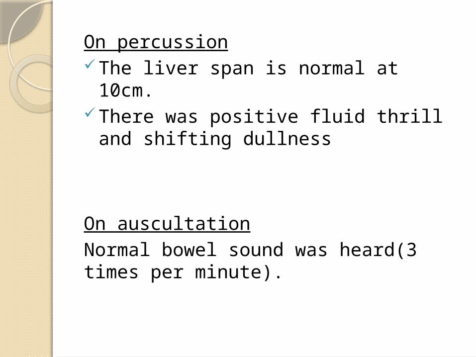
On percussionThe liver span is normal at 10cm. There was positive fluid thrill and shifting
dullness
On auscultationNormal bowel sound was heard(3 times per minute).

SUMMARYA 59 years old Chinese man was admitted to hospital due to hematemesis associated with loose black stool(melena), dizziness, fatigue, abdominal pain and loss of appetite.On physical examination the abdomen was distended. There was positive fluid thrill and shifting dullness.

Provisional DiagnosisDiagnosis Points to support
Esophageal Varices 2⁰ to Liver Disease
- Age (> 60 years old)- Past history of hematemesis and melena- History of Hepatitis C (known history of
liver disease)- Intravenous drug user- Alcohol consumer and smoker- Abdominal pain- Loss of appetite- Unknown loss of weight- Loose stool (diarrhea)- Weakness & Fatigue

Differential DiagnosisDIAGNOSIS POINTS TO SUPPORT POINTS AGAINST
Peptic Ulcer Disease - Nausea & vomiting- Unknown weight loss- Loss of appetite- Melena & hematemesis
(severe case)- Smoking
- No abdominal bloating- No burning pain before
or after meal- No heartburn- No indigestion
Esophageal Carcinoma - Gender - male- Unknown weight loss- Melena- Hematemesis- Vomiting- Smoking
- Age (usually >65yrs)- No persistent cough- No hoarseness- No indigestion- No dyspepsia- No heartburn- No dysphagia- No chest or back pain

Gastric Carcinoma - Age (common in elderly)- Nausea or vomiting- Loss of appetite- Melena- Hematemesis- Weight loss- Diarrhea- Weakness and fatigue
- No indigestion- No dysphagia- No postprandial
fullness- No palpable mass on
abdomen- No heartburn- No bloated feeling
after eating
DIAGNOSIS POINTS TO SUPPORT POINTS AGAINST

Investigations
Self revised Done by hospitalFull Blood Count Haemotological TestLiver Function Test Renal Function TestRenal Function Test Liver Function TestEndoscopy (OGDS) Arterial Blood GasesCoagulation Profile Chest X-RayChest X-Ray Abdominal X-RayAbdominal X-Ray HIV TestHepatitis C Screening Hepatitis C Screening
Hepatitis B ScreeningRapid Plasma Reagin

1) Haemotological Test
Test Patient’s Result Unit Normal Range InterpretationRBC 2.68 10^6/uL 4.5 – 6.5 Low
WBC 19.6 10^3/uL 4 - 11 High
HGB 8.1 g/dL 13 - 18 Low
HCT 23.9 % 40 – 54 Low
MCV 89.2 fL 76 – 96 Normal
MCH 30.2 pg 27 – 32 Normal
PLT 207 10^3/uL 150 - 400 Normal
.

2) Renal Function Test
Test Result Unit Range Interpretation
Urea 15.4 mmol/L 2.8 – 7.2 High
Sodium 140 mmol/L 136 – 145 Normal
Potassium 4.6 mmol/L 3.5 – 5.1 Normal
Chloride 104 mmol/L 98 – 107 Normal
Creatinine 95 umol/L 45 - 84 Normal

3) Liver Function Test
Test Patient’s Result Unit Normal
Range Interpretation
Total Protein 59 g/L 66 – 83 Low
Albumin 28 g/L 35 – 52 Low
Globulin 31 g/L 25 – 39 Normal
A/G Ratio 0.9 0:9 – 1:8 Normal
ALP 53 IU/L 30 – 120 Normal
ALT 79 IU/L 0 – 50 High
Total Bilirubin
20.1 umol/L 5 - 21 Normal

4) Arterial Blood Gas
Test Result Unit Range
pH 7.382 - 7.350 – 7.450
pCO2 37.1 mmHg 35.0 – 48.0
pO2 21.3 mmHg 83.0 - 108
5) HIV Test & Rapid Plasma Reagin
Test ResultHIV Ag/Ab Non-Reactive
Syphilis Reagin Ab Non-Reactive

6) Hepatitis B Screening
Test Result
Hepatitis B e Ab Reactive
Hepatitis B e Ag Non-Reactive
Hepatitis B s Ag Non-Reactive
7) Hepatitis C Screening
Test ResultAnti HCV Reactive
HCV particle Agglutination Detected

8) Chest X-Ray
Impression :
Shows no abnormalities (normal)

9) Abdominal X-Ray
Impression :
Shows no abnormalities (normal)

CHILD-PUGH-SCORE (SEVERITY OF CIRRHOSIS)
CATEGORY 1 2 3
ENCEPHALOPATHY 0 I/II III/IV
ASCITES ABSENT MILD-MODERATE SEVERE
BILIRUBIN (umol/L)
<34 34 – 51 >51
ALBUMIN(g/L) >35 28 – 35 <28
INR <1.3 1.3 – 1.5 >1.5
Score A : 6 or lessScore B : 7-9Score C : 10 and greater
EncephalopathyGrade I : confusion, altered mood and behaviourGrade II : drowsy, inappropriate behaviourGrade III : stuporous but obeys simple commands, slurred speech, marked confusionGrade IV : unarousable coma

CATEGORY RESULT SCOREENCEPHALOPATHY No 1
ASCITES Mild 2BILIRUBIN (umol/L) 20 1
ALBUMIN(g/L) 28 2INR 1.7 3
Total score : 9Grade : Child Pugh B

RESUSCITATION
• Vitals are monitored (pulse, BP, cardiac monitor, urine output)• Assessment of haemodynamic stability – severity of blood loss• IV Route - Fluid resuscitation such as normal saline• Oxygen support to prevent hypoxia of tissues – nasal cannula
or facemask.• Correction of coagulopathy and thrombocytopenia• Urine output (through an indwelling catheter)• Blood transfusion is administered to patients who are shocked
and are actively bleeding.
Management

• Pharmacological therapy : vasoconstrictors to arrest the bleeding (terlipressin, octreotide, vasopressin, somatostatin)
• Banding• Sclerotherapy• Non-selective ß-adrenergic antagonists such as propranolol and
nadolol to prevent rebleeding.• Transjugular Intrahepatic Portosystemic Shunt (TIPS)• Distal Splenorenal Shunt (DSRS).• Liver transplant• Devascularization• Esophageal transection
Management


TIPS DSRS

a) Currently, no treatment can prevent the development of esophageal varices in people with liver disease.
b) While beta blocker drugs are effective in preventing bleeding in many people who have esophageal varices, they do not keep esophageal varices from forming.
c) If one have been diagnosed with liver disease, strategies to avoid liver disease complications should be taken.
- Don't drink alcohol.- Eat a healthy diet.- Reduce your risk of hepatitis.
Prevention

Discussion
• Portal venous pressure = portal venous flow x portal venous resistance
• Hepatic venous pressure gradient (HVPG) is the difference in pressure between the portal vein (wedged hepatic venous pressure) and free hepatic vein pressure.
• HVPG = surrogate for portal pressure• Increased HVPG > 5mmHg = Portal Hypertension• Clinical manifestation of portal hypertension are many
included Esophageal Varices.

Increased portal pressure(HVPG > 10mmHg)
Formation of varices
Dilatation of varices
Rupture of varices(HVPG > 12mmHg)
Natural History


Anatomy of esophagus• The esophagus is a 25-cm long muscular tube that connects the pharynx to
the stomach.• The esophagus extends from the lower border of the cricoid cartilage (at
the level of the 6th cervical vertebra) to the cardiac orifice of the stomach at the side of the body of the 11th thoracic vertebra.
• The esophagus has 3 constrictions in its vertical course.• These measurements are clinically important for endoscopy and
endoscopic surgeries of the esophagus.• The esophagus has been subdivided into 3 portions:
a) Cervical
b) Thoracic
c) Abdominal• Histologically, the esophagus has 4 concentric layers. (Mucosa, Submucosa,
Muscular, Adventitia)


Blood supply• Cervical portion : inferior thyroid artery• Thoracic portion : bronchial and esophageal branches of the thoracic
aorta• Abdominal portion : ascending branches of the left phrenic and left
gastric arteries.
Venous drainage• Venous blood from the esophagus drains into a submucosal plexus. From
this plexus, blood drains to the periesophageal venous plexus. Esophageal veins arise from this plexus and drain in a segmental way similar to the arterial supply, as follows:
• From the cervical esophagus, veins drain into the inferior thyroid vein• From the thoracic esophagus, veins drain into the azygos veins,
hemiazygos, intercostal, and bronchial veins• From the abdominal portion, esophagus veins drain into the left gastric
vein; the left gastric vein is a tributary of the portal system.

Risk Factors• High portal vein pressure. The risk of bleeding increases with the
amount of pressure in the portal vein (portal hypertension).• Large varices. The larger the varices, the more likely they are to
bleed.• Red marks on the varices. When viewed through an endoscope
passed down your throat, some varices show long, red streaks or red spots. These marks indicate a high risk of bleeding.
• Severe cirrhosis or liver failure. Most often, the more severe your liver disease, the more likely varices are to bleed.
• Continued alcohol use. Your risk of variceal bleeding is far greater if you continue to drink than if you stop, especially if your disease is alcohol related.
• Bacterial infection - schistosomiasis

Sign & Symptoms1) Vomiting blood (in large amount) can cause
- Dizziness- Loss consciousness- Pallor- Anaemia
2) Some patient bleed in smaller amount over a longer period, they swallow the blood rather than vomit it, so they may have :
- Black, tarry or bloody stools3) Shock and sepsis(in severe cases)4) Low blood pressure (hypotension)5) Rapid heart rate (tachycardia)6) Abdominal pain7) Confusion secondary to encephalopathy

Signs of liver disease :1) Jaundice (yellow discoloration)2) Spider nevi (cluster of tiny blood vessels shaped like spider)3) Palmar erythema (Reddening on palm)4) Splenomegaly5) Hand deformity known as Dupuytren’s contracture6) Testicular atrophy7) Ascites (fluid build up)8) Gynaecomastia 9) Hair Loss10) Pruritus11) Muscle loss

• Encephalopathy (sometimes called hepatic encephalopathy)• Esophageal stricture after surgery or endoscopic therapy• Hypovolemic shock• Infection (pneumonia, bloodstream infection, peritonitis)• Rebleeding after treatment• Kidney failure
Possible Complications

References
• Bailey & Love's Short Practice of Surgery, 25th edn
• http://www.hopkinsmedicine.org• http://www.slideshare.net• http://www.nlm.nih.gov• http://emedicine.medscape.com

THANK YOU!


![Small Esophageal Varices in Patients with Cirrhosis—Should ... · Varices of medium/large size (>5-mm diameter), or small varices with red spot signs [3†]. According to the](https://static.fdocuments.us/doc/165x107/5fa0286b8b7f711ce374a04d/small-esophageal-varices-in-patients-with-cirrhosisashould-varices-of-mediumlarge.jpg)