Equol,aMetaboliteoftheSoybeanIsoflavoneDaidzein ... · more practical than those intervening at the...
12
Equol, a Metabolite of the Soybean Isoflavone Daidzein, Inhibits Neoplastic Cell Transformation by Targeting the MEK/ERK/p90RSK/Activator Protein-1 Pathway * Received for publication, February 20, 2007, and in revised form, July 11, 2007 Published, JBC Papers in Press, August 27, 2007, DOI 10.1074/jbc.M701459200 Nam Joo Kang ‡§1,2 , Ki Won Lee ‡¶1 , Evgeny A. Rogozin ‡ , Yong-Yeon Cho ‡ , Yong-Seok Heo , Ann M. Bode ‡ , Hyong Joo Lee §3 , and Zigang Dong ‡4 From the ‡ Hormel Institute, University of Minnesota, Austin, Minnesota 55912, § Department of Agricultural Biotechnology, Seoul National University, Seoul 151-742, Republic of Korea, ¶ Department of Bioscience and Biotechnology, Konkuk University, Seoul 143-701, Republic of Korea, and Department of Chemistry, Konkuk University, Seoul 143-701, Republic of Korea Daidzein and genistein are isoflavones found in soybean. Genistein is known to exhibit anticarcinogenic activities and inhibit tyrosine kinase activity. However, the underlying molec- ular mechanisms of the chemopreventive activities of daidzein and its metabolite, equol, are not understood. Here we report that equol inhibits 12-O-tetradecanoylphorbol-13-acetate (TPA)-induced neoplastic transformation of JB6 P mouse epi- dermal cells by targeting the MEK/ERK/p90RSK/activator pro- tein-1 signaling pathway. TPA-induced neoplastic cell transfor- mation was inhibited by equol, but not daidzein, at noncytotoxic concentrations in a dose-dependent manner. Equol dose- dependently attenuated TPA-induced activation of activator protein-1 and c-fos, whereas daidzein did not exert any effect when tested at the same concentrations. The TPA-induced phosphorylation of ERK1/2, p90RSK, and Elk, but not MEK or c-Jun N-terminal kinase, was inhibited by equol but not by daid- zein. In vitro kinase assays revealed that equol greatly inhibited MEK1, but not Raf1, kinase activity, and an ex vivo kinase assay also demonstrated that equol suppressed TPA-induced MEK1 kinase activity in JB6 P cell lysates. Equol dose-dependently inhibited neoplastic transformation of JB6 P cells induced by epidermal growth factor or H-Ras. Both in vitro and ex vivo pull- down assays revealed that equol directly bound with glutathione S-transferase-MEK1 to inhibit MEK1 activity without compet- ing with ATP. These results suggested that the antitumor-pro- moting effect of equol is due to the inhibition of cell transforma- tion mainly by targeting a MEK signaling pathway. These findings are the first to reveal a molecular basis for the antican- cer action of equol and may partially account for the reported chemopreventive effects of soybean. Carcinogenesis is characterized as a multistage process that includes initiation, promotion, and progression stages. Cancer prevention strategies that involve intervention at the tumor promotion stage, a reversible and long-term process, seem to be more practical than those intervening at the tumor initiation stage, which is an irreversible and short-term process. Activator protein-1 (AP-1) 5 is a well characterized transcription factor composed of homodimers and/or heterodimers of the Fos and Jun protein families and plays a key role in “preneoplastic-to- neoplastic” transformation and proliferation (1). A diverse vari- ety of stimuli induce AP-1 binding to various genes that govern cellular processes such as transformation and proliferation (2). In particular, 12-O-tetradecanoylphorbol-13-acetate (TPA), H-Ras, and epidermal growth factor (EGF) are the most com- mon experimental stimuli used to activate AP-1 and induce cellular transformation in many different cell types and animal models (3). Many mechanisms are involved in the up- and down-regula- tion of AP-1 activity (2). The mitogen-activated protein (MAP) kinases are the most common signaling pathways known to mediate AP-1 function (4), and the blocking of MAP kinases leads to the inhibition of AP-1 transactivation and subsequent cell transformation (5). These kinase families include extracel- lular-signal-regulated protein kinases (ERKs), c-Jun N-terminal kinases, and the p38 family of kinases. Although the proteins have some redundancy in function, ERKs generally play a crit- ical role in transmitting signals initiated by tumor promoters such as TPA, EGF, and platelet-derived growth factor (6, 7). MEK is a dual-specificity protein kinase that phosphorylates its downstream target ERK on specific tyrosine and threonine res- idues. The constitutive activation of MEK1 results in cellular transformation, whereas a small molecular inhibitor of MEK was shown to suppress transformation and tumor growth in both cell culture and mouse models (6, 8). Moreover, a mutant H-ras gene perpetually activates the MEK/ERK signaling path- way and drives cells to develop a more aggressive cancer-like * The work is supported in part by The Hormel Foundation and National Insti- tutes of Health Grants CA120388, CA111536, CA88961, and CA81064 and by BioGreen21 Program Grant 20070301-034-042, Rural Development Administration, Republic of Korea. The costs of publication of this article were defrayed in part by the payment of page charges. This article must therefore be hereby marked “advertisement” in accordance with 18 U.S.C. Section 1734 solely to indicate this fact. 1 These authors contributed equally to this work. 2 Recipient of a BK21 fellowship from the Korea Ministry of Education. 3 To whom correspondence may be addressed. Tel.: 82-2-880-4860; Fax: 82-2- 873-5095; E-mail: [email protected]. 4 To whom correspondence may be addressed: Hormel Institute, University of Minnesota, 801 16th Ave. NE, Austin, MN 55912. Tel.: 507-437-9600; Fax: 507-437-9606; E-mail: [email protected]. 5 The abbreviations used are: AP-1, activator protein-1; MEK, mitogen- activated protein kinase/extracellular signal-regulated kinase kinase; ERK, extracellular signal-regulated kinase; TPA, 12-O-tetradecanoyl- phorbol-13-acetate; EGF, epidermal growth factor; FBS, fetal bovine serum; MOPS, 4-morpholinepropanesulfonic acid; ROS, reactive oxy- gen species; GST, glutathione S-transferase; MEM, Eagle’s minimum essential medium; MTS, 3-(4,5-dimethylthiazol-2-yl)-5-(3-carboxy- methoxy-phenyl)-2-(4-sulfonyl)-2H-tetrazolium. THE JOURNAL OF BIOLOGICAL CHEMISTRY VOL. 282, NO. 45, pp. 32856 –32866, November 9, 2007 © 2007 by The American Society for Biochemistry and Molecular Biology, Inc. Printed in the U.S.A. 32856 JOURNAL OF BIOLOGICAL CHEMISTRY VOLUME 282 • NUMBER 45 • NOVEMBER 9, 2007 by guest on July 15, 2018 http://www.jbc.org/ Downloaded from
Transcript of Equol,aMetaboliteoftheSoybeanIsoflavoneDaidzein ... · more practical than those intervening at the...
Equol, a Metabolite of the Soybean Isoflavone Daidzein,Inhibits Neoplastic Cell Transformation by Targeting theMEK/ERK/p90RSK/Activator Protein-1 Pathway*
Received for publication, February 20, 2007, and in revised form, July 11, 2007 Published, JBC Papers in Press, August 27, 2007, DOI 10.1074/jbc.M701459200
Nam Joo Kang‡§1,2, Ki Won Lee‡¶1, Evgeny A. Rogozin‡, Yong-Yeon Cho‡, Yong-Seok Heo�, Ann M. Bode‡,Hyong Joo Lee§3, and Zigang Dong‡4
From the ‡Hormel Institute, University of Minnesota, Austin, Minnesota 55912, §Department of Agricultural Biotechnology, SeoulNational University, Seoul 151-742, Republic of Korea, ¶Department of Bioscience and Biotechnology, Konkuk University,Seoul 143-701, Republic of Korea, and �Department of Chemistry, Konkuk University, Seoul 143-701, Republic of Korea
Daidzein and genistein are isoflavones found in soybean.Genistein is known to exhibit anticarcinogenic activities andinhibit tyrosine kinase activity. However, the underlyingmolec-ular mechanisms of the chemopreventive activities of daidzeinand its metabolite, equol, are not understood. Here we reportthat equol inhibits 12-O-tetradecanoylphorbol-13-acetate(TPA)-induced neoplastic transformation of JB6 P�mouse epi-dermal cells by targeting the MEK/ERK/p90RSK/activator pro-tein-1 signaling pathway. TPA-induced neoplastic cell transfor-mationwas inhibited by equol, but not daidzein, at noncytotoxicconcentrations in a dose-dependent manner. Equol dose-dependently attenuated TPA-induced activation of activatorprotein-1 and c-fos, whereas daidzein did not exert any effectwhen tested at the same concentrations. The TPA-inducedphosphorylation of ERK1/2, p90RSK, and Elk, but not MEK orc-JunN-terminal kinase, was inhibited by equol but not by daid-zein. In vitro kinase assays revealed that equol greatly inhibitedMEK1, but not Raf1, kinase activity, and an ex vivo kinase assayalso demonstrated that equol suppressed TPA-induced MEK1kinase activity in JB6 P� cell lysates. Equol dose-dependentlyinhibited neoplastic transformation of JB6 P� cells induced byepidermal growth factor orH-Ras. Both in vitro and ex vivo pull-down assays revealed that equol directly boundwith glutathioneS-transferase-MEK1 to inhibit MEK1 activity without compet-ing with ATP. These results suggested that the antitumor-pro-moting effect of equol is due to the inhibition of cell transforma-tion mainly by targeting a MEK signaling pathway. Thesefindings are the first to reveal a molecular basis for the antican-cer action of equol and may partially account for the reportedchemopreventive effects of soybean.
Carcinogenesis is characterized as a multistage process thatincludes initiation, promotion, and progression stages. Cancerprevention strategies that involve intervention at the tumorpromotion stage, a reversible and long-termprocess, seem to bemore practical than those intervening at the tumor initiationstage,which is an irreversible and short-termprocess. Activatorprotein-1 (AP-1)5 is a well characterized transcription factorcomposed of homodimers and/or heterodimers of the Fos andJun protein families and plays a key role in “preneoplastic-to-neoplastic” transformation and proliferation (1). A diverse vari-ety of stimuli induce AP-1 binding to various genes that governcellular processes such as transformation and proliferation (2).In particular, 12-O-tetradecanoylphorbol-13-acetate (TPA),H-Ras, and epidermal growth factor (EGF) are the most com-mon experimental stimuli used to activate AP-1 and inducecellular transformation in many different cell types and animalmodels (3).Many mechanisms are involved in the up- and down-regula-
tion of AP-1 activity (2). The mitogen-activated protein (MAP)kinases are the most common signaling pathways known tomediate AP-1 function (4), and the blocking of MAP kinasesleads to the inhibition of AP-1 transactivation and subsequentcell transformation (5). These kinase families include extracel-lular-signal-regulated protein kinases (ERKs), c-JunN-terminalkinases, and the p38 family of kinases. Although the proteinshave some redundancy in function, ERKs generally play a crit-ical role in transmitting signals initiated by tumor promoterssuch as TPA, EGF, and platelet-derived growth factor (6, 7).MEK is a dual-specificity protein kinase that phosphorylates itsdownstream target ERK on specific tyrosine and threonine res-idues. The constitutive activation of MEK1 results in cellulartransformation, whereas a small molecular inhibitor of MEKwas shown to suppress transformation and tumor growth inboth cell culture and mouse models (6, 8). Moreover, a mutantH-ras gene perpetually activates the MEK/ERK signaling path-way and drives cells to develop a more aggressive cancer-like
* The work is supported in part by The Hormel Foundation and National Insti-tutes of Health Grants CA120388, CA111536, CA88961, and CA81064 andby BioGreen21 Program Grant 20070301-034-042, Rural DevelopmentAdministration, Republic of Korea. The costs of publication of this articlewere defrayed in part by the payment of page charges. This article musttherefore be hereby marked “advertisement” in accordance with 18 U.S.C.Section 1734 solely to indicate this fact.
1 These authors contributed equally to this work.2 Recipient of a BK21 fellowship from the Korea Ministry of Education.3 To whom correspondence may be addressed. Tel.: 82-2-880-4860; Fax: 82-2-
873-5095; E-mail: [email protected] To whom correspondence may be addressed: Hormel Institute, University of
Minnesota, 801 16th Ave. NE, Austin, MN 55912. Tel.: 507-437-9600; Fax:507-437-9606; E-mail: [email protected].
5 The abbreviations used are: AP-1, activator protein-1; MEK, mitogen-activated protein kinase/extracellular signal-regulated kinase kinase;ERK, extracellular signal-regulated kinase; TPA, 12-O-tetradecanoyl-phorbol-13-acetate; EGF, epidermal growth factor; FBS, fetal bovineserum; MOPS, 4-morpholinepropanesulfonic acid; ROS, reactive oxy-gen species; GST, glutathione S-transferase; MEM, Eagle’s minimumessential medium; MTS, 3-(4,5-dimethylthiazol-2-yl)-5-(3-carboxy-methoxy-phenyl)-2-(4-sulfonyl)-2H-tetrazolium.
THE JOURNAL OF BIOLOGICAL CHEMISTRY VOL. 282, NO. 45, pp. 32856 –32866, November 9, 2007© 2007 by The American Society for Biochemistry and Molecular Biology, Inc. Printed in the U.S.A.
32856 JOURNAL OF BIOLOGICAL CHEMISTRY VOLUME 282 • NUMBER 45 • NOVEMBER 9, 2007
by guest on July 15, 2018http://w
ww
.jbc.org/D
ownloaded from

phenotype, such as anchorage-independent growth (9–11).These results indicated that MEK is important in the transfor-mation of cells and development of tumors.The National Cancer Institute of the National Institutes of
Health has identified about 40 plant-based foods, includingsoybean, green tea, red wine, onion, ginger, broccoli, cabbage,cauliflower, Brussels sprouts, and turmeric, which have beenreported to possess cancer preventive properties (7, 12, 13).Naturally occurring dietary phenolic phytochemicals originat-ing from these plant-based foods have been suggested to havechemopreventive effects in carcinogenesis, particularly in thestage of promotion (7, 14, 15). Soybean is an excellent source ofdietary phenolic substances in the Asian diet (13, 16). The twomajor isoflavones in soybean are genistein and daidzein (seeFig. 1A), which together compose 100–300 mg/100 g of soy-bean and are found in tissues as glycoside conjugates (17).These compounds are heat-stable and, hence, are also abun-dant in many processed and fermented soybean products (18).Genistein was originally described as a protein-tyrosine kinaseinhibitor (19), and it also has been reported to suppress angio-genesis (20) and cell cycle progression (21) and stimulateapoptosis (22). Daidzein is another prominent isoflavone foundin soybean foods but has consistently been reported to be lessactive then genistein (23). This conclusion is probably not accu-rate because, unlike genistein, daidzein is converted to equol(Fig. 1B) by the intestinal microflora, which can lead to bioac-tivation (24). Specifically, after ingestion, daidzein is convertedto equol (70%), dihydrodaidzein (10–25%), and O-desmethyl-angolensin (5–20%) (25). The half-life in the body for equol issignificantly longer compared with daidzein or genistein (26),and equol is consistently reported to be present at high levels inthe blood (27). Several independent lines of evidence indicatethat equol is one of the most biologically active metabolites ofdaidzein.In many cell types equol is more potent than daidzein in
terms of its antioxidant properties (28). For example, equol atconcentrations within the physiological range were shown toprotect against hydrogen peroxide-mediated DNA damage inhuman lymphocytes as determined by alkaline single-cell gelelectrophoresis (29). Equol was reported to exhibit protectiveeffects against UV-induced DNA damage in hairless mice (30).Moreover, equol was shown to protect against UV-inducedskin cancer in the hairless mouse model (31). The UV-inducedactivation of ornithine decarboxylase, a skin tumor promotionbiomarker enzyme, was attenuated by equol treatment, indicat-ing that the anticancer activity of equol may be attributed to itsinhibition of the tumor promotion phase of carcinogenesis (31).These accumulated data represent evidence suggesting thatequol is a potent chemopreventive agent against carcinogene-sis, particularly skin cancer, but the underlying molecularmechanisms and molecular target(s) remain unclear.The skin is highly sensitive to TPA induction of MEK/ERK/
AP-1 signaling activation and tumorigenesis (32). In JB6mouseepidermal cell lines, TPA and EGF were shown to induce AP-1transcriptional activity in promotion-sensitive (P�), but not inpromotion-resistant (P�), cellular phenotypes (1).When AP-1induction was blocked, P� cells reverted to the P� phenotype,indicating that AP-1 activity is required for TPA- or EGF-in-
duced cell transformation (1). The JB6 P� mouse epidermalcell line provides a validated model for screening cancer che-mopreventive agents and elucidating their molecular mecha-nisms of action (33). In the present study we compared theeffects of equol and daidzein onTPA-inducedAP-1 activity andcell transformation in JB6 P� cells. We found that equol, butnot daidzein, was a potent inhibitor of MEK activity and subse-quently inhibited AP-1 transactivation and cell transformation.The results of this investigation suggested a molecular mecha-nism that underlies the chemopreventive activity of equol andmight partially account for the chemopreventive effects of soy-bean foods.
EXPERIMENTAL PROCEDURES
Chemicals—Equol, daidzein, TPA, and trypan blue solutionwere obtained from Sigma. Eagle’s minimum essential medium(MEM), basalmediumEagle, gentamicin, and L-glutaminewerepurchased from Invitrogen; fetal bovine serum (FBS) was pur-chased from Gemini Bio-Products (Calabasas, CA). PD098059was purchased from Calbiochem. The antibodies against phos-phorylatedMEK (Ser-217/221), phosphorylatedERK (Tyr-202/Tyr-204), total ERK, phosphorylated c-Jun N-terminal kinase(Thr-183/Tyr-185), total c-Jun N-terminal kinase, phosphoryl-ated p90RSK (Thr-359/Ser-363), total p90RSK, phosphorylatedElk-1 (Ser-383), and total Elk-1 were purchased from Cell Sig-nal Biotechnology (Beverly, MA). The antibody against totalMEK was from Santa Cruz Biotechnology (Santa Cruz, CA).TheMEK1 andRaf1 kinase assay kit was obtained fromUpstateBiotechnology (Lake Placid, NY). CNBr-Sepharose 4B, gluta-thione-Sepharose 4B, and [�-32P]ATP were purchased fromAmershamBiosciences, and the protein assay kit was fromBio-Rad. G418, the CellTiter96 Aqueous One Solution Cell Prolif-eration Assay kit, and the luciferase assay substrate were pur-chased from Promega (Madison, WI).Cell Culture—The JB6 P� mouse epidermal (JB6 P�) cell
line and H-Ras-transformed JB6 P� mouse epidermal (H-RasJB6 P�) cell line were cultured in monolayers at 37 °C in a 5%CO2 incubator in MEM containing 5% FBS, 2 mM L-glutamine,and 25 �g/ml gentamicin. The JB6 mouse epidermal cell linewas stably transfected with an AP-1 luciferase reporter plasmidandmaintained inMEMsupplementedwith 5%FBS containing200 �g/ml G418.Cell Proliferation Assay—To estimate cell proliferation, JB6
P� cells were seeded (103 cells/well) in 96-well plates with 5%FBS, MEM at 37 °C in a 5% CO2 incubator. After culturing forthe indicated times, 20�l of CellTiter 96AqueousOne Solution(Promega) were added to each well, and the cells were thenincubated for 1 h at 37 °C in a 5% CO2 incubator. Absorbancewas measured at 492 and 690 nm.Cell Viability Assay—The trypan blue exclusion assay was
based on the capability of viable cells to exclude the dye.Because viable JB6 P� cells maintain membrane integrity, thecells do not allow trypan blue dye to pass through the cell mem-brane. Cells with damaged membrane appeared blue due totheir accumulation of dye and were counted as dead. Trypanblue (0.4%) was added to JB6 P� cells, and after 5 min, cellswere loaded into a hematocytometer and counted. The number
Inhibition of Neoplastic Transformation and MEK Activity
NOVEMBER 9, 2007 • VOLUME 282 • NUMBER 45 JOURNAL OF BIOLOGICAL CHEMISTRY 32857
by guest on July 15, 2018http://w
ww
.jbc.org/D
ownloaded from

of viable cells was calculated as percent of the total cellpopulation.Anchorage-independent Transformation Assay—The effects
of daidzein or equol on TPA-induced cell transformation wereinvestigated in JB6 P� cells. Cells (8� 103/ml) were exposed toTPA with or without daidzein (0–20 �M) or equol (0–100 �M)in 1ml of 0.33%basalmediumEagle agar containing 10%FBSorin 3.5 ml of 0.5% basal medium Eagle agar containing 10% FBS.The cultures were maintained at 37 °C in a 5% CO2 incubatorfor 14 days, and the cell colonies were counted under a micro-scope with the aid of the Image-Pro Plus software (Version.4)program (Media Cybernetics, Silver Spring, MD) as describedby Colburn et al. (34).Luciferase Assay for AP-1 Transactivation—Confluent
monolayers of JB6 P� cells stably transfected with an AP-1luciferase reporter plasmidwere trypsinized, and 8� 103 viablecells suspended in 100 �l of 5% FBS, MEM were added to eachwell of a 96-well plate. Plates were incubated at 37 °C in ahumidified atmosphere of 5% CO2. When cells reached80–90% confluence, they were starved by culturing them in0.1% FBS, MEM for another 24 h. The cells were then treatedfor 1 hwith daidzein (0–100�M) or equol (0–100�M) and thenexposed to 20 ng/ml TPA for 24 h. After treatment, cells weredisruptedwith 100�l of lysis buffer (0.1 M potassiumphosphatebuffer (pH7.8), 1%TritonX-100, 1mMdithiothreitol, and 2mMEDTA), and the luciferase activity was measured using a lumi-nometer (Luminoskan Ascent, Thermo Electron, Helsinki,Finland).Reporter-gene Assay—The reporter gene assay for firefly
luciferase activity was performed using lysates from transfectedcells. In addition, the reporter gene vector pRL-SV40 (Pro-mega) was co-transfected into each cell line, with the transfec-tion efficiencies normalized to the Renilla luciferase activitygenerated by this vector. Cell lysates were prepared by firstwashing the transfected JB6 P� cells once in phosphate-buff-ered saline at 37 °C. After removing the phosphate-bufferedsaline completely, 500 �l of passive lysis buffer (Promega DualLuciferase Reporter Assay system) were added, and the cellswere incubated for 1 h with gentle shaking. The lysate was thentransferred to a reaction tube, and the cellular debris wasremoved by centrifugation. The supernatant fraction was usedfor the measurement of firefly and Renilla luciferase activities.Cell lysates (20 �l each) were mixed with 100 �l of LuciferaseAssay II reagent, and the emitted firefly luciferase light wasmeasured (Luminoskan Ascent). Subsequently, the coelentera-zine reagent (100 �l) containing the substrate for the emissionof Renilla luciferase light was mixed to normalize the fireflyluciferase data. The c-fos luciferase promoter (pFos-WT GL3)and constructs were kindly provided by Dr. Ron Prywes(Columbia University, New York, NY).Western Blotting—After the cells (1.5� 106) were cultured in
a 10-cm dish for 48 h, they were starved in serum-free mediumfor another 24 h to eliminate the influence of FBS on the acti-vation of mitogen-activated protein kinases. The cells werethen treatedwith daidzein (0–100�M) or equol (0–100�M) for1 h before theywere exposed to 20 ng/mlTPA for different timeperiods. The harvested cells were disrupted, and the superna-tant fractions were boiled for 5 min. The protein concentration
was determined using a dye-binding protein assay kit (Bio-Rad)as described in the manufacturer’s manual. Lysate protein (20�g) was subjected to 10% SDS-PAGE and electrophoreticallytransferred to a polyvinylidene difluoride membrane (Amer-sham Biosciences). After blotting, the membrane was incu-bated with the specific primary antibody at 4 °C overnight. Pro-tein bandswere visualized by the chemiluminescence detectionkit (AmershamBiosciences) after hybridization with the horse-radish peroxidase-conjugated secondary antibody. The relativeamounts of proteins associated with specific antibodies werequantified using Sicon Image (NIH, Bethesda, MD).In Vitro MEK1 and Raf1 Kinase Assay—The in vitro kinase
assays were performed in accordancewith the instructions pro-vided by Upstate Biotechnology. In brief, every reaction con-tained 20 �l of assay dilution buffer (20 mM MOPS (pH 7.2),25 mM �-glycerol phosphate, 5 mM EGTA, 1 mM sodiumorthovanadate, and 1 mM dithiothreitol) and a magnesium-ATPmixture buffer. For MEK1, 1 �g of the inactive ERK2 sub-strate peptide was also included. For Raf1, 0.4 �g of inactiveMEK1 and 1 �g of inactive ERK2 were included. A 4-�l aliquotwas removed from the reaction mixture containing 20 �g ofmyelin basic protein substrate peptide and 10 �l of diluted[�-32P]ATP solution and incubated at 30 °C for 30 min. Thismixture was incubated for 10 min at 30 °C, and then 25-�l ali-quots were transferred onto p81 paper andwashed 3 times with0.75% phosphoric acid for 5 min per wash and once with ace-tone for 2 min. The radioactive incorporation was determinedusing a scintillation counter. The effects of daidzein (0–100�M) or equol (0–100�M)were evaluated by separately incubat-ing each compound with the reaction mixtures at 30 °C for 30min. Each experiment was performed 3 times.Ex Vivo MEK1 Immunoprecipitation and Kinase Assay—JB6
P� cells were cultured to 80% confluence and then serum-starved in 0.1% FBS, MEM for 24 h at 37 °C. Cells were eithertreated or not treated with daidzein (0–100 �M) or equol(0–100�M) for 1 h, then treated with 20 ng/ml TPA for 30min,disrupted with lysis buffer (20 mM Tris/HCl (pH 7.4), 1 mM
EDTA, 150 mM NaCl, 1 mM EGTA, 1% Triton X-100, 1 mM
�-glycerophosphate, 1 mg/ml leupeptin, 1 mM Na3VO4, and 1mM phenylmethylsulfonyl fluoride), and finally centrifuged at14,000 rpm for 10 min in a microcentrifuge. The lysates con-taining 500 �g of protein were used for immunoprecipitationwith an antibody against MEK1 and then incubated at 4 °Covernight. Protein A/G Plus-agarose beads were then added,and the mixture was continuously rotated for another 3 h at4 °C. The beads were washed 3 times with kinase buffer (20 mM
MOPS (pH 7.2), 25 mM �-glycerol phosphate, 5 mM EGTA, 1mM sodium orthovanadate, and 1 mM dithiothreitol) and thenresuspended in 20 �l of 1� kinase buffer supplemented with 1�g of inactive ERK2 and incubated for an additional 30 min at30 °C. Then myelin basic protein (20 �g) and 10 �l of diluted[�-32P]ATP solution were added, and the mixture was incu-bated for 10 min at 30 °C. A 20-�l aliquot was transferred ontop81 paper and washed 3 times with 0.75% phosphoric acid for 5min per wash and once with acetone for 2 min. The radioactiveincorporation was determined using a scintillation counter.Each experiment was performed 3 times.
Inhibition of Neoplastic Transformation and MEK Activity
32858 JOURNAL OF BIOLOGICAL CHEMISTRY VOLUME 282 • NUMBER 45 • NOVEMBER 9, 2007
by guest on July 15, 2018http://w
ww
.jbc.org/D
ownloaded from
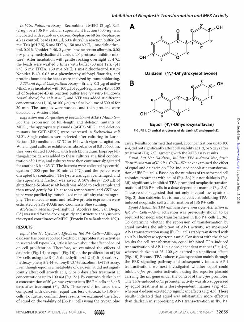
In Vitro Pulldown Assay—Recombinant MEK1 (2 �g), Raf1(2 �g), or a JB6 P� cellular supernatant fraction (500 �g) wasincubatedwith equol- or daidzein-Sepharose 4B (or -Sepharose4B as control) beads (100 �l, 50% slurry) in reaction buffer (50mMTris (pH 7.5), 5 mM EDTA, 150mMNaCl, 1 mM dithiothre-itol, 0.01% Nonidet P-40, 2 �g/ml bovine serum albumin, 0.02mM phenylmethylsulfonyl fluoride, 1� protease inhibitor mix-ture). After incubation with gentle rocking overnight at 4 °C,the beads were washed 5 times with buffer (50 mM Tris, (pH7.5), 5 mM EDTA, 150 mM NaCl, 1 mM dithiothreitol, 0.01%Nonidet P-40, 0.02 mM phenylmethylsulfonyl fluoride), andproteins bound to the beads were analyzed by immunoblotting.ATP and Equol Competition Assay—Briefly, 0.2 �g of active
MEK1was incubated with 100 �l of equol-Sepharose 4B or 100�l of Sepharose 4B in reaction buffer (see “In vitro PulldownAssay” above) for 12 h at 4 °C, and ATP was added at differentconcentrations (1, 10, or 100 �M) to a final volume of 500 �l for30 min. The samples were washed, and then proteins weredetected by Western blot.Expression and Purification of Recombinant MEK1 Mutants—
For the expression of full-length and deletion mutants ofMEK1, the appropriate plasmids (pGEX-MEK1 and deletionmutants for GST-MEK1) were expressed in Escherichia coliBL21. Single colonies were selected after culturing in Luria-Bertani (LB) medium at 37 °C for 16 h with vigorous agitation.When liquid cultures exhibited an absorbance of 0.8 at 600 nm,they were diluted 100-fold with fresh LBmedium. Isopropyl-D-thiogalactoside was added to these cultures at a final concen-tration of 0.1mM, and cultures were then continuously agitatedfor another 3 h at 25 °C. The bacteria were collected by centrif-ugation (4000 rpm for 10 min at 4 °C), and the pellets weredisrupted by sonication. The lysate was again centrifuged, andthe supernatant fraction was saved. A 50% slurry (250 �l) ofglutathione-Sepharose 4B beads was added to each sample andthen mixed gently for 1 h at room temperature, and GST pro-teins were purified by immobilizedmetal affinity chromatogra-phy. The molecular mass and relative protein expression wereestimated by SDS-PAGE and Coomassie Blue staining.Molecular Modeling—Insight II (Accelrys Inc., San Diego,
CA) was used for the docking study and structure analysis withthe crystal coordinates ofMEK1 (ProteinData Bank code 1S9J).
RESULTS
Equol Has No Cytotoxic Effects on JB6 P� Cells—Althoughdaidzein has been reported to exhibit antiproliferative activitiesin several cell types (35), little is known about the effect of equolon cell proliferation. Therefore, we examined the effects ofdaidzein (Fig. 1A) or equol (Fig. 1B) on the proliferation of JB6P� cells using the 3-(4,5-dimethylthiazol-2-yl)-5-(3-carboxy-methoxy-phenyl)-2-(4-sulfonyl)-2H-tetrazolium (MTS) assay.Even though equol is a metabolite of daidzein, it did not signif-icantly affect cell growth at 1, 3, or 5 days after treatment atconcentrations up to 100 �M (Fig. 2A). By contrast, daidzein ata concentration of 50 �Mwas cytotoxic to JB6 P� cells at 3 or 5days after treatment (Fig. 2B). These results indicated that,compared with daidzein, equol was less cytotoxic to JB6 P�cells. To further confirm these results, we examined the effectof equol on the viability of JB6 P� cells using the trypan blue
assay. Results confirmed that equol, at concentrations up to 100�M, did not significantly affect cell viability at 1, 3, or 5 days aftertreatment (Fig. 2C), agreeing with the MTS assay results.Equol, but Not Daidzein, Inhibits TPA-induced Neoplastic
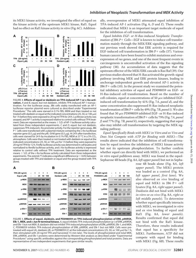
Transformation of JB6 P� Cells—We next examined the effectof equol and daidzein on TPA-induced neoplastic transforma-tion of JB6 P� cells. Based on the numbers of transformed cellcolonies, treatment with equol (Fig. 3A) but not daidzein (Fig.3B), significantly inhibited TPA-promoted neoplastic transfor-mation of JB6 P� cells in a dose-dependent manner (Fig. 3A).These results suggested that not only is equol less cytotoxic(Fig. 2) than daidzein, but is more effective at inhibiting TPA-induced neoplastic cell transformation of JB6 P� cells.Equol Attenuates TPA-induced AP-1 and c-fos Activation in
JB6 P� Cells—AP-1 activation was previously shown to berequired for neoplastic transformation in JB6 P� cells (1, 36).To determine whether the repression of transformation byequol involves the inhibition of AP-1 activity, we measuredAP-1 transactivation using JB6 P� cells stably transfected withan AP-1 luciferase reporter plasmid. Consistent with the aboveresults for cell transformation, equol inhibited TPA-inducedtransactivation of AP-1 in a dose-dependent manner (Fig. 4A),whereas daidzein at 25–100 �M exerted no significant effects(Fig. 4B). BecauseTPA induces c-fos expressionmainly throughthe ERK signaling pathway and subsequently induces AP-1transactivation, we next investigated whether equol couldinhibit c-fos promoter activation using the reporter plasmidcarrying the luc gene under the control of the c-fos promoter.The TPA-induced c-fos promoter activity was also suppressedby equol treatment in a dose-dependent manner (Fig. 4C),whereas daidzein exerted no significant effects (Fig. 4D). Theseresults indicated that equol was substantially more effectivethan daidzein in suppressing AP-1 transactivation in JB6 P�
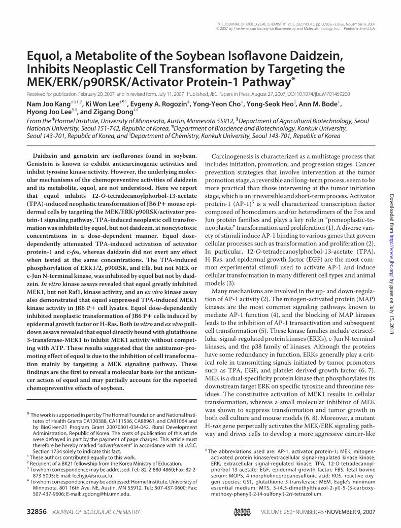
FIGURE 1. Chemical structures of daidzein (A) and equol (B).
Inhibition of Neoplastic Transformation and MEK Activity
NOVEMBER 9, 2007 • VOLUME 282 • NUMBER 45 JOURNAL OF BIOLOGICAL CHEMISTRY 32859
by guest on July 15, 2018http://w
ww
.jbc.org/D
ownloaded from
cells, which may contribute to the antitumor-promoting activ-ity of equol.Equol Suppresses TPA-induced Phosphorylation of ERK,
p90RSK, and Elk-1 but NotMEK or c-Jun N-terminal Kinase, inJB6 P�Cells—Previous studies have shown that theMEK/ERKsignaling pathway is strongly involved in TPA-induced cell
transformation andAP-1 transactivation in JB6 P� cells (1, 36).Thus, we next investigated the effect of equol on theMEK/ERKsignaling pathway and found that equol suppressed TPA-in-duced phosphorylation of ERK, one of its well known sub-strates, p90RSK, and Elk-1 in JB6 P� cells in a dose-dependentmanner (Fig. 5A). However, equol had no effect on TPA-in-duced phosphorylation of MEK in these cells (Fig. 5A). Addi-tionally, equol also had no effect on TPA-induced phosphoryl-ation of c-JunN-terminal kinase in these cells (data not shown).The TPA-induced activation of the MEK/ERK signaling path-way was inhibited by PD098059, a well known MEK inhibitorthat was used as a positive control (Fig. 5C). In contrast, evenwhen used at concentrations up to 100 �M, daidzein had noeffect on TPA-induced activation of the MEK/ERK/p90RSK/Elk signaling pathway (Fig. 5B). Overall these results confirmedthat equol is markedly more effective than daidzein as a sup-pressor of the TPA-induced phosphorylation of ERK, Elk-1,and p90RSK.Equol Specifically Inhibits MEK1 Kinase Activity but Not
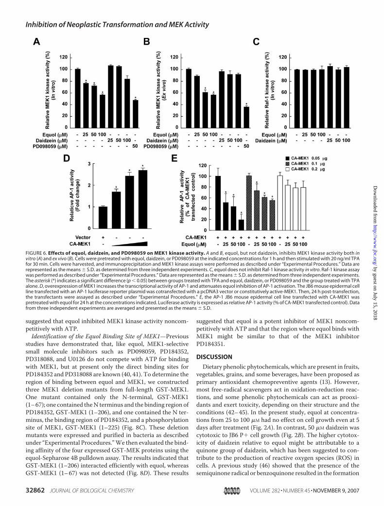
Raf1 Kinase Activity—We next investigated whether MEK1might be a molecular target of equol for the inhibition of celltransformation. Results of an in vitro kinase assay indicated thatequol, but not daidzein, strongly inhibitedMEK1kinase activity(Fig. 6A). Furthermore, equol, but not daidzein, dose-depend-ently inhibited TPA-stimulated MEK1 activity in JB6 P� celllysates (Fig. 6B). To determinewhether equol specifically inhib-
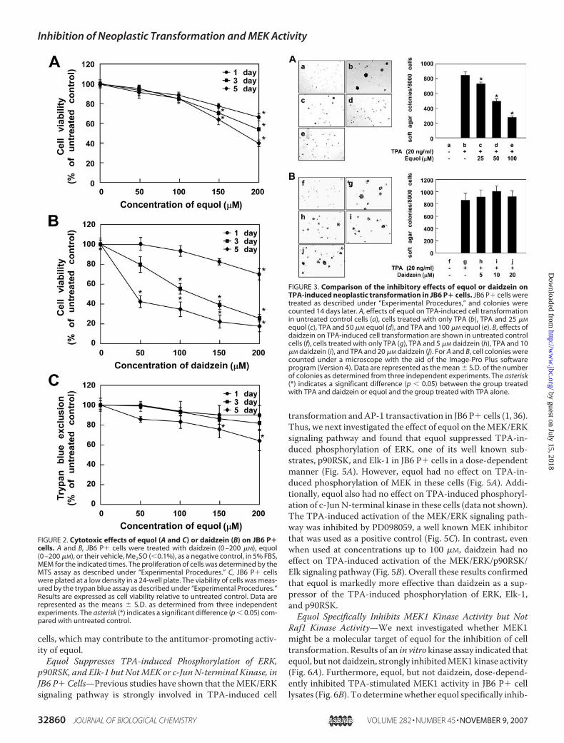
FIGURE 2. Cytotoxic effects of equol (A and C) or daidzein (B) on JB6 P�cells. A and B, JB6 P� cells were treated with daidzein (0 –200 �M), equol(0 –200 �M), or their vehicle, Me2SO (�0.1%), as a negative control, in 5% FBS,MEM for the indicated times. The proliferation of cells was determined by theMTS assay as described under “Experimental Procedures.” C, JB6 P� cellswere plated at a low density in a 24-well plate. The viability of cells was meas-ured by the trypan blue assay as described under “Experimental Procedures.”Results are expressed as cell viability relative to untreated control. Data arerepresented as the means � S.D. as determined from three independentexperiments. The asterisk (*) indicates a significant difference (p � 0.05) com-pared with untreated control.
FIGURE 3. Comparison of the inhibitory effects of equol or daidzein onTPA-induced neoplastic transformation in JB6 P� cells. JB6 P� cells weretreated as described under “Experimental Procedures,” and colonies werecounted 14 days later. A, effects of equol on TPA-induced cell transformationin untreated control cells (a), cells treated with only TPA (b), TPA and 25 �M
equol (c), TPA and 50 �M equol (d), and TPA and 100 �M equol (e). B, effects ofdaidzein on TPA-induced cell transformation are shown in untreated controlcells (f), cells treated with only TPA (g), TPA and 5 �M daidzein (h), TPA and 10�M daidzein (i), and TPA and 20 �M daidzein (j). For A and B, cell colonies werecounted under a microscope with the aid of the Image-Pro Plus softwareprogram (Version 4). Data are represented as the mean � S.D. of the numberof colonies as determined from three independent experiments. The asterisk(*) indicates a significant difference (p � 0.05) between the group treatedwith TPA and daidzein or equol and the group treated with TPA alone.
Inhibition of Neoplastic Transformation and MEK Activity
32860 JOURNAL OF BIOLOGICAL CHEMISTRY VOLUME 282 • NUMBER 45 • NOVEMBER 9, 2007
by guest on July 15, 2018http://w
ww
.jbc.org/D
ownloaded from
its MEK1 kinase activity, we investigated the effect of equol onthe kinase activity of the upstream MEK1 kinase, Raf1. Equolhad no effect on Raf1 kinase activity in vitro (Fig. 6C). Addition-
ally, overexpression of MEK1 attenuated equol inhibition ofTPA-induced AP-1 activation (Fig. 6, D and E). These resultsindicated that MEK1 is an important target molecule of equolfor the inhibition of cell transformation.Equol Inhibits EGF- or H-Ras-induced Neoplastic Transfor-
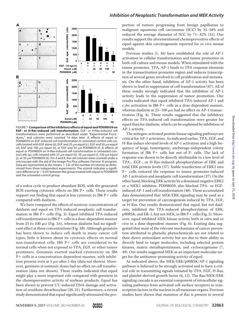
mation of JB6 P� Cells—EGF is known to induce cell transfor-mation mainly through the MEK/ERK signaling pathway, andour previous work showed that ERK activity is required forEGF-induced cell transformation in JB6 P� cells (37). Varioushuman cancers have been found to exhibit mutations and over-expression of ras genes, and one of the most frequent events incarcinogenesis is uncontrolled activation of the Ras signalingpathway (38). An accumulation of data suggests that theupstream effector of theMEK/ERK cascade is Ras/Raf (39). Ourprevious studies showed thatH-Ras activated the growth-signalpathway involving MEK and ERK protein kinases, leading toanchorage-independent growth and elevated AP-1 activity inJB6 P� cells (10). In the present study we examined the poten-tial inhibitory activities of equol and PD098059 on EGF- orH-Ras-induced cell transformation. Based on the number ofcell colonies, equol at concentrations of 50 �M inhibited EGF-induced cell transformation by 41% (Fig. 7A, panel d), and thesame concentration also suppressed H-Ras-induced neoplastictransformation of JB6 P� cells by 49% (Fig. 7B, panel i).We alsofound that 50 �M PD098059 inhibited EGF- or H-Ras-inducedneoplastic transformation of JB6 P� cells by 79% (Fig. 7A, panelf) and 77% (Fig. 7B, panel k), respectively, suggesting that equolalso may inhibit cell transformation by targeting the MEK sig-naling pathway.Equol Specifically Binds withMEK1 in Vitro and ex Vivo and
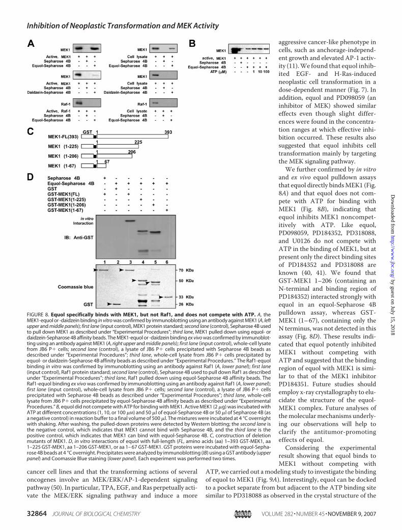
Does Not Compete with ATP for Binding with MEK1—Theresults above indicated that the inhibition of cell transforma-tion by equol involves the inhibition of MEK1 kinase activitybut not its upstream phosphorylation. To further confirmwhether equol can directly bind with MEK1, we performed anin vitro equol pulldown assay. MEK1 was found in the equol-Sepharose 4B beads (Fig. 8A, left upper panel) but not in Sepha-
rose 4B beads alone (Fig. 8A, leftupper panel). The MEK1 proteinwas loaded as a control (Fig. 8A,left upper panel, first lane). Wealso observed en vivo binding ofequol and MEK1 in JB6 P� celllysates (Fig. 8A, right upper panel).Daidzein did not bind with MEK1in vitro or ex vivo (Fig. 8A, right orleft middle panels). To determinewhether equol specifically interactswith MEK1, we investigated in vitroand ex vivo binding of equol andRaf1 (Fig. 8A, lower panels).Results confirmed that equol didnot bind with the Raf1 kinase.Therefore, these results indicatedthat equol has a specificity forMEK1. Furthermore, ATP did notcompete with equol for bindingwith MEK1 (Fig. 8B). These results
FIGURE 4. Effects of equol or daidzein on TPA-induced AP-1 or c-fos acti-vation. A and B, equol, but not daidzein, inhibits TPA-induced AP-1 transac-tivation. For the luciferase assay, JB6 cells stably transfected with an AP-1luciferase reporter plasmid were cultured as described under “ExperimentalProcedures.” The cells were starved in 0.1% FBS, MEM and then treated or nottreated with daidzein or equol at the indicated concentrations (25, 50, or 100 �M)for 1 h before they were exposed to 20 ng/ml TPA for 24 h. Luciferase activity wasassayed, and AP-1 activity is expressed relative to control cells without TPA treat-ment. Data are represented as the means � S.D. of AP-1 luciferase activity calcu-lated from three independent experiments. C and D, equol, but not daidzein,suppresses TPA-induced c-fos promoter activity. For the reporter-gene assay, JB6P� cells were transfected with a plasmid mixture containing the c-fos-luciferasereporter gene (0.5 �g) and the pRL-SV40 gene (0.5 �g). At 24 h after transfection,cells were starved for 24 h by incubation in 0.1% FBS, MEM at 37 °C in a 5% CO2atmosphere. Cells were then treated or not treated with daidzein or equol at theindicated concentrations (25, 50, or 100 �M) for 1 h before they were exposed to20 ng/ml TPA for 12 h. Firefly luciferase activity was determined in cell lysates andnormalized to Renilla luciferase activity, and c-fos-luciferase activity is expressedrelative to control cells without TPA treatment. Data are represented as themeans � S.D. of the c-fos-luciferase activity calculated from three independentexperiments. The asterisk (*) indicates a significant difference (p � 0.05) betweengroups treated with TPA and daidzein or equol and the group treated with TPAalone.
FIGURE 5. Effects of equol, daidzein, and PD098059 on TPA-induced phosphorylation of ERK, p90RSK,Elk-1, MEK, and c-Jun N-terminal kinase. A, equol inhibits TPA-induced phosphorylation (p-) of ERK, p90RSK,and Elk-1 but not MEK. B, daidzein did not inhibit TPA-induced phosphorylation of ERK, p90RSK, Elk-1, and MEK.C, PD098059 inhibits TPA-induced phosphorylation of ERK, p90RSK, and Elk-1 but not MEK. Cells were pre-treated with equol (A), daidzein (B), or PD098059 (C) at the indicated concentrations (25, 50, or 100 �M) for 1 h,then stimulated with 20 ng/ml TPA and harvested 15 min later. The levels of phosphorylated and total MEK,ERK, p90RSK, and Elk-1 proteins were determined by Western blot analysis as described under “ExperimentalProcedures” using specific antibodies against the corresponding phosphorylated and total proteins. Data arerepresentative of two independent experiments that gave similar results.
Inhibition of Neoplastic Transformation and MEK Activity
NOVEMBER 9, 2007 • VOLUME 282 • NUMBER 45 JOURNAL OF BIOLOGICAL CHEMISTRY 32861
by guest on July 15, 2018http://w
ww
.jbc.org/D
ownloaded from
suggested that equol inhibited MEK1 kinase activity noncom-petitively with ATP.Identification of the Equol Binding Site of MEK1—Previous
studies have demonstrated that, like equol, MEK1-selectivesmall molecule inhibitors such as PD098059, PD184352,PD318088, and U0126 do not compete with ATP for bindingwith MEK1, but at present only the direct binding sites forPD184352 and PD318088 are known (40, 41). To determine theregion of binding between equol and MEK1, we constructedthree MEK1 deletion mutants from full-length GST-MEK1.One mutant contained only the N-terminal, GST-MEK1(1–67); one contained theN terminus and the binding region ofPD184352, GST-MEK1 (1–206), and one contained the N ter-minus, the binding region of PD184352, and a phosphorylationsite of MEK1, GST-MEK1 (1–225) (Fig. 8C). These deletionmutants were expressed and purified in bacteria as describedunder “Experimental Procedures.”We then evaluated the bind-ing affinity of the four expressed GST-MEK proteins using theequol-Sepharose 4B pulldown assay. The results indicated thatGST-MEK1 (1–206) interacted efficiently with equol, whereasGST-MEK1 (1–67) was not detected (Fig. 8D). These results
suggested that equol is a potent inhibitor of MEK1 noncom-petitively with ATP and that the region where equol binds withMEK1 might be similar to that of the MEK1 inhibitorPD184351.
DISCUSSION
Dietary phenolic phytochemicals, which are present in fruits,vegetables, grains, and some beverages, have been proposed asprimary antioxidant chemopreventive agents (13). However,most free-radical scavengers act in oxidation-reduction reac-tions, and some phenolic phytochemicals can act as prooxi-dants and exert toxicity, depending on their structure and theconditions (42–45). In the present study, equol at concentra-tions from 25 to 100 �M had no effect on cell growth even at 5days after treatment (Fig. 2A). In contrast, 50 �M daidzein wascytotoxic to JB6 P� cell growth (Fig. 2B). The higher cytotox-icity of daidzein relative to equol might be attributable to aquinone group of daidzein, which has been suggested to con-tribute to the production of reactive oxygen species (ROS) incells. A previous study (46) showed that the presence of thesemiquinone radical or benzoquinone resulted in the formation
FIGURE 6. Effects of equol, daidzein, and PD098059 on MEK1 kinase activity. A and B, equol, but not daidzein, inhibits MEK1 kinase activity both invitro (A) and ex vivo (B). Cells were pretreated with equol, daidzein, or PD098059 at the indicated concentrations for 1 h and then stimulated with 20 ng/ml TPAfor 30 min. Cells were harvested, and immunoprecipitation and MEK1 kinase assays were performed as described under “Experimental Procedures.” Data arerepresented as the means � S.D. as determined from three independent experiments. C, equol does not inhibit Raf-1 kinase activity in vitro. Raf-1 kinase assaywas performed as described under “Experimental Procedures.” Data are represented as the means � S.D. as determined from three independent experiments.The asterisk (*) indicates a significant difference (p � 0.05) between groups treated with TPA and equol, daidzein, or PD098059 and the group treated with TPAalone. D, overexpression of MEK1 increases the transcriptional activity of AP-1 and attenuates equol inhibition of AP-1 activation. The JB6 mouse epidermal cellline transfected with an AP-1 luciferase reporter plasmid was cotransfected with a pcDNA3 vector or constitutively active-MEK1. Then, 24 h post-transfection,the transfectants were assayed as described under “Experimental Procedures.” E, the AP-1 JB6 mouse epidermal cell line transfected with CA-MEK1 waspretreated with equol for 24 h at the concentrations indicated. Luciferase activity is expressed as relative AP-1 activity (% of CA-MEK1 transfected control). Datafrom three independent experiments are averaged and presented as the means � S.D.
Inhibition of Neoplastic Transformation and MEK Activity
32862 JOURNAL OF BIOLOGICAL CHEMISTRY VOLUME 282 • NUMBER 45 • NOVEMBER 9, 2007
by guest on July 15, 2018http://w
ww
.jbc.org/D
ownloaded from
of a redox cycle to produce abundant ROS, with the generatedROS exerting cytotoxic effects on JB6 P� cells. These resultssupport our finding that equol is less cytotoxic to JB6 P� cellscompared with daidzein.We have compared the effects of nontoxic concentrations of
daidzein and equol on TPA-induced neoplastic cell transfor-mation in JB6 P� cells (Fig. 3). Equol inhibited TPA-inducedcell transformation in JB6P� cells in a dose-dependentmannerfrom 25 to 100 �M (Fig. 3A), whereas daidzein had no signifi-cant effect at these concentrations (Fig. 3B). Although genisteinhas been shown to induce cell death in many cancer celltypes, little is known about its cytotoxic effects on normalnon-transformed cells. JB6 P� cells are considered to benormal cells when not exposed to TPA, EGF, or other tumorpromoters. Genistein exerted marked cytotoxicity on JB6P� cells in a concentration-dependent manner, with inhibi-tion present even at 5 �M after 1 day (data not shown). More-over, genistein at nontoxic doses had no effect on cell transfor-mation (data not shown). These results indicated that equolmight play a more important role compared with genistein inthe chemopreventive activity of soybean products. Equol hasbeen shown to prevent UV-induced DNA damage and activa-tion of ornithine decarboxylase (30, 31). Furthermore, a recentstudy demonstrated that equol significantly attenuated the pro-
portion of tumors progressing from benign papillomas tomalignant squamous cell carcinomas (SCC) by 33–58% andreduced the average diameter of SCC by 71–82% (31). Ourresults support the aforementioned chemopreventive effects ofequol against skin carcinogenesis reported for in vivo mousemodels.Previous studies (1, 36) have established the role of AP-1
activation in cellular transformation and tumor promotion inboth cell culture andmouse models. When stimulated with thetumor promoter, TPA, AP-1 binds to TPA response elementsin the transactivation promoter region and induces transcrip-tion of several genes involved in cell proliferation and metasta-sis. On the other hand, inhibition of AP-1 activity has beenshown to lead to suppression of cell transformation (47). All ofthese results strongly indicated that the inhibition of AP-1activity leads to the suppression of tumor promotion. Ourresults indicated that equol inhibited TPA-induced AP-1 andc-fos activation in JB6 P� cells in a dose-dependent manner,whereas daidzein at 25–100 �M had no affect on AP-1 transac-tivation (Fig. 4). These results suggested that the inhibitoryeffects on TPA-induced cell transformation were greater forequol than for daidzein, which can be explained by inhibition ofAP-1 activity.Themitogen-activated protein kinase signaling pathways are
critical for AP-1 activation. As indicated earlier, TPA, EGF, andH-Ras induce elevated levels of AP-1 activation and a high fre-quency of large, tumorigenic, anchorage-independent colonyformation of JB6 P� cells but not of P- cells. The lack ofresponse was shown to be directly attributable to a low level ofTPA-, EGF-, or H-Ras-induced phosphorylation of ERK andtotal ERK protein levels (37). Stable transfection of ERK2 intoP� cells restored the response to tumor promoter-inducedAP-1 activation and neoplastic cell transformation (37). On theother hand, blocking ERK activity by a dominant negative ERK2or a MEK1 inhibitor, PD098059, also blocked TPA- or EGF-induced AP-1 and cell transformation (48). These accumulateddata demonstrated that MEK/ERK signaling may be used as atarget for prevention of carcinogenesis induced by TPA, EGF,or H-Ras. Our results demonstrated that equol, but not daid-zein, inhibited the TPA-induced phosphorylation of ERK,p90RSK, and Elk-1, but notMEK, in JB6 P� cells (Fig. 5).More-over, equol inhibited MEK kinase activity both in vitro and exvivo in a dose-dependent manner (Fig. 6). Existing data sug-gested that most of the relevant mechanisms of cancer preven-tion attributed to phenolic phytochemicals are not related totheir direct antioxidant activity but are due to their ability todirectly bind to target molecules, including selected proteinkinases, matrix metalloproteinases, and cyclooxygenases (7,49). Our results suggestedMEK as an important molecular tar-get for the antitumor-promoting activity of equol.As indicated above, the MEK/ERK/p90RSK/AP-1 signaling
pathway is believed to be strongly activated and to have a crit-ical role in transmitting signals initiated by TPA, EGF, H-Ras,and platelet-derived growth factor (6, 12). The Ras/MEK/ERKsignaling cascade is an essential component of intracellular sig-naling pathways from activated cell-surface receptors to tran-scription factors in the nucleus in allmetazoan organs. Previousstudies have shown that mutation of Ras is present in several
FIGURE 7. Comparison of the inhibitory effects of equol and PD098059 onEGF- or H-Ras-induced cell transformation. EGF- or H-Ras-induced celltransformations were performed as described under “Experimental Proce-dures,” and colonies were counted 14 days later. A, effects of equol orPD098059 on EGF-induced cell transformation in untreated control cells (a),cells treated with EGF alone (b), EGF and 25 �M equol (c), EGF and 50 �M equol(d), EGF and 100 �M equol (e), or EGF and 50 �M PD098059 (f). B, effects ofequol or PD098059 on H-Ras-induced cell transformation in untreated con-trol cells (g), cells treated with 25 �M equol (h), 50 �M equol (i), 100 �M equol(j), or 50 �M PD098059 (k). For A and B, the cell colonies were counted under amicroscope with the aid of the Image-Pro Plus software (Version 4) program.Data are represented as the means � S.D. of the number of colonies as deter-mined from three independent experiments. The asterisk indicates a signifi-cant difference (p � 0.05) between the group treated with equol or PD098059and the untreated control group.
Inhibition of Neoplastic Transformation and MEK Activity
NOVEMBER 9, 2007 • VOLUME 282 • NUMBER 45 JOURNAL OF BIOLOGICAL CHEMISTRY 32863
by guest on July 15, 2018http://w
ww
.jbc.org/D
ownloaded from
cancer cell lines and that the transforming actions of severaloncogenes involve an MEK/ERK/AP-1-dependent signalingpathway (50). In particular, TPA, EGF, andRas perpetually acti-vate the MEK/ERK signaling pathway and induce a more
aggressive cancer-like phenotype incells, such as anchorage-independ-ent growth and elevated AP-1 activ-ity (11).We found that equol inhib-ited EGF- and H-Ras-inducedneoplastic cell transformation in adose-dependent manner (Fig. 7). Inaddition, equol and PD098059 (aninhibitor of MEK) showed similareffects even though slight differ-ences were found in the concentra-tion ranges at which effective inhi-bition occurred. These results alsosuggested that equol inhibits celltransformation mainly by targetingthe MEK signaling pathway.We further confirmed by in vitro
and ex vivo equol pulldown assaysthat equol directly bindsMEK1 (Fig.8A) and that equol does not com-pete with ATP for binding withMEK1 (Fig. 8B), indicating thatequol inhibits MEK1 noncompet-itively with ATP. Like equol,PD098059, PD184352, PD318088,and U0126 do not compete withATP in the binding of MEK1, but atpresent only the direct binding sitesof PD184352 and PD318088 areknown (40, 41). We found thatGST-MEK1 1–206 (containing anN-terminal and binding region ofPD184352) interacted strongly withequol in an equol-Sepharose 4Bpulldown assay, whereas GST-MEK1 (1–67), containing only theN terminus, was not detected in thisassay (Fig. 8D). These results indi-cated that equol potently inhibitedMEK1 without competing withATP and suggested that the bindingregion of equol with MEK1 is simi-lar to that of the MEK1 inhibitorPD184351. Future studies shouldemploy x-ray crystallography to elu-cidate the structure of the equol-MEK1 complex. Future analyses ofthemolecularmechanisms underly-ing our observations will help toclarify the antitumor-promotingeffects of equol.Considering the experimental
result showing that equol binds toMEK1 without competing with
ATP, we carried out amodeling study to investigate the bindingof equol to MEK1 (Fig. 9A). Interestingly, equol can be dockedto a pocket separate from but adjacent to the ATP binding sitesimilar to PD318088 as observed in the crystal structure of the
FIGURE 8. Equol specifically binds with MEK1, but not Raf1, and does not compete with ATP. A, theMEK1-equol or -daidzein binding in vitro was confirmed by immunoblotting using an antibody against MEK1 (A, leftupper and middle panels); first lane (input control), MEK1 protein standard; second lane (control), Sepharose 4B usedto pull down MEK1 as described under “Experimental Procedures”; third lane, MEK1 pulled down using equol- ordaidzein-Sepharose 4B affinity beads. The MEK1-equol or -daidzein binding ex vivo was confirmed by immunoblot-ting using an antibody against MEK1 (A, right upper and middle panels); first lane (input control), whole-cell lysatefrom JB6 P� cells; second lane (control), a lysate of JB6 P� cells precipitated with Sepharose 4B beads asdescribed under “Experimental Procedures”; third lane, whole-cell lysate from JB6 P� cells precipitated byequol- or daidzein-Sepharose 4B affinity beads as described under “Experimental Procedures.” The Raf1-equolbinding in vitro was confirmed by immunoblotting using an antibody against Raf1 (A, lower panel); first lane(input control), Raf1 protein standard; second lane (control), Sepharose 4B used to pull down Raf1 as describedunder “Experimental Procedures”; third lane, Raf1 pulled down using equol-Sepharose 4B affinity beads. TheRaf1-equol binding ex vivo was confirmed by immunoblotting using an antibody against Raf1 (A, lower panel);first lane (input control), whole-cell lysate from JB6 P� cells; second lane (control), a lysate of JB6 P� cellsprecipitated with Sepharose 4B beads as described under “Experimental Procedures”; third lane, whole-celllysate from JB6 P� cells precipitated by equol-Sepharose 4B affinity beads as described under “ExperimentalProcedures.” B, equol did not compete with ATP for binding with MEK1. Active MEK1 (2 �g) was incubated withATP at different concentrations (1, 10, or 100 �M) and 50 �l of equol-Sepharose 4B or 50 �l of Sepharose 4B (asa negative control) in reaction buffer to a final volume of 500 �l. The mixtures were incubated at 4 °C overnightwith shaking. After washing, the pulled-down proteins were detected by Western blotting; the second lane isthe negative control, which indicates that MEK1 cannot bind with Sepharose 4B, and the third lane is thepositive control, which indicates that MEK1 can bind with equol-Sepharose 4B. C, construction of deletionmutants of MEK1. D, in vitro interactions of equol with full-length (FL, amino acids (aa) 1–393 GST-MEK1, aa1–225 GST-MEK1, aa 1–206 GST-MEK1, or aa 1– 67 GST-MEK1. GST proteins were incubated with equol-Sepha-rose 4B beads at 4 °C overnight. Precipitates were analyzed by immunoblotting (IB) using a GST antibody (upperpanel) and Coomassie Blue staining (lower panel). Each experiment was performed two times.
Inhibition of Neoplastic Transformation and MEK Activity
32864 JOURNAL OF BIOLOGICAL CHEMISTRY VOLUME 282 • NUMBER 45 • NOVEMBER 9, 2007
by guest on July 15, 2018http://w
ww
.jbc.org/D
ownloaded from
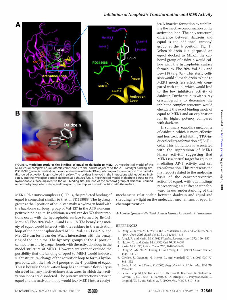
MEK1-PD318088 complex (41). Thus, the predicted binding ofequol is somewhat similar to that of PD318088. The hydroxylgroup at the 7 position of equol canmake a hydrogen bondwiththe backbone carbonyl group of Val-127 in the ATP noncom-petitive binding site. In addition, several van derWaals interac-tions occur with the hydrophobic surface formed by Ile-141,Met-143, Phe-209, Val-211, and Leu-118. The benzyl ring moi-ety of equol would interact with the residues in the activationloop of the nonphosphorylated MEK1. Val-211, Leu-215, andMet-219 can form van der Waals interactions with the benzylring of the inhibitor. The hydroxyl groups at the 4� positioncannot form any hydrogen bondswith the activation loop in themodel structure of MEK1. However, we cannot exclude thepossibility that the binding of equol to MEK1 would induce aslight structural change of the activation loop to form a hydro-gen bond with the hydroxyl groups at the 4� position of equol.This is because the activation loop has an intrinsic flexibility asobserved inmany inactive kinase structures, inwhich their acti-vation loops are disordered. The putative interactions betweenequol and the activation loop would lock MEK1 into a catalyt-
ically inactive formation by stabiliz-ing the inactive conformation of theactivation loop. The only structuraldifference between daidzein andequol is the additional carbonylgroup at the 4 position (Fig. 1).When daidzein is superposed onequol docked to MEK1, the car-bonyl group of daidzein would col-lide with the hydrophobic surfaceformed by Phe-209, Val-211, andLeu-118 (Fig. 9B). This steric colli-sion would allow daidzein to bind toMEK1 much less effectively com-pared with equol, which would leadto the low inhibitory activity ofdaidzein. Further studies with x-raycrystallography to determine theinhibitor complex structure wouldelucidate the exact binding mode ofequol to MEK1 and an explanationfor its higher potency comparedwith daidzein.In summary, equol is ametabolite
of daidzein, which is more effectiveand less toxic at inhibiting TPA-in-duced cell transformation of JB6 P�cells. This inhibition is associatedwith the suppression of MEK1kinase activity, suggesting thatMEK1 is a critical target for equol inmediating AP-1 activity and celltransformation. This represents thefirst report related to the molecularbasis of the cancer-preventiveaction of equol, with our findingsrepresenting a significant step for-ward in our understanding of the
mechanistic relationship between daidzein and equol andshedding new light on the molecular mechanisms of equol inchemoprevention.
Acknowledgment—We thank Andria Hansen for secretarial assistance.
REFERENCES1. Dong, Z., Birrer, M. J., Watts, R. G., Matrisian, L. M., and Colburn, N. H.
(1994) Proc. Natl. Acad. Sci. U. S. A. 91, 609–6132. Angel, P., and Karin, M. (1991) Biochim. Biophys. Acta 1072, 129–1573. Hunter, T., and Karin, M. (1992) Cell 70, 375–3874. Karin, M. (1995) J. Biol. Chem. 270, 16483–164865. Dong, Z., Ma, W. Y., Huang, C., and Yang, C. S. (1997) Cancer Res. 57,
4414–44196. Cowley, S., Paterson, H., Kemp, P., and Marshall, C. J. (1994) Cell 77,
841–8527. Bode, A. M., and Dong, Z. (2005) Prog. Nucleic Acid Res. Mol. Biol. 79,
237–2978. Sebolt-Leopold, J. S., Dudley, D. T., Herrera, R., Becelaere, K., Wiland, A.,
Gowan, R. C., Tecle, H., Barrett, S. D., Bridges, A., Przybranowski, S.,Leopold, W. R., and Saltiel, A. R. (1999) Nat. Med. 5, 810–816
FIGURE 9. Modeling study of the binding of equol or daidzein to MEK1. A, hypothetical model of theMEK1-equol complex. Equol (atomic color) binds to the pocket adjacent to the ATP (orange) binding site.PD318088 (green) is overlaid on the model structure of the MEK1-equol complex for comparison. The partiallydisordered activation loop is colored in yellow. The residues involved in the interactions with equol are indi-cated, and the hydrogen bond is depicted as a dashed line. B, hypothetical model of daidzein bound to thehydrophobic surface adjacent to the ATP binding site. The end of the carbonyl group of daidzein is buriedunder the hydrophobic surface, and the green arrow implies its steric collision with the surface.
Inhibition of Neoplastic Transformation and MEK Activity
NOVEMBER 9, 2007 • VOLUME 282 • NUMBER 45 JOURNAL OF BIOLOGICAL CHEMISTRY 32865
by guest on July 15, 2018http://w
ww
.jbc.org/D
ownloaded from
9. Shapiro, P. (2002) Crit. Rev. Clin. Lab. Sci. 39, 285–33010. Chung, J. Y., Park, J. O., Phyu, H., Dong, Z., and Yang, C. S. (2001) FASEB
J. 15, 2022–202411. Chung, J. Y., Huang, C., Meng, X., Dong, Z., and Yang, C. S. (1999)Cancer
Res. 59, 4610–461712. Bode, A. M., and Dong, Z. (2000) Lancet Oncol. 1, 181–18813. Surh, Y.-J. (2003) Nat. Rev. Cancer 3, 768–78014. Lee, K. W., and Lee, H. J. (2006)Mech. Ageing Dev. 127, 424–43115. Lee, K. W., and Lee, H. J. (2006) Biofactors 26, 105–12116. Birt, D. F., Hendrich, S., and Wang, W. (2001) Pharmacol. Ther. 90,
157–17717. Eldridge, A. C., andKwolek,W. F. (1983) J. Agric. FoodChem. 31, 394–39618. Setchell, K. D. (1998) Am. J. Clin. Nutr. 68, 1333–134619. Akiyama, T., Ishida, J., Nakagawa, S., Ogawara, H., Watanabe, S., Itoh, N.,
Shibuya, M., and Fukami, Y. (1987) J. Biol. Chem. 262, 5592–559520. Peterson, G. (1995) J. Nutr. 125, (Suppl. 3) 784–78921. Kim,H., Peterson, T.G., andBarnes, S. (1998)Am. J. Clin. Nutr.68, (Suppl.
6) 1418–142522. Spinozzi, F., Pagliacci, M. C., Migliorati, G., Moraca, R., Grignani, F., Ric-
cardi, C., and Nicoletti, I. (1994) Leuk. Res. 18, 431–43923. Verma, S. P., and Goldin, B. R. (1998) Nutr. Cancer 30, 232–23924. Xu, X., Harris, K. S., Wang, H. J., Murphy, P. A., and Hendrich, S. (1995) J.
Nutr. 125, 2307–231525. Chang, Y. C., Nair, M. G., and Nitiss, J. L. (1995) J. Nat. Prod. 58,
1901–190526. Kelly, G. E., Joannou, G. E., Reeder, A. Y., Nelson, C., and Waring, M. A.
(1995) Proc. Soc. Exp. Biol. Med. 208, 40–4327. Rowland, I. R., Wiseman, H., Sanders, T. A., Adlercreutz, H., and Bowey,
E. A. (2000) Nutr. Cancer 36, 27–3228. Arora, A., Nair, M. G., and Strasburg, G. M. (1998) Arch. Biochem. Bio-
phys. 356, 133–14129. Sierens, J., Hartley, J. A., Campbell, M. J., Leathem, A. J., and Woodside,
J. V. (2001)Mutat. Res. 485, 169–17630. Widyarini, S. (2006) J. Vet. Sci. 7, 217–22331. Widyarini, S., Husband, A. J., and Reeve, V. E. (2005) Photochem. Photo-
biol. 81, 32–3732. Zhong, S., Quealy, J. A., Bode, A. M., Nomura, M., Kaji, A., Ma,W. Y., and
Dong, Z. (2001) Cancer Res. 61, 4084–4091
33. Dong, Z., and Cmarik, J. L. (2002) Sci. STKE 2002, PL734. Colburn, N.H.,Wendel, E. J., andAbruzzo, G. (1981) Proc. Natl. Acad. Sci.
U. S. A. 78, 6912–691635. Iwashita, K., Kobori, M., Yamaki, K., and Tsushida, T. (2000) Biosci. Bio-
technol. Biochem. 64, 1813–182036. Huang, C., Ma,W. Y., Dawson, M. I., Rincon, M., Flavell, R. A., and Dong,
Z. (1997) Proc. Natl. Acad. Sci. U. S. A. 94, 5826–583037. Huang, C.,Ma,W.Y., Young,M. R., Colburn,N., andDong, Z. (1998)Proc.
Natl. Acad. Sci. U. S. A. 95, 156–16138. Zachos, G., and Spandidos, D. A. (1997) Crit. Rev. Oncol. Hematol. 26,
65–7539. Kyriakis, J. M., and Avruch, J. (2001) Physiol. Rev. 81, 807–86940. Alessi, D. R., Cuenda, A., Cohen, P., Dudley, D. T., and Saltiel, A. R. (1995)
J. Biol. Chem. 270, 27489–2749441. Ohren, J. F., Chen, H., Pavlovsky, A., Whitehead, C., Zhang, E., Kuffa, P.,
Yan, C., McConnell, P., Spessard, C., Banotai, C., Mueller,W. T., Delaney,A., Omer, C., Sebolt-Leopold, J., Dudley, D. T., Leung, I. K., Flamme, C.,Warmus, J., Kaufman, M., Barrett, S., Tecle, H., and Hasemann, C. A.(2004) Nat. Struct. Mol. Biol. 11, 1192–1197
42. Galati, G., Lin, A., Sultan, A. M., and O’Brien, P. J. (2006) Free Radic. Biol.Med. 40, 570–580
43. Galati, G., and O’Brien, P. J. (2004) Free Radic. Biol. Med. 37, 287–30344. Elbling, L., Weiss, R. M., Teufelhofer, O., Uhl, M., Knasmueller, S.,
Schulte-Hermann, R., Berger, W., and Micksche, M. (2005) FASEB J. 19,807–809
45. Lee, K. W., Hur, H. J., Lee, H. J., and Lee, C. Y. (2005) J. Agric. Food Chem.53, 1990–1995
46. Oikawa, S., Furukawaa, A., Asada, H., Hirakawa, K., and Kawanishi, S.(2003) Free Radic. Res. 37, 881–890
47. Dong, Z., Huang, C., Brown, R. E., andMa,W. Y. (1997) J. Biol. Chem. 272,9962–9970
48. Watts, R. G., Huang, C., Young,M. R., Li, J. J., Dong, Z., Pennie,W. D., andColburn, N. H. (1998) Oncogene 17, 3493–3498
49. Sang, S., Hou, Z., Lambert, J. D., and Yang, C. S. (2005) Antioxid. RedoxSignal. 7, 1704–1714
50. Abrams, S. I., Hand, P.H., Tsang, K. Y., and Schlom, J. (1996) Semin.Oncol.23, 118–134
Inhibition of Neoplastic Transformation and MEK Activity
32866 JOURNAL OF BIOLOGICAL CHEMISTRY VOLUME 282 • NUMBER 45 • NOVEMBER 9, 2007
by guest on July 15, 2018http://w
ww
.jbc.org/D
ownloaded from
Ann M. Bode, Hyong Joo Lee and Zigang DongNam Joo Kang, Ki Won Lee, Evgeny A. Rogozin, Yong-Yeon Cho, Yong-Seok Heo,
PathwayTransformation by Targeting the MEK/ERK/p90RSK/Activator Protein-1
Equol, a Metabolite of the Soybean Isoflavone Daidzein, Inhibits Neoplastic Cell
doi: 10.1074/jbc.M701459200 originally published online August 27, 20072007, 282:32856-32866.J. Biol. Chem.
10.1074/jbc.M701459200Access the most updated version of this article at doi:
Alerts:
When a correction for this article is posted•
When this article is cited•
to choose from all of JBC's e-mail alertsClick here
http://www.jbc.org/content/282/45/32856.full.html#ref-list-1
This article cites 49 references, 12 of which can be accessed free at
by guest on July 15, 2018http://w
ww
.jbc.org/D
ownloaded from