Epithelial/myoepithelial carcinoma of breast: A report of ...
5
http://crcp.sciedupress.com Case Reports in Clinical Pathology 2016, Vol. 3, No. 4 CASE REPORT Epithelial/myoepithelial carcinoma of breast: A report of hitherto unreported case Tadashi Terada * Departments of Pathology, Shizuoka City Shimizu Hospital, Shizuoka, Japan Received: February 28, 2016 Accepted: May 20, 2016 Online Published: June 6, 2016 DOI: 10.5430/crcp.v3n4p15 URL: http://dx.doi.org/10.5430/crcp.v3n4p15 ABSTRACT Herein reported is the first case of epithelial/myoepithelial carcinoma (EMC) of breast. An 83-year-old woman noticed a circa 1.0 cm tumor of left breast. Core needle biopsies were diagnosed as invasive ductal carcinoma. Mastectomy with axillary lymph node dissection was performed, and gross examination revealed a circa 1 cm × 1 cm tumor. Microscopically, the tumor was composed of invasive malignant cells forming tubular or cords structures with a scirrhous collagenous stroma. The malignant cells were composed by the following two elements; one was malignant epithelial cells and the other was malignant myoepithelial cells with clear or vacuolated cytoplasm. Immunohistochemically, the myoepithelial element was positive for p63, CD10, cytokeratin (CK) 34BE12, and CK5/6, S100 protein and alpha-smooth muscle actin. Both elements were positive for CK7, p53, and Ki67 (labeling: 16%), estrogen receptor, progesterone receptor. Both elements were negative for CK20 and HER2/neu. The sentinel and level 1 lymph node were negative for metastasis (0/6). The tumor was pT1pN0cM0 and the stage was stage 1. The patient had booster hormone therapy and is now alive 14 months without recurrence after the operation. Key Words: Breast, Epithelium, Myoepithelium, Epithelial/myoepithelial carcinoma, Histopathology 1. I NTRODUCTION Mammary glands are composed of ductal epithelium, lobular epithelium, myoepithelium, and stromal cells; many kinds of tumors arise from them. [1–4] Epithelial/myoepithelial car- cinoma (EMC) composed of malignant ductal epithelium and outer myoepithelium and showing an organoid pattern with clear separation between epithelial and myoepithelial elements is not listed in 2012 WHO blue book of tumor classification of breast, [1] and has not been described in the English literature, to the author’s best knowledge. The author reports the first case. 2. CASE REPORT An 83-year-old woman noticed a circa 1.0 cm tumor of her left breast. Mammography also identified the tumor with relatively well demarcation; no spicules were noted. Blood laboratory data and other images including chest and abdom- inal X-ray showed no significant findings. Breast needle cytology showed atypical cells but was decided to be inde- terminate. Core biopsies showed malignant epithelial cells with clear cytoplasm and vacuoles in a collagenous stroma, and they were diagnosed as invasive ductal carcinoma (scir- rhous carcinoma) by the author. Retrospective inspection showed that the carcinoma in core biopsies are the same as that of mastectomy specimens; therefore the diagnosis of core biopsy was not correct. Mastectomy and axillary lymph node dissection, but not breast-preserving wide excision, was performed; this procedure is recommended for resectable small breast carcinoma in NCI guidelines, to the best of the author’s knowledge. A gross examination revealed a circa * Correspondence: Tadashi Terada, MD, PhD; Email: [email protected]; Address: Department of Pathology, Shizuoka City Shimizu Hospital, Miyakami 1231 Shimizu-Ku, Shizuoka 424-8636, Japan. Published by Sciedu Press 15
Transcript of Epithelial/myoepithelial carcinoma of breast: A report of ...
http://crcp.sciedupress.com Case Reports in Clinical Pathology 2016, Vol. 3, No. 4
CASE REPORT
Epithelial/myoepithelial carcinoma of breast: A reportof hitherto unreported case
Tadashi Terada∗
Departments of Pathology, Shizuoka City Shimizu Hospital, Shizuoka, Japan
Received: February 28, 2016 Accepted: May 20, 2016 Online Published: June 6, 2016DOI: 10.5430/crcp.v3n4p15 URL: http://dx.doi.org/10.5430/crcp.v3n4p15
ABSTRACT
Herein reported is the first case of epithelial/myoepithelial carcinoma (EMC) of breast. An 83-year-old woman noticed a circa1.0 cm tumor of left breast. Core needle biopsies were diagnosed as invasive ductal carcinoma. Mastectomy with axillary lymphnode dissection was performed, and gross examination revealed a circa 1 cm × 1 cm tumor. Microscopically, the tumor wascomposed of invasive malignant cells forming tubular or cords structures with a scirrhous collagenous stroma. The malignant cellswere composed by the following two elements; one was malignant epithelial cells and the other was malignant myoepithelial cellswith clear or vacuolated cytoplasm. Immunohistochemically, the myoepithelial element was positive for p63, CD10, cytokeratin(CK) 34BE12, and CK5/6, S100 protein and alpha-smooth muscle actin. Both elements were positive for CK7, p53, and Ki67(labeling: 16%), estrogen receptor, progesterone receptor. Both elements were negative for CK20 and HER2/neu. The sentineland level 1 lymph node were negative for metastasis (0/6). The tumor was pT1pN0cM0 and the stage was stage 1. The patienthad booster hormone therapy and is now alive 14 months without recurrence after the operation.
Key Words: Breast, Epithelium, Myoepithelium, Epithelial/myoepithelial carcinoma, Histopathology
1. INTRODUCTIONMammary glands are composed of ductal epithelium, lobularepithelium, myoepithelium, and stromal cells; many kindsof tumors arise from them.[1–4] Epithelial/myoepithelial car-cinoma (EMC) composed of malignant ductal epitheliumand outer myoepithelium and showing an organoid patternwith clear separation between epithelial and myoepithelialelements is not listed in 2012 WHO blue book of tumorclassification of breast,[1] and has not been described in theEnglish literature, to the author’s best knowledge. The authorreports the first case.
2. CASE REPORTAn 83-year-old woman noticed a circa 1.0 cm tumor of herleft breast. Mammography also identified the tumor with
relatively well demarcation; no spicules were noted. Bloodlaboratory data and other images including chest and abdom-inal X-ray showed no significant findings. Breast needlecytology showed atypical cells but was decided to be inde-terminate. Core biopsies showed malignant epithelial cellswith clear cytoplasm and vacuoles in a collagenous stroma,and they were diagnosed as invasive ductal carcinoma (scir-rhous carcinoma) by the author. Retrospective inspectionshowed that the carcinoma in core biopsies are the same asthat of mastectomy specimens; therefore the diagnosis ofcore biopsy was not correct. Mastectomy and axillary lymphnode dissection, but not breast-preserving wide excision, wasperformed; this procedure is recommended for resectablesmall breast carcinoma in NCI guidelines, to the best of theauthor’s knowledge. A gross examination revealed a circa
∗Correspondence: Tadashi Terada, MD, PhD; Email: [email protected]; Address: Department of Pathology, Shizuoka City Shimizu Hospital,Miyakami 1231 Shimizu-Ku, Shizuoka 424-8636, Japan.
Published by Sciedu Press 15
http://crcp.sciedupress.com Case Reports in Clinical Pathology 2016, Vol. 3, No. 4
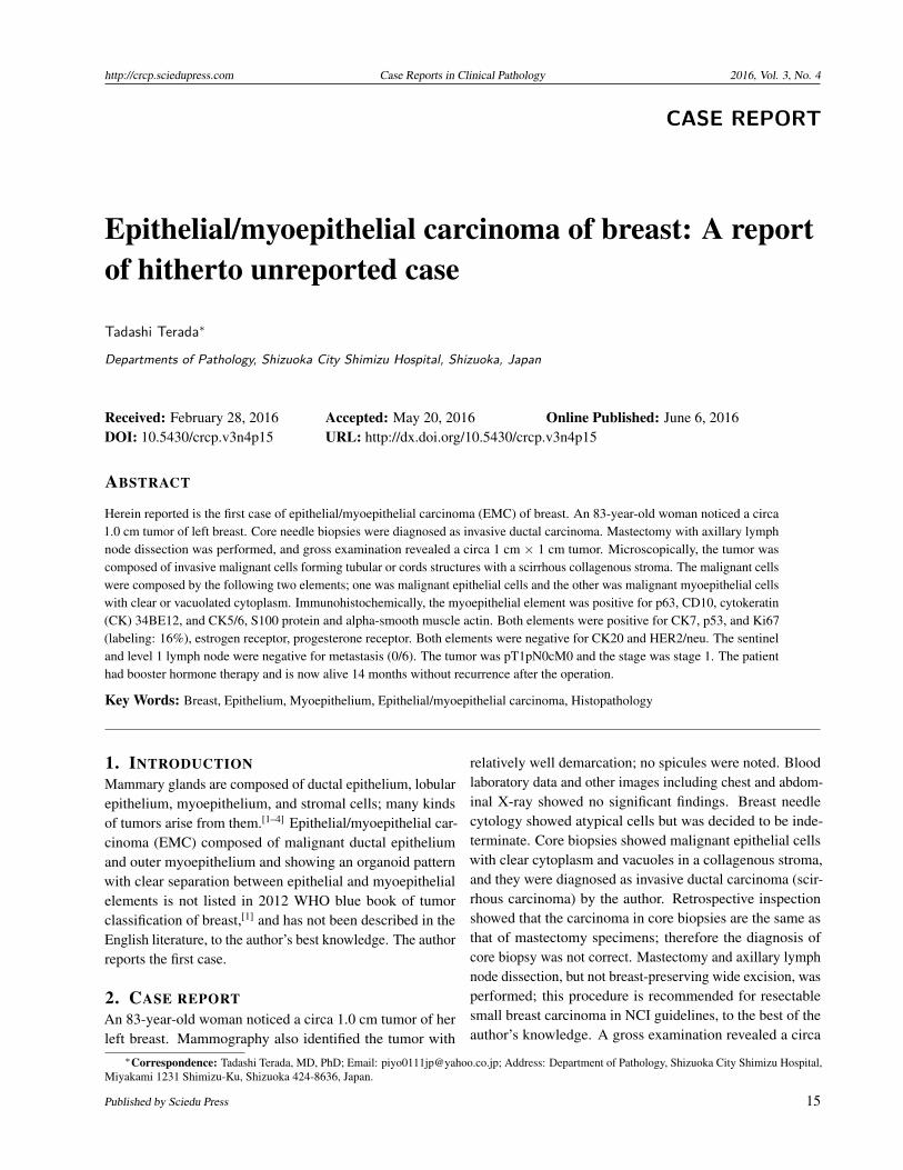
1 cm × 1 cm solid, rigid tumor (see Figure 1). No re-construction surgery of the breast was carried out. Mas-tectomy without re-construction was selected because of theold age of the patient.
Figure 1. Gross features of the tumor (arrows)It measures circa 1cm, and solid with relatively well demarcation
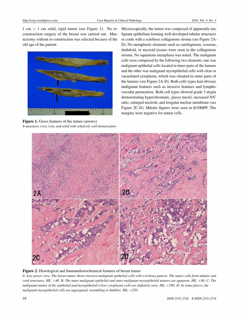
Microscopically, the tumor was composed of apparently ma-lignant epithelium forming well-developed tubular structuresor cords with a scirrhous collagenous stroma (see Figure 2A-D). No metaplastic elements such as cartilaginous, osseous,rhabdoid, or myxoid tissues were seen in the collagenousstroma. No squamous metaplasia was noted. The malignantcells were composed by the following two elements; one wasmalignant epithelial cells located in inner parts of the lumensand the other was malignant myoepithelial cells with clear orvacuolated cytoplasm, which was situated in outer parts ofthe lumens (see Figure 2A-D). Both cells types had obviousmalignant features such as invasive features and lympho-vascular permeation. Both cell types showed grade 3 atypiademonstrating hyperchromatic, glassy nuclei, increased N/Cratio, enlarged nucleoli, and irregular nuclear membrane (seeFigure 2C-D). Mitotic figures were seen in 8/10HPF. Themargins were negative for tumor cells.
Figure 2. Histological and Immunohistochemical features of breast tumorA: Low power view. The breast tumor shows invasive malignant epithelial cells with scirrhous pattern. The tumor cells form tubular andcord structures. HE, ×40. B: The inner malignant epithelial and outer malignant myoepithelial natures are apparent. HE, ×80. C: Themalignant nature of the epithelial and myoepithelial (clear cytoplasm) cells are definitely seen. HE, ×200. D: In some places, themalignant myoepithelial cells are aggregated, resembling to bubbles. HE, ×250.
16 ISSN 2331-2726 E-ISSN 2331-2734

http://crcp.sciedupress.com Case Reports in Clinical Pathology 2016, Vol. 3, No. 4
An Immunohistochemical study was carried out with the useof Envision method.[5–7] The malignant myoepithelial ele-ment was positive for p63 (see Figure 3A), CD10 (see Figure3B), cytokeratin (CK) 34BE12 (see Figure 3C), CK5/6 (seeFigure 3D), S100 protein, and alpha-smooth muscle actin(see Figure 3E). In contrast, the epithelial component wasnegative for these myoepithelium-related antigens.[3, 4, 8–10]
Both elements were positive for CK7, p53 (see Figure 3F),and Ki67 (labeling index: 16%) (see Figure 3G), estrogenreceptor (ER), and progesterone receptor (PgR). Both el-ements were negative for CK20 and HER2/neu. The pat-
tern of companion diagnosis was ER = 8, PgR = 6, HER= 0, and Ki67 labeling index = 16%; therefore the SaintGallen pattern[11] was Luminal A, which is suitable for anti-ER therapy (Tamoxifen). The sentinel and level 1 lymphnodes were negative for metastasis (0/6). The tumor waspT1pN0cM0 and the stage was stage 1. The patient hadbooster hormone therapy (Tamoxifen: 20 mg/day × 40= 800 mg in total) because of positive ER and PgR, andis now alive without recurrence 14 months after the opera-tion.
Figure 3. Immunohistochremically, the myoepithelial element is positive for p63 (A), CD10 (B), CK34BE12 (C), CK5/6(D), alpha-smooth muscle antigen (E), p53 (diffuse) (F), and Ki67 (labeling index = 14%) (G). A-G: ×200.
3. DISCUSSION
The pathological diagnosis of the tumor made by the authorwas EMC. No description of EMC is present in the updatedWHO 2012 blue book of breast tumors, nor in the Englishliterature. Therefore, the current tumor should be differen-tiated from mimics of EMC. It is obvious that the presenttumor is made up of epithelial cells and myoepithelial cells.The inner location in the tumor glands of the epithelial cells
and the negative myoepithelial markers (smooth muscle anti-gen, S100 protein, CK34BE12, CK5/6, CD10, and p63) inthe epithelial cells suggest that the inner epithelial cells areductal cells not myoepithelial cells. In general, the outerlocation in the tumor glands, clear cell changes, bubbly ap-pearances, and nuclear hyperchromasia were histologicalfeatures of myoepithelial cells; therefore the outer cells ofthe tumor glands of the current tumor are truly myoepithelial
Published by Sciedu Press 17

http://crcp.sciedupress.com Case Reports in Clinical Pathology 2016, Vol. 3, No. 4
cells. In addition, they were positive for various myoepithe-lial antigens. Thus, the tumor is composed of epithelial andmyoepithelial cells. Both the epithelial cells showed malig-nant features such as invasive growth, vascular permeation,nuclear hyperchromasia, increased N/C ratio, prominent nu-cleoli, high mitotic activity, increased Ki67 labeling index,and diffuse p53 expression. The most latter suggest p53gene mutation.[12] Thus, it is malignant tumor composed ofepithelial and myoepithelial cells. It is most problematic toterm this tumor. The author labeled this tumor as EMC. Thisis largely because the tumor consisted of malignant epithelialcells and malignant myoepithelial cells, whose separationis clear and organoid with inner epithelial cells and outermyoepithelial cells in tumor tubules and cords. If the twoepithelial cells were much more mixed up, the author hadtermed it as malignant adeno-myoepithelioma[3] or breastcarcinoma with mixed ductal and myoepithelial differentia-tion or myoepithelial cell-rich carcinoma after Coiny et al.[13]
The present tumor is not adenoid cystic carcinoma becauseof the lack of cribriform pattern.[10]
In 2012 WHO blue book, the classification of breast tumorsincludes epithelial-myoepithelial tumors which in turn con-sists of adeno-myoepithelioma with carcinoma and adenoidcystic carcinoma (ACC).[14] The former is benign adeno-myoepithelioma with malignant carcinomatous focus, andthe latter is composed of malignant epithelial cells and ma-lignant myoepithelial cells both forming a characteristic crib-riform pattern. The current tumor is composed entirelyof carcinoma cells and lacked element of benign adeno-myoepithelioma, thus being different from the former. Thetumor did not present the cribriform pattern, thus being dif-ferent from ACC.
In 2004, Coiny et al[13] reported 6 cases of breast carcinomawith dual differentiation into epithelial cells and myoepithe-lial cells, and they termed these “breast carcinomas withmixed ductal and myoepithelial differentiation”. The tu-mors were characterized by high nuclear grade (Bloom andRichardson grade 3), solid/nodular/sheet-like tumor cell ar-rangements, and rarity of duct formation. In their four cases,segregation between epithelial cells and myoepithelial cellsare much more obscure than the current case. Immunohis-tochemically, all cases showed strong positivity for CK14,calponin, smooth actin, and muscle specific antigen. Epithe-lial membrane antigen and CK8 were positive in a similarproportion of cells. Of interest, foci of metaplastic carcinomawere seen in three cases (3/6: 50%). Their term is not listedin the current 2012 WHO blue book. The preset case seemssomewhat different from their 6 cases.[13]
The present case should be differentiated from metaplastic
carcinoma. In 2012 WHO blue book of breast tumors,[15]
metaplastic carcinoma is defined as a group of neoplasmscharacterized by differentiations of the neoplastic epitheliuminto squamous cells and/or mesenchymal-looking elementsincluding spindle, chondroid, osseous, and rhabdomyoidcells. In the current tumor, there were no areas of squamousdifferentiation and no areas of these mesenchymal differen-tiation; therefore the tumor is not metaplastic carcinoma ofbreast. Myoepithelial carcinoma is listed in one category ofWHO 2012 blue book.[15] It is a pure carcinoma composedonly of malignant myoepithelial cells; therefore the currentcase is not myoepithelial carcinoma.
The clinical behaviors of carcinoma composed of epithelialand myoepithelial cells are obscure because of their rarity.Coyne et al[13] reported the clinical course of 6 cases ofbreast carcinoma with dual differentiations into epithelialand myoepithelial cells. According to them, one patient died23 months following diagnosis with metastatic carcinoma,another patient died of unrelated disease and four patientsare alive with follow-up ranging from 18 months to 25 years.The biological behaviors of the neoplasms are not clear. Ac-cumulation of much more cases are needed. In the presentcase, the tumor is small (1 cm, pT1), no lymph node metas-tasis was seen (pN0) and clinically no metastasis was found(cM0), thus being at stage 1. Because no data of treatmentof EMC were available, the patient was treated using con-ventional NCI recommended regimen, in this case partialmastectomy and dissection of sentinel and level 1 lymphnodes. After confirming negativity of cut margins, boosteradministration of Tamoxifen was added because the stainingpattern was Saint Gallen Luminal A.
The myoepithelial cells are among interesting cell typeswhich are distributed in mammary glands (a kind of apoc-rine glands), cutaneous sweat and apocrine glands, and sali-vary glands (major and minor). Prostatic gland has similarcells called basal cells. This kind of cell is located outsidethe glandular epithelium of above-mentioned glands andhas both contractile and supportive functions. They con-tain actin filaments and other characteristic proteins such asp63, high-molecular weight CK including CK14, CK5, CK6and CK33BE12, calponin, caldesmon, alpha-smooth muscleactin, S100 protein, and others. Thus, they are highlightedby immunohistochemistry for these antigens. These cellsalso can produce extracellular matrix proteins such as carti-lage, bone, myxoid tissue, collagens, glycose-amino-glycan,proteoglycan and others, and can transform into specificcells producing these matrix proteins. This phenomenonis typically seen in pleomorphic adenoma (mixed tumor) ofsalivary glands in which cartilage and osseous tissue made byneoplastic myometrium are recognizable. In breast tumors
18 ISSN 2331-2726 E-ISSN 2331-2734

http://crcp.sciedupress.com Case Reports in Clinical Pathology 2016, Vol. 3, No. 4
also, cells of myoepithelial carcinoma and breast carcinomawith dual epithelial and myoepithelial differentiations canproduce such extracellular matrix proteins. At present, thebiology of myoepithelium is not extensively studied. Further
studies remains to be done.
CONFLICTS OF INTEREST DISCLOSUREThe author declares no conflicts of interest.
REFERENCES[1] Tan PH, Ellis IO. Myoepithelial and epithelial-myoepithelial, mes-
enchymal and fibroepithelial breast lesions: updates from the WHOClassification of Tumours of the Breast 2012. J Clin Pathol. 2013; 66:465-70. PMid: 23533258. http://dx.doi.org/10.1136/jclinpath-2012-201078
[2] Terada T. Clear cell variant of ductal carcinoma in situ of the breast.Breast J. 2012; 18: 279-80. PMid: 22530727. http://dx.doi.org/10.1111/j.1524-4741.2012.01237.x
[3] Terada T. Malignant myoepithelioma of the breast. Pathol Int. 2011;61: 99-103. PMid: 21255187. http://dx.doi.org/10.1111/j.1440-1827.2010.02638.x
[4] Terada T. Ductal adenoma of the breast: immunohistochemistry oftwo cases. Pathol Int. 2008; 58: 801-5. PMid: 19067857. http://dx.doi.org/10.1111/j.1440-1827.2008.02315.x
[5] Terada T, Kawaguchi M, Furukawa K, et al. Minute mixed ductal-endocrine carcinoma of the pancreas with predominant intraduc-tal growth. Pathol Int. 2002; 52: 740-6. PMid: 12685552. http://dx.doi.org/10.1046/j.1440-1827.2002.01416.x
[6] Terada T, Taniguchi M. Intraductal oncocytic papillary neoplasmof the liver. Pathol Int. 2004; 54: 116-23. PMid: 14720143. http://dx.doi.org/10.1111/j.1440-1827.2004.01594.x
[7] Terada T, Kawaguchi M. Primary clear cell adenocarcinoma of theperitoneum. Tohoku J Exp Med. 2005; 206: 271-5. PMid: 15942157.http://dx.doi.org/10.1620/tjem.206.271
[8] Terada T. Myoepithelial carcinoma of pharynx expressing KIT andPDGFRA. Int J Clin Exp Pathol. 2013; 6: 314-7. PMid: 23330018.
[9] Terada T. Primary cutaneous neuroendocrine tumor (atypical carci-noid) expressing KIT and PDGFRA with myoepithelial differentia-
tion: a case report with immunohistochemical and molecular geneticstudies. Int J Clin Exp Pathol. 2013; 6: 802-9. PMid: 23573331.
[10] Terada T. Pigmented adenoid cystic carcinoma of the ear skin arisingfrom the epidermis: a case report with immunohistochemical studies.Int J Clin Exp Pathol. 2012; 5: 254-9. PMid: 22558481.
[11] Goldhirsch A, Ingle JN, Gelber RD, et al. Thresholds for therapies:highlights of the St Gallen International Expert Consensus on theprimary therapy of early breast cancer 2009. Ann Oncol. 2009; 20:1319-29. PMid: 19535820. http://dx.doi.org/10.1093/annonc/mdp322
[12] Terada T, Shimizu K, Izumi R, et al. Methods in pathology. p53expression in formalin-fixed, paraffin-embedded archival specimensof intrahepatic cholangiocarcinoma: retrieval of p53 antigenicity bymicrowave oven heating of tissue sections. Mod Pathol. 1994; 7:249-52. PMid: 8008749.
[13] Coyne JD, Dervan PA, Barr L. High-grade carcinomas of thebreast showing patterns of mixed ductal and myoepithelial dif-ferentiation (including myoepithelial cell-rich carcinoma of thebreast. Histopathology. 2004; 44: 580-4. PMid: 15186273. http://dx.doi.org/10.1111/j.1365-2559.2004.01891.x
[14] Schmitt F, Tan PH, Dabbs D, et al. Myoepithelial and epithelial-myoepithelial lesions. In Lakhani SR, Ellis IO, Schnitt SJ, Tan PH,van de Vijver, M.J (Eds) WHO classification of Tumours of theBreast, Fourth Edition. IARC. Lyon: 2012. p120-3.
[15] Reis-Filho JS, Lakhani SR, Gobbi H, et al. Metaplastic carcinoma.In Lakhani SR, Ellis IO, Schnitt SJ, Tan PH, van de Vijver, M.J (Eds)WHO classification of Tumours of the Breast, Fourth Edition. Lyon:IARC; 2012. p48-51.
Published by Sciedu Press 19