Epithelial–mesenchymal transition confers resistance to ... · PDF filePostgraduate...
10
ORIGINAL ARTICLE Epithelial–mesenchymal transition confers resistance to selective FGFR inhibitors in SNU-16 gastric cancer cells Paulina Grygielewicz • Barbara Dymek • Anna Bujak • Pawel Gunerka • Aleksandra Stanczak • Monika Lamparska-Przybysz • Maciej Wieczorek • Karolina Dzwonek • Daria Zdzalik Received: 24 June 2014 / Accepted: 1 November 2014 / Published online: 19 November 2014 Ó The Author(s) 2014. This article is published with open access at Springerlink.com Abstract Background Up to 10 % of primary gastric cancers are characterized by FGFR2 amplification, and fibroblast growth factor receptor (FGFR) inhibitors may represent therapeutic agents for patients with these malignancies. However, long-term benefits of the treatment might be limited owing to the occurrence of drug resistance. Methods To investigate the mechanisms of resistance to selective FGFR inhibitors, we established three FGFR2- amplified SNU-16 gastric cancer cell lines resistant to AZD4547, BGJ398, and PD173074, respectively. Results The resistant cell lines (SNU-16R) demonstrated changes characteristic of epithelial-to-mesenchymal tran- sition (EMT). In addition, they displayed loss of expression of FGFR2 and other tyrosine kinase receptors concurrent with activation of downstream signaling proteins and upregulation of the transforming growth factor b (TGF-b) level. However, treatment of parental SNU-16 cells with TGF-b 1 did not evoke EMT, and pharmacological inhibi- tion of TGF-b receptor I was not sufficient to reverse EMT changes in the resistant cells. Finally, we showed that the SNU-16R cell lines were sensitive to the human epidermal growth factor receptor 2 inhibitor mubritinib and the heat shock protein 90 inhibitor AUY922. Conclusion In conclusion, we provide experimental evi- dence that EMT-mediated resistance might emerge in gastric cancer patients following treatment with FGFR inhibitors, and mubritinib or AUY922 treatment may be an alternative therapeutic strategy for these patients. Keywords Fibroblast growth factor receptor Á Epithelial– mesenchymal transition Á Drug resistance Á Fibroblast growth factor receptor inhibitor Á AZD4547 Á BGJ398 Introduction The fibroblast growth factor (FGF) receptors (FGFRs) constitute one of the most extensively studied novel ther- apeutic targets in the field of anticancer drug development. The FGFR family comprises four members (FGFR1– FGFR4) which serve as high-affinity receptors for 22 dif- ferent FGF ligands [1]. The FGF/FGFR pathway plays an important role in a myriad of key cellular processes, such as cell proliferation, differentiation, migration, survival, and angiogenesis [1]. Aberrant activation of FGFR kinases owing to gene amplification, chromosomal translocation, or gain-of-function mutation has been implicated in the pathogenesis of a variety of human cancers, including gastric cancer [2], breast cancer [3], bladder cancer [4], Electronic supplementary material The online version of this article (doi:10.1007/s10120-014-0444-1) contains supplementary material, which is available to authorized users. P. Grygielewicz Á B. Dymek Á A. Bujak Á P. Gunerka Á A. Stanczak Á M. Lamparska-Przybysz Á M. Wieczorek Á K. Dzwonek Á D. Zdzalik Innovative Drugs R&D Department, Celon Pharma Inc., Mokra 41a, 05-092 Lomianki/Kielpin, Poland P. Grygielewicz (&) Á A. Bujak Postgraduate School of Molecular Medicine, Zwirki i Wigury 61, 02-091 Warsaw, Poland e-mail: [email protected] P. Gunerka Department of Medical Biotechnology, Medical University of Lodz, Al. Kosciuszki 4, 90-419 Lodz, Poland K. Dzwonek Department of Immunology, Center for Biostructure Research, Medical University of Warsaw, Banacha 1a, F Building, 02-097 Warsaw, Poland 123 Gastric Cancer (2016) 19:53–62 DOI 10.1007/s10120-014-0444-1
Transcript of Epithelial–mesenchymal transition confers resistance to ... · PDF filePostgraduate...

ORIGINAL ARTICLE
Epithelial–mesenchymal transition confers resistance to selectiveFGFR inhibitors in SNU-16 gastric cancer cells
Paulina Grygielewicz • Barbara Dymek • Anna Bujak • Pawel Gunerka •
Aleksandra Stanczak • Monika Lamparska-Przybysz • Maciej Wieczorek •
Karolina Dzwonek • Daria Zdzalik
Received: 24 June 2014 / Accepted: 1 November 2014 / Published online: 19 November 2014
� The Author(s) 2014. This article is published with open access at Springerlink.com
Abstract
Background Up to 10 % of primary gastric cancers are
characterized by FGFR2 amplification, and fibroblast
growth factor receptor (FGFR) inhibitors may represent
therapeutic agents for patients with these malignancies.
However, long-term benefits of the treatment might be
limited owing to the occurrence of drug resistance.
Methods To investigate the mechanisms of resistance to
selective FGFR inhibitors, we established three FGFR2-
amplified SNU-16 gastric cancer cell lines resistant to
AZD4547, BGJ398, and PD173074, respectively.
Results The resistant cell lines (SNU-16R) demonstrated
changes characteristic of epithelial-to-mesenchymal tran-
sition (EMT). In addition, they displayed loss of expression
of FGFR2 and other tyrosine kinase receptors concurrent
with activation of downstream signaling proteins and
upregulation of the transforming growth factor b (TGF-b)
level. However, treatment of parental SNU-16 cells with
TGF-b1 did not evoke EMT, and pharmacological inhibi-
tion of TGF-b receptor I was not sufficient to reverse EMT
changes in the resistant cells. Finally, we showed that the
SNU-16R cell lines were sensitive to the human epidermal
growth factor receptor 2 inhibitor mubritinib and the heat
shock protein 90 inhibitor AUY922.
Conclusion In conclusion, we provide experimental evi-
dence that EMT-mediated resistance might emerge in
gastric cancer patients following treatment with FGFR
inhibitors, and mubritinib or AUY922 treatment may be an
alternative therapeutic strategy for these patients.
Keywords Fibroblast growth factor receptor � Epithelial–
mesenchymal transition � Drug resistance � Fibroblast
growth factor receptor inhibitor � AZD4547 � BGJ398
Introduction
The fibroblast growth factor (FGF) receptors (FGFRs)
constitute one of the most extensively studied novel ther-
apeutic targets in the field of anticancer drug development.
The FGFR family comprises four members (FGFR1–
FGFR4) which serve as high-affinity receptors for 22 dif-
ferent FGF ligands [1]. The FGF/FGFR pathway plays an
important role in a myriad of key cellular processes, such
as cell proliferation, differentiation, migration, survival,
and angiogenesis [1]. Aberrant activation of FGFR kinases
owing to gene amplification, chromosomal translocation,
or gain-of-function mutation has been implicated in the
pathogenesis of a variety of human cancers, including
gastric cancer [2], breast cancer [3], bladder cancer [4],
Electronic supplementary material The online version of thisarticle (doi:10.1007/s10120-014-0444-1) contains supplementarymaterial, which is available to authorized users.
P. Grygielewicz � B. Dymek � A. Bujak � P. Gunerka �A. Stanczak � M. Lamparska-Przybysz � M. Wieczorek �K. Dzwonek � D. Zdzalik
Innovative Drugs R&D Department, Celon Pharma Inc.,
Mokra 41a, 05-092 Lomianki/Kielpin, Poland
P. Grygielewicz (&) � A. Bujak
Postgraduate School of Molecular Medicine,
Zwirki i Wigury 61, 02-091 Warsaw, Poland
e-mail: [email protected]
P. Gunerka
Department of Medical Biotechnology, Medical
University of Lodz, Al. Kosciuszki 4, 90-419 Łodz, Poland
K. Dzwonek
Department of Immunology, Center for Biostructure Research,
Medical University of Warsaw, Banacha 1a, F Building,
02-097 Warsaw, Poland
123
Gastric Cancer (2016) 19:53–62
DOI 10.1007/s10120-014-0444-1

endometrial cancer [5], squamous cell lung cancer [6],
multiple myeloma [7], and glioblastoma [8].
Since aberrantly activated FGFR kinases serve as an
oncogenic ‘‘driver,’’ a great number of FGFR inhibitors are
currently in clinical development. However, the vast
majority of them are multikinase inhibitors such as dovi-
tinib, nintedanib, or ponatinib [9]. To date, no selective
small-molecule FGFR inhibitor has been approved for
clinical use. The first selective FGFR inhibitor developed
was PD173074, which, despite its high selectivity and
cellular activity, has never entered clinical use [10]. At
present, only a few selective FGFR inhibitors are under
clinical investigation, and the two in the most advanced
stage are AZD4547 and BGJ398, developed by AstraZen-
eca and Novartis, respectively [11, 12].
According to the World Health Organization, gastric
cancer accounts for nearly one in ten of all deaths of cancer
patients [13]. Mean survival for patients with stage IV
metastatic gastric cancer is only 10 months [14]. Given
this, there is an urgent need to improve gastric cancer
therapy. FGFR2 amplification occurs in 3–10 % of primary
gastric cancers, and has been reported to be more frequent
in the undifferentiated diffuse subtype [2]. The effective-
ness of FGFR inhibitors against gastric cancer in vitro and
in vivo suggests that the use of these compounds may be a
therapeutic strategy for the selected group of patients with
FGFR2-amplified gastric cancer [15, 16]. However, from
experience with other kinase inhibitors used in clinical
practice, there is a high probability that resistance will
develop in patients following FGFR inhibitor treatment.
To date, there is a lack of clinical data concerning drug
resistance in FGFR-inhibitor-based therapy. Nevertheless,
recently published results from in vitro models cast some light
on the possible mechanisms of resistance to FGFR inhibitors.
It has been shown that the heterozygous gatekeeper mutation
FGFR3 V555M emerged as a mechanism of acquired resis-
tance to the selective FGFR inhibitor AZ12908010 in KMS-
11 myeloma cells, and the resistant cells were cross-resistant
to other FGFR inhibitors, AZD4547 and PD173074 [17].
Additionally, several activating mutations were identified in
FGFR2-expressing Ba/F3 cells treated with high concentra-
tions of dovitinib. Recognized mutations conferred cross-
resistance to the selective FGFR inhibitor PD173074 but not
to the multikinase inhibitor ponatinib, whose inhibitory
activity was affected only by the V565I gatekeeper mutation
[18]. In addition to secondary mutations in the kinase domain,
resistance to kinase inhibitors may also occur through acti-
vation of alternative signaling pathways. With use of a high-
throughput ‘‘secretome’’ screening platform, it has been
established that ligand-mediated activation of compensatory
kinases such as epidermal growth factor receptor (EGFR),
human EGFR (HER), or MET may compensate for loss of
FGFR kinase activity [19]. Additionally, using parallel RNA
interference genetic screens, Herrera–Abreu et al. [20] dis-
covered that EGFR is a key mediator of resistance in FGFR3-
dependent cancer cells.
Given this, to predict the mechanisms of acquired
resistance which may emerge in patients with FGFR2-
dependent gastric cancer following treatment with selective
FGFR inhibitors, we generated three SNU-16 cell lines
resistant (SNU-16R) to AZD4547, BGJ398, and PD173074,
respectively. It has been well established that SNU-16 cells
harbor FGFR2 amplification, and thereby they are highly
sensitive to FGFR inhibitors in vitro and in vivo [2, 21–23].
However, long-term exposure to increasing concentrations
of selected inhibitors induced resistance accompanied by
loss of FGFR2 expression and epithelial-to-mesenchymal
transition (EMT), a phenomenon that has previously been
shown to be involved in resistance to other kinase inhibitors
[24–27]. Finally, on the basis of our results, we propose
possible therapeutic strategies to overcome EMT-mediated
resistance to FGFR inhibitors.
Materials and methods
Compounds and cell lines
AT9283, AZD4547, AZD6244, BGJ398, crizotinib, pictilisib,
ibrutinib, PD173074, ruxolitinib, TAE684, and gandotinib
were all purchased from Selleck Chemicals, AUY922, dovi-
tinib, everolimus, gefitinib, mubritinib, saracatinib, sunitinib,
and ZSTK474 were from LC Laboratories, CH5424802 was
from Active Biochem, SB525334 was from Tocris, and fed-
ratinib was from Axon Medchem. The human gastric cancer
cell line SNU-16 was obtained from ATCC and was cultured
in RPMI 1640 medium supplemented with 10 % fetal bovine
serum according to the manufacturer’s instructions.
Generation of AZD4547-, BGJ398-, and PD173074-
resistant SNU-16 cells
To generate drug-resistant cells, SNU-16 cells were
exposed to increasing concentrations of AZD4547,
BGJ398, or PD173074 for 1 month. The concentration of
each inhibitor was doubled at every second passage to a
final concentration of 0.6 lM for AZD4547 and 1.2 lM for
BGJ398 and PD173074. The resistant cells were cultured
until they again had growth kinetics similar to the growth
kinetics of untreated parental cells.
Treatment of parental SNU-16 cells with transforming
growth factor b1
SNU-16 cells were cultured in a medium containing human
recombinant transforming growth factor (TGF)-b1 (Merck
54 P. Grygielewicz et al.
123

Millipore) in a final concentration of 0.5 ng/ml. Fresh
TGF-b1 was added every 72 h for 3 weeks.
Cell viability assays
For viability tests, 6 9 103 cells per well were seeded in
96-well plates and exposed to increasing doses of the tested
compounds. After 72 h of drug treatment, cell viability was
determined by the ATPlite assay (Perkin Elmer) according
to the manufacturer’s instructions.
Half maximal inhibitory concentrations (IC50) of mub-
ritinib were calculated with GraphPad Prism using the
sigmoid dose–response function. All experiments were
performed at least twice.
Immunoblot analysis
SNU-16 parental and resistant cells were seeded in six-well
plates at a density of 0.5 9 106 cells per milliliter in an
inhibitor-free medium. After 24 h, cells were lysed and the
level of proteins was examined by Western blotting
according to the protocols provided by the antibody sup-
pliers. The antibodies against MET, EGFR, HER2, extra-
cellular-signal-regulated kinase (ERK), phosphorylated
AKT, phosphorylated EGFR, phosphorylated ERK
(pERK), phosphorylated FGFR, phosphorylated FGFR
substrate, and phosphorylated signal transducer and acti-
vator of transcription (STAT3) were purchased from Cell
Signaling Technology, b-tubulin and AKT were from
Millipore, E-cadherin, STAT3, and b-catenin were from
BD Biosciences, FGFR2 was from Abnova, and vimentin
was from Calbiochem. All experiments were performed at
least twice.
Phosphoprotein array analysis
SNU-16 parental and resistant cells were seeded in T25
bottles at a density of 1.5 9 106 cells per bottle in an
inhibitor-free medium. The assay was conducted in
accordance with the commercial protocol of the PathScan�
receptor tyrosine kinase signaling antibody array kit (Cell
Signaling Technology).
Results
SNU-16R cell lines exhibited a substantial decrease
in sensitivity to all FGFR inhibitors tested
To generate SNU-16 gastric cancer cell lines resistant to
AZD4547 (AZDR), BGJ398 (BGJR), and PD173074
(PDR), SNU-16 cells were cultured with increasing con-
centrations of the respective FGFR inhibitor. In the
viability assay, the parental cell line was shown to be
sensitive to FGFR inhibitors, with 50 % inhibition of cell
viability at a concentration of approximately 150 nM for
AZD4547 and BGJ398, and 380 nM for PD173074
(Fig. 1). The observed growth inhibition was dose inde-
pendent in range from 0.153 to 6 lM, and the viability of
SNU-16 cells was completely inhibited only at high
micromolar concentrations of the FGFR inhibitors. Given
this, we were unable to fit sigmoid dose–response curves to
the data and calculate true IC50 values. Consistent with the
ability of resistant cells to grow at high concentrations of a
particular FGFR inhibitor, AZDR, BGJR, and PDR cells
were highly resistant to AZD4547, BGJ398, or PD173074,
respectively, with an IC50 of approximately 10 lM
(Fig. 1). Moreover, each resistant cell line exhibited cross-
resistance to all FGFR inhibitors tested, and the resistance
to AZD4547, BGJ398, and PD173074 was comparable
among all resistant cell lines. Altogether, the substantial
decrease in sensitivity of SNU-16R cell lines to FGFR
inhibitors observed in the cell viability assay confirmed the
emergence of drug resistance in SNU-16R cells.
SNU-16R cells lose their dependency on FGFR2
signaling and display alterations in other signaling
pathways
To examine the mechanism of resistance to FGFR inhibi-
tors in SNU-16R cell lines, firstly we determined the status
of FGFR2 signaling by immunoblot analysis. It revealed
that the level of FGFR2 was undetectable in resistant cells
in contrast to parental ones (Fig. 2a). Consistent with these
findings, the decrease of phosphorylation of phosphory-
lated FGFR substrate 2, a direct downstream target of
FGFR2, was also observed (Fig. 2a). Given this, all SNU-
16R cell lines became independent of FGFR2 signaling.
However, despite loss of FGFR2 expression, phosphory-
lation of downstream proteins such as ERK, AKT, and
STAT3 was upregulated in AZDR, BGJR, and PDR cells
(Fig. 2a). This suggested that activation of alternative
pathways could be responsible for the observed increase in
phosphorylation status of downstream proteins.
To verify whether acquired resistance of AZDR, BGJR,
and PDR cells is the effect of the induction of an alterna-
tive signaling pathway, we used the slide-based PathScan�
receptor tyrosine kinase signaling antibody array kit.
Phosphoprotein array analysis confirmed that all SNU-16R
cell lines exhibit alterations in diverse signaling pathways
in comparison with parental SNU-16 cells. We found a
slight increase in the phosphorylation status of SRC and
RET concomitantly with downregulation of phosphory-
lated MET and phosphorylated HER3 in all three SNU-16R
cell lines compared with wild-type SNU-16 cells (Fig. 2b).
Additional immunoblot analysis showed that the decrease
EMT confers resistance to FGFR-TKI 55
123

Fig. 1 Sensitivity of parental
SNU-16 and resistant SNU-16
(SNU-16R) cells to selective
fibroblast growth factor receptor
(FGFR) inhibitors. Cells were
treated with increasing
concentrations of AZD4547,
BGJ398, or PD173074 for 72 h,
and cell viability was measured
with the ATPlite assay. Each
data point represents the mean
of two independent triplicate
measurements, and error bars
indicate the standard deviation.
AZDR SNU-16 cell line
resistant to AZD4547, BGJR
SNU-16 cell line resistant to
BGJ398, PDR SNU-16 cell line
resistant to PD173074
Fig. 2 Alterations in signaling pathways in resistant SNU-16 (SNU-
16R) cell lines. Parental SNU-16 and SNU-16R cells were incubated
in inhibitor-free medium for 24 h, and then cell lysates from each cell
line were subjected to immunoblot analysis. A representative result
from one of two separate experiments is shown. a Immunoblot
analysis of FGFR-dependent signaling and downstream proteins.
b Analysis of phosphorylation status of key signaling molecules
assessed by the PathScan� receptor tyrosine kinase signaling antibody
array. c Immunoblot analysis of the expression of the receptors
epidermal growth factor receptor (EGFR) and MET. AZDR SNU-16
cell line resistant to AZD4547, BGJR SNU-16 cell line resistant to
BGJ398, ERK extracellular-signal-regulated kinase, FGFR fibroblast
growth factor receptor, HER2 human epidermal growth factor
receptor 2, HER3 human epidermal growth factor receptor 3, PDR
SNU-16 cell line resistant to PD173074, pERK phosphorylated
extracellular-signal-regulated kinase, pFGFR phosphorylated fibro-
blast growth factor receptor, pFRS2 phosphorylated fibroblast growth
factor receptor substrate 2, pSTAT3 phosphorylated signal transducer
and activator of transcription 3, STAT3 signal transducer and activator
of transcription 3
56 P. Grygielewicz et al.
123

in the phosphorylated MET level was correlated with loss
of total MET protein expression, and loss of other trans-
membrane proteins such as EGFR also occurred (Fig. 2c).
EMT-like changes are induced in SNU-16R cell lines
We observed a remarkable morphologic difference
between parental SNU-16 and SNU-16R cell lines. As
shown in Fig. 3a, SNU-16R cells acquired a fibroblast-
like, spindle-shaped morphology, suggesting that resistant
cells might have undergone EMT-like changes. Recently,
growing evidence has suggested that EMT is implicated
in resistance to kinase-inhibitor-based therapy [24–27].
Since EMT is characterized by downregulation of epi-
thelial phenotype markers such as E-cadherin and
upregulation of mesenchymal phenotype markers such as
vimentin [28], we tested the level of these markers in
AZDR, BGJR, and PDR cells. This analysis revealed that
E-cadherin expression was significantly reduced and
vimentin expression was upregulated in all SNU-16R cell
lines when compared with the parental SNU-16 cells
(Fig. 3b). Additionally, we observed a substantial
decrease in total b-catenin level in resistant cells, which
was probably the result of acquisition of a mesenchymal
phenotype associated with loss of E-cadherin/b-catenin–
based adherens junctions (Fig. 3b). However, we cannot
exclude intranuclear accumulation of b-catenin, a well-
known EMT marker, in AZDR, BGJR, and PDR cells
[29, 30]. Taken together, our findings indicate that EMT
characteristics accompanied the acquisition of resistance
to AZD4547, BGJ398, and PD173074.
SNU-16R cell lines have enhanced expression of
TGF-b
It has previously been shown that TGF-b signaling plays an
important role in EMT, and treatment of cells with TGF-b1
can trigger EMT changes [24]. To verify the involvement
of TGF-b signaling in EMT changes in SNU-16R cell
lines, we analyzed the expression of TGF-b and activation
of its downstream molecules SMAD2 and SMAD3. As
shown in Fig. 3b, TGF-b expression was upregulated in all
Fig. 3 Induction of epithelial-to-mesenchymal transition (EMT) in
resistant SNU-16 (SNU-16R) cell lines. a The morphology of parental
SNU-16 cells, SNU-16R cells, and SNU-16 cells treated with
transforming growth factor (TGF) b1 observed under a light
microscope (magnification 9200). b Immunoblot analysis of the
expression of EMT markers and TGF-b1 signaling proteins in parental
SNU-16 cells and SNU-16R cells following incubation in inhibitor-
free medium for 24 h and in parental SNU-16 cells treated with TGF-
b1 for 3 weeks. c Immunoblot analysis of the expression of EMT
marker proteins in parental SNU-16 and SNU-16R cell lines
following incubation with the TGF-b receptor I inhibitor SB525334
for 24 h. pSMAD2 phosphorylated SMAD2, pSMAD3 phosphorylated
SMAD3
EMT confers resistance to FGFR-TKI 57
123
resistant SNU-16R cells, and this was accompanied by a
slight increase in the phosphorylation of SMAD2.
SNU-16 cells treated with TGF-b1 do not mimic EMT
changes displayed in resistant cell lines
To evoke EMT in SNU-16 cells and mimic conditions
which occur in in vivo tumor microenvironment [31] as
well as to promote a sustained EMT and an invasive phe-
notype [32], cells were treated with TGF-b1 for 3 weeks.
As shown in Fig. 3b, TGF-b1 treatment induced an
increase in the phosphorylation level of SMAD2 and
SMAD3 (Fig. 3b). However, despite observed activation of
the TGF-b signaling pathway, SNU-16 cells did not
acquire characteristics associated with EMT, following
3 weeks of incubation with TGF-b1 (Fig. 3a, b).
EMT characteristics of SNU-16R cell lines are
not abrogated by inhibition of TGF-b receptor I
Owing to the essential role of TGF-b signaling in induction
and maintaining EMT, TGF-b signaling pathway inhibitors
are able to reverse EMT [33]. Given this, in order to evoke
induction of EMT in SNU-16 cells, we treated SNU-16R
cell lines with SB525334—an inhibitor of activin-receptor-
like kinase 5, an isoform of TGF-b receptor I (TbRI).
However, 24 h incubation with SB525334 did not reverse
EMT since we did not observe an increase in E-cadherin
level and a decrease in vimentin level (Fig. 3c). Addi-
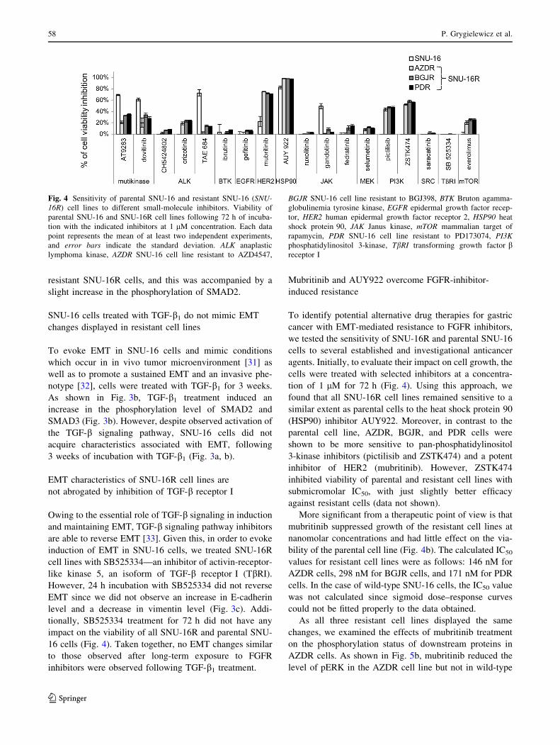
tionally, SB525334 treatment for 72 h did not have any
impact on the viability of all SNU-16R and parental SNU-
16 cells (Fig. 4). Taken together, no EMT changes similar
to those observed after long-term exposure to FGFR
inhibitors were observed following TGF-b1 treatment.
Mubritinib and AUY922 overcome FGFR-inhibitor-
induced resistance
To identify potential alternative drug therapies for gastric
cancer with EMT-mediated resistance to FGFR inhibitors,
we tested the sensitivity of SNU-16R and parental SNU-16
cells to several established and investigational anticancer
agents. Initially, to evaluate their impact on cell growth, the
cells were treated with selected inhibitors at a concentra-
tion of 1 lM for 72 h (Fig. 4). Using this approach, we
found that all SNU-16R cell lines remained sensitive to a
similar extent as parental cells to the heat shock protein 90
(HSP90) inhibitor AUY922. Moreover, in contrast to the
parental cell line, AZDR, BGJR, and PDR cells were
shown to be more sensitive to pan-phosphatidylinositol
3-kinase inhibitors (pictilisib and ZSTK474) and a potent
inhibitor of HER2 (mubritinib). However, ZSTK474
inhibited viability of parental and resistant cell lines with
submicromolar IC50, with just slightly better efficacy
against resistant cells (data not shown).
More significant from a therapeutic point of view is that
mubritinib suppressed growth of the resistant cell lines at
nanomolar concentrations and had little effect on the via-
bility of the parental cell line (Fig. 4b). The calculated IC50
values for resistant cell lines were as follows: 146 nM for
AZDR cells, 298 nM for BGJR cells, and 171 nM for PDR
cells. In the case of wild-type SNU-16 cells, the IC50 value
was not calculated since sigmoid dose–response curves
could not be fitted properly to the data obtained.
As all three resistant cell lines displayed the same
changes, we examined the effects of mubritinib treatment
on the phosphorylation status of downstream proteins in
AZDR cells. As shown in Fig. 5b, mubritinib reduced the
level of pERK in the AZDR cell line but not in wild-type
Fig. 4 Sensitivity of parental SNU-16 and resistant SNU-16 (SNU-
16R) cell lines to different small-molecule inhibitors. Viability of
parental SNU-16 and SNU-16R cell lines following 72 h of incuba-
tion with the indicated inhibitors at 1 lM concentration. Each data
point represents the mean of at least two independent experiments,
and error bars indicate the standard deviation. ALK anaplastic
lymphoma kinase, AZDR SNU-16 cell line resistant to AZD4547,
BGJR SNU-16 cell line resistant to BGJ398, BTK Bruton agamma-
globulinemia tyrosine kinase, EGFR epidermal growth factor recep-
tor, HER2 human epidermal growth factor receptor 2, HSP90 heat
shock protein 90, JAK Janus kinase, mTOR mammalian target of
rapamycin, PDR SNU-16 cell line resistant to PD173074, PI3K
phosphatidylinositol 3-kinase, TbRI transforming growth factor breceptor I
58 P. Grygielewicz et al.
123

SNU-16 cells. These data suggest that mubritinib-mediated
inhibition of SNU-16R cell proliferation might be the
effect of suppression of ERK phosphorylation. Addition-
ally, the activation of HER2 was markedly reduced in
AZDR cells in comparison with wild-type SNU-16 cells
(Figs. 2b, 5b). Thus, it is possible that the downregulation
of pERK and substantial sensitivity of AZDR cells to
mubritinib treatment are not the effect of HER2 inhibition.
Furthermore, applying the HER2 inhibitor to SNU-16R cell
lines for 48 h and 72 h did not reverse EMT since we did
not observe an increase in E-cadherin level or a decrease in
vimentin level (Fig. S1).
Discussion
To predict the mechanisms of resistance to selective FGFR
inhibitors, we established three resistant cell lines—AZDR,
BGJR, and PDR—by long-term exposure of the human
gastric cancer cell line SNU-16 to AZD4547, BGJ398, and
PD173074, respectively. Compared with the parental SNU-
16 cells, the resistant sublines generated were less sensitive
to the FGFR inhibitors tested, with approximately two
orders of magnitude higher concentration being needed to
inhibit cell viability by 50 %. Moreover, they displayed
cross-resistance to other FGFR inhibitors. The morpho-
logical changes in resistant cells suggested EMT, which
was further confirmed by a decrease of the level of the
epithelial marker E-cadherin and an increase of the level of
vimentin, a mesenchymal marker. It is important to note
that AZD4547, BGJ398, or PD173074, three different
FGFR inhibitors of diverse selectivity and physicochemical
properties, induced the same EMT changes in SNU-16
cells. This suggests that the resistance described here is
specific to FGFR inhibition and does not depend on the
FGFR inhibitor itself. It is highly probable that the
Fig. 5 Resistant SNU-16
(SNU-16R) cell lines are
sensitive to the human
epidermal growth factor
receptor 2 (HER2) inhibitor
mubritinib. a Cell viability
assay of a parental cell line and
resistant cell lines following
72 h of incubation with
increasing concentrations of
mubritinib. Each data point
represents the mean of two
independent triplicate
measurements, and error bars
indicate the standard deviation.
b Immunoblot analysis of
protein phosphorylation status
in parental SNU-16 cells and
SNU-16 cells resistant to
AZD4547 (ADZR) treated for
2 h with the indicated
concentrations of mubritinib.
BGJR SNU-16 cell line resistant
to BGJ398, ERK extracellular-
signal-regulated kinase, PDR
SNU-16 cell line resistant to
PD173074, pERK
phosphorylated extracellular-
signal-regulated kinase, pHER2
phosphorylated HER2, pSTAT3
phosphorylated signal
transducer and activator of
transcription 3, STAT3 signal
transducer and activator of
transcription 3
EMT confers resistance to FGFR-TKI 59
123

occurrence of the resistance associated with EMT depends
on the cellular context since we did not observe EMT in
UM-UC-14 bladder cell lines with FGFR3 mutation
resistant to AZD4547, BGJ398, and PD173074, respec-
tively (our unpublished data).
EMT refers to a complex molecular and cellular reor-
ganization that in cancer is associated with metastasis, poor
prognosis, and drug resistance. The changes from epithelial
to mesenchymal status have been implicated in resistance
to conventional and targeted anticancer drugs. It was
confirmed that EMT induction confers resistance to EGFR
inhibitors [26, 27], anaplastic lymphoma kinase inhibitors
[34], and the multikinase inhibitor sorafenib [35]. Our
study for the first time demonstrated that EMT is also a
mechanism involved in acquired resistance to selective
FGFR inhibitors in gastric cancer cells. At the time the
manuscript was being written, Wang et al. [36] had dis-
covered that acquired resistance to BGJ398 is characterized
by EMT and a switch in dependency from FGFR to HER2/
HER3 in the FGFR3-dependent bladder cancer cell line
RT112. There are also some reports in the literature con-
cerning the correlation between epithelial/mesenchymal
status and sensitivity to FGFR inhibitors; however, none of
them refer to acquired resistance. The multitarget FGFR
inhibitor dovitinib was shown to be more effective in
bladder cancer cell lines with an epithelial phenotype [37].
Furthermore, PD173074 treatment induced mesenchymal-
to-epithelial transition in head and neck squamous carci-
noma cells [38].
In our SNU-16-based resistance models the activation of
EMT was associated with loss of FGFR2 expression.
FGFR2 has previously been shown to be implicated in
regulation of EMT. It was demonstrated that FGFR2IIIb,
an isoform of FGFR2, occurs mainly in epithelial cells, in
contrast to the FGFR2IIIc isoform, which is expressed in
mesenchymal cells [39]. The switch from FGFR2IIIb to
FGFR2IIIc was confirmed to promote an EMT phenotype
[40]. Finally, loss of FGFR2IIIb was accompanied by loss
of E-cadherin and acquisition of a mesenchymal phenotype
in bladder carcinomas [41]. Our studies revealed that loss
of FGFR2 expression is also implicated in resistance-
associated EMT induction in SNU-16 gastric cancer cells,
in which the FGFR2IIIb isoform dominates [42]. In addi-
tion to loss of FGFR2 expression, we observed reduced
activation/expression of other transmembrane receptors,
such as MET, HER2, HER3, and EGFR. In our FGFR-
inhibitor-induced resistance models, the downregulation of
these receptor kinases was accompanied by activation of
SRC and downstream proteins such as ERK, AKT, and
STAT3. Rho et al. [24] also observed reduced expression
of EGFR, HER3, and MET and significantly enhanced
activation of AKT in a gefitinib-resistant non-small-cell
lung cancer cell line (A549) with EMT characteristics.
However, in contrast to our results, phosphorylation of
ERK was downregulated in that model [24]. The loss of
expression of receptors such as FGFR2, MET, and EGFR
observed here in SNU-16R cell lines suggests that another
pathway is responsible for activation of downstream
proteins.
It was established that TGF-b signaling is a key player
in EMT-related chemotherapy and targeted therapy resis-
tance in a number of malignancies [25, 43, 44]. TGF-bactivates its signaling via binding with high-affinity type II
TGF-b receptor (TbRII), which dimerizes with TbRI,
which facilitates TbRI phosphorylation. The activation of
TbRI leads to the propagation of signaling by the canonical
SMAD-dependent pathway and the noncanonical SMAD-
independent pathway. The mesenchymal features and
increase in phosphorylation level of proteins such as AKT,
ERK, and SRC suggested activation of noncanonical TGF-
b signaling in SNU-16R cell lines [45, 46]. However, we
failed to induce EMT changes in parental SNU-16 cells by
TGF-b1 treatment. The only evidence of the induction of
the TGF-b signaling pathway in the resistant cells was
upregulated TGF-b ligand expression and slight activation
of SMAD2. These differences between TGF-b1-treated
parental SNU-16 cells and SNU-16R cells suggest that, in
addition to TGF-b-activated signaling, other unidentified
pathways might be responsible for induction of an EMT-
like phenotype of SNU-16R cells. A similar conclusion
was drawn by Suda et al. [47], who also failed to prove the
involvement of TGF-b in acquired resistance to erlotinib.
Moreover, applying SB525334—an inhibitor of TbRI—to
SNU-16R cell lines neither reversed EMT nor reduced cell
viability. Our results suggest that the application of a single
inhibitor of one of the isoforms of TbRI is not sufficient to
reverse EMT-like changes in SNU-16R cell lines.
Since we did not observe any impact of the TbRI
inhibitor on the viability of the SNU-16R cell lines, we
decided to test other known anticancer agents in order to
determine an alternative therapeutic strategy for over-
coming EMT-mediated resistance to selective FGFR
inhibitors. We found that SNU-16R cells remained sensi-
tive to the HSP90 inhibitor AUY922. In support of our
results, HSP90 inhibitors effectively suppress growth of
crizotinib-resistant H2228 lung cancer cells with EMT
changes [34]. Kim et al. [34] suggested that the overcom-
ing of resistance by HSP90 inhibitors resulted from
reversal of EMT due to degradation of TbRII and further
the restoration of E-cadherin expression. Finally, we found
that an inhibitor of HER2 can overcome FGFR inhibitor
resistance mediated by the EMT since all the SNU-16R cell
lines, in contrast to parental cells, were highly sensitive to
mubritinib. Moreover, the observed marked sensitivity of
resistant cells to mubritinib suggests that use of this
inhibitor might be an alternative therapeutic strategy for the
60 P. Grygielewicz et al.
123

management of gastric cancer resistant to FGFR inhibitors
owing to EMT. The exact mechanism of mubritinib-med-
iated growth inhibition of the SNU-16R cell lines is
unclear, and it might be the effect of inhibition of targets
other than HER2, since we observed a decrease in activa-
tion of HER2 in SNU-16R cells, in contrast to results
reported by Wang et al. [36]. Furthermore, unlike in the
wild-type SNU-16 cells, mubritinib treatment of AZDR
cells suppressed phosphorylation of ERK. However, fur-
ther investigations are needed to clarify the mechanism of
action of mubritinib in the SNU-16R cells. In conclusion,
we found that EMT was induced as a result of long-term
exposure of a gastric cancer cell line in vitro to selective
FGFR inhibitors. Additionally, EMT induction was asso-
ciated with reduction in expression of FGFR2 and other
transmembrane receptors. On the basis of our study, EMT
should be considered as a possible acquired resistance
mechanism contributing to the decreased efficacy of
FGFR-inhibitor-based therapy in cancer patients. Further-
more, our data suggest that the use of mubritinib or
AUY922 treatment may be an alternative therapeutic
approach for treating cancer patients with EMT-mediated
resistance to FGFR inhibitors.
Grant Support This study was supported by Celon Pharma’s own
funds.
Conflict of interest Paulina Grygielewicz, Barbara Dymek, Anna
Bujak, Pawel Gunerka, and Aleksandra Stanczak are full-time
employees of Celon Pharma. Maciej Wieczorek is Chief Executive
Officer of Celon Pharma, and Monika Lamparska-Przybysz, Karolina
Dzwonek, and Daria Zdzalik were full-time employees of Celon
Pharma during the experimental phase of this work. The authors
declare that they have no conflict of interest.
Open Access This article is distributed under the terms of the
Creative Commons Attribution License which permits any use, dis-
tribution, and reproduction in any medium, provided the original
author(s) and the source are credited.
References
1. Eswarakumar VP, Lax I, Schlessinger J. Cellular signaling by
fibroblast growth factor receptors. Cytokine Growth Factor Rev.
2005;16(2):139–49.
2. Kunii K, et al. FGFR2-amplified gastric cancer cell lines require
FGFR2 and Erbb3 signaling for growth and survival. Cancer Res.
2008;68(7):2340–8.
3. Gru AA, Allred DC. FGFR1 amplification and the progression of
non-invasive to invasive breast cancer. Breast Cancer Res.
2012;14(6):116.
4. Billerey C, et al. Frequent FGFR3 mutations in papillary non-
invasive bladder (pTa) tumors. Am J Pathol. 2001;158(6):1955–9.
5. Pollock PM, et al. Frequent activating FGFR2 mutations in
endometrial carcinomas parallel germline mutations associated
with craniosynostosis and skeletal dysplasia syndromes. Onco-
gene. 2007;26(50):7158–62.
6. Liao RG, et al. Inhibitor-sensitive FGFR2 and FGFR3 mutations
in lung squamous cell carcinoma. Cancer Res. 2013;73(16):
5195–205.
7. Chesi M, et al. Frequent translocation t(4;14)(p16.3;q32.3) in
multiple myeloma is associated with increased expression and
activating mutations of fibroblast growth factor receptor 3. Nat
Genet. 1997;16(3):260–4.
8. Singh D, et al. Transforming fusions of FGFR and TACC genes
in human glioblastoma. Science. 2012;337(6099):1231–5.
9. Dieci MV, et al. Fibroblast growth factor receptor inhibitors as a
cancer treatment: from a biologic rationale to medical perspec-
tives. Cancer Discov. 2013;3(3):264–79.
10. Mohammadi M, et al. Crystal structure of an angiogenesis
inhibitor bound to the FGF receptor tyrosine kinase domain.
EMBO J. 1998;17(20):5896–904.
11. Gavine PR, et al. AZD4547: an orally bioavailable, potent, and
selective inhibitor of the fibroblast growth factor receptor tyrosine
kinase family. Cancer Res. 2012;72(8):2045–56.
12. Guagnano V, et al. Discovery of 3-(2,6-dichloro-3,5-dimethoxy-
phenyl)-1-{6-[4-(4-ethyl-piperazin-1-yl)-phenylamino]-pyrimi-
din-4-yl}-1-methyl-urea (NVP-BGJ398), a potent and selective
inhibitor of the fibroblast growth factor receptor family of
receptor tyrosine kinase. J Med Chem. 2011;54(20):7066–83.
13. Jemal A, et al. Global cancer statistics. CA Cancer J Clin.
2011;61(2):69–90.
14. Roukos DH. Targeting gastric cancer with trastuzumab: new
clinical practice and innovative developments to overcome
resistance. Ann Surg Oncol. 2010;17(1):14–7.
15. Zhao G, et al. A novel, selective inhibitor of fibroblast growth
factor receptors that shows a potent broad spectrum of antitumor
activity in several tumor xenograft models. Mol Cancer Ther.
2011;10(11):2200–10.
16. Guagnano V, et al. FGFR genetic alterations predict for sensi-
tivity to NVP-BGJ398, a selective pan-FGFR inhibitor. Cancer
Discov. 2012;2(12):1118–33.
17. Chell V, et al. Tumour cell responses to new fibroblast growth
factor receptor tyrosine kinase inhibitors and identification of a
gatekeeper mutation in FGFR3 as a mechanism of acquired
resistance. Oncogene. 2013;32(25):3059–70.
18. Byron SA, et al. The N550 K/H mutations in FGFR2 confer
differential resistance to PD173074, dovitinib, and ponatinib
ATP-competitive inhibitors. Neoplasia. 2013;15(8):975–88.
19. Harbinski F, et al. Rescue screens with secreted proteins reveal
compensatory potential of receptor tyrosine kinases in driving
cancer growth. Cancer Discov. 2012;2(10):948–59.
20. Herrera-Abreu MT, et al. Parallel RNA interference screens
identify EGFR activation as an escape mechanism in FGFR3-
mutant cancer. Cancer Discov. 2013;3(9):1058–71.
21. Bar-Am I, et al. Detection of amplified DNA sequences in human
tumor cell lines by fluorescence in situ hybridization. Genes
Chromosomes Cancer. 1992;4(4):314–20.
22. Xie L, et al. FGFR2 gene amplification in gastric cancer predicts
sensitivity to the selective FGFR inhibitor AZD4547. Clin Cancer
Res. 2013;19(9):2572–83.
23. Ku JL, Park JG. Biology of SNU cell lines. Cancer Res Treat.
2005;37(1):1–19.
24. Rho JK, et al. Epithelial to mesenchymal transition derived from
repeated exposure to gefitinib determines the sensitivity to EGFR
inhibitors in A549, a non-small cell lung cancer cell line. Lung
Cancer. 2009;63(2):219–26.
25. Brunen D, et al. TGF-b: an emerging player in drug resistance.
Cell Cycle. 2013;12(18):2960–8.
26. Yauch RL, et al. Epithelial versus mesenchymal phenotype
determines in vitro sensitivity and predicts clinical activity of
erlotinib in lung cancer patients. Clin Cancer Res. 2005;11(24 Pt
1):8686–98.
EMT confers resistance to FGFR-TKI 61
123

27. Thomson S, et al. Epithelial to mesenchymal transition is a
determinant of sensitivity of non-small-cell lung carcinoma cell
lines and xenografts to epidermal growth factor receptor inhibi-
tion. Cancer Res. 2005;65(20):9455–62.
28. Voulgari A, Pintzas A. Epithelial-mesenchymal transition in
cancer metastasis: mechanisms, markers and strategies to over-
come drug resistance in the clinic. Biochim Biophys Acta.
2009;1796(2):75–90.
29. Chaw SY, et al. Epithelial to mesenchymal transition (EMT)
biomarkers – E-cadherin, beta-catenin, APC and vimentin – in
oral squamous cell carcinogenesis and transformation. Oral
Oncol. 2012;48(10):997–1006.
30. Kato N, et al. b-Catenin activation and epithelial-mesenchymal
transition in the pathogenesis of pterygium. Invest Ophthalmol
Vis Sci. 2007;48(4):1511–7.
31. Oft M, et al. TGF-beta1 and Ha-Ras collaborate in modulating
the phenotypic plasticity and invasiveness of epithelial tumor
cells. Genes Dev. 1996;10(19):2462–77.
32. Gal A, et al. Sustained TGF beta exposure suppresses Smad and
non-Smad signalling in mammary epithelial cells, leading to
EMT and inhibition of growth arrest and apoptosis. Oncogene.
2008;27(9):1218–30.
33. Kim YJ, et al. Transforming growth factor beta receptor I
inhibitor sensitizes drug-resistant pancreatic cancer cells to
gemcitabine. Anticancer Res. 2012;32(3):799–806.
34. Kim HR, et al. Epithelial-mesenchymal transition leads to criz-
otinib resistance in H2228 lung cancer cells with EML4-ALK
translocation. Mol Oncol. 2013;7(6):1093–102.
35. van Malenstein H, et al. Long-term exposure to sorafenib of liver
cancer cells induces resistance with epithelial-to-mesenchymal
transition, increased invasion and risk of rebound growth. Cancer
Lett. 2013;329(1):74–83.
36. Wang J et al. Ligand-associated ERBB2/3 activation confers
acquired resistance to FGFR inhibition in FGFR3-dependent
cancer cells. Oncogene. 2014. doi:10.1038/onc.2014.161.
37. Hanze J, et al. Epithelial mesenchymal transition status is asso-
ciated with anti-cancer responses towards receptor tyrosine-
kinase inhibition by dovitinib in human bladder cancer cells.
BMC Cancer. 2013;13:589.
38. Nguyen PT, et al. The FGFR1 inhibitor PD173074 induces
mesenchymal-epithelial transition through the transcription factor
AP-1. Br J Cancer. 2013;109(8):2248–58.
39. Warzecha CC, et al. ESRP1 and ESRP2 are epithelial cell-type-
specific regulators of FGFR2 splicing. Mol Cell. 2009;33(5):
591–601.
40. Shirakihara T, et al. TGF-b regulates isoform switching of FGF
receptors and epithelial-mesenchymal transition. EMBO J.
2011;30(4):783–95.
41. De Medina SG, et al. Relationship between E-cadherin and
fibroblast growth factor receptor 2b expression in bladder carci-
nomas. Oncogene. 1999;18(41):5722–6.
42. Bai A, et al. GP369, an FGFR2-IIIb-specific antibody, exhibits
potent antitumor activity against human cancers driven by acti-
vated FGFR2 signaling. Cancer Res. 2010;70(19):7630–9.
43. Huang S, et al. MED12 controls the response to multiple cancer
drugs through regulation of TGF-b receptor signaling. Cell.
2012;151(5):937–50.
44. Yao Z, et al. TGF-b IL-6 axis mediates selective and adaptive
mechanisms of resistance to molecular targeted therapy in lung
cancer. Proc Natl Acad Sci U S A. 2010;107(35):15535–40.
45. Akhurst RJ, Hata A. Targeting the TGFb signalling pathway in
disease. Nat Rev Drug Discov. 2012;11(10):790–811.
46. Morrison CD, Parvani JG, Schiemann WP. The relevance of the
TGF-b paradox to EMT-MET programs. Cancer Lett.
2013;341(1):30–40.
47. Suda K, et al. Epithelial to mesenchymal transition in an epidermal
growth factor receptor-mutant lung cancer cell line with acquired
resistance to erlotinib. J Thorac Oncol. 2011;6(7):1152–61.
62 P. Grygielewicz et al.
123