Epidemiology of Normal Pressure Hydrocephalus · anatomy and physiology of the central nervous...
114
Epidemiology of Normal Pressure Hydrocephalus Prevalence, Risk Factors, Diagnosis and Prognosis Daniel Jaraj Department of Psychiatry and Neurochemistry Institute of Neuroscience and Physiology Sahlgrenska Academy at University of Gothenburg 2016
Transcript of Epidemiology of Normal Pressure Hydrocephalus · anatomy and physiology of the central nervous...
Epidemiology of Normal Pressure Hydrocephalus
Prevalence, Risk Factors, Diagnosis and Prognosis
Daniel Jaraj
Department of Psychiatry and Neurochemistry Institute of Neuroscience and Physiology
Sahlgrenska Academy at University of Gothenburg
2016

ii
Abc
Epidemiology of Normal Pressure Hydrocephalus © Daniel Jaraj 2016 [email protected] ISBN 978-91-628-9784-0 (Print) ISBN 978-91-628-9785-7 (PDF) Printed in Gothenburg, Sweden 2016 By Ineko AB

iii
To my beloved Rebecka, Mother, Father and brother David

iv
v
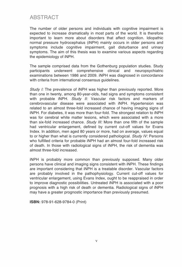
ABSTRACT
The number of older persons and individuals with cognitive impairment is expected to increase dramatically in most parts of the world. It is therefore important to learn more about disorders that affect cognition. Idiopathic normal pressure hydrocephalus (iNPH) mainly occurs in older persons and symptoms include cognitive impairment, gait disturbance and urinary symptoms. The aim of this thesis was to examine various aspects regarding the epidemiology of iNPH.
The sample comprised data from the Gothenburg population studies. Study participants underwent comprehensive clinical and neuropsychiatric examinations between 1986 and 2009. iNPH was diagnosed in concordance with criteria from international consensus guidelines.
Study I: The prevalence of iNPH was higher than previously reported. More than one in twenty, among 80-year-olds, had signs and symptoms consistent with probable iNPH. Study II: Vascular risk factors and markers of cerebrovascular disease were associated with iNPH. Hypertension was related to an almost three-fold increased chance of having imaging signs of iNPH. For diabetes, it was more than four-fold. The strongest relation to iNPH was for cerebral white matter lesions, which were associated with a more than six-fold increased chance. Study III: More than one fifth of the sample had ventricular enlargement, defined by current cut-off values for Evans Index. In addition, men aged 80 years or more, had on average, values equal to or higher than what is currently considered pathological. Study IV: Persons who fulfilled criteria for probable iNPH had an almost four-fold increased risk of death. In those with radiological signs of iNPH, the risk of dementia was almost three-fold increased.
iNPH is probably more common than previously supposed. Many older persons have clinical and imaging signs consistent with iNPH. These findings are important considering that iNPH is a treatable disorder. Vascular factors are probably involved in the pathophysiology. Current cut-off values for ventricular enlargement, using Evans Index, ought to be reappraised in order to improve diagnostic possibilities. Untreated iNPH is associated with a poor prognosis with a high risk of death or dementia. Radiological signs of iNPH may have a greater prognostic importance than previously presumed.
ISBN: 978-91-628-9784-0 (Print)

vi
SAMMANFATTNING PÅ SVENSKA
Antalet äldre och personer med nedsatt kognitiv förmåga förväntas öka dramatiskt de kommande åren. Det är således av stor vikt att studera sjukdomar som påverkar intellektuella funktioner. Idiopatisk normaltryckshydrocefalus (iNPH) drabbar framförallt äldre personer. Symptomen innefattar försämrad kognitiv funktion, gångsvårigheter och vattenkastningsbesvär. Syftet med denna avhandling var att undersöka epidemiologiska aspekter av denna sjukdom.
Materialet utgörs av ett befolkningsmaterial från populationsstudierna i Göteborg. Studiedeltagare genomgick omfattande undersökningar, inklusive datortomografi av hjärnan, mellan åren 1986 och 2009.
Delstudie I: Förekomsten av iNPH var högre än vad man tidigare uppskattat. Mer än var tjugonde 80-åring uppvisade kliniska och radiologiska fynd förenliga med iNPH. Delstudie II: Vaskulära riskfaktorer och markörer för cerebrovaskulär sjukdom var kopplat till iNPH. Hypertoni ökade sannolikheten att ha radiologiska fynd förenliga med iNPH nästan trefaldigt. Gällande diabetes var sannolikheten mer än fyrfaldigt ökad. Starkast koppling var till vitsubstansförändringar som gav en mer än sexfaldigt ökad sannolikhet. Delstudie III: Mer än en femtedel av alla hade förstorade ventriklar enligt gällande definition, baserat på Evans Index. Dessutom hade män, i åldrarna 80 år och äldre, i genomsnitt ett värde på Evans Index som utifrån dagens kriterier skulle klassas som sjukligt. Delstudie IV: De som uppfyllde kriterierna för iNPH hade en nästan fyrfaldigt ökad risk för död. Dessutom var risken för demens nästan trefaldigt ökad hos de som hade radiologiska fynd förenliga med iNPH.
Många äldre har kliniska och radiologiska fynd förenliga med iNPH, vilket talar för att diagnosen är betydligt vanligare än vad man tidigare har trott. Dessa resultat kan vara av betydelse med tanke på att sjukdomen är behandlingsbar. Vidare pekar den höga förekomsten av kärlsjukdomar mot att cerebrovaskulära förändringar är involverade i sjukdomsmekanismen. För att förbättra diagnostiken bör nuvarande kriterier för ventrikelvidgning, baserat på Evans Index, ses över. Vidare, förefaller obehandlad iNPH vara kopplat till en dålig prognos med ökad risk för demens och tidig död. Slutligen, radiologiska fynd verkar vara av större prognostisk betydelse än vad man tidigare har trott.

vii
LIST OF PAPERS
This thesis is based on the following studies, referred to in the text by their Roman numerals.
I. D Jaraj, K Rabiei, T Marlow, C Jensen, I Skoog, C Wikkelsø. Prevalence of Idiopathic Normal-Pressure Hydrocephalus. Neurology 2014;82:1449-1454. © American Academy of Neurology
II. D Jaraj, S Agerskov, K Rabiei, T Marlow, C Jensen, X Guo, S Kern, C Wikkelsø, I Skoog. Vascular Factors in Suspected Normal-Pressure Hydrocephalus: A Population-based Study. Neurology 2016;86:592-9. © American Academy of Neurology
III. D Jaraj, K Rabiei, T Marlow, C Jensen, I Skoog, C Wikkelsø. Estimated Ventricle Size Using Evans Index In a Population-based Sample. Manuscript.
IV. D Jaraj, C Wikkelsø, K Rabiei, T Marlow, C Jensen, S Östling, I Skoog. Mortality and Risk of Dementia in Normal-Pressure Hydrocephalus: A Population Study. Submitted.

viii

ix
CONTENT 1 NORMAL PRESSURE HYDROCEPHALUS - AN INTRODUCTION ................ 1
Classification of Hydrocephalus ........................................................ 1History of iNPH .................................................................................. 4Clinical Features ................................................................................ 4Anatomy and Physiology of the CSF Circulation ............................... 8Pathophysiology .............................................................................. 13Prevalence and Incidence ............................................................... 20Diagnosis ......................................................................................... 21Treatment ........................................................................................ 31Prognosis ......................................................................................... 34
2 THE AGING SOCIETY AND THE AGING BRAIN .................................... 373 AIM ................................................................................................. 414 METHODS AND STUDY DESIGN ......................................................... 43
Study sample - The Gothenburg Population Studies ....................... 43Radiological Examinations ............................................................... 49Diagnosis of iNPH ............................................................................ 49Papers I – IV .................................................................................... 50
5 RESULTS AND DISCUSSION .............................................................. 546 CONCLUSIONS AND FUTURE PERSPECTIVES ..................................... 79ACKNOWLEDGEMENTS ......................................................................... 82REFERENCES ....................................................................................... 84

Hydrocephalus –
From the Greek words “Hydor”= water and “Kéfale”= skull

Daniel Jaraj
1
1 NORMAL PRESSURE HYDROCEPHALUS - AN INTRODUCTION
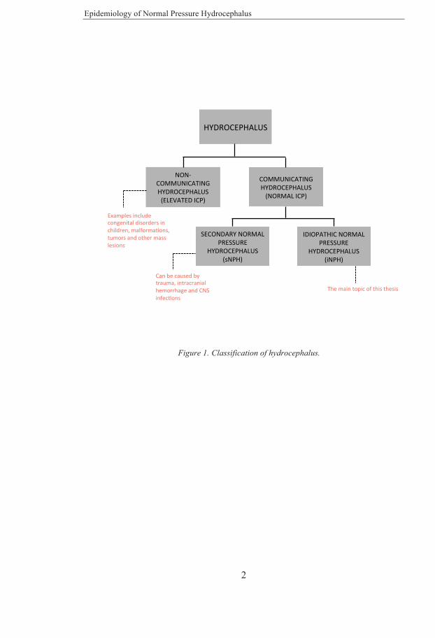
Classification of Hydrocephalus Hydrocephalus is a term for various conditions characterized by impaired cerebrospinal fluid (CSF) dynamics. Enlargement of the cerebral ventricles is one of the main hallmarks.1 Hydrocephalus includes several different disorders with varying causes and clinical presentations, and can therefore occur in all ages.2 The topic of this thesis is idiopathic normal pressure hydrocephalus, which is an adult form.
The classification of the hydrocephalic disorders is largely based on the anatomy and physiology of the central nervous system (figure 1). Hydrocephalus can be classified into two main forms: non-communicating (obstructive) hydrocephalus, and communicating (non-obstructive) hydrocephalus. Non-communicating hydrocephalus is characterized by an obstruction of the CSF flow, somewhere between the point of production and absorption. Causes can include congenital malformations, hemorrhage, tumors and various other intracranial mass lesions. Non-communicating hydrocephalus can affect persons of all ages and have an acute or chronic onset. Clinical features depend on the underlying cause.2 Because CSF flow is obstructed, intracranial pressure (ICP) can be increased. In contrast, in the communicating form of hydrocephalus, there are no visible obstructions of CSF flow, and the underlying mechanisms are not fully understood. There is no increase in intracranial pressure, hence the name normal pressure hydrocephalus (NPH).3 NPH affects adults, and is further divided into two groups. When it is caused by certain precipitants, such as trauma, meningitis or subarachnoid hemorrhage, NPH is classified as secondary (sNPH). In cases where no identifiable cause can be found, NPH is labeled idiopathic (iNPH).3, 4 This thesis involves iNPH and discussions regarding other forms of hydrocephalus are beyond the scope of the present work.
Epidemiology of Normal Pressure Hydrocephalus
2
Figure 1. Classification of hydrocephalus.
HYDROCEPHALUS
NON-COMMUNICATINGHYDROCEPHALUS (ELEVATEDICP)
SECONDARYNORMALPRESSURE
HYDROCEPHALUS(sNPH)
IDIOPATHICNORMALPRESSURE
HYDROCEPHALUS(iNPH)
COMMUNICATINGHYDROCEPHALUS (NORMALICP)
Examplesincludecongenitaldisordersinchildren,malformaJons,tumorsandothermasslesions
Canbecausedbytrauma,intracranialhemorrhageandCNSinfecJons
Themaintopicofthisthesis

In order to fully understand the present, we must first have knowledge about the past.

Epidemiology of Normal Pressure Hydrocephalus
4
History of iNPH The first modern description of iNPH was made by the Colombian neurosurgeon Salomón Hakim in 1965.5, 6 In the original publications, two cases of secondary NPH, following traumatic brain injury, and one case of idiopathic NPH were described. The patients presented with cognitive impairment, gait disturbance and urinary incontinence and were noted to have distended ventricles on angiography and pneumoencephalogram. Lumbar puncture was performed and intracranial pressure was found to be within normal limits. Interestingly, after the lumbar puncture, clinical improvement was noted. Shunt surgery was performed and further improvement occurred.
After having published his original finding, Salomón Hakim devoted himself to further research on NPH and its potential causes. In particular, he was interested in intracranial biomechanics. Thus, he spent much time on the hydrodynamic aspects of CSF and the cerebral ventricles.7 He theorized that, because pressure is defined as force per unit area, the ventricles in NPH could be enlarged due to increased force acting on the brain tissue while the CSF pressure remains constant.
Although, neither change in intracranial pressure nor ventricle size have subsequently been found to relate to postoperative improvement, much of the research has so far been focused on pressure-volume relationships. However, it might be that the complexities involved in the CSF circulation have been underestimated. Also, compared to the hydrodynamic aspects, other research areas in iNPH, such as the overlap between other neurodegenerative disorders have thus far received less attention.
Clinical Features iNPH mainly affects older persons and symptoms often develop insidiously. Core signs and symptoms include gait and balance disturbance, cognitive impairment and urinary symptoms.1 These are summarized in figure 2. The symptoms are sometimes referred to as the “typical triad”. However, this is
Daniel Jaraj
5
probably not an appropriate description for several reasons. Approximately half of all patients might only have one or two of these symptoms.8 Thus, the constellation of clinical characteristics is likely more complex than implied by the term “triad”. Furthermore, there is uncertainty regarding the exact distinctive features for each symptom. Gait and balance problems, cognitive dysfunction and urinary symptoms are common in older persons and may sometimes be due comorbidities.9, 10 Therefore, ascribing these symptoms to patients with suspicions of iNPH should be made in a thoughtful manner.
Gait and Balance Gait disturbance is common in iNPH. It is often stated to be the first symptom to develop, and the most characteristic. However, it is not known whether this is truly the case. For example, it might be that walking difficulties are more easily detected at an initial stage than subtle cognitive symptoms. Nevertheless, gait disturbance is an important part of the clinical picture. Typically the abnormal gait pattern is characterized as broad-based, magnetic with reduced cadence, step height and step width.11, 12 In classic literature, the gait is sometimes described as apraxic. Sometimes the gait pattern can have features of Parkinsonism, such as hesitation, freezing and reduced arm-swing. However, iNPH-patients more typically have retropulsion, i.e. a tendency towards a backward-extended posture.13

Epidemiology of Normal Pressure Hydrocephalus
6
Figure 2.
The balance and posture dysfunction seems to be related to a backward-displaced center of pressure and a defective vertical visual perception.14 Other characteristics of the gait pattern include outward rotation of the feet, and difficulties turning.11 Impaired balance often coexists with gait disturbance in iNPH, and can result in an increased risk of falls. Although the mechanisms underlying gait and balance disturbance in iNPH are not precisely known, these symptoms may be similar to those seen in patients with vascular cognitive impairment.15
Cognitive impairment Symptoms and signs of cognitive impairment are common among patients with iNPH. Often cognitive impairment in iNPH is of the frontal-subcortical type. Thus, common features include psychomotor slowing, inattention,
MAINSIGNSANDSYMPTOMSINiNPH
GAITANDBALANCEDISTURBANCE
COGNITIVEIMPAIRMENT
URINARYSYMPTOMS
Hypokine<cgait,broadbasedpaFernwithreducedcadence,stridelengthandstepheight.OutwardrotatedfeetDecreasedbalancewithretropulsion
Mainlyofsubcor<caltype,Signsoffrontaldysfunc<on.InaFen<veness,forgeNulness,lossofmo<va<onanddecreasedwakefulness
MostoPendescribedasurgencywithorwithoutincon<nence.Possiblyduetocentraldisinhibi<onofdetrusorac<vity
Daniel Jaraj
7
forgetfulness and impaired executive functions.1, 16 In a study comparing iNPH with patients with Alzheimer’s disease, those with iNPH had more frontal lobe symptoms.17 They were found to have a more pronounced impairment regarding attention and psychomotor speed, whereas the patients with Alzheimer’s disease had worse memory. It was speculated that the frontal lobe symptoms in iNPH might be secondary to subcortical, periventricular, white matter disease. In another study, compared to healthy individuals, iNPH patients had impaired functions in several domains, as described above, and also exhibited signs of impaired dexterity and fine motor skills.18 It was also found that neuropsychological impairment was associated with gait disturbance, incontinence and increased daily sleep.18 iNPH patients with concomitant vascular risk factors performed worse on neuropsychological tests. Several of these findings were later confirmed in a rather large, multicenter study.19 The authors speculate that the signs and symptoms in iNPH might be due to multifocal periventricular hypometabolism in combination with impaired connectivity in cortical-subcortical circuits. It was also theorized that the reduced wakefulness might be caused by dysfunction in the ascending activating systems.
Urinary Symptoms Urinary symptoms are common in iNPH. It is said that patients often experience increased frequency and urgency early on, and develop incontinence at later stages.1 However, urinary symptoms are very common in both men and women in the general population. Thus, it can be difficult to differentiate these symptoms of iNPH from other causes. Urgency and incontinence in iNPH is thought to occur from a central disinhibition leading to hyperactivity of the detrusor, as this has been found in urodynamic studies.20,
21 Impaired cognition and locomotion may however also contribute.

Epidemiology of Normal Pressure Hydrocephalus
8
Anatomy and Physiology of the CSF Circulation CSF (Liquor cerebrospinalis) is a clear and colorless fluid that surrounds the central nervous system. CSF has several vital physiological functions, such as protective cushioning, regulation of intracranial pressure and transport of metabolites and waste products.22 The total volume of CSF (surrounding the brain and spinal cord) is approximately 200 ml. Turnover is rather high given that the production rate is around 500 ml per day (20 ml per hour).23 Thus, daily formation is two to three times higher than the total CSF volume. The basic anatomy of the ventricular system is shown in figure 3.
The arachnocentric view on CSF circulation The traditional approach to CSF circulation is largely based on anatomical descriptions and experiments made in the nineteenth century and beginning of twentieth century, i.e. more than 100-150 years ago.24 Much emphasis has been placed on the absorptive pathways through the arachnoid villi. Therefore, the traditional view on CSF circulation could be said to have an arachnocentric focus. Accordingly, the main production site of CSF is the choroid plexus.25 CSF flows in a pulsatile manner with each cardiac cycle. A net flow occurs from the lateral ventricles, through the foramina of Monro to the third ventricle, from there, through the aqueduct of Sylvius down to the fourth ventricle and eventually exits through foramen of Magendie and Luschkae. The CSF enters the subarachnoid space of the cisterna magna. Traditionally CSF is said to travel down the subarachnoid space of the spinal cord and also directly up along the cerebral convexities where it is absorbed into the venous blood of sinuses through the arachnoid granulations.

Daniel Jaraj
9
Figure 3. Schematic illustration of the cerebral ventricles and outflow tracts
A more contemporary approach to CSF dynamics The traditional view on CSF circulation is rather straightforward and easy to grasp. However, recent findings indicate a more complex process, and there is now increasing evidence that the traditional view on CSF circulation is over-simplified and outdated. For example, the brain parenchyma, capillaries including perivascular spaces and interstitial fluid might be more important for CSF production and absorption than the choroid plexus and arachnoid granulations.22, 26, 27 In upright active individuals up to two thirds of CSF absorption can occur through the spinal subarachnoid space.28 Furthermore, perineural sheaths of pia and arachnoid mater along cranial- and spinal nerves have been found to constitute important pathways of lymphatic CSF
LateralventriclesMainsiteofChoroidPlexus
FrontalhornoftheLateralventricles
ForaminaofMonro
Occipitalhornsofthelateralventricles
AqueductofSylvius
FourthventricleOu>lowtractsincludeForamenofMagendieandforaminaofLuschkaeThirdventricle
Temporalhornsofthelateralventricles

Epidemiology of Normal Pressure Hydrocephalus
10
drainageA. 29 Several studies have shown that CSF tracers readily enter extra-cranial lymphatics, such as the cervical lymph nodes, and ultimately the blood stream. In one study performed on sheep, the cribriform plate was obstructed, blocking absorption to the nasal mucosa lymphatics.30 This led to an increased intracranial pressure and an almost three-fold increase in CSF outflow resistance.
To complicate things further… In 2013, a landmark paper was published in Science that provided a possible explanation for the function of sleep. 31 As it turns out, the findings may also have implications for our understanding of the CSF circulation, and possibly the pathophysiology of iNPH.
Real-time imaging was made, in vivo, using fluorescent tracers, injected into the CSF. This was performed in asleep and awake mice and in anesthetized mice. Intriguingly, in the sleeping and anesthetized mice, a substantial influx of CSF occurred along the para-arterial spaces into the brain parenchyma. Upon arousal, however, this influx decreased by approximately 95 %. This was then repeated using radiolabeled amyloid beta (Aβ). It was found that during sleep, Aβ was cleared from the interstitial space two-times faster than during the awake state. Furthermore, cortical interstitial volume was also measured and found to increase almost twofold during sleep.
These findings indicate that during sleep, the interstitial volume shrinks. This in turn, allows influx of CSF along the para-arterial spaces, and clearance of metabolites and waste products. The pathway of waste clearance, from the extracellular space, occurred as a convective flow through a complex astroglial network referred to as the glymphatic system.
A The lymphatic system has an important role in the clearance of metabolic waste. In almost all organs, the number of lymphatic pathways is proportional to the metabolic activity. The brain is however an exception. It is interesting that despite having the highest metabolic activity, and thus an enormous need for waste clearance, the CNS itself lacks lymphatic vessels.
Daniel Jaraj
11
These processes are believed to require a high energy expenditure, which cannot be accommodated in the aroused state. While awake, the brain has to take in, filter, and process vast amounts of new data. Therefore, maintenance and household functions in the brain, i.e. elimination of waste, takes place during sleep.
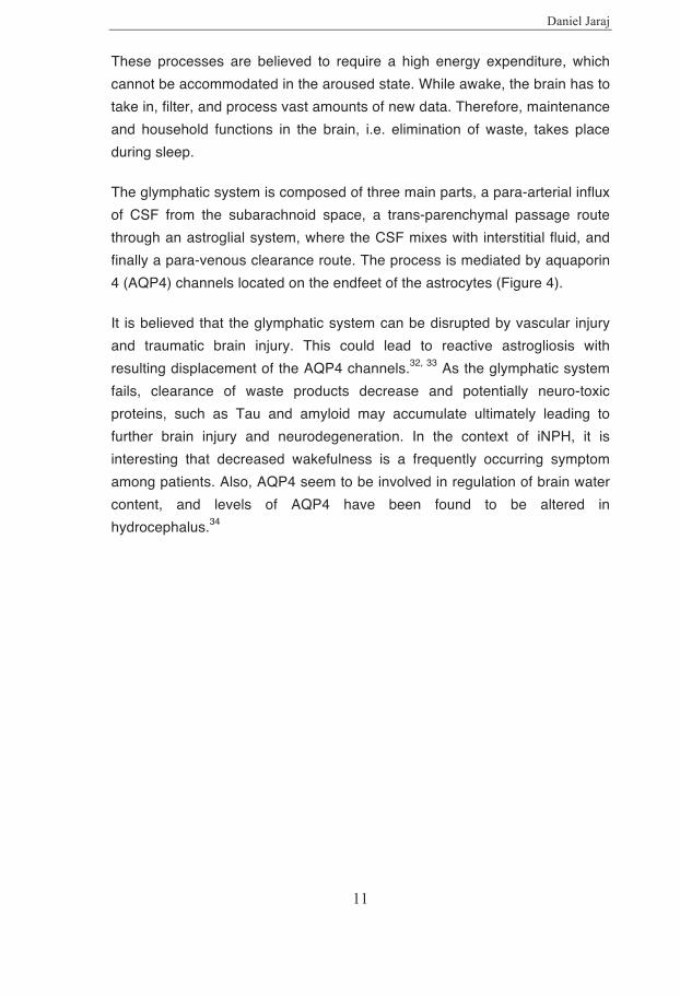
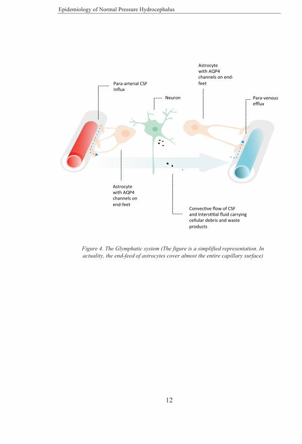
The glymphatic system is composed of three main parts, a para-arterial influx of CSF from the subarachnoid space, a trans-parenchymal passage route through an astroglial system, where the CSF mixes with interstitial fluid, and finally a para-venous clearance route. The process is mediated by aquaporin 4 (AQP4) channels located on the endfeet of the astrocytes (Figure 4).
It is believed that the glymphatic system can be disrupted by vascular injury and traumatic brain injury. This could lead to reactive astrogliosis with resulting displacement of the AQP4 channels.32, 33 As the glymphatic system fails, clearance of waste products decrease and potentially neuro-toxic proteins, such as Tau and amyloid may accumulate ultimately leading to further brain injury and neurodegeneration. In the context of iNPH, it is interesting that decreased wakefulness is a frequently occurring symptom among patients. Also, AQP4 seem to be involved in regulation of brain water content, and levels of AQP4 have been found to be altered in hydrocephalus.34
Epidemiology of Normal Pressure Hydrocephalus
12
Figure 4. The Glymphatic system (The figure is a simplified representation. In actuality, the end-feed of astrocytes cover almost the entire capillary surface)
������������ ��������� �������������� ����"������������ ������� � ������� �����
��$��������������!�
��$�����������!�
�������
������"��� �������%�������������� $�����
������"��� �������%�������������� $�����

Daniel Jaraj
13
Pathophysiology It is often believed that iNPH is primarily caused by an alteration of CSF dynamics, such as decreased absorption.35 This might very well be true. However, there is so far little evidence that directly supports this notion. Indeed CSF diversion does improve symptoms. Though, it is theoretically possible that the effect of shunt surgery is due to secondary changes in the cerebral microcirculation. It might be that the previously oversimplified concepts of CSF circulation have led to an underestimation of the actual complexities involved in the pathophysiology.
Morphological changes of CSF outflow tracts, such as arachnoid fibrosis have been noted in some patients on autopsy.36 This might implicate an impaired outflow as a pathological basis. However, most studies included only a few cases, and more recent papers have provided contradictory results.37 Also, patients with iNPH are said to have an increased resistance to CSF outflow.38 However, most patients with iNPH are older, and it is known that both production and absorption of CSF decreases with age.39, 40 Furthermore, values of CSF outflow resistance (Rout) that are considered pathological may be found in as many as 25 % of healthy elderly.41 Moreover, other measures of CSF dynamics, such as continuous monitoring of intracranial pressure and radionuclide cisternography, that are often said to show characteristic features of iNPH, lack evidence-based support.42 For example, intermittent alterations of intracranial pressure have been reported to be diagnostic of iNPH and useful as a prognostic marker in terms of shunt surgery.43, 44 however, similar findings are seen in healthy individuals and might actually be physiological occurrences.28, 45 Thus, regarding the pathophysiology, mechanisms other than those involving CSF dynamics should also be considered.
The role of vascular disease There are numerous reports linking iNPH to various vascular risk factors including hypertension, diabetes mellitus and heart disease.46-50 These studies were made using hospital-based samples. Thus population-based epidemiological studies have so far been lacking. Also, in a review issued by
Epidemiology of Normal Pressure Hydrocephalus
14
a task force for the International Society for Hydrocephalus and Cerebrospinal Fluid Disorders (ISHCSF), it was noted that the previous studies were based on small samples and were more than twenty years old.10 Furthermore, these papers do not provide data for a direct causal relation. Nevertheless, a possible causal association between vascular disease processes and iNPH is further supported by several additional studies.
Arterial hypertension is more common in patients with iNPH, and a previous prospective cohort study found that systolic blood pressure and pulse pressure were related to development of increased ventricle size.51 Interestingly, the relation between hypertension and ventricular enlargement is also supported by animal studies. Spontaneously hypertensive rats have been found to develop enlarged ventricles.52 In addition, another study on sheep found that hydrocephalus developed quickly after that balloons were inserted into the ventricles and set to inflate during systole and deflate during diastole, thereby increasing the pulse pressure.53
Small vessel disease has also been implicated in the pathogenesis of iNPH. Neuropathological examinations have revealed signs of cerebrovascular disease in patients with iNPH.54 In addition, cerebral white matter lesions (WMLs), which are associated with small vessel disease and white matter ischemia 55, have been found to be more common in iNPH compared to controls.56, 57 Furthermore, WMLs are related to similar subcortical symptoms, such as gait disturbance, urinary incontinence and cognitive impairment.58 Moreover, WMLs have been found to decrease after shunt surgery, with reductions correlating with clinical improvement.59, 60 Additional support for a vascular pathology underlying iNPH comes from numerous studies that have found reduced blood flow in several areas including the periventricular white matter.61-63 Also, cerebral blood flow has been found to increase after CSF removal via lumbar puncture or shunt surgery.64, 65
Some authors have reported that WMLs in iNPH might be associated with a poor response to shunt surgery.66 However, this may be contradicted by a randomized, double-blinded sham-controlled study that included iNPH patients with severe white matter disease, who also fulfilled criteria for
Daniel Jaraj
15
Binswanger’s disease.67 Half of the patients received a standard ventriculoperitoneal shunt, and half received a ligated shunt. After three months, the patients in the treatment group improved while no improvement was noted in the control group. After the three months, the ligated shunts were opened in the control patients, after which they to improved.
Further support for a vascular disease mechanism might come from a recent study in which iNPH patients were found to have a higher number of cerebral microbleeds than healthy controls.68 The association between iNPH and cerebral microbleeds might be due to small vessel disease.
Two studies have reported interesting results regarding the reduction of white matter hyperintensities and clinical improvement in iNPH patients treated with Acetazolamide.69, 70 Acetazolamide, a carbonic anhydrase inhibitor, is the only potential pharmacological treatment that has been proposed for iNPH. It is currently approved for treatment of glaucoma, idiopathic intracranial hypertension and acute mountain sickness. Acetazolamide is a diuretic and has been shown to reduce CSF production. More importantly, Acetazolamide is also a vasodilator that has been shown to increase cerebral blood flow.71, 72 Among eight, non-shunted, iNPH patients treated with Acetazolamide, five responded positively with improved gait.69 Furthermore, a significant reduction in periventricular hyperintensities was seen. In another study, non-shunted iNPH patients, treated with Acetazolamide underwent repeated MRI and clinical assessments.70 These were compared to iNPH patients who underwent external lumbar drainage (ELD) and controls consisting of iNPH patients without intervention. White matter changes decreased in those treated with Acetazolamide and in the patients who underwent ELD, but not among controls. It is important to note that WMLs in iNPH might have other causes than ischemia, such edema or CSF stagnation, and the authors state the positive effects of Acetazolamide might be due to decreased interstitial brain water, reduced transependymal CSF flow, or increased cerebral blood flow. Regardless, the findings of these studies are highly intriguingB, and a
B Of interest, Acetazolamide has also been suggested to be a blocker of aquaporin channels (Owler et al. Cerebrospinal Fluid Research 2010, 7:15)
Epidemiology of Normal Pressure Hydrocephalus
16
randomized controlled trial on Acetazolamide in iNPH would probably be of value.
As mentioned in the previous section, it is conceivable that vascular disturbances and ischemia might contribute to partially reversible changes in the microcirculation. However, the exact mechanisms linking vascular disease to iNPH are currently not clear.
Other potential causes Interestingly, iNPH patients have been found to have larger head size than healthy controls.73, 74 This might suggest that iNPH is a congenital disorder that becomes symptomatic in late life. Given these findings, it has been hypothesized iNPH might be a “two-hit disease”.75, 76 Accordingly it was theorized that iNPH begins with benign external hydrocephalus during infancy (the first hit), which causes an increased head size and ventricle size. After this, the person may be asymptomatic until late-life, when white matter disease develops (the second hit). The authors state that white matter ischemia, with resulting myelin loss, may give rise to a more hydrophilic environment that impedes CSF flow through the extracellular space. This process ultimately leads to further decrease of CSF absorption with clinical decompensation causing the person to become symptomatic.
However, another study found that head circumference was only increased in a subset of iNPH patients.77 The authors found that the number of patients with iNPH who had a head circumference above the 90th percentile was significantly increased compared to controls. Though, this was not the case for the rest of the iNPH sample. The distribution among those with a head circumference below the 90th percentile was similar to that of the control population. These results imply that a congenital cause of iNPH might be the case only in a minority of the patients. Of further importance, it has been suggested that iNPH, in fact is a rather heterogeneous syndrome with many potential causes and that persons with suspected iNPH might also have other neurodegenerative disorders or various concomitant comorbidities.10, 78 This might be true, at least to some extent, given the considerable overlap between iNPH and other neurodegenerative diseases.

Daniel Jaraj
17
Cerebral amyloid pathology in iNPH and overlapping features with Alzheimer’s disease iNPH and Alzheimer’s disease have several common features. These include clinical, radiological and biochemical findings. For example, patients with Alzheimer’s disease often have enlarged ventricles, gait disturbance and urinary problems. Also, patients with iNPH have reduced levels of Amyloid- β (Aβ42) in CSF, similar to those with Alzheimer’s disease.79 In addition, Amyloid-β is frequently found in cortical brain biopsies in iNPH.80 In fact the two diseases are thought to often coexist. Some authors have therefore hypothesized that iNPH and Alzheimer’s disease might have a common etiological mechanism.81 It was previously postulated that both diseases are the result of hydrodynamic disturbances of CSF flow. Alzheimer’s disease was theorized to be due reduced CSF production, with decreased turnover of CSF ultimately leading to accumulation of Aβ42 and in turn causing neurodegeneration. iNPH, on the other hand was thought to occur from a decreased CSF absorption which would also lead to decreased turnover of CSF and accumulation of Aβ42 giving rise to a similar syndrome as Alzheimer’s disease. Based on this, a pilot study was conducted in order to examine the possible therapeutic effects of shunt surgery in patients with Alzheimer’s disease.82 However, despite initially promising results, a randomized, double-blinded, sham-controlled trial later showed no benefit of CSF shunting in patients with Alzheimer’s disease.83
There might also be other explanations for the existence of cerebral amyloid pathology in iNPH. It might be that the development of iNPH is related to interstitial CSF stagnation with reduced periventricular metabolism leading to decreased CSF clearance and accumulation of Aβ42. A study conducted in Gothenburg, Sweden found that patients with iNPH, compared to healthy individuals, had not only low levels of Aβ42 but also other types of amyloid proteins (Aβ38, Aβ40, sAPPα and sAPPβ) were low.84 This finding is typically not seen in Alzheimer’s disease. The authors found also increased levels of neurofilament light protein (NFL), which might be due to degeneration of periventricular axons. Moreover, all amyloid proteins increased in ventricle CSF after shunt surgery. It has, since then, been theorized that that the low

Epidemiology of Normal Pressure Hydrocephalus
18
levels of CSF Aβ42, in iNPH, are not due to deposition (as in Alzheimer’s disease) but instead caused by decreased clearance via the glymphatic system.85
In a study from Finland, cortical biopsies were obtained, during shunt surgery or postmortem, from patients with iNPH and Alzheimer’s disease.86 iNPH patients with amyloid pathology had higher levels of γ-secretase activity compared to iNPH patients without amyloid pathology. Patients with Alzheimer’s disease, on the other hand, are known to have increased activity of β-secretase. These findings might indicate different pathophysiological mechanisms. In addition, in a relatively large cohort of iNPH patients from the same registry, no association was found between Apolipoprotein E-status and iNPH.87 Furthermore, patients with iNPH seem to have larger hippocampus volumes than those with Alzheimer’s disease.88 Another study is also worth mentioning. Positron emission tomography (PET) imaging was made, using 11C-labeled radiotracer Pittsburgh compound B imaging (PIB) to detect cerebral amyloid pathology and compare patients with iNPH and those with Alzheimer’s disease.89 Three out of ten iNPH patients had increased cortical amyloid. However, the amyloid distribution was different in those with iNPH compared to those with Alzheimer’s disease. Those with Alzheimer’s disease showed increased levels in the frontal and temporoparietal areas, while those with iNPH had a distribution limited to the high convexity and parasagittal areas. The authors state that these regions might be mechanically more compressed in iNPH, resulting in decreased clearance of Amyloid-β.
Although both patients with iNPH and Alzheimer’s disease may have cerebral amyloid pathology, altogether, previous results suggest that the pathophysiological mechanisms are different. Nevertheless, many of the common features are interesting and perhaps worthy of further investigation.
Why are the ventricles enlarged? In obstructive hydrocephalus intracranial pressure (ICP) is increased and the reason for ventricular enlargement could be regarded as rather intuitive.

Daniel Jaraj
19
However, in iNPH ICP appears to be within normal range and the cause of ventricular enlargement has puzzled the scientific community for decades.
Originally it was postulated that an increase in ventricle size might result in a concomitant decrease in pressure.7 This reasoning was partly based on Pascal’s principle, in which pressure is equal to force per unit area. Thus ICP could be normal due to having an increased area (ventricle volume) and increased force acting on the ventricle walls. Owing to this, it has been theorized that iNPH might be due to microscopic obstructions of CSF flow such as scarring or fibrosis of the arachnoid granulations. However, histopathological studies have been unable to confirm this.37 Furthermore, attempts to predict response to shunt surgery based on pressure-volume variables have been futile. Some patients have normal resistance to CSF outflow, but improve after CSF diversion while others have elevated outflow resistance but remain unchanged.90 These findings are difficult to explain using the previous models. Therefore, the exact reason for ventricular enlargement remains highly unclear.
It is well known that patients with cerebral atrophy can have enlarged ventricles (a condition sometimes termed hydrocephalus ex vacuo). However, not all patients with brain atrophy have enlarged ventricles. Therefore, it is theoretically possible that ventricular enlargement in some patients with Alzheimer’s disease and vascular dementia is not entirely attributable to loss of brain parenchyma. For instance, it might be that there are concurrent changes in the cerebral microcirculation similar to that in iNPH. This could possibly also explain part of the overlap regarding symptoms. However, other typical radiological signs of iNPH such as narrowing of high convexity sulci and subarachnoid space do not seem to be common in other neurodegenerative disorders. Although, this has, so far, not been investigated using population samples.
As mentioned earlier, hypertension and diabetes are common in iNPH. It might also be that stiffening of large vessels, due to atherosclerotic processes, lead to an increased pulse-pressure. This could, in turn, result in increased mechanical force acting on the brain parenchyma leading to

Epidemiology of Normal Pressure Hydrocephalus
20
ventricular enlargement. However, in order to gain knowledge on this topic, epidemiological studies using population-based samples with longitudinal data are needed. For example, the order of events regarding development of different radiological signs and clinical features are currently not known.
Prevalence and Incidence Epidemiological studies in iNPH are scarce and there are few population studies on the prevalence and incidence. Most previous studies were made using smaller samples and included few people above age 80 years. Also, several authors have stated that iNPH is a rare disorder.91, 92 Although, there is some uncertainty regarding prevalence estimatesC, existing data does not support the notion that iNPH is uncommon.
In Germany, a door-to-door survey was conducted in order to examine the prevalence of Parkinsonism.93 The authors found a prevalence of iNPH, in persons above age 65 years, of 0.4%. However, because iNPH was only examined in those who screened positive for Parkinsonism, the study probably underestimated the prevalence. A study from Norway reported a prevalence of probable iNPH of approximately 0.1 % in persons older than 65 years.94 However, this study was not populations based. Participants were recruited from an advertisement campaign directed to the general population and primary care physicians. It is thus possible that the low prevalence was due to recruitment bias.
A population-based study in Tajiri, Japan found a prevalence of NPH of 2.9 % among 170 men and women aged 65 years or older.95 Another population-based study from the same area examined 497 persons aged 65 years or more and reported a prevalence of possible iNPH of 1.4%.96 A third population-based study from Japan included 790 persons and found that C One of the reasons for having uncertainty in the prevalence estimates might be that iNPH, like many other neurological diseases is rather difficult to diagnose. The brain is a highly complex organ shielded by thick bone and isolated from the rest of the body by a blood-brain-barrier. Advanced imaging techniques and extensive workup are often needed to make an accurate diagnosis. Thus, making a precise diagnosis on a population-level is difficult and may require substantial resources.

Daniel Jaraj
21
1.5% had features of NPH on MRI, and 0.5% met the criteria of possible iNPH.97 These studies included mainly younger elderly. A study, conducted in Umeå, Sweden examined the occurrence of ventricular enlargement and symptoms of iNPH among patients with TIA admitted to a stroke unit.98 The authors found that 3.9 % of the patients fulfilled radiological and clinical criteria for possible iNPH. Several other studies have been aimed to assess the prevalence of iNPH.99-101 However, these were not population-based. Instead, they were conducted on specific samples, such as patients from memory clinics or nursing homes and thus had inherent limitations. A recent systematic review pooled prevalence data from earlier population studies and found that the prevalence of iNPH, among persons aged 60 years or more, was 1.3 % (95 % CI; 0.96-1.71).102
Two previous studies have examined the incidence of iNPH 94, 103, one of which was population-based.103 The first study was conducted in Norway and estimated the incidence among all ages to 5.5/100,000. The second one, conducted in Japan followed a cohort of 70-year-olds for ten years and estimated the incidence to 120/100,000. In Sweden, the annual incidence of shunt surgery for hydrocephalus is 3.33/100,000 104 (of which approximately half are for iNPH). The incidence of shunt surgery in Norway has been reported to be 1.09/100,000.105 Thus, extrapolating from earlier studies, existing data suggests that iNPH is highly underdiagnosed and undertreated. Using even the most modest estimates, it seems that less than 20 % of patients receive treatment. However, it should be pointed out that the number of shunt surgeries seems to have increased, at least in Japan, since these studies were published.106
Diagnosis The diagnosis of iNPH is mainly based on the combination clinical signs and brain imaging findings from CT or MRI.1, 16 Certain additional tests of CSF dynamics may sometimes also be applied. Evidence-based guidelines for the diagnosis of iNPH have previously been developed in order to aid clinicians and provide consensus in research.

Epidemiology of Normal Pressure Hydrocephalus
22
The first guidelines were created by several experts from Japan and published in 2004 (The English version was published in 2008).107 A second edition was made in conjunction with the Japanese Ministry of Health, and was published in 2011 (The English version, published in 2012).16
In 2005, researchers from the USA and Europe also created separate guidelines for management of iNPH.1 According to these, iNPH should be diagnosed by careful review of the patient history, possibly also from a close informant, thorough clinical examination and neuroimaging. If diagnostic uncertainty remains, additional tests of CSF dynamics can be performed. However, despite meticulous review of the literature, the authors acknowledge the uncertainty and difficulties in diagnosing iNPH. For this reason, a classification system of “probable”, “possible” and “improbable” iNPH was proposed. The main signs and symptoms and diagnostic criteria are summarized in figure 5.
Overall, the Japanese and American-European guidelines are rather similar. One differences is that gait disturbance is not mandatory for the classification of “probable iNPH” according to the Japanese guidelines. Also, according to the Japanese criteria, persons who improve after shunt surgery can be labeled “definite iNPH”. This is not the case in the American-European guidelines.
So far, the guideline criteria have not been validated regarding reliability, validity, sensitivity and specificity. Nevertheless, they have been of value in providing consensus and an evidence-based approach to the management of iNPH. Also, it is worth mentioning that in japan, the number of shunt surgeries for iNPH seems to have increased dramatically after the publication of the guidelines in 2004.106
Patient history Clinical features of iNPH are also discussed in previous sections. Signs and symptoms of iNPH typically develop gradually.1, 16 A more acute onset should prompt the clinician to consider other diagnoses. Patients often present with gait and balance difficulties, unsteadiness and or increased number of falls.3
Daniel Jaraj
23
They may also present with signs of cognitive dysfunction, such as inattention, forgetfulness, drowsiness, increased sleep, lack of motivation as described earlier.9 Obtaining a medical and surgical history from a relative or other close informant is often of value considering that patients can sometimes have trouble recalling. Urinary symptoms, such as urgency and or incontinence are also common.4 However, urinary problems frequently occur in older persons without iNPH, due to other reasons. It is therefore important to distinguish symptoms in iNPH, which are thought to occur from overactivity of the detrusor, from other causes such as benign prostatic hyperplasia or stress incontinence. One should also bear in mind that some patients may find it difficult to discuss these types of symptoms. In addition, it is important to consider causes of secondary NPH, such as previous head trauma, meningitis or subarachnoid hemorrhage. Thus, past episodes of possible precipitants should be inquired. The possibility of congenital causes should also be considered. Treating physicians can therefore ask about neurological symptoms during childhood. Finally, there are several somatic and psychiatric disorders that can mimic iNPH. The presence of any comorbidities should thus be carefully reviewed.10

Epidemiology of Normal Pressure Hydrocephalus
24
Figure 5. Overview and summary of the American-European guidelines 1 for the diagnosis of iNPH.
MAINSIGNSANDSYMPTOMSACCORDINGTOGUIDELINES
Pa3entHistory
ProgressivesymptomswithonsetaCertheageof40.Minimumdura3onof3months.NoevidenceofsecondaryNPH.Nootherconcurrentcondi3onscansufficientlyexplainsymptoms
Neuroimaging
Ventricularenlargement(EvansIndex>0,3).Noobstruc3onofCSFflow.Inaddi3on,atleastoneofthefollowingfeatures:Enlargementofthetemporalhorns.Periventricularsignalchanges.FlowvoidonMRI.
ClinicalFeatures
Gait:Decreasedstepheight,length,cadance.Increasedtrunksway,widenedstance.Outwardrotatedfeet,retropulsion.Impairedbalance.Cogni3on:Decreasedpsycho-motorspeed,finemotorskill.Decreaseda[en3on,recall,andexecu3vedysfunc3on.Urinarysymptoms:Episodicorpersistentincon3nence,nota[ributabletourologicaldisease.
PROBABLEiNPH
GaitandBalancedisturbance+
Cogni3veImpairmentorUrinaryIncon3nenceorboth
POSSIBLEiNPH
Atleastoneofthefollowing:GaitandBalance
disturbance,Cogni3veImpairmentorUrinary
Incon3nence
UNLIKELYiNPH
Nosignsofventricularenlargement,Signsofincreasedintracranial
pressure,noneoftheclassicsymptomspresent,signsandsymptomsa[ributableto
othercondi3ons
ImagingsignsconsistentwithiNPHmustbepresent
SUMMARYOFGUIDELINECRITERIA

Daniel Jaraj
25
Assessment of gait and neuropsychology Gait and balance can be assessed in several ways. A clinical examination, in which a physician examines gait, including tandem gait and Romberg’s test, is of value. Additional examination by a physiotherapist may probably add more information. Also, formal testing, by measuring gait speed and number of steps is important in order to compare pre- and postoperative values. There are also many ways in which neuro-psychological evaluations can be done. Often the MMSE is performed. More specific testing of frontal-subcortical, executive functions and fine motor skills can be performed using the Stroop test, Grooved Pegboard test and the Rey Auditory Verbal Learning Test.18 Several other similar cognitive tests can also be applied.108
Imaging in iNPH All patients with suspected iNPH must undergo imaging of the brain in order to examine ventricle size and exclude possible obstructions. A normal scan probably rules out iNPH rather effectively. According to the guidelines, ventricular enlargement is a mandatory criterion.1, 16 It is stated in the guidelines that ventricular enlargement should be evidenced by an Evans Index greater than 0.3, or by an equivalent measure of ventricles size. However, no such alternative measure is suggested.
Regarding the choice of imaging,MRI is superior to CT in many ways. Although the guidelines do state that CT is probably sufficient for routine diagnosis of iNPH. Nevertheless, MRI allows for a better visualization of small obstructive lesions, including possible thin membranes.109 More advanced imaging techniques, such as measurements of cerebral perfusion by CT, MRI, SPECT, PET or pseudo-continuous arterial spin labeling (pCASL) have also been described.110-113 These have provided interesting results from an academic standpoint, but are currently not clinically applied. Additional methods, such as isotope cisternography114, have previously also been advocated, but currently lack evidence.1
One of the major radiological hallmarks of iNPH is the presence of ventricular enlargement disproportional to subarachnoid space volume, i.e. ventricular enlargement not due to atrophy.8, 115 More specifically, the radiological
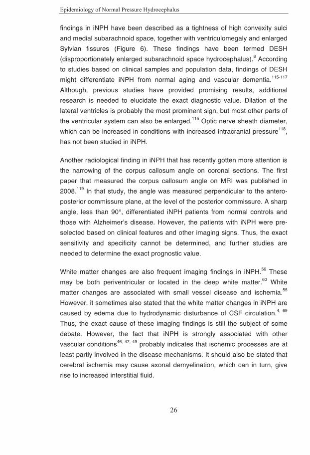
Epidemiology of Normal Pressure Hydrocephalus
26
findings in iNPH have been described as a tightness of high convexity sulci and medial subarachnoid space, together with ventriculomegaly and enlarged Sylvian fissures (Figure 6). These findings have been termed DESH (disproportionately enlarged subarachnoid space hydrocephalus).8 According to studies based on clinical samples and population data, findings of DESH might differentiate iNPH from normal aging and vascular dementia.115-117 Although, previous studies have provided promising results, additional research is needed to elucidate the exact diagnostic value. Dilation of the lateral ventricles is probably the most prominent sign, but most other parts of the ventricular system can also be enlarged.115 Optic nerve sheath diameter, which can be increased in conditions with increased intracranial pressure118, has not been studied in iNPH.
Another radiological finding in iNPH that has recently gotten more attention is the narrowing of the corpus callosum angle on coronal sections. The first paper that measured the corpus callosum angle on MRI was published in 2008.119 In that study, the angle was measured perpendicular to the antero-posterior commissure plane, at the level of the posterior commissure. A sharp angle, less than 90°, differentiated iNPH patients from normal controls and those with Alzheimer’s disease. However, the patients with iNPH were pre-selected based on clinical features and other imaging signs. Thus, the exact sensitivity and specificity cannot be determined, and further studies are needed to determine the exact prognostic value.
White matter changes are also frequent imaging findings in iNPH.56 These may be both periventricular or located in the deep white matter.60 White matter changes are associated with small vessel disease and ischemia.55 However, it sometimes also stated that the white matter changes in iNPH are caused by edema due to hydrodynamic disturbance of CSF circulation.4, 69 Thus, the exact cause of these imaging findings is still the subject of some debate. However, the fact that iNPH is strongly associated with other vascular conditions46, 47, 49 probably indicates that ischemic processes are at least partly involved in the disease mechanisms. It should also be stated that cerebral ischemia may cause axonal demyelination, which can in turn, give rise to increased interstitial fluid.
Daniel Jaraj
27
Figure 6. Morphological changes in iNPH
An increased CSF flow through the cerebral aqueduct, termed flow void, can sometimes be seen on MRI.120 This has been said to be suggestive of iNPH and have prognostic importance. However, previous studies have been contradictory121 and the exact value of this radiological sign is therefore not known. Regardless, the presence of a flow void is probably useful in differentiating communicating, from non-communicating hydrocephalus.
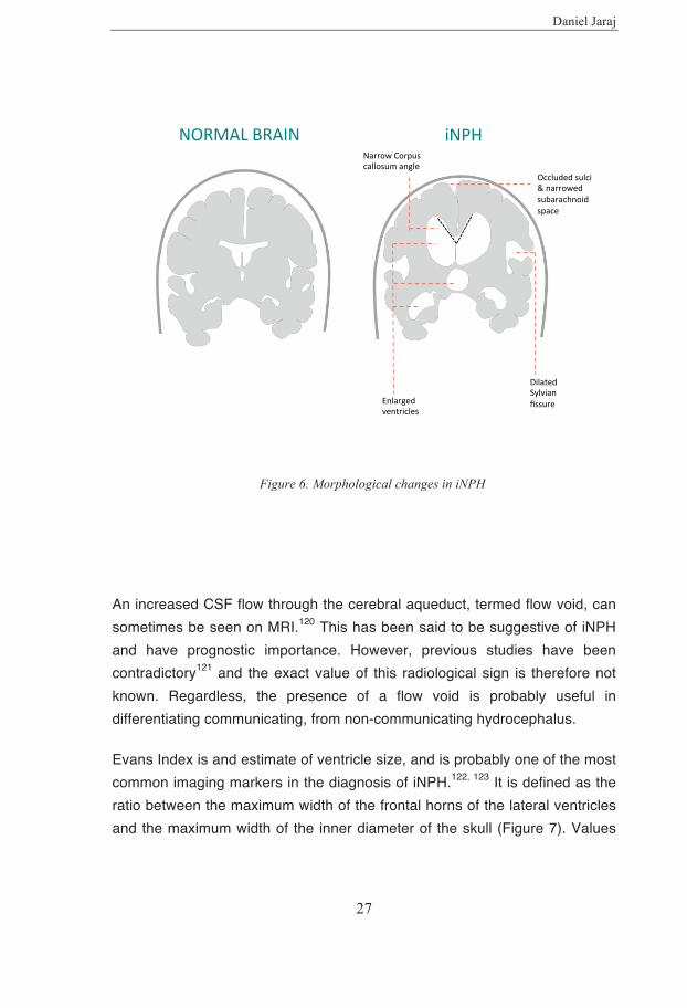
Evans Index is and estimate of ventricle size, and is probably one of the most common imaging markers in the diagnosis of iNPH.122, 123 It is defined as the ratio between the maximum width of the frontal horns of the lateral ventricles and the maximum width of the inner diameter of the skull (Figure 7). Values
Enlargedventricles
DilatedSylvianfissure
NarrowCorpuscallosumangle
Occludedsulci&narrowedsubarachnoidspace
NORMALBRAIN iNPH

Epidemiology of Normal Pressure Hydrocephalus
28
higher than 0.3 are regarded pathological, and are currently required by international consensus guidelines for the diagnosis.1, 16 However, despite the fact that Evans Index is used extensively in both research and clinical practice, exact values in the adult population are not precisely known. Earlier studies reporting values on Evans Index were made using small samples or non-representative populations. Despite having a major role in the diagnosis of iNPH, no previous population-based epidemiological studies have reported reference values.
Evans Index was first described in 1942 in children, using sagittal views on pneumoencephalograms.122 Later, in 1976, it was adapted for CT images.124 In the original paper, Evans examined 53 children and concluded that a value, using the ratio between the frontal horns and the inner diameter of the skull, higher than 0.3 represents ventricular enlargement. Since then, this cut-off value has been applied for the diagnosis of iNPH in older adults. Also, of interest, in the original paper Evans stated that there was less variation in ratio between the frontal horns and the skull, as apposed to just measuring the frontal horns. However, subsequently it was found that this was merely due to a miscalculation and that in fact the opposite was true.125
In studies on healthy elderly, mean values for Evans Index have varied between 0.25 and 0.31.119, 126-128 In addition, population-based studies have reported prevalence values, of Evans Index higher than 0.3, varying between 6.5 and 16.1 %.95-97, 116 However, these studies included mainly younger elderly and did not report mean values.
Of further note, the exact method of measuring Evans Index has not been specified. In a study comprising ten iNPH patients, Evans Index was measured at different angles and sections in each patient and a considerable variation was noted.129 Another limitation regarding Evans Index is that it is not a direct measure ventricular volume, instead it might be regarded a surrogate marker. A previous study found that although Evans Index correlated highly with ventricular volume, it was not a reliable estimate of ventricle volume due to a wide prediction interval.126
Daniel Jaraj
29
Figure 7. Schematic illustration of Evans Index
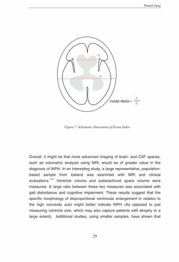
Overall, it might be that more advanced imaging of brain- and CSF spaces, such as volumetric analysis using MRI, would be of greater value in the diagnosis of iNPH. In an interesting study, a large representative, population-based sample from Iceland was examined with MRI and clinical evaluations.117 Ventricle volume and subarachnoid space volume were measured. A large ratio between these two measures was associated with gait disturbance and cognitive impairment. These results suggest that the specific morphology of disproportional ventricular enlargement in relation to the high convexity sulci might better indicate iNPH (As opposed to just measuring ventricle size, which may also capture patients with atrophy to a large extent). Additional studies, using smaller samples, have shown that
Epidemiology of Normal Pressure Hydrocephalus
30
measurement of ventricular volume in relation to cortical thickness and subarachnoid space volume might be of value in differentiating iNPH from other neurodegenerative diseases.130, 131
Finally it is important to discuss the fact that ventricle size does not appear to correlate with improvement after CSF diversion.132, 133 If this is true, then it might not be meaningful at all to have a have a certain defined cut-off for either Evans Index or any other measure of ventricle size. For example, it is not known whether patients with symptoms of iNPH who have values of Evans Index less than 0.3 could respond to treatment.
CSF tap-test The CSF tap test is a well-known diagnostic test for iNPH. CSF is removed through a lumbar puncture after which the patient is evaluated for possible improvement.134 The CSF tap test has been used for the diagnosis of iNPH, and prediction of who will benefit from treatment, for several decades. However, the predictive value of the CSF tap test is not precisely known. The exact way of carrying out the test has not been standardized.42 For example, the amount of CSF removed has varied between 30 and 50 ml. Also, the specific types of clinical evaluations and time between CSF removal and clinical evaluation have varied. Different authors have also used different cut-off criteria for the classifying patients as improved or not. According to a recent systematic review 135 the average sensitivity, for a favorable treatment outcome, was 58 % (ranging from 26 % to 87 %). The average specificity was 75 % (ranging from 33 % to 100 %). Thus, current data suggests that the CSF tap test is not suitable for ruling out patients from treatment and has, an overall, rather limited clinical value.
CSF Infusion tests Various aspects of CSF dynamics can be measured using so-called infusion tests. More precisely, the resistance of CSF outflow (Rout) can be calculated by infusing saline through a lumbar puncture. Increased CSF outflow resistance, i.e. a high Rout, has been said to be an important diagnostic marker and predictor of shunt response.38 It is thus often used in the clinical work-up of iNPH. However, when considering existing data, CSF infusion
Daniel Jaraj
31
studies are probably not reliable for diagnosing iNPH and should not be used for selecting candidates for shunt surgery. Values of Rout, that are believed to indicate iNPH, may be found in as many as 25 % of healthy elderly.41 In addition, a more recent, large prospective cohort study found no relation between CSF outflow resistance and outcome after shunt surgery.90 Therefore, similar to the tap-test, measurement of CSF outflow resistance is probably of limited value.
Treatment Diversion of CSF by a surgically placed shunt catheter is currently the only evidence-based treatment of iNPH.136 Different methods of CSF drainage can be applied. The most common include placement of a ventriculo-peritoneal shunt, in which the proximal catheter tip is inserted in the lateral ventricles and the distal end within the peritoneal cavity.137 Similarly, in ventriculo-atrial shunts the proximal tip is within the lateral ventricles, but the distal end is placed in the right atrium of the heart.138 In lumbo-peritoneal shunts, the proximal end of the catheter is placed within the dura mater in the spinal canal, and the distal end within the peritoneal cavity.139 Many other locations for shunt placement have previously been described, but have not gained acceptance.140
So far most studies on the treatment in iNPH have been non-blinded and made without control groups. High-level evidence, such as randomized controlled trials (RCT’s), has so far been scare.141 However, several well-conducted observational studies have indicated that iNPH patients may benefit substantially from shunt surgery. It might also be of interest to point out that the difference between observational studies and RCT’s may possibly be overstated. RCT’s are often considered to be the gold standard of evidence. However, two earlier papers have compared RCT’s to observational studies by examining several different disorders in various research areas.142, 143 The studies found that the effects estimated by RCT’s and those estimated by observational studies were highly similar.

Epidemiology of Normal Pressure Hydrocephalus
32
Of further note, there is one previous RCT that has compared ventriculo-peritoneal shunting to placebo.67 However, the study has so far received surprisingly little attention. In that study, iNPH patients with concomitant Binswanger’s disease were randomized to receive either a standard ventriculo-peritoneal shunt, or a ligated shunt. Both patients and caregivers were blinded to the intervention. Three months post-randomization, patients in the treatment arm improved while no improvement was noted in the control group. When the ligated shunts were opened in the control patients they to improved. Despite the fact that only fourteen patients were included, significant differences were detected. The study was stopped early after interim analysis.
One important observational study has provided further evidence for CSF diversion.144 The study sample consisted of 33 iNPH patients who were inadvertently subjected to a severe delay of treatment (more than six months, due to a major administrative failure of the hospital). These were compared to 69 patients who were treated in normal fashion, within three months. A substantial deterioration occurred in those with delayed treatment, while those treated within three months improved. Although the study actually intended to examine the natural history of iNPH, it may also be regarded as a study on the effect of treatment based on a within-subject design. It is reasonable to assume that that the administrative failure leading to delayed treatment affected patient groups at random. Therefore, the study could be considered a natural experimentD, which might allow for causal inference, at least to some extent.145, 146
A systematic review, published in 2013, examined 64 studies comprising more than 3,000 patients.147 According to the results of this paper, the percentage of patients improving after shunt surgery has increased considerably over the past decades. According to the studies published
D Natural experiments can be described as studies in which randomization is not performed by the researchers, but instead occurs due to an exogenous, chance phenomenon. They can provide important information and allow for causal inference in circumstances when randomization is not ethical or impractical for various other reasons. These types of studies are common in economics.

Daniel Jaraj
33
during the last five years, an estimated 82 % of patients improved after treatment. Moreover, during the past decades, shunt-related mortality, morbidity and revision rates have decreased substantially.
Improved cognition after shunt surgery has been reported in several studies43, 148, 149 including a recent meta-analysis150 that showed improved global cognitive function as well as enhanced memory and psychomotor speed.
In a European multicenter study, 142 iNPH patients, from thirteen centers in nine countries were included.151 All patients were treated with shunt. At follow-up, after one year, 69 % had improved at least one level on the modified Rankin scale152 (mRS). Almost one third of the sample improved two or three levels. The percentage of patients being able to live independently increased from 53 %, before surgery to 82 % after. Based on the outcome of an iNPH-scale153, 84 % of the patients were classified as improved (≥ 5 points).
In the SINPHONI-study8, another large multicenter study, consisting of 26 centers in Japan, 100 iNPH patients were treated with a ventriculo-peritoneal shunt. The primary outcome was improvement of the mRS. Secondary outcome measures were based on an iNPH grading scale140, timed “Up & Go” test, and the mini mental state examination (MMSE). Follow-up examinations were conducted at 3, 6 and 12 months after shunt surgery. Almost 70 % of the patients improved at least one level on the mRS. The percentage of patients who improved to mRS ≤ 1 (i.e. no functional impairment) increased from 7 % to 44 % after treatment. The mean value of MMSE increased from 23 to 25 after treatment. Significant improvement was also noted in all other secondary outcome measures.
The SINPHONI-2, a subsequent study, was a prospective randomized controlled trial that included 93 patients from 20 different centers.154 Study participants were randomly assigned to receive shunt surgery or conservative management over a three-month period. At follow-up, three months after randomization, 65 % of those treated with a lumbo-peritoneal shunt improved

Epidemiology of Normal Pressure Hydrocephalus
34
≥ 1 level on the mRS, compared to only 5 % in those with conservative therapy. Almost all other tests of gait, cognition and caregiver burden also indicated significant improvement in the treatment arm. However, it is important to note that the study was neither sham-controlled nor blinded. Nevertheless the findings are important and indicate that CSF diversion is beneficial. Comparing results from the SINPHONI and SINPHONI-2 trials, lumbo-peritoneal shunts seem to have a slightly higher revision rate, but similar safety and efficacy compared to ventriculo-peritoneal shunts.155 Head-to-head studies have however so far not been conducted.
Assessment of outcome after shunt surgery There is no standard measure of outcome in iNPH. Thus improvement after treatment has been defined in different ways in different studies. A commonly used method to evaluate outcome is the mRS. However, it is important to bear in mind that change in mRS is a rather crude outcome measure. This implies that only substantial changes can be detected. It is therefore plausible that the mRS is less prone to placebo-effect and observer bias. Evaluation can also be made using a specific outcome scale for iNPH.153 This covers four symptom domains (gait, balance, cognition and urinary incontinence) and includes both ordinal and continuous variables. The scale is calibrated and norm-based. Several other scales156, 157 have also been developed, but have not gained acceptance.
Prognosis Mortality in iNPH patients, treated with shunt, has been found to be approximately two to three times higher than in the general population, and similar to patients with stroke.158-160 So far, almost all studies regarding the prognosis in iNPH have been based on convenience samples.161-163 Furthermore, there is little data on the natural course, i.e. prognosis in untreated patients.164
As mentioned in the previous section, an earlier study examined the progression of hydrocephalic symptoms in a group of iNPH patients who were unable to undergo treatment due to a severe delay by the hospital.144

Daniel Jaraj
35
The study found that untreated patients deteriorated considerably, with worsening gait and cognition. However, there are currently no epidemiological studies regarding the natural course. Furthermore, it is not known whether treatment with shunt surgery increases survival.
Although cognitive impairment is one of the main symptoms in iNPH, the risk of developing dementia is not precisely known. A recent study from a large registry-based sample consisting of patients with suspected iNPH showed a high risk of cognitive impairment and dementia in both shunted and non-shunted individuals during a follow-up period of over 4 years (46 % of shunt responders were eventually diagnosed with dementia).165 However, all patients who were diagnosed with iNPH received shunts. Thus, the non-shunted patients had other diagnoses, such as Alzheimer’s disease or vascular dementia. Also, the decision to shunt was based on results from intracranial pressure monitoring which might have biased the results. Nevertheless, the findings indicate that iNPH is an important cause of dementia. Early diagnosis and treatment is probably crucial.

Never before in human history has the population been as aged as currently. Over the last fifty years, the number of older persons has tripled. Furthermore, the number of older persons is expected to triple again over the next fifty years.
Daniel Jaraj
37
2 THE AGING SOCIETY AND THE AGING BRAIN
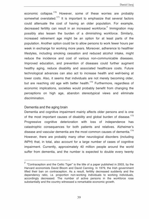
The world’s older population is increasing rapidly in almost all countries. Currently, approximately 12 % of the world’s population is aged 60 years or more.166 By 2050, this number is expected to increase to more than 21 % (Figure 8). Furthermore, the older population is aging as well. The number of persons aged 80 years or more, is projected to triple over the next thirty to forty years.167 Several major challenges lie ahead for societies throughout the world. However, given improved health and increased longevity, population aging can also be viewed as a success for mankind.
The causes of population aging include decreased mortality, in particular due to decreased tobacco use and decreased mortality from cardiovascular diseases.168 Declining fertility is also an important cause.166 Interestingly, there seems to be a continuing trend for decreasing mortalityE and current data does not support the theory that humans are reaching a theoretical upper age-limit.168 For more than 160 years, life expectancy in the record-holding countries has been increasing by almost three months per year.169 Also, based on long-term trends, there is no indication that the increase in life expectancy is abating.169 Indeed, this raises an intriguing question. What is the maximum life span of a human being?
E In the year 1800, average life expectancy in Sweden was less than 40 years. In 1900, this number had increased to slightly higher than 50 years. Today, the average life expectancy is more than 80 years (Source: UN Demographic yearbook and Statistics Sweden, SCB).

Epidemiology of Normal Pressure Hydrocephalus
38
Figure 8. Population aging (Adapted from World Population Ageing 2013 by
United Nations, Department of Economic and Social Affairs, Population Division, © 2013 United Nations. Reprinted with the permission of the United Nations)
Potential Social and Economic Consequences Increased longevity and population aging poses several important economic and societal challenges. Indeed, major interventions are needed to cope with these concerns. Such interventions might be to raise retirement age, decrease benefits or increase taxes. However, the rapid increase in population age is historically unprecedented. Therefore, fundamental reforms with innovative reorganization of health care policies and welfare systems are required.
Population aging has been described as a major threat to societies with potential devastating consequences such as increased suffering and possible
�����
������������������ �
������������������� �
������������������ �
�������� ����
������
���
����
����
��������� ������������� ����� ������!��"��������#���
Daniel Jaraj
39
economic collapse.170 However, some of these worries are probably somewhat overstated.171 It is important to emphasize that several factors could attenuate the cost of having an older population. For example, decreased fertility can result in an increased workforce.F Immigration may possibly also lessen the burden of a diminishing workforce. Similarly, increased retirement age might be an option for at least parts of the population. Another option could be to allow persons to work fewer hours per week in exchange for working more years. Moreover, adherence to healthier lifestyles, including smoking cessation and reduced alcohol intake, might reduce the incidence and cost of various non-communicable diseases. Improved education, and prevention of diseases could further augment healthy aging, reduce disability and associated healthcare costs. Future technological advances can also act to increase health and well-being at lower costs. Also, it seems that individuals are not merely becoming older, but are reaching old age with better health.172 Furthermore, regardless of economic implications, societies would probably benefit from changing the perceptions on high age, abandon stereotypical views and eliminate discrimination.
Dementia and the aging brain Dementia and cognitive impairment mainly affects older persons and is one of the most important causes of disability and global burden of disease.173 Progressive cognitive deterioration with loss of independence has catastrophic consequences for both patients and relatives. Alzheimer’s disease and vascular dementia are the most common causes of dementia.174 However, there are probably many other neurological disorders (including iNPH) that, in total, also account for a large number of cases of cognitive impairment. Currently, approximately 40 million people around the world suffer from dementia, and the number is expected to double every twenty
F ”Contraception and the Celtic Tiger” is the title of a paper published in 2003, by the Harvard economists David Bloom and David Canning. In 1979, the Irish government lifted their ban on contraception. As a result, fertility decreased suddenly and the dependency ratio, i.e. proportion non-working individuals to working individuals, accordingly decreased. The number of active persons in the workforce rose substantially and the country witnessed a remarkable economic growth.

Epidemiology of Normal Pressure Hydrocephalus
40
years, reaching more than 115 million by 2050.175 The cost of dementia, in 2010, was estimated to be equivalent of 1 % of the worlds GDP.176, 177 However, there are reasons for optimism. For example, the occurrence of dementia seems to be decreasing despite population aging.178, 179 Also, more recently possibilities for primary and secondary prevention have been emerging.180 As we continue to fill knowledge gaps, hopefully lifestyle changes and enhanced living conditions as well as increase in education and socioeconomic reforms may lead us to a better future with improved well-being throughout higher age.

Daniel Jaraj
41
3 AIM The overall aim of this thesis was to examine the epidemiology of iNPH. This included the prevalence, risk factors, estimated ventricle size in the general population, and long-term outcome among untreated individuals. The thesis is based a representative population-based sample of men and women aged 70 years or more.
Paper 1 The aim of the first paper was to determine the prevalence of probable iNPH, and occurrence of radiological signs consistent with iNPH.
Paper 2 The aim of the second paper was to examine vascular risk factors and WMLs in relation to clinical and imaging signs of iNPH, using a nested case-control analysis.
Paper 3 In paper three, the aim was to examine ventricle size and provide reference values for Evans Index (a diagnostic marker for iNPH).
Paper 4 The aim of the fourth paper was to study long-term outcome among untreated individuals with probable iNPH and persons with imaging signs of iNPH. Outcomes were mortality, dementia and progression of hydrocephalic symptoms.

Epidemiology of Normal Pressure Hydrocephalus
42
Daniel Jaraj
43
4 METHODS AND STUDY DESIGN
Study sample - The Gothenburg Population Studies The Gothenburg population studies include several representative cohorts, followed for almost fifty years. The studies consist of detailed examinations designed to investigate aging and age-related disorders. All participants were systematically obtained from the Swedish population register based on birth dates, and included people living in private households and in residential care. The examinations comprised physical and psychiatric examinations including laboratory and radiological work-up. Also, house-visits, telephone interviews, close-informant interviews and non-respondent analyses were made. In addition, the studies were complemented with data from the Swedish national inpatient register and Swedish cause of death register. For the present thesis, data from the following cohorts were included.
The Population Studies of Women The Population studies of women (PPSW)181, 182 began in 1968 and baseline examinations included women aged 38 to 65 (born between 1914 and 1930). The PPSW is currently still ongoing.
The Longitudinal Gerontological and Geriatric Population Studies and H85 Studies in Gothenburg The Longitudinal Gerontological and Geriatric Population Studies (H70) and H85-cohorts183-186 are population-based cohort studies that began in 1971. Similar to the PPSW, the examinations included comprehensive assessments of somatic and psychiatric disorders, physical examinations including blood test, lumbar puncture, ECG, radiological examinations, anthropometric measurements and data on psychosocial background factors and psychometric tests. The first examinations included 70-year old men and women born in 1901-02. Follow-up examinations have been conducted at various ages. In 1986, a follow-up examination was conducted and the prevalence of dementia was determined with the help of CT imaging of the
Epidemiology of Normal Pressure Hydrocephalus
44
brain.183 At the time, the sample included approximately half of all 85-year olds in Gothenburg. The cohort examined in 2000 (70-year-olds born in 1930) was merged with PPSW cohort, i.e. they were examined together.
The Nordic Research on Ageing Studies The Nordic Research on Ageing (NORA)187 studies began in 1990 and were conducted in Gothenburg, Sweden, and additional sites in Denmark and Finland. The sample included 75-year-olds at baseline, and follow-up was made five years later at age 80. For the present thesis, only those examined in Gothenburg were included.
Study population in papers I-VI We merged data from the studies described above, and included all persons who underwent one or more CT examination of the brain between 1986 and 2000. All participants were aged 70 years or more at baseline. An overview of the cohorts is shown in the figure 9. A total of 3246 individuals were invited to take part and 2182 accepted (response rate 67 %), with no significant difference in response rate between men and women (65 % versus 68 %; p=0.107). Of those who took part in the clinical examinations, 1238G underwent a CT scan of the brain (response rate 58 %; 60 % for men and 56 % for women; p=0.067). The selection of study participants is shown in figure 10. Participants in the CT study were on average slightly younger and performed better on the Mini Mental State Examination (MMSE). There were no significant differences in the prevalence of dementia or major depression between the CT group and non-CT group (table 1). Also, the participants in each cohort were similar to non-participants regarding several demographic factors such as age, sex, marital status, mortality and various psychiatric illnesses. Thus, the sample can considered
G Correction: Subsequently it was noted that two persons had undergone CT imaging but had no other clinical data. These were therefore excluded. Also, one person was found to be less than 70 years at the time of CT. This person was therefore also excluded. After re-analyzing the data, these changes were did not alter any results significantly. However, in paper I results are presented based on a sample of 1238 while subsequent studies included 1235 participants.

Daniel Jaraj
45
representative of the population. However, due to having merged individual cohorts, unbalance occurred in the total data set, in particular regarding age groups and sex. The distribution of the merged sample is shown in table 2.
Figure 9. Gothenburg population studies, overview of the cohorts included in this study
The sample comprised data from four prospective cohort studies. NORA = Nordic Research On Ageing, PPSW = Prospective Population study of Women, H85-H90 and H70 = Longitudinal Gerontological and Geriatric Population Studies in Gothenburg, Sweden. Baseline examination was considered at time of first CT. All persons were aged 70 years or more at baseline. Data from the Swedish National Inpatient Register and the Population Register was available until 2012
2000
2005
2009
2000
1990, 1991
1995, 1996
PPSW Studies
Birth cohorts 1908, 1914, 1918, 1922, 1930
2005 Women aged 70 + years
Women aged 70 + years
Women aged 70 + years
1986, 1987
1989, 1990
1991, 1992
H85 Studies
Birth cohorts 1901, 1902
85 year-olds
88 year-olds
90 year-olds
NORA Studies Birth cohorts 1915, 1916
75year-olds
80 year-olds
Women aged 70 + years 2009
H70 Studies Birth cohort 1930
70year-olds
75 year-olds
1992
79 year-olds
Pooled sample (n = 1235)
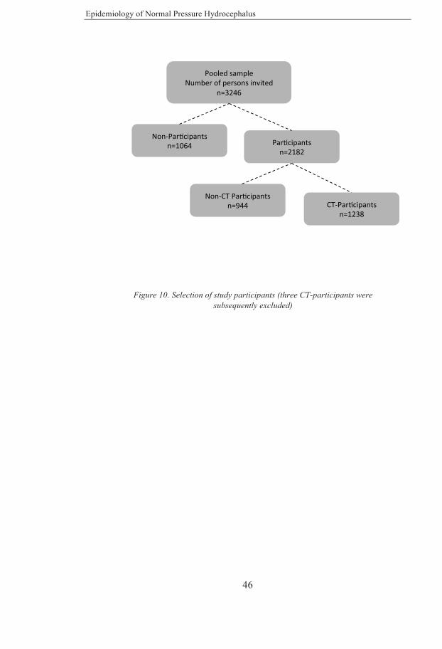
Epidemiology of Normal Pressure Hydrocephalus
46
Figure 10. Selection of study participants (three CT-participants were subsequently excluded)
PooledsampleNumberofpersonsinvited
n=3246
Par9cipantsn=2182
Non-Par9cipantsn=1064
CT-Par9cipantsn=1238
Non-CTPar9cipantsn=944

Daniel Jaraj
47
Table 1. Comparison between CT participants and non-CT participants
Non-CT Group (N=944)
CT Group (N=1235)a p-value
Women, % 76.4 72.9 0.067
Dementia (DSM-III-R), % 12.0 10.8 0.401
Major depression, % 8.4 6.7 0.136
Mean age (SD) 77.2 (6.4) 74.7 (6.0) <0.001
Mean MMSE score (SD) 26.1 (5.9) 26.8 (5.0) 0.002
Legend: DSM-III-R=Diagnostic And Statistical Manual of Mental Disorders, 3rd edition revised, MMSE= Mini-Mental State Examination, SD=Standard Deviation. a=Two persons were excluded due to complete lack of data. In addition, one individual was subsequently noted to be less than 70 years at baseline. This person was therefore also excluded.
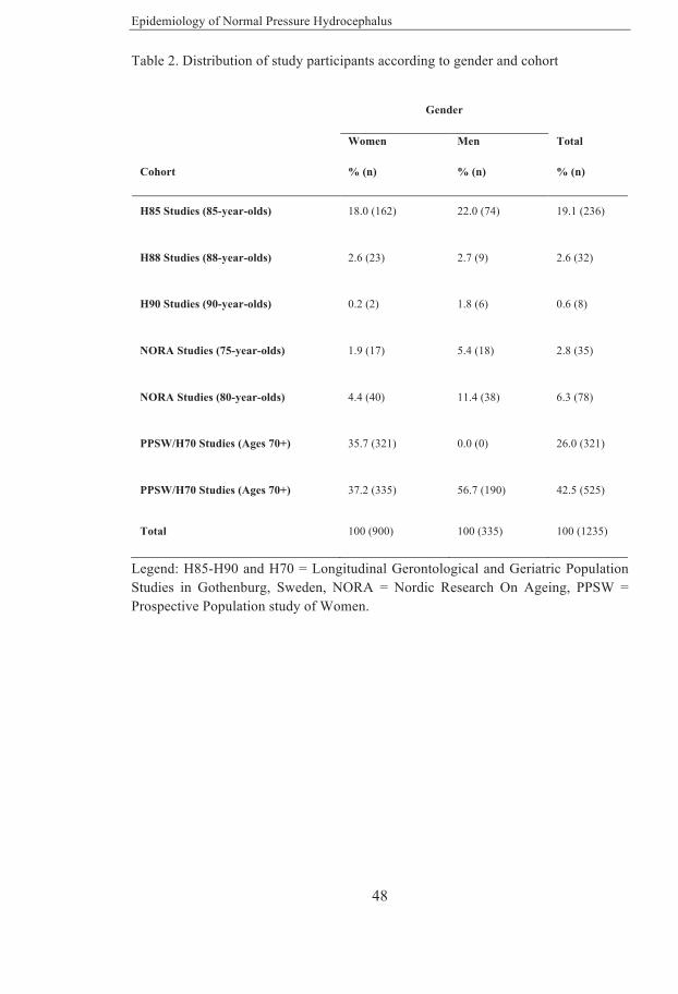
Epidemiology of Normal Pressure Hydrocephalus
48
Table 2. Distribution of study participants according to gender and cohort
Gender
Cohort
Women
% (n)
Men
% (n)
Total
% (n)
H85 Studies (85-year-olds) 18.0 (162) 22.0 (74) 19.1 (236)
H88 Studies (88-year-olds) 2.6 (23) 2.7 (9) 2.6 (32)
H90 Studies (90-year-olds) 0.2 (2) 1.8 (6) 0.6 (8)
NORA Studies (75-year-olds) 1.9 (17) 5.4 (18) 2.8 (35)
NORA Studies (80-year-olds) 4.4 (40) 11.4 (38) 6.3 (78)
PPSW/H70 Studies (Ages 70+) 35.7 (321) 0.0 (0) 26.0 (321)
PPSW/H70 Studies (Ages 70+) 37.2 (335) 56.7 (190) 42.5 (525)
Total 100 (900) 100 (335) 100 (1235)
Legend: H85-H90 and H70 = Longitudinal Gerontological and Geriatric Population Studies in Gothenburg, Sweden, NORA = Nordic Research On Ageing, PPSW = Prospective Population study of Women.

Daniel Jaraj
49
Radiological Examinations Between 1986 and 2000, study participants underwent CT imaging of the brain. All scans were in the transverse plane, and no contrast was used. Three observers, a medical studentH, a resident in neurosurgeryI and a consultant in neurologyJ, collectively made an initial screening assessment of all CT images. The observers were, at the time of the image assessments, unaware of clinical data. Each case was evaluated for hydrocephalic ventricular enlargement, i.e. radiological findings consistent with iNPH. This was based on previous descriptions in the literature 1, 8, 59, 188, and was defined as enlargement of all four ventricles without equivalent widening of cortical sulci or obstruction of CSF flow or structural lesions. Persons with extracerebral mass lesions or other pathologies influencing ventricular morphology were excluded. The presence of WMLs was not considered for the diagnosis. Cases in which ventricular dilation was caused by conditions other than NPH, such as brain atrophy, obstructive hydrocephalus or loss of brain parenchyma were not considered as hydrocephalic ventricular enlargement. Evans Index was measured in all cases, and defined as the ratio between the largest width of the frontal horns and the largest inner diameter of the skull. Occluded sulci at the high convexity (OccSul) was defined as not having any sulcus extending to the midline at the falx cerebri on the two uppermost CT slices. A consultant neuroradiologistK re-evaluated all images that in the initial assessment screened positive for hydrocephalic ventricular enlargement or had uncertain findings. The neuroradiologist made the final diagnosis in all cases.
Diagnosis of iNPH The diagnosis of iNPH was based on the results of the imaging findings, clinical examinations and data from interviews. This was made in concordance with criteria from the American-European iNPH guidelines.1
H Daniel Jaraj (Then medical student, now licensed physician) I Katrin Rabiei (Then Neurosurgical resident, now specialist in Neurosurgery) J Carsten WikkelsØ (Consultant physician and Professor of Neurology) K Christer Jensen (Consultant physician, specialist in Neuroradiology)

Epidemiology of Normal Pressure Hydrocephalus
50
History of severe head trauma, meningitis or subarachnoid bleeding were exclusion criteria.
Gait disturbance was assessed by evaluating data from clinical examinations and interviews. Physicians, specializing in geriatrics, performed physical examinations in which a general assessment of gait was made. The examiner graded walking difficulties as non-existent, slight or extensive. Participants had also been asked questions regarding gait and walking difficulties on interviews. Gait disturbance was defined as presence of any walking difficulty on examination and/or presence of self-reported walking difficulty. Cognitive function was evaluated with the Mini Mental State Examination (MMSE). A score of 25 or less was defined as cognitive impairment. Urinary incontinence was assessed by self-report, and defined as involuntary leakage of urine occurring more than once per week.
Probable iNPH was diagnosed in participants who had hydrocephalic ventricular together with gait disturbance and one of either MMSE ≤ 25 or urinary incontinence. Possible iNPH was diagnosed in persons with hydrocephalic ventricular enlargement and at least one other core clinical sign of iNPH, i.e. gait disturbance, MMSE ≤ 25 or urinary incontinence.
Papers I – IV Paper I The prevalence of probable iNPH, and radiological signs of iNPH, was calculated by dividing the number of persons with each finding by the total number of study participants. Crude prevalence rates regarding age group and sex were compared using the Fisher’s exact test. In addition, the association between radiological signs, and symptoms of iNPH were examined using multiple binary logistic regression models adjusting for age and sex.
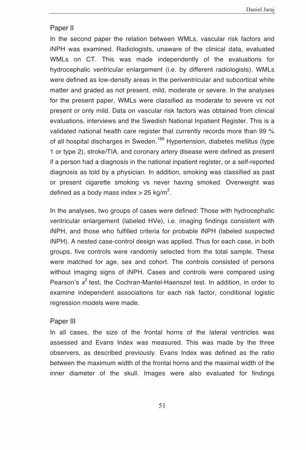
Daniel Jaraj
51
Paper II In the second paper the relation between WMLs, vascular risk factors and iNPH was examined. Radiologists, unaware of the clinical data, evaluated WMLs on CT. This was made independently of the evaluations for hydrocephalic ventricular enlargement (i.e. by different radiologists). WMLs were defined as low-density areas in the periventricular and subcortical white matter and graded as not present, mild, moderate or severe. In the analyses for the present paper, WMLs were classified as moderate to severe vs not present or only mild. Data on vascular risk factors was obtained from clinical evaluations, interviews and the Swedish National Inpatient Register. This is a validated national health care register that currently records more than 99 % of all hospital discharges in Sweden.189 Hypertension, diabetes mellitus (type 1 or type 2), stroke/TIA, and coronary artery disease were defined as present if a person had a diagnosis in the national inpatient register, or a self-reported diagnosis as told by a physician. In addition, smoking was classified as past or present cigarette smoking vs never having smoked. Overweight was defined as a body mass index > 25 kg/m2.
In the analyses, two groups of cases were defined: Those with hydrocephalic ventricular enlargement (labeled HVe), i.e. imaging findings consistent with iNPH, and those who fulfilled criteria for probable iNPH (labeled suspected iNPH). A nested case-control design was applied. Thus for each case, in both groups, five controls were randomly selected from the total sample. These were matched for age, sex and cohort. The controls consisted of persons without imaging signs of iNPH. Cases and controls were compared using Pearson’s x2 test, the Cochran-Mantel-Haenszel test. In addition, in order to examine independent associations for each risk factor, conditional logistic regression models were made.
Paper III In all cases, the size of the frontal horns of the lateral ventricles was assessed and Evans Index was measured. This was made by the three observers, as described previously. Evans Index was defined as the ratio between the maximum width of the frontal horns and the maximal width of the inner diameter of the skull. Images were also evaluated for findings
Epidemiology of Normal Pressure Hydrocephalus
52
consistent with iNPH as described above. All uncertain cases were reevaluated by a neuroradiologist.
Descriptive statistics were examined for the total sample and subgroups based on sex and age. Mean and median values as well as dispersion, distribution and outliers were assessed using the descriptive data, histograms and box-plots. Independent sample t-tests were used to compare mean values between groups. Evans Index was also dichotomized as > 0.3 vs < 0.3 and differences were tested using the Pearson’s x2 test. Statistical analyses were made using SPSS version 22.0 (SPSS Inc., Chicago, IL) and R version 3.2.2.190
Paper IV Baseline examinations were performed between 1986 and 2000. One, or more, follow-up examination was made for all cohorts until 2009 (figure 9). Median follow-up time was 11.5 years (interquartile range 6.5 years) with a maximum follow-up of 25 years. Data was obtained from the somatic and psychiatric examinations, interviews, close-informant interviews and complemented with data from the Swedish National Inpatient Register.
Possible iNPH and probable iNPH was diagnosed in accordance with criteria from international consensus guidelines, as described previously. Dementia was diagnosed by geriatric psychiatrist using the Diagnostic and Statistical Manual of Mental Disorders (third edition, revised) criteria. Date of death was obtained from the Swedish Population Register (National Board of Health and Welfare’s Cause of Death Register). This is a national register that is complete regarding mortality among all Swedish residents both in Sweden and abroad.191
Progression to probable iNPH, from possible iNPH or asymptomatic hydrocephalic ventricular enlargement, was evaluated using criteria the American-European iNPH guidelines. This was made using data from the clinical examinations and the Swedish National Inpatient Register. The development of gait disturbance cognitive impairment or urinary incontinence was also assessed in those with hydrocephalic ventricular enlargement who
Daniel Jaraj
53
were asymptomatic at baseline.
Among those with hydrocephalic ventricular enlargement (n=55), two persons had previously been treated with shunt surgery and were therefore excluded from all analyses. In the analyses, outcomes were examined for all those with hydrocephalic ventricular enlargement. In addition, they were divided into the following subgroups: those with probable iNPH (n=24), and those with possible iNPH or asymptomatic ventricular enlargement (n=29).
The date of the first CT examination was considered as baseline. All comparisons were made with individuals without hydrocephalic ventricular enlargement (n=1180). Crude five-year mortality was compared between groups using the Pearson’s x2 test. Mortality and risk of dementia was also examined using Kaplan-Meier survival curves, the Log-rank test and Cox proportional hazard models for each group. The models were adjusted for baseline age, sex, and cohort. In addition, a binary logistic regression models was made to examine the association between hydrocephalic ventricular enlargement (all cases, n=53) and risk of dementia at any time during the study, i.e. baseline and follow-up. Adjustment was made for baseline age, sex and cohort. Statistical analyses were made using SPSS version 22.0 (SPSS Inc., Chicago, IL) and R version 3.2.2.190

Epidemiology of Normal Pressure Hydrocephalus
54
5 RESULTS AND DISCUSSION Paper I The prevalence of iNPH, in the total sample (n=1238) was 2.1 %. Stratifying by age groups, the prevalence was 0.2 % in those aged 70-79 years, and 5.9 % in those aged 80 years or more (Table 3). There was no difference in the prevalence regarding sex. However, the prevalence was strongly related to being older than 80 years (OR 35.5; 95 % CI 4.8-262.7). The prevalence of imaging findings of iNPH is shown in table 4. The relation between symptoms of iNPH and radiological findings is shown in table 5.
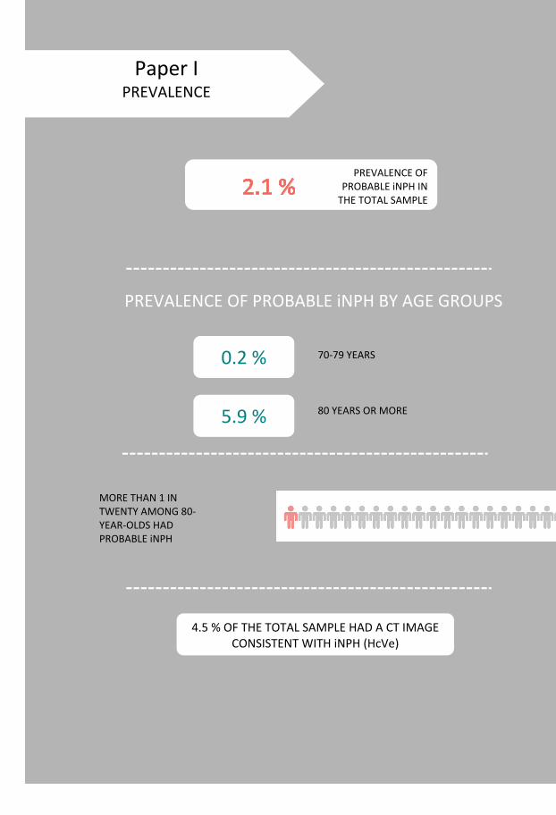
Paper I PREVALENCE
80 YEARS OR MORE 5.9 %
0.2 % 70-79 YEARS
PREVALENCE OF PROBABLE iNPH BY AGE GROUPS
MORE THAN 1 IN TWENTY AMONG 80-YEAR-OLDS HAD PROBABLE iNPH
4.5 % OF THE TOTAL SAMPLE HAD A CT IMAGE CONSISTENT WITH iNPH (HcVe)
PREVALENCE OF PROBABLE iNPH IN
THE TOTAL SAMPLE

Epidemiology of Normal Pressure Hydrocephalus
56
Table 3. Prevalence of probable iNPH by age group and gender (N=1238)
Age groups Men %(n/N) Women % (n/N) Total % (n/N)
70-79 0.5 (1/208) 0.2 (1/626) 0.2 (2/834)
80 + 7.1 (9/127) 5.4 (15/277) 5.9 (24/404)
Total 3.0 (10/335) 1.8 (16/903) 2.1 (26/1238)
Legend: n= The number of persons with probable iNPH. N= The number of persons included in this study. Prevalence in 70-79 vs 80+ p <0.001, Prevalence in men vs women p = 0.186
Table 4. Prevalence of Hydrocephalic Ventricular Enlargement (HcVe), Evans Index (EI) > 0.3, and Occluded sulci by sex and age groups (N=1238) Men % (n=335) Women % (n=903) Total % (n=1238)
70-79 80+ Tot. 70-79 80+ Tot. 70-79 80+ Tot.
HcVe 5.3
(11)
11.0
(14)NS
7.5
(25)
1.4
(9)
7.4
(22)d
3.4
(31) a
2.4
(20)
8.9
(36) d
4.5
(56)
EI>0.3 23.6
(49)
46.5
(59) d
32.2
(108)
9.9
(62)
31.0
(86)d
16.4
(148)b
13.3
(111)
35.9
(145) d
20.7
(256)
Occluded
Sulci
6.3
(13)
11.0
(14) NS
8.1
(27)
3.8
(24)
5.8
(16) NS
4.4
(40) a
4.4
(37)
7.4
(30)c
5.4
(67)
Legend: Data are in percentages a = p<0.005, b = p<0.001: differences between sex, NS = p≥0.05, c = p <0.05, d = p<0.001: differences between age groups
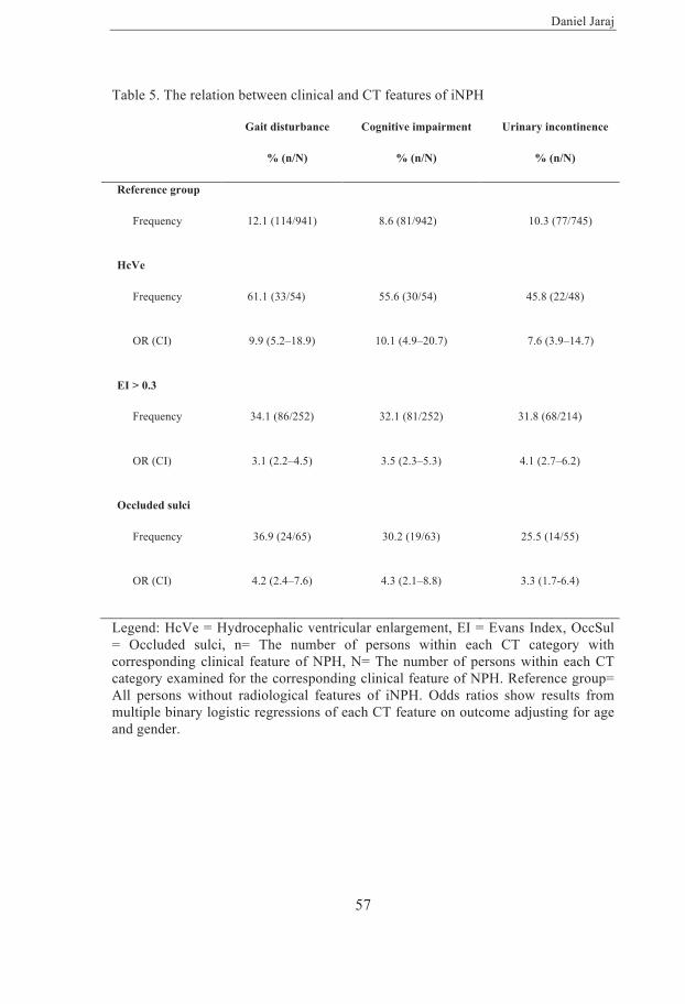
Daniel Jaraj
57
Table 5. The relation between clinical and CT features of iNPH Gait disturbance
% (n/N)
Cognitive impairment
% (n/N)
Urinary incontinence
% (n/N)
Reference group
Frequency 12.1 (114/941) 8.6 (81/942) 10.3 (77/745)
HcVe
Frequency 61.1 (33/54) 55.6 (30/54) 45.8 (22/48)
OR (CI) 9.9 (5.2–18.9) 10.1 (4.9–20.7) 7.6 (3.9–14.7)
EI > 0.3
Frequency 34.1 (86/252) 32.1 (81/252) 31.8 (68/214)
OR (CI) 3.1 (2.2–4.5) 3.5 (2.3–5.3) 4.1 (2.7–6.2)
Occluded sulci
Frequency 36.9 (24/65) 30.2 (19/63) 25.5 (14/55)
OR (CI) 4.2 (2.4–7.6) 4.3 (2.1–8.8) 3.3 (1.7-6.4)
Legend: HcVe = Hydrocephalic ventricular enlargement, EI = Evans Index, OccSul = Occluded sulci, n= The number of persons within each CT category with corresponding clinical feature of NPH, N= The number of persons within each CT category examined for the corresponding clinical feature of NPH. Reference group= All persons without radiological features of iNPH. Odds ratios show results from multiple binary logistic regressions of each CT feature on outcome adjusting for age and gender.

Epidemiology of Normal Pressure Hydrocephalus
58
This is, to date, the largest population-based study on the prevalence of iNPH. According to the results, more than one in twenty, among 80-year-olds have signs and symptoms consistent with probable iNPH. Furthermore, 4.5 % of the total sample, and almost 9 % of 80-year-olds, had hydrocephalic ventricular enlargement, i.e. radiological findings consistent with iNPH. This finding is important considering that ventricular enlargement might be the earliest sign of iNPH.103 If the results of this study are true, an estimated 700,000 persons in the United States, and two million persons in Europe might be suffering from iNPH. Earlier estimates have shown that less than one in twenty are being treated94, 104 and the results from this study further indicate that iNPH is highly underdiagnosed and undertreated.
The most important limitation of this study concerns the diagnosis of probable iNPH. The diagnosis was made retrospectively, in a rather crude way, using data from interviews and clinical examinations. Although cases were diagnosed using criteria from international consensus guidelines, there were several important clinical features that were not assessed. Gait disturbance, cognitive impairment and urinary incontinence are common among older persons and often non-specific. Therefore, data on more detailed and specific examinations would have been desirable. However, the diagnostic bluntness might have also lead to an underestimation of the prevalence, as some iNPH patients might only have subtle symptoms. Furthermore, all cases diagnosed with probable iNPH had a rather characteristic CT image, i.e. enlarged ventricles disproportional to the subarachnoid space. In addition, the association between symptoms of iNPH and CT findings were rather strong.
Additional limitations of this study include those that pertain to observational studies in general. These include issues such as low participation rate and possible selection bias. Indeed, these factors might have influenced the results. However, a selection bias and a low participation rate would probably have led to an underestimation of the prevalence. Another limitation is the use of CT. The image quality was not optimal in some of the older scans. This is due to the technical standard at the time they were made. MRI might

Daniel Jaraj
59
have been somewhat better, but was unavailable. Nevertheless, despite several limitations, the results of this study indicate that many older persons have clinical and imaging features of iNPH and are in need of further evaluations. iNPH is probably more common than previously supposed. Given the progressive nature and debilitating symptoms, it is crucial to increase the possibilities for early diagnosis and treatment.

Epidemiology of Normal Pressure Hydrocephalus
60
Paper II In this population-based study, WMLs, Hypertension, diabetes, stroke/TIA and overweight were more common in cases with hydrocephalic ventricular enlargement and suspected iNPH compared to matched controls (Table 6). In the regression models, suspected iNPH was associated with moderate to severe WMLs, while hydrocephalic ventricular enlargement was related to history of hypertension, moderate to severe WMLs, and diabetes (Table 7). Among cases with hydrocephalic ventricular enlargement, all but four persons (51/55, 93%) had hypertension, diabetes, or WMLs on CT. Of those with suspected iNPH, all but two persons (24/26, 92%) had hypertension, diabetes, or WMLs on CT.

Paper II RISK FACTORS
HVe (Radiological iNPH)
Hypertension
WMLs
Diabetes
Odds Ratio (95 % CI)
2.7 (1.1-6.8)
6.5 (2.1-20.3)
4.3 (1.1-16.3)
Suspected iNPH (Probable iNPH)
WMLs
Odds Ratio (95 % CI)
5.2 (1.1-6.8)
AMONG CASES WITH RADIOLOGICAL iNPH, ALL BUT 4 PERSONS, i.e. 97 % HAD HYPERTENSION, DIABETES OR WMLs
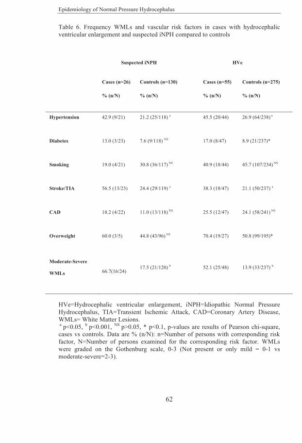
Epidemiology of Normal Pressure Hydrocephalus
62
Table 6. Frequency WMLs and vascular risk factors in cases with hydrocephalic ventricular enlargement and suspected iNPH compared to controls
Suspected iNPH
HVe
Cases (n=26)
% (n/N)
Controls (n=130)
% (n/N)
Cases (n=55)
% (n/N)
Controls (n=275)
% (n/N)
Hypertension 42.9 (9/21) 21.2 (25/118) a 45.5 (20/44) 26.9 (64/238) a
Diabetes 13.0 (3/23) 7.6 (9/118) NS 17.0 (8/47) 8.9 (21/237)*
Smoking 19.0 (4/21) 30.8 (36/117) NS 40.9 (18/44) 45.7 (107/234) NS
Stroke/TIA 56.5 (13/23) 24.4 (29/119) a 38.3 (18/47) 21.1 (50/237) a
CAD 18.2 (4/22) 11.0 (13/118) NS 25.5 (12/47) 24.1 (58/241) NS
Overweight 60.0 (3/5) 44.8 (43/96) NS 70.4 (19/27) 50.8 (99/195)*
Moderate-Severe
WMLs 66.7(16/24) 17.5 (21/120) b 52.1 (25/48) 13.9 (33/237) b
HVe=Hydrocephalic ventricular enlargement, iNPH=Idiopathic Normal Pressure Hydrocephalus, TIA=Transient Ischemic Attack, CAD=Coronary Artery Disease, WMLs= White Matter Lesions. a p<0.05, b p<0.001, NS p>0.05, * p<0.1, p-values are results of Pearson chi-square, cases vs controls. Data are % (n/N): n=Number of persons with corresponding risk factor, N=Number of persons examined for the corresponding risk factor. WMLs were graded on the Gothenburg scale, 0-3 (Not present or only mild = 0-1 vs moderate-severe=2-3).

Daniel Jaraj
63
Table 7. Multivariable analysis of vascular risk factors in hydrocephalic ventricular enlargement and suspected iNPH
Suspected iNPH (n=26) HVe (n=55)
OR (95% CI)
p-value
OR (95% CI)
p-value
Hypertension
2.1 (0.5-8.7)
0.286
2.7 (1.1-6.8)
0.039
WMLs
5.2 (1.5-17.6)
0.009
6.5 (2.1-20.3)
0.001
Stroke/TIA
2.3 (0.7-7.8)
0.178
0.8 (0.2-3.3)
0.741
Diabetes
-
-
4.3 (1.1-16.3)
0.031
Overweight
-
-
1.8 (0.6-5.4)
0.293
HVe=Hydrocephalic ventricular enlargement, iNPH=Idiopathic Normal Pressure Hydrocephalus, OR=Odds Ratio, CI=Confidence Interval, WMLs=White Matter Lesions. WMLs graded on the Gothenburg scale, 0-3 (Not present or only mild = 0-1 vs moderate-severe=2-3). OR’s and p-values show results of conditional logistic regression models. The model for suspected iNPH included history of hypertension WMLs and stroke/TIA. For HVe, the model included History of hypertension, WMLs, Stroke/TIA, Diabetes mellitus and Overweight.

Epidemiology of Normal Pressure Hydrocephalus
64
The results of this study suggest that vascular mechanisms might be involved in the pathophysiology of iNPH. The fact that vascular risk factors are common in iNPH might also be important from a clinical standpoint. Health care professionals ought to be aware of the high occurrence of vascular risk factors in iNPH, considering that they are treatable.
The main strength of this study is the population-based sample. Previous investigations regarding the risk factors in iNPH have been made using hospital-based samples. These studies might have included more severe cases and had a higher risk of selection bias, given that cases and controls were selected separately. In the present study, a large representative sample was examined, and a nested case-control analysis was performed. Thus, cases and controls were obtained from the same population. Furthermore, exposure data was collected prospectively from comprehensive clinical examinations and the Swedish National Inpatient Register. Nevertheless there are several important sources of potential bias that should be considered. Similar to the first study in this thesis, there was a diagnostic bluntness regarding the cases. This might have led to bias and confounding. However, the persons included in this study were not scanned due to symptoms or suspicions of iNPH, yet a strong association between imaging signs of iNPH and several vascular factors was found. The group with hydrocephalic ventricular enlargement should theoretically have been subjected to less bias, considering that they were selected solely on the basis of imaging findings by blinded observers. Also, the number of cases was relatively small. A larger sample would probably have provided more precise outcome estimates. Furthermore, the use of CT is an important limitation, in particular regarding analysis of WMLs. MRI would have been better. Although, it has been suggested that CT might be more specific for detection of clinically relevant WMLs.192 Another important limitation is the fact that the study design does not allow for causal inference. It is not known whether the WMLs cause iNPH, or vice versa. For example, it might be that white matter changes in iNPH are not merely due to small vessel disease, but could also be caused by CSF stagnation or edema due to iNPH. However, it is not particularly common for any single study to, by itself, provide causal

Daniel Jaraj
65
evidence. Thus, it is important to view the results in context. When taking into consideration that several previous, clinical and animal, studies have provided concurring data, the results of this study suggest that vascular disease processes are probably involved in the mechanisms underlying iNPH.

Epidemiology of Normal Pressure Hydrocephalus
66
Paper III Evans Index ranged between 0.11 and 0.46, in the total sample. The mean value was 0.28 (SD 0.04). Descriptive statistics are shown in Table 8. The distribution of Evans Index in the total sample is shown in figure 11.
On average, men had higher values than women, 0.29 vs 0.27 (p<0.001). Also, higher age was associated with larger values of Evans Index in age groups 70-79 years vs 80 years or older (0.27 vs 0.29, p<0.001). The mean value of Evans Index, in men aged 80 years or more was 0.3 (SD 0.03). The number of persons with Evans Index > 0.3, in the total sample was 255 (20.6 %). These findings are shown in table 9 and figure 12. The mean value of Evans Index among those with hydrocephalic ventricular enlargement (n=55) was 0.36 (SD 0.04).

Paper III EVANS INDEX
MEAN VALUES OF EVANS INDEX BY AGE GROUPS
70-74 years 75-79 years 80-84 years 85 + years
More than 1 in 5 individuals above the age of 70 fulfilled current criteria for ventricular enlargement
THE FREQUENCY OF EVANS INDEX > 0.3 IN THOSE AGED 85 YEARS OR MORE
THE FREQUENCY OF EVANS INDEX > 0.3 IN THE TOTAL SAMPLE
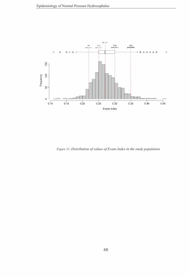
Epidemiology of Normal Pressure Hydrocephalus
68
Figure 11. Distribution of values of Evans Index in the study population
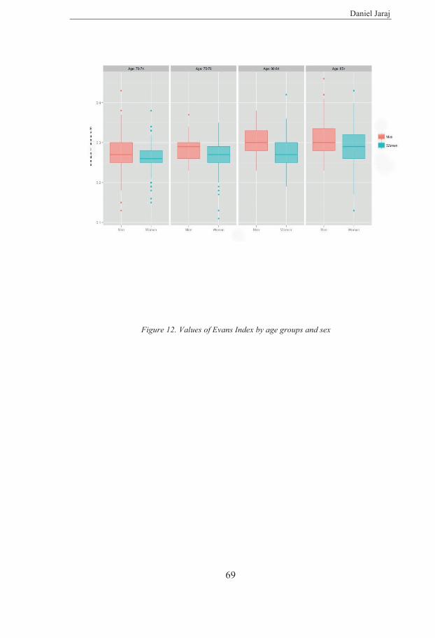
Daniel Jaraj
69
Figure 12. Values of Evans Index by age groups and sex

Epidemiology of Normal Pressure Hydrocephalus
70
Table 8. Descriptive statistics for Evans Index in a population-based sample of 1235 men and women aged 70 years or more
Descriptive variable Value (Total sample n=1235)
Mean 0.275
95 % CI 0.273-0.277
Median 0.270
Interquartile range 0.05
Std. Deviation 0.04
Minimum-Maximum value of EI 0.11-0.46
Percentiles
5th 0.22
10th 0.23
25th 0.25
50th 0.27
75th 0.30
90th 0.33
95th 0.35
CI= Confidence Interval, Std. Deviation= Standard Deviation, EI=Evans Index.

Daniel Jaraj
71
Table 9. Mean values of Evans Index and frequency of values higher than 0.3 by sex and age group in a population based sample of 1235 men and women
Age groups (years)
70-74 75-79 80-84 85+ Total
sample
Mean Evans Index
Men (SD) 0.28 (0.04) 0.29 (0.04) 0.30 (0.03) 0.31 (0.05) 0.29 (0.05)
Women (SD) 0.26 (0.03) 0.27 (0.04) 0.28 (0.04) 0.29 (0.05) 0.27 (0.04)
Total (SD) 0.27 (0.03) 0.27 (0.04) 0.28 (0.04) 0.30 (0.05) 0.28 (0.04)
Evans Index >0.3
Men % (n) 23.7 (45) 22.2 (4) 42.5 (17) 48.3 (42) 32.2 (108)
Women % (n) 10.0 (52) 9.4 (10) 21.4 (18) 35.1 (67) 16.3 (147)
Total % (n) 13.7 (97) 11.3 (14) 28.2 (35) 39.2 (109) 20.6 (255)
Epidemiology of Normal Pressure Hydrocephalus
72
This is the first population-based study to report reference values on Evans Index. Evans Index is extensively used in both research and clinical practice, despite the fact that normal values in adults are not precisely known. A correct, and early diagnosis of iNPH is crucial considering the progressive nature of the disorder. The results of this study indicate that the current cut-off criterion, of 0.3, has a low specificity. The mean value of Evans Index, in the total sample was close to what is currently considered pathological. Also, more than one fifth of the study population would be classified as having ventricular enlargement if the definition of Evans Index > 0.3 is used. Moreover, men aged 80 years or more, had on average, values equal to or higher than 0.3.
One of the main limitations of this study is the fact that Evans Index was measured by non-radiologists. However, considering that Evans Index is a simple linear measure based on rather obvious anatomical structures, it could reasonable to suppose that even non-specialists, i.e. persons with basic training in neurology and neuroradiology, can perform the measurement correctly. Other potential sources of bias may be unbalance in the data and possible cohort effects. In addition, it is important to mention the possibility of selection bias. Among those in the original sample, many did not undergo CT imaging. This might have affected the results. However, when comparing CT-participants vs non-CT-participants, only small differences were noted.

Daniel Jaraj
73

Epidemiology of Normal Pressure Hydrocephalus
74
Paper IV Crude 5-year mortality was higher in those with probable iNPH, compared to individuals without iNPH (87.5 % vs 19.1 %, p<0.001). The crude five-year mortality and adjusted hazard ratios for each group is shown in table 10. Figure 13 shows the Kaplan-Meier survival curves for each group. Mortality was increased in those with probable iNPH (n=24), and in the total group with hydrocephalic ventricular enlargement (n=53) in Cox proportional hazard models. There was no significant difference in mortality between those with possible iNPH/asymptomatic hydrocephalic ventricular enlargement (n=29) and those without imaging signs of iNPH.
The risk of dementia during follow-up, among those with possible iNPH or asymptomatic hydrocephalic ventricular enlargement (n=29), was also examined. In total, 40.0 % of those with possible iNPH or asymptomatic hydrocephalic ventricular enlargement developed dementia, compared to 20.6 % of those without (p=0.019). The estimated hazard ratio for dementia, adjusted for baseline age, sex and cohort, was 2.8 (95% CI: 1.5-5.2). Among all those with hydrocephalic ventricular enlargement (n=53), 68 % (n=36) had dementia at baseline, or developed dementia during follow-up (adjusted odds ratio 4.9; 95% CI: 2.5-9.6, compared to those without imaging findings of iNPH).
Regarding the progression of symptoms, 45 % of those with possible iNPH or asymptomatic hydrocephalic ventricular enlargement developed probable iNPH during follow-up. Only two persons (3.8 %) with hydrocephalic ventricular enlargement remained asymptomatic throughout follow-up.
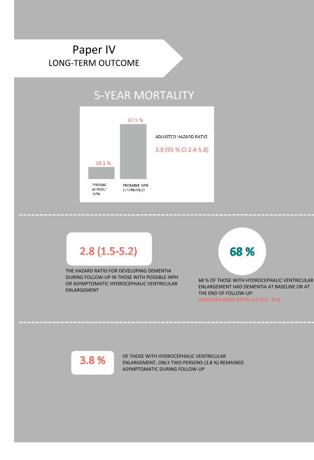
Paper IV LONG-TERM OUTCOME
5-YEAR MORTALITY
THE HAZARD RATIO FOR DEVELOPING DEMENTIADURING FOLLOW-UP IN THOSE WITH POSSIBLE iNPH OR ASYMPTOMATIC HYDROCEPHALIC VENTRICULAR ENLARGEMENT
68 % OF THOSE WITH HYDROCEPHALIC VENTRICULAR ENLARGEMENT HAD DEMENTIA AT BASELINE OR AT THE END OF FOLLOW-UP: ADJUSTED ODDS RATIO 4.9 (2.5 - 9.6)
OF THOSE WITH HYDROCEPHALIC VENTRICULAR ENLARGEMENT, ONLY TWO PERSONS (3.8 %) REMAINED ASYMPTOMATIC DURING FOLLOW-UP
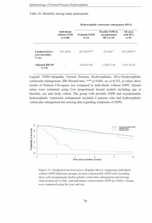
Epidemiology of Normal Pressure Hydrocephalus
76
Table 10. Mortality among study participants
Hydrocephalic ventricular enlargement (HVe)
Individuals without iNPH
n=1180 Probable iNPH
n=24
Possible iNPH & Asymptomatic
HVe n=29
All cases with HVe
n=53
Unadjusted five-year mortality, % (n)
19.1 (225)
87.5 (21)***
27.6 (8) ns
54.7 (29)***
Adjusted HR (95 % CI)
- 3.8 (2.4-5.8) 1.2 (0.7-1.8) 1.9 (1.4-2.5)
Legend: iNPH=idiopathic Normal Pressure Hydrocephalus, HVe=Hydrocephalic ventricular enlargement, HR=Hazard ratio, *** p<0.001, ns: p=0.251, p-values show results of Pearson Chi-square test compared to individuals without iNPH. Hazard ratios were estimated using Cox proportional hazard models including age at baseline, sex and study cohort. The group with possible iNPH and asymptomatic hydrocephalic ventricular enlargement included 4 persons who had hydrocephalic ventricular enlargement but missing data regarding symptoms of iNPH.
Figure 13. Unadjusted survival curves (Kaplan-Meier), comparing individuals without iNPH (Reference group), persons with possible iNPH (also including those with asymptomatic hydrocephalic ventricular enlargement and missing clinical data) (p=0.284), and individuals with probable iNPH (p<0.001). Groups were compared using the Log rank test.

Daniel Jaraj
77
According to the results of this study, untreated individuals with signs and symptoms of iNPH have a substantially increased risk of mortality and dementia. Furthermore, almost all persons with imaging signs of iNPH developed dementia or other symptoms of iNPH. These findings indicate that imaging signs of iNPH are important markers for cognitive impairment and neurological disability. The results of this study suggest that clinical and radiological signs of iNPH are associated with a markedly poor prognosis with a high risk of death and dementia among untreated persons.
The results of this study also suggest that radiological signs of iNPH might precede clinical symptoms. This has previously been indicated by a study that examined six cases.103 If it is true that radiological signs are the first to develop, then current diagnostic criteria might only capture individuals at a later stage when the disorder has manifested with more severe symptoms. Therefore, the results of this study might also have implications regarding the diagnosis of iNPH.
There are no previous epidemiological studies on long-term outcome in iNPH. Previous studies have mainly used hospital-based samples and there is very little data on the natural course. Furthermore, the risk of dementia and progression of symptoms over time is not precisely known.
Main strengths of this study include the population-based sample, comprehensive examinations and duration of follow-up. There are, however, also several important limitations that should be discussed. As described previously, the diagnostic uncertainty regarding iNPH is important to consider. Although this may have influenced the validity of the study, it is also reasonable to suppose that there might have been less bias regarding the associations between imaging signs of iNPH and outcomes. The reason for this is that those with hydrocephalic ventricular enlargement were diagnosed by a consultant neuroradiologist, unaware of clinical data. Therefore, results of this study suggest that imaging findings of iNPH are associated with a poor prognosis regardless of symptoms. Other limitations, that have also been discussed earlier, include unbalance in the data, possible cohort effects and small number of cases. The group with possible iNPH and asymptomatic

Epidemiology of Normal Pressure Hydrocephalus
78
hydrocephalic ventricular enlargement (n=29) did not have a significantly increased mortality. These persons were, on average, younger and part of the more recent cohorts, thus having less follow-up time. It is plausible that significant differences might have been detected using a larger sample with longer follow-up. Cases were diagnosed with CT, and MRI would probably have been somewhat better. However, this was not available at the time of baseline examinations. Small obstructive lesions can be missed on CT. Nevertheless, it can be argued that CT imaging allows for an overall sufficient assessment of the ventricles and high convexity sulci.

Daniel Jaraj
79
6 CONCLUSIONS AND FUTURE PERSPECTIVES
iNPH is probably more common than previously supposed. Furthermore, a large number of older persons have imaging features consistent with iNPH. This finding is important considering that radiological features might be an early sign of iNPH.
Many elderly are in need of further evaluations. However, despite being a treatable disorder, the number of persons being diagnosed and treated is likely far less than the actual number of person suffering from iNPH. Considering that life expectancy is rising, the number of patients with iNPH might also increase. There is a need for future studies to improve diagnosis and selection of candidates for shunt surgery.
Vascular factors, such as cerebral WMLs, hypertension and diabetes are rather strongly associated with iNPH. The findings indicate that vascular disease mechanisms are involved in the pathophysiology. Considering also earlier data, a causal association may be supposed. Clinicians ought to be aware of the high occurrence of vascular diseases in iNPH since concurrent management of risk factors is important. The findings may also have importance from a perspective of preventive measures. Future studies should aim to further elucidate the role of vascular mechanisms in iNPH. This might not only improve understanding of the pathophysiology but also provide insight into disease features that might be of diagnostic importance. It is also important to learn more about potential causes in order to improve treatment. Also, future investigations regarding other mechanisms including non-vascular risk factors would be of value.
Mean values of Evans Index, among older persons in the general population, are higher than previously reported. Current criteria for ventricular enlargement might capture as many as one in five, of those aged 70 years or more. Thus, the current cut-off value of 0.3 probably has a low specificity for
Epidemiology of Normal Pressure Hydrocephalus
80
the diagnosis of iNPH. Persons with hydrocephalic ventricular enlargement had, on average, values above the 95th percentile. However, it is difficult to determine diagnostic cut-off values. For example, it is not known whether or not persons with Evans Index below 0.3 could still have iNPH. Considering that ventricle size does seem not predict outcome after shunt surgery, more advanced imaging such as volumetric assessment of the ventricles, subarachnoid space and cortical thickness might be of greater value. Given the risks of shunt surgery, a correct diagnosis of iNPH is crucial. However, previous attempts to predict shunt response based on various hydrodynamic characteristics have so far been rather futile. Further research on clinical variables and other imaging features, such as white matter changes, blood flow and metabolism might add more knowledge and insight. Also, considering the high degree of overlap between iNPH and other neurodegenerative conditions, future studies on diagnostic measures should include not only iNPH patients and healthy controls, but also patients with other diagnoses such as Alzheimer’s disease and vascular dementia. It is also important for future studies to have adequate power given the heterogeneity of iNPH.
Untreated individuals who fulfill criteria for probable iNPH have substantial excess mortality. Considering these findings, individuals with signs and symptoms of iNPH should be thoroughly evaluated and treated without delay if the diagnosis is confirmed. Furthermore, persons with radiological signs of iNPH appear to have a very high risk of developing dementia or other symptoms. Radiological signs are therefore probably more important than previously thought. It remains to be elucidated whether pre-symptomatic iNPH should be treated. Additional epidemiological studies, using population-based data, are needed in the field. However, given the estimated prevalence of iNPH, even relatively large samples would likely yield a somewhat small number of cases. One way to circumvent this problem might be to merge population data from several centers.

81

Epidemiology of Normal Pressure Hydrocephalus
82
ACKNOWLEDGEMENTS I would like to express my most sincere gratitude to all of those who have offered their help, encouragement and kind support. I wish to thank my supervisors. Needless to say, without whom this thesis would not exist. Ingmar Skoog, thank you for taking me on as your PhD student and embracing me into your research group. Your devotion and endless curiosity has been a genuine source of inspiration. Among many things, you taught me the value of patience and precision in research. I owe you my deepest gratitude. Carsten WikkelsØ, you took me on as a medical student and trusted me with meaningful work. For this I am forever grateful. Your creativeness, level of perception and constant drive has been an inspiration. Thank you for showing me what true leadership is like. Katrin Rabiei, colleague, co-author, and friend. You taught me how to work hard, be passionate and still have fun. (As a medical student, you also taught me the meaning of the surgeons promise). Thank you for all your support and kind friendship. Tina Jacobsson, I cannot thank you enough for everything that you have done. I feel that this work would not be possible without you. To say that your contributions have been invaluable would be an understatement. You helped me feel at home in the group, and your professionalism has been an inspiration from day one. Thank you so much for everything. Cecilia Mellqvist, thank you for all your help. You also showed me what it means to be professional and devoted. I wish to express my most sincere appreciation for all of your contributions to this research. Tom Marlow, thank you for your patience, calmness and keen-sightedness. This work would not have been possible without you. Our discussions on statistics and study design have been one of the more joyful parts of this work. Thank you, to all my co-authors: Christer Jensen, Svante Östling and Simon Agerskov.

83
Kristoffer Bäckman, thank you for all your help. Working with you has been a constant enjoyment. I treasure our many discussions on data sciences and truly admire your high level of knowledge. Valter Sundh and Erik Joas, thank you for teaching me about statistics and allowing me to ask all the senseless questions. Having you as colleagues has been a great satisfaction. Mikael Edsbagge, thank you so much for giving me a place in the neurology clinic. Thank you also for teaching me about hydrocephalus and general neurology, and being such a fantastic mentor. You are, by far, the best boss I have ever had. I also want to thank the best nurses in the world, Christel Olivefors, Tove Bulic Rasmussen and Eva Niemi. It was a delight to work with you and experience the high level care that you provide each patient. Also, thank you, Gunilla Ahl-Börjesson, for all you kindness, support and help. To those who are not mentioned, please forgive me, and know that I am grateful to everyone in the hydrocephalus research unit and neuropsychiatric epidemiology research group. I would like to thank my dear friend and main man, Hasif Hakim. Without you, I wouldn’t even have the “M” in the M.D. Slutligen, vill jag tacka min familj. Min mamma Katalin, min pappa Janos och min kära bror David. Ni har gett mig allt. Genom hela min uppväxt har jag överväldigats av den varmaste kärlek man kan tänka sig. Min älskade storebror David, som är den smartaste och mest perfekta förebild som finns. Inget av detta vore möjligt utan er. Min fantastiska Rebecka, och hennes underbara familj, Charlotte, Staffan, Henrik, Magrethe, Ulrika, Mathias, Lovisa, Markus, Nils-Gunnar och Greta, samt Jan-Erik och Ylva. Tack för allt ni har gett mig genom åren. Min underbara, älskade, vackra Rebecka. Vi har vuxit upp tillsammans och du har lärt mig så mycket, stöttat mig och alltid funnits vid min sida. Jag har det aldrig så roligt som när jag är med dig. Ingen är klokare än du, och jag kan inte ens beskriva den tacksamhet jag känner.
Epidemiology of Normal Pressure Hydrocephalus
84
REFERENCES
1.Relkin N, Marmarou A, Klinge P, Bergsneider M, Black PM. Diagnosing idiopathic normal-pressure hydrocephalus. Neurosurgery 2005;57:S4-16; discussion ii-v.
2.Mori K, Shimada J, Kurisaka M, Sato K, Watanabe K. Classification of hydrocephalus and outcome of treatment. Brain & development 1995;17:338-348.
3.Gallia GL, Rigamonti D, Williams MA. The diagnosis and treatment of idiopathic normal pressure hydrocephalus. Nature clinical practice Neurology 2006;2:375-381. 4.Kiefer M, Unterberg A. The differential diagnosis and treatment of normal-pressure hydrocephalus. Deutsches Arzteblatt international 2012;109:15-25; quiz 26. 5.Hakim S, Adams RD. The special clinical problem of symptomatic hydrocephalus with normal cerebrospinal fluid pressure. Observations on cerebrospinal fluid hydrodynamics. Journal of the neurological sciences 1965;2:307-327. 6.Adams RD, Fisher CM, Hakim S, Ojemann RG, Sweet WH. SYMPTOMATIC OCCULT HYDROCEPHALUS WITH "NORMAL" CEREBROSPINAL-FLUID PRESSURE.A TREATABLE SYNDROME. The New England journal of medicine 1965;273:117-126. 7.Wallenstein MB, McKhann GM, 2nd. Salomon Hakim and the discovery of normal-pressure hydrocephalus. Neurosurgery 2010;67:155-159; discussion 159. 8.Hashimoto M, Ishikawa M, Mori E, Kuwana N. Diagnosis of idiopathic normal pressure hydrocephalus is supported by MRI-based scheme: a prospective cohort study. Cerebrospinal fluid research 2010;7:18. 9.Graff-Radford NR. Normal pressure hydrocephalus. Neurologic clinics 2007;25:809-832, vii-viii.
85
10.Malm J, Graff-Radford NR, Ishikawa M, et al. Influence of comorbidities in idiopathic normal pressure hydrocephalus - research and clinical care. A report of the ISHCSF task force on comorbidities in INPH. Fluids and barriers of the CNS 2013;10:22. 11.Stolze H, Kuhtz-Buschbeck JP, Drucke H, Johnk K, Illert M, Deuschl G. Comparative analysis of the gait disorder of normal pressure hydrocephalus and Parkinson's disease. Journal of neurology, neurosurgery, and psychiatry 2001;70:289-297. 12.Stolze H, Kuhtz-Buschbeck JP, Drucke H, et al. Gait analysis in idiopathic normal pressure hydrocephalus--which parameters respond to the CSF tap test? Clinical neurophysiology : official journal of the International Federation of Clinical Neurophysiology 2000;111:1678-1686. 13.Blomsterwall E, Svantesson U, Carlsson U, Tullberg M, Wikkelso C. Postural disturbance in patients with normal pressure hydrocephalus. Acta neurologica Scandinavica 2000;102:284-291. 14.Wikkelso C, Blomsterwall E, Frisen L. Subjective visual vertical and Romberg's test correlations in hydrocephalus. Journal of neurology 2003;250:741-745. 15.L. Pantoni DI, A. Wallin, ed. The Matter of White Matter, Clinical and Pathophysiological Aspects of White Matter Disease Related to Cognitive Decline and Vascular Dementia: Academic Pharmaceutical Productions 2000. 16.Mori E, Ishikawa M, Kato T, et al. Guidelines for management of idiopathic normal pressure hydrocephalus: second edition. Neurologia medico-chirurgica 2012;52:775-809. 17.Ogino A, Kazui H, Miyoshi N, et al. Cognitive impairment in patients with idiopathic normal pressure hydrocephalus. Dementia and geriatric cognitive disorders 2006;21:113-119. 18.Hellstrom P, Edsbagge M, Archer T, Tisell M, Tullberg M, Wikkelso C. The neuropsychology of patients with clinically diagnosed idiopathic normal

Epidemiology of Normal Pressure Hydrocephalus
86
pressure hydrocephalus. Neurosurgery 2007;61:1219-1226; discussion 1227-1218. 19.Hellstrom P, Klinge P, Tans J, Wikkelso C. The neuropsychology of iNPH: findings and evaluation of tests in the European multicentre study. Clinical neurology and neurosurgery 2012;114:130-134. 20.Jonas S, Brown J. Neurogenic bladder in normal pressure hydrocephalus. Urology 1975;5:44-50. 21.Sakakibara R, Kanda T, Sekido T, et al. Mechanism of bladder dysfunction in idiopathic normal pressure hydrocephalus. Neurourology and urodynamics 2008;27:507-510. 22.Brodbelt A, Stoodley M. CSF pathways: a review. British journal of neurosurgery 2007;21:510-520. 23.Cutler RW, Page L, Galicich J, Watters GV. Formation and absorption of cerebrospinal fluid in man. Brain : a journal of neurology 1968;91:707-720. 24.Kapoor KG, Katz SE, Grzybowski DM, Lubow M. Cerebrospinal fluid outflow: an evolving perspective. Brain research bulletin 2008;77:327-334. 25.Arthur C. Guyton JEH. Textbook of Medical Physiology, 11th ed: Elsevier Saunders, 2006. 26.Abbott NJ. Evidence for bulk flow of brain interstitial fluid: significance for physiology and pathology. Neurochemistry international 2004;45:545-552. 27.Brinker T, Stopa E, Morrison J, Klinge P. A new look at cerebrospinal fluid circulation. Fluids and barriers of the CNS 2014;11:10. 28.Edsbagge M, Tisell M, Jacobsson L, Wikkelso C. Spinal CSF absorption in healthy individuals. American journal of physiology Regulatory, integrative and comparative physiology 2004;287:R1450-1455. 29.Erlich SS, McComb JG, Hyman S, Weiss MH. Ultrastructural morphology of the olfactory pathway for cerebrospinal fluid drainage in the rabbit. Journal of neurosurgery 1986;64:466-473.

87
30.Silver I, Kim C, Mollanji R, Johnston M. Cerebrospinal fluid outflow resistance in sheep: impact of blocking cerebrospinal fluid transport through the cribriform plate. Neuropathology and applied neurobiology 2002;28:67-74. 31.Xie L, Kang H, Xu Q, et al. Sleep Drives Metabolite Clearance from the Adult Brain. Science 2013;342:373-377. 32.Ren Z, Iliff JJ, Yang L, et al. 'Hit & Run' model of closed-skull traumatic brain injury (TBI) reveals complex patterns of post-traumatic AQP4 dysregulation. Journal of cerebral blood flow and metabolism : official journal of the International Society of Cerebral Blood Flow and Metabolism 2013;33:834-845. 33.Iliff JJ, Nedergaard M. Is there a cerebral lymphatic system? Stroke; a journal of cerebral circulation 2013;44:S93-95. 34.Skjolding AD, Rowland IJ, Sogaard LV, Praetorius J, Penkowa M, Juhler M. Hydrocephalus induces dynamic spatiotemporal regulation of aquaporin-4 expression in the rat brain. Cerebrospinal fluid research 2010;7:20. 35.Rekate HL. The definition and classification of hydrocephalus: a personal recommendation to stimulate debate. Cerebrospinal fluid research 2008;5:2. 36.DeLand FH, James AE, Jr., Ladd DJ, Konigsmark BW. Normal pressure hydrocephalus: a histologic study. American journal of clinical pathology 1972;58:58-63. 37.Bech RA, Waldemar G, Gjerris F, Klinken L, Juhler M. Shunting effects in patients with idiopathic normal pressure hydrocephalus; correlation with cerebral and leptomeningeal biopsy findings. Acta neurochirurgica 1999;141:633-639. 38.Boon AJ, Tans JT, Delwel EJ, et al. Dutch normal-pressure hydrocephalus study: prediction of outcome after shunting by resistance to outflow of cerebrospinal fluid. Journal of neurosurgery 1997;87:687-693. 39.May C, Kaye JA, Atack JR, Schapiro MB, Friedland RP, Rapoport SI. Cerebrospinal fluid production is reduced in healthy aging. Neurology 1990;40:500-503.

Epidemiology of Normal Pressure Hydrocephalus
88
40.Albeck MJ, Skak C, Nielsen PR, Olsen KS, Borgesen SE, Gjerris F. Age dependency of resistance to cerebrospinal fluid outflow. Journal of neurosurgery 1998;89:275-278. 41.Malm J, Jacobsson J, Birgander R, Eklund A. Reference values for CSF outflow resistance and intracranial pressure in healthy elderly. Neurology 2011;76:903-909. 42.Marmarou A, Bergsneider M, Klinge P, Relkin N, Black PM. The value of supplemental prognostic tests for the preoperative assessment of idiopathic normal-pressure hydrocephalus. Neurosurgery 2005;57:S17-28; discussion ii-v. 43.Raftopoulos C, Deleval J, Chaskis C, et al. Cognitive recovery in idiopathic normal pressure hydrocephalus: a prospective study. Neurosurgery 1994;35:397-404; discussion 404-395. 44.Eide PK, Sorteberg W. Diagnostic intracranial pressure monitoring and surgical management in idiopathic normal pressure hydrocephalus: a 6-year review of 214 patients. Neurosurgery 2010;66:80-91. 45.Stephensen H, Andersson N, Eklund A, Malm J, Tisell M, Wikkelso C. Objective B wave analysis in 55 patients with non-communicating and communicating hydrocephalus. Journal of neurology, neurosurgery, and psychiatry 2005;76:965-970. 46.Casmiro M, D'Alessandro R, Cacciatore FM, Daidone R, Calbucci F, Lugaresi E. Risk factors for the syndrome of ventricular enlargement with gait apraxia (idiopathic normal pressure hydrocephalus): a case-control study. Journal of neurology, neurosurgery, and psychiatry 1989;52:847-852. 47.Graff-Radford NR, Godersky JC. Idiopathic normal pressure hydrocephalus and systemic hypertension. Neurology 1987;37:868-871. 48.Jacobs L. Diabetes mellitus in normal pressure hydrocephalus. Journal of neurology, neurosurgery, and psychiatry 1977;40:331-335.

89
49.Krauss JK, Regel JP, Vach W, Droste DW, Borremans JJ, Mergner T. Vascular risk factors and arteriosclerotic disease in idiopathic normal-pressure hydrocephalus of the elderly. Stroke; a journal of cerebral circulation 1996;27:24-29. 50.Eide PK, Pripp AH. Increased prevalence of cardiovascular disease in idiopathic normal pressure hydrocephalus patients compared to a population-based cohort from the HUNT3 survey. Fluids and Barriers of the CNS 2014;11:19. 51.Graff-Radford NR, Knopman DS, Penman AD, Coker LH, Mosley TH. Do systolic BP and pulse pressure relate to ventricular enlargement? European journal of neurology : the official journal of the European Federation of Neurological Societies 2013;20:720-724. 52.Ritter S, Dinh TT. Progressive postnatal dilation of brain ventricles in spontaneously hypertensive rats. Brain research 1986;370:327-332. 53.Di Rocco C, Pettorossi VE, Caldarelli M, Mancinelli R, Velardi F. Experimental hydrocephalus following mechanical increment of intraventricular pulse pressure. Experientia 1977;33:1470-1472. 54.Akai K, Uchigasaki S, Tanaka U, Komatsu A. Normal pressure hydrocephalus. Neuropathological study. Acta pathologica japonica 1987;37:97-110. 55.Pantoni L, Garcia JH. Pathogenesis of leukoaraiosis: a review. Stroke; a journal of cerebral circulation 1997;28:652-659. 56.Bradley WG, Jr., Whittemore AR, Watanabe AS, Davis SJ, Teresi LM, Homyak M. Association of deep white matter infarction with chronic communicating hydrocephalus: implications regarding the possible origin of normal-pressure hydrocephalus. AJNR American journal of neuroradiology 1991;12:31-39. 57.Krauss JK, Regel JP, Vach W, et al. White matter lesions in patients with idiopathic normal pressure hydrocephalus and in an age-matched control group: a comparative study. Neurosurgery 1997;40:491-495; discussion 495-496.

Epidemiology of Normal Pressure Hydrocephalus
90
58.O'Brien JT, Erkinjuntti T, Reisberg B, et al. Vascular cognitive impairment. The Lancet Neurology 2003;2:89-98. 59.Tullberg M, Hultin L, Ekholm S, Mansson JE, Fredman P, Wikkelso C. White matter changes in normal pressure hydrocephalus and Binswanger disease: specificity, predictive value and correlations to axonal degeneration and demyelination. Acta Neurol Scand 2002;105:417-426. 60.Tullberg M, Jensen C, Ekholm S, Wikkelso C. Normal pressure hydrocephalus: vascular white matter changes on MR images must not exclude patients from shunt surgery. AJNR American journal of neuroradiology 2001;22:1665-1673. 61.Momjian S, Owler BK, Czosnyka Z, Czosnyka M, Pena A, Pickard JD. Pattern of white matter regional cerebral blood flow and autoregulation in normal pressure hydrocephalus. Brain : a journal of neurology 2004;127:965-972. 62.Ziegelitz D, Starck G, Kristiansen D, et al. Cerebral perfusion measured by dynamic susceptibility contrast MRI is reduced in patients with idiopathic normal pressure hydrocephalus. Journal of magnetic resonance imaging : JMRI 2014;39:1533-1542. 63.Owler BK, Pickard JD. Normal pressure hydrocephalus and cerebral blood flow: a review. Acta neurologica Scandinavica 2001;104:325-342. 64.Hertel F, Walter C, Schmitt M, et al. Is a combination of Tc-SPECT or perfusion weighted magnetic resonance imaging with spinal tap test helpful in the diagnosis of normal pressure hydrocephalus? Journal of neurology, neurosurgery, and psychiatry 2003;74:479-484. 65.Kimura M, Tanaka A, Yoshinaga S. Significance of periventricular hemodynamics in normal pressure hydrocephalus. Neurosurgery 1992;30:701-704; discussion 704-705. 66.Krauss JK, Droste DW, Vach W, et al. Cerebrospinal fluid shunting in idiopathic normal-pressure hydrocephalus of the elderly: effect of

91
periventricular and deep white matter lesions. Neurosurgery 1996;39:292-299; discussion 299-300. 67.Tisell M, Tullberg M, Hellstrom P, Edsbagge M, Hogfeldt M, Wikkelso C. Shunt surgery in patients with hydrocephalus and white matter changes. Journal of neurosurgery 2011;114:1432-1438. 68.Johansson E, Ambarki K, Birgander R, Bahrami N, Eklund A, Malm J. Cerebral microbleeds in idiopathic normal pressure hydrocephalus. Fluids and barriers of the CNS 2016;13:4. 69.Alperin N, Oliu CJ, Bagci AM, et al. Low-dose acetazolamide reverses periventricular white matter hyperintensities in iNPH. Neurology 2014;82:1347-1351. 70.Ivkovic M, Reiss-Zimmermann M, Katzen H, et al. MRI assessment of the effects of acetazolamide and external lumbar drainage in idiopathic normal pressure hydrocephalus. Fluids and barriers of the CNS 2015;12:9. 71.Vorstrup S, Henriksen L, Paulson OB. Effect of acetazolamide on cerebral blood flow and cerebral metabolic rate for oxygen. The Journal of clinical investigation 1984;74:1634-1639. 72.Okazawa H, Yamauchi H, Sugimoto K, Toyoda H, Kishibe Y, Takahashi M. Effects of acetazolamide on cerebral blood flow, blood volume, and oxygen metabolism: a positron emission tomography study with healthy volunteers. Journal of cerebral blood flow and metabolism : official journal of the International Society of Cerebral Blood Flow and Metabolism 2001;21:1472-1479. 73.Krefft TA, Graff-Radford NR, Lucas JA, Mortimer JA. Normal pressure hydrocephalus and large head size. Alzheimer disease and associated disorders 2004;18:35-37. 74.Bradley WG, Safar FG, Furtado C, Ord J, Alksne JF. Increased intracranial volume: a clue to the etiology of idiopathic normal-pressure hydrocephalus? AJNR American journal of neuroradiology 2004;25:1479-1484.
Epidemiology of Normal Pressure Hydrocephalus
92
75.Bradley WG, Jr., Bahl G, Alksne JF. Idiopathic normal pressure hydrocephalus may be a "two hit" disease: benign external hydrocephalus in infancy followed by deep white matter ischemia in late adulthood. Journal of magnetic resonance imaging : JMRI 2006;24:747-755. 76.Bradley WG, Jr. CSF Flow in the Brain in the Context of Normal Pressure Hydrocephalus. AJNR American journal of neuroradiology 2015;36:831-838. 77.Wilson RK, Williams MA. Evidence that congenital hydrocephalus is a precursor to idiopathic normal pressure hydrocephalus in only a subset of patients. Journal of neurology, neurosurgery, and psychiatry 2007;78:508-511. 78.Koivisto AM, Alafuzoff I, Savolainen S, et al. Poor cognitive outcome in shunt-responsive idiopathic normal pressure hydrocephalus. Neurosurgery 2013;72:1-8;discussion 8. 79.Kapaki EN, Paraskevas GP, Tzerakis NG, et al. Cerebrospinal fluid tau, phospho-tau181 and beta-amyloid1-42 in idiopathic normal pressure hydrocephalus: a discrimination from Alzheimer's disease. European journal of neurology : the official journal of the European Federation of Neurological Societies 2007;14:168-173. 80.Leinonen V, Koivisto AM, Alafuzoff I, et al. Cortical brain biopsy in long-term prognostication of 468 patients with possible normal pressure hydrocephalus. Neuro-degenerative diseases 2012;10:166-169. 81.Silverberg GD, Mayo M, Saul T, Rubenstein E, McGuire D. Alzheimer's disease, normal-pressure hydrocephalus, and senescent changes in CSF circulatory physiology: a hypothesis. The Lancet Neurology 2003;2:506-511. 82.Silverberg GD, Levinthal E, Sullivan EV, et al. Assessment of low-flow CSF drainage as a treatment for AD: results of a randomized pilot study. Neurology 2002;59:1139-1145. 83.Silverberg GD, Mayo M, Saul T, Fellmann J, Carvalho J, McGuire D. Continuous CSF drainage in AD: results of a double-blind, randomized, placebo-controlled study. Neurology 2008;71:202-209.
93
84.Jeppsson A, Zetterberg H, Blennow K, Wikkelso C. Idiopathic normal-pressure hydrocephalus: pathophysiology and diagnosis by CSF biomarkers. Neurology 2013;80:1385-1392. 85.Graff-Radford NR. Alzheimer CSF biomarkers may be misleading in normal-pressure hydrocephalus. Neurology 2014;83:1573-1575. 86.Laitera T, Sarajarvi T, Haapasalo A, et al. Increased gamma-secretase activity in idiopathic normal pressure hydrocephalus patients with beta-amyloid pathology. PloS one 2014;9:e93717. 87.Pyykkö OT, Helisalmi S, Koivisto AM, et al. APOE4 predicts amyloid-β in cortical brain biopsy but not idiopathic normal pressure hydrocephalus. Journal of Neurology, Neurosurgery & Psychiatry 2012;83:1119-1124. 88.Savolainen S, Laakso MP, Paljarvi L, et al. MR imaging of the hippocampus in normal pressure hydrocephalus: correlations with cortical Alzheimer's disease confirmed by pathologic analysis. AJNR American journal of neuroradiology 2000;21:409-414. 89.Kondo M, Tokuda T, Itsukage M, et al. Distribution of amyloid burden differs between idiopathic normal pressure hydrocephalus and Alzheimer's disease. The neuroradiology journal 2013;26:41-46. 90.Wikkelso C, Hellstrom P, Klinge PM, Tans JT. The European iNPH Multicentre Study on the predictive values of resistance to CSF outflow and the CSF Tap Test in patients with idiopathic normal pressure hydrocephalus. Journal of neurology, neurosurgery, and psychiatry 2013;84:562-568. 91.Harrison's Neurology in Clinical Medicine, Second Edition ed: McGraw-Hill 2010. 92.Vanneste J, Augustijn P, Dirven C, Tan WF, Goedhart ZD. Shunting normal-pressure hydrocephalus: do the benefits outweigh the risks? A multicenter study and literature review. Neurology 1992;42:54-59. 93.Trenkwalder C, Schwarz J, Gebhard J, et al. Starnberg trial on epidemiology of Parkinsonism and hypertension in the elderly. Prevalence of Parkinson's disease and related disorders assessed by a door-to-door survey of inhabitants older than 65 years. Archives of neurology 1995;52:1017-1022.
Epidemiology of Normal Pressure Hydrocephalus
94
94.Brean A, Eide PK. Prevalence of probable idiopathic normal pressure hydrocephalus in a Norwegian population. Acta neurologica Scandinavica 2008;118:48-53. 95.Hiraoka K, Meguro K, Mori E. Prevalence of idiopathic normal-pressure hydrocephalus in the elderly population of a Japanese rural community. Neurologia medico-chirurgica 2008;48:197-199; discussion 199-200. 96.Tanaka N, Yamaguchi S, Ishikawa H, Ishii H, Meguro K. Prevalence of possible idiopathic normal-pressure hydrocephalus in Japan: the Osaki-Tajiri project. Neuroepidemiology 2009;32:171-175. 97.Iseki C, Kawanami T, Nagasawa H, et al. Asymptomatic ventriculomegaly with features of idiopathic normal pressure hydrocephalus on MRI (AVIM) in the elderly: a prospective study in a Japanese population. Journal of the neurological sciences 2009;277:54-57. 98.Israelsson H, Birgander R, Ambarki K, Eklund A, Malm J. Ventriculomegaly and balance disturbances in patients with TIA. Acta neurologica Scandinavica 2012;125:163-170. 99.Vale FA, Miranda SJ. Clinical and demographic features of patients with dementia attended in a tertiary outpatient clinic. Arquivos de neuro-psiquiatria 2002;60:548-552. 100.Marmarou A, Young HF, Aygok GA. Estimated incidence of normal pressure hydrocephalus and shunt outcome in patients residing in assisted-living and extended-care facilities. Neurosurgical focus 2007;22:E1. 101.Razay G, Wimmer M. Normal pressure hydrocephalus: Treatable dementia, missed diagnosis—The Launceston Prevalence Study. Alzheimer's & Dementia: The Journal of the Alzheimer's Association;9:P755. 102.Martin-Laez R, Caballero-Arzapalo H, Lopez-Menendez LA, Arango-Lasprilla JC, Vazquez-Barquero A. Epidemiology of Idiopathic Normal Pressure Hydrocephalus: A Systematic Review of the Literature. World neurosurgery 2015;84:2002-2009.
95
103.Iseki C, Takahashi Y, Wada M, Kawanami T, Adachi M, Kato T. Incidence of idiopathic normal pressure hydrocephalus (iNPH): a 10-year follow-up study of a rural community in Japan. Journal of the neurological sciences 2014;339:108-112. 104.Tisell M, Hoglund M, Wikkelso C. National and regional incidence of surgery for adult hydrocephalus in Sweden. Acta neurologica Scandinavica 2005;112:72-75. 105.Brean A, Fredo HL, Sollid S, Muller T, Sundstrom T, Eide PK. Five-year incidence of surgery for idiopathic normal pressure hydrocephalus in Norway. Acta neurologica Scandinavica 2009;120:314-316. 106.Mori E. DESH vs. Tap Test: The implications of DESH on clinical practive of NPH. International Hydrocephalus Imaging Working Group; 2013; San Diego. 107.Ishikawa M, Hashimoto M, Kuwana N, et al. Guidelines for management of idiopathic normal pressure hydrocephalus. Neurologia medico-chirurgica 2008;48 Suppl:S1-23. 108.Devito EE, Pickard JD, Salmond CH, Iddon JL, Loveday C, Sahakian BJ. The neuropsychology of normal pressure hydrocephalus (NPH). British journal of neurosurgery 2005;19:217-224. 109.Dincer A, Kohan S, Ozek MM. Is all "communicating" hydrocephalus really communicating? Prospective study on the value of 3D-constructive interference in steady state sequence at 3T. AJNR American journal of neuroradiology 2009;30:1898-1906. 110.Ziegelitz D, Starck G, Mikkelsen IK, et al. Absolute quantification of cerebral blood flow in neurologically normal volunteers: dynamic-susceptibility contrast MRI-perfusion compared with computed tomography (CT)-perfusion. Magnetic resonance in medicine 2009;62:56-65. 111.Owler BK, Momjian S, Czosnyka Z, et al. Normal pressure hydrocephalus and cerebral blood flow: a PET study of baseline values. Journal of cerebral blood flow and metabolism : official journal of the International Society of Cerebral Blood Flow and Metabolism 2004;24:17-23.

Epidemiology of Normal Pressure Hydrocephalus
96
112.Granado JM, Diaz F, Alday R. Evaluation of brain SPECT in the diagnosis and prognosis of the normal pressure hydrocephalus syndrome. Acta neurochirurgica 1991;112:88-91. 113.Virhammar J, Laurell K, Ahlgren A, Cesarini KG, Larsson EM. Idiopathic normal pressure hydrocephalus: cerebral perfusion measured with pCASL before and repeatedly after CSF removal. Journal of cerebral blood flow and metabolism : official journal of the International Society of Cerebral Blood Flow and Metabolism 2014;34:1771-1778. 114.Larsson A, Moonen M, Bergh AC, Lindberg S, Wikkelso C. Predictive value of quantitative cisternography in normal pressure hydrocephalus. Acta neurologica Scandinavica 1990;81:327-332. 115.Kitagaki H, Mori E, Ishii K, Yamaji S, Hirono N, Imamura T. CSF spaces in idiopathic normal pressure hydrocephalus: morphology and volumetry. AJNR American journal of neuroradiology 1998;19:1277-1284. 116.Akiguchi I, Shirakashi Y, Budka H, et al. Disproportionate subarachnoid space hydrocephalus-outcome and perivascular space. Annals of clinical and translational neurology 2014;1:562-569. 117.Palm WM, Saczynski JS, van der Grond J, et al. Ventricular dilation: association with gait and cognition. Annals of neurology 2009;66:485-493. 118.Kimberly HH, Shah S, Marill K, Noble V. Correlation of optic nerve sheath diameter with direct measurement of intracranial pressure. Academic emergency medicine : official journal of the Society for Academic Emergency Medicine 2008;15:201-204. 119.Ishii K, Kanda T, Harada A, et al. Clinical impact of the callosal angle in the diagnosis of idiopathic normal pressure hydrocephalus. European radiology 2008;18:2678-2683. 120.Gideon P, Stahlberg F, Thomsen C, Gjerris F, Sorensen PS, Henriksen O. Cerebrospinal fluid flow and production in patients with normal pressure hydrocephalus studied by MRI. Neuroradiology 1994;36:210-215. 121.Krauss JK, Regel JP, Vach W, Jungling FD, Droste DW, Wakhloo AK. Flow void of cerebrospinal fluid in idiopathic normal pressure hydrocephalus
97
of the elderly: can it predict outcome after shunting? Neurosurgery 1997;40:67-73; discussion 73-64. 122.Evans WA, Jr. An encephalographic ratio for estimating the size of the cerebral ventricles: Further experience with serial observations. American Journal of Diseases of Children 1942;64:820-830. 123.Williams MA, Relkin NR. Diagnosis and management of idiopathic normal-pressure hydrocephalus. Neurology Clinical practice 2013;3:375-385. 124.Synek V, Reuben JR, Du Boulay GH. Comparing Evans' index and computerized axial tomography in assessing relationship of ventricular size to brain size. Neurology 1976;26:231-233. 125.Zatz LM. The Evans ratio for ventricular size: a calculation error. Neuroradiology 1979;18:81. 126.Ambarki K, Israelsson H, Wahlin A, Birgander R, Eklund A, Malm J. Brain ventricular size in healthy elderly: comparison between Evans index and volume measurement. Neurosurgery 2010;67:94-99; discussion 99. 127.Jacoby RJ, Levy R, Dawson JM. Computed tomography in the elderly: I. The normal population. The British journal of psychiatry : the journal of mental science 1980;136:249-255. 128.Inatomi Y, Yonehara T, Hashimoto Y, Hirano T, Uchino M. Correlation between ventricular enlargement and white matter changes. Journal of the neurological sciences 2008;269:12-17. 129.Toma AK, Holl E, Kitchen ND, Watkins LD. Evans' index revisited: the need for an alternative in normal pressure hydrocephalus. Neurosurgery 2011;68:939-944. 130.Moore DW, Kovanlikaya I, Heier LA, et al. A pilot study of quantitative MRI measurements of ventricular volume and cortical atrophy for the differential diagnosis of normal pressure hydrocephalus. Neurology research international 2012;2012:718150. 131.Matsumae M, Kikinis R, Morocz I, et al. Intracranial compartment volumes in patients with enlarged ventricles assessed by magnetic

Epidemiology of Normal Pressure Hydrocephalus
98
resonance-based image processing. Journal of neurosurgery 1996;84:972-981. 132.Palm WM, Walchenbach R, Bruinsma B, et al. Intracranial compartment volumes in normal pressure hydrocephalus: volumetric assessment versus outcome. AJNR American journal of neuroradiology 2006;27:76-79. 133.Lenfeldt N, Hansson W, Larsson A, Birgander R, Eklund A, Malm J. Three-day CSF drainage barely reduces ventricular size in normal pressure hydrocephalus. Neurology 2012;79:237-242. 134.Wikkelso C, Andersson H, Blomstrand C, Lindqvist G. The clinical effect of lumbar puncture in normal pressure hydrocephalus. Journal of neurology, neurosurgery, and psychiatry 1982;45:64-69. 135.Mihalj M, Dolic K, Kolic K, Ledenko V. CSF tap test - Obsolete or appropriate test for predicting shunt responsiveness? A systemic review. Journal of the neurological sciences 2016;362:78-84. 136.Bergsneider M, Black PM, Klinge P, Marmarou A, Relkin N. Surgical management of idiopathic normal-pressure hydrocephalus. Neurosurgery 2005;57:S29-39; discussion ii-v. 137.Patwardhan RV, Nanda A. Implanted ventricular shunts in the United States: the billion-dollar-a-year cost of hydrocephalus treatment. Neurosurgery 2005;56:139-144; discussion 144-135. 138.Liu A, Sankey EW, Jusue-Torres I, et al. Clinical outcomes after ventriculoatrial shunting for idiopathic normal pressure hydrocephalus. Clinical neurology and neurosurgery 2016;143:34-38. 139.Aoki N. Lumboperitoneal Shunt: Clinical Applications, Complications, and Comparison with Ventriculoperitoneal Shunt. Neurosurgery 1990;26:998-1004. 140.McCullough DC. A history of the treatment of hydrocephalus. Fetal therapy 1986;1:38-45. 141.Halperin JJ, Kurlan R, Schwalb JM, Cusimano MD, Gronseth G, Gloss D. Practice guideline: Idiopathic normal pressure hydrocephalus: Response to
99
shunting and predictors of response: Report of the Guideline Development, Dissemination, and Implementation Subcommittee of the American Academy of Neurology. Neurology 2015;85:2063-2071. 142.Concato J, Shah N, Horwitz RI. Randomized, controlled trials, observational studies, and the hierarchy of research designs. The New England journal of medicine 2000;342:1887-1892. 143.Benson K, Hartz AJ. A comparison of observational studies and randomized, controlled trials. The New England journal of medicine 2000;342:1878-1886. 144.Andren K, Wikkelso C, Tisell M, Hellstrom P. Natural course of idiopathic normal pressure hydrocephalus. Journal of neurology, neurosurgery, and psychiatry 2014;85:806-810. 145.Rockers PC, Rottingen JA, Shemilt I, Tugwell P, Barnighausen T. Inclusion of quasi-experimental studies in systematic reviews of health systems research. Health policy (Amsterdam, Netherlands) 2015;119:511-521. 146.Shadish WR, Cook TD, Campbell DT. Experimental and Quasi-experimental Designs for Generalized Causal Inference: Houghton Mifflin, 2002. 147.Toma AK, Papadopoulos MC, Stapleton S, Kitchen ND, Watkins LD. Systematic review of the outcome of shunt surgery in idiopathic normal-pressure hydrocephalus. Acta neurochirurgica 2013;155:1977-1980. 148.Hellstrom P, Edsbagge M, Blomsterwall E, et al. Neuropsychological effects of shunt treatment in idiopathic normal pressure hydrocephalus. Neurosurgery 2008;63:527-535; discussion 535-526. 149.Thomas G, McGirt MJ, Woodworth G, et al. Baseline neuropsychological profile and cognitive response to cerebrospinal fluid shunting for idiopathic normal pressure hydrocephalus. Dementia and geriatric cognitive disorders 2005;20:163-168. 150.Peterson KA, Savulich G, Jackson D, Killikelly C, Pickard JD, Sahakian BJ. The effect of shunt surgery on neuropsychological performance in normal

Epidemiology of Normal Pressure Hydrocephalus
100
pressure hydrocephalus: a systematic review and meta-analysis. Journal of neurology 2016. 151.Klinge P, Hellstrom P, Tans J, Wikkelso C. One-year outcome in the European multicentre study on iNPH. Acta neurologica Scandinavica 2012;126:145-153. 152.van Swieten JC, Koudstaal PJ, Visser MC, Schouten HJ, van Gijn J. Interobserver agreement for the assessment of handicap in stroke patients. Stroke; a journal of cerebral circulation 1988;19:604-607. 153.Hellstrom P, Klinge P, Tans J, Wikkelso C. A new scale for assessment of severity and outcome in iNPH. Acta neurologica Scandinavica 2012;126:229-237. 154.Kazui H, Miyajima M, Mori E, Ishikawa M. Lumboperitoneal shunt surgery for idiopathic normal pressure hydrocephalus (SINPHONI-2): an open-label randomised trial. The Lancet Neurology 2015. 155.Miyajima M, Kazui H, Mori E, Ishikawa M. One-year outcome in patients with idiopathic normal-pressure hydrocephalus: comparison of lumboperitoneal shunt to ventriculoperitoneal shunt. Journal of neurosurgery 2016:1-10. 156.Boon AJ, Tans JT, Delwel EJ, et al. Dutch normal pressure hydrocephalus study: baseline characteristics with emphasis on clinical findings. European journal of neurology : the official journal of the European Federation of Neurological Societies 1997;4:39-47. 157.Meier U. The grading of normal pressure hydrocephalus. Biomedizinische Technik Biomedical engineering 2002;47:54-58. 158.Malm J, Kristensen B, Stegmayr B, Fagerlund M, Koskinen LO. Three-year survival and functional outcome of patients with idiopathic adult hydrocephalus syndrome. Neurology 2000;55:576-578. 159.Tisell M, Hellstrom P, Ahl-Borjesson G, et al. Long-term outcome in 109 adult patients operated on for hydrocephalus. British journal of neurosurgery 2006;20:214-221.

101
160.Kahlon B, Sjunnesson J, Rehncrona S. Long-term outcome in patients with suspected normal pressure hydrocephalus. Neurosurgery 2007;60:327-332; discussion 332. 161.Savolainen S, Hurskainen H, Paljarvi L, Alafuzoff I, Vapalahti M. Five-year outcome of normal pressure hydrocephalus with or without a shunt: predictive value of the clinical signs, neuropsychological evaluation and infusion test. Acta neurochirurgica 2002;144:515-523; discussion 523. 162.Raftopoulos C, Massager N, Baleriaux D, Deleval J, Clarysse S, Brotchi J. Prospective analysis by computed tomography and long-term outcome of 23 adult patients with chronic idiopathic hydrocephalus. Neurosurgery 1996;38:51-59. 163.Mirzayan MJ, Luetjens G, Borremans JJ, Regel JP, Krauss JK. Extended long-term (> 5 years) outcome of cerebrospinal fluid shunting in idiopathic normal pressure hydrocephalus. Neurosurgery 2010;67:295-301. 164.Toma AK, Stapleton S, Papadopoulos MC, Kitchen ND, Watkins LD. Natural history of idiopathic normal-pressure hydrocephalus. Neurosurgical review 2011;34:433-439. 165.Koivisto AM, Kurki MI, Alafuzoff I, et al. High Risk of Dementia in Ventricular Enlargement with Normal Pressure Hydrocephalus Related Symptoms1. Journal of Alzheimer's disease : JAD 2016. 166.World Population Ageing 2013: United Nations, Department of Economic and Social Affairs, Population Division (2013), ST/ESA/SER.A/348. 167.Wan H, Goodkind D, Kowal P. An Aging World: 2015. United States Census Bureau, 2016. 168.Mathers CD, Stevens GA, Boerma T, White RA, Tobias MI. Causes of international increases in older age life expectancy. Lancet 2015;385:540-548. 169.Oeppen J, Vaupel JW. Demography. Broken limits to life expectancy. Science 2002;296:1029-1031.

Epidemiology of Normal Pressure Hydrocephalus
102
170.Gruenberg EM. The failures of success. The Milbank Memorial Fund quarterly Health and society 1977;55:3-24. 171.Bloom DE, Chatterji S, Kowal P, et al. Macroeconomic implications of population ageing and selected policy responses. Lancet 2015;385:649-657. 172.Vaupel JW. Biodemography of human ageing. Nature 2010;464:536-542. 173.Sousa RM, Ferri CP, Acosta D, et al. Contribution of chronic diseases to disability in elderly people in countries with low and middle incomes: a 10/66 Dementia Research Group population-based survey. Lancet 2009;374:1821-1830. 174.Fratiglioni L, Launer LJ, Andersen K, et al. Incidence of dementia and major subtypes in Europe: A collaborative study of population-based cohorts. Neurologic Diseases in the Elderly Research Group. Neurology 2000;54:S10-15.
175.Prince M, Bryce R, Albanese E, Wimo A, Ribeiro W, Ferri CP. The global prevalence of dementia: a systematic review and metaanalysis. Alzheimer's & dementia : the journal of the Alzheimer's Association 2013;9:63-75.e62. 176.Wimo A, Jonsson L, Bond J, Prince M, Winblad B. The worldwide economic impact of dementia 2010. Alzheimer's & dementia : the journal of the Alzheimer's Association 2013;9:1-11.e13. 177.Prince MJ, Wu F, Guo Y, et al. The burden of disease in older people and implications for health policy and practice. Lancet 2015;385:549-562. 178.Wu YT, Fratiglioni L, Matthews FE, et al. Dementia in western Europe: epidemiological evidence and implications for policy making. The Lancet Neurology 2016;15:116-124. 179.Satizabal CL, Beiser AS, Chouraki V, Chene G, Dufouil C, Seshadri S. Incidence of Dementia over Three Decades in the Framingham Heart Study. The New England journal of medicine 2016;374:523-532. 180.Ngandu T, Lehtisalo J, Solomon A, et al. A 2 year multidomain intervention of diet, exercise, cognitive training, and vascular risk monitoring

103
versus control to prevent cognitive decline in at-risk elderly people (FINGER): a randomised controlled trial. Lancet 2015;385:2255-2263. 181.Bengtsson C, Blohme G, Hallberg L, et al. The study of women in Gothenburg 1968-1969--a population study. General design, purpose and sampling results. Acta medica Scandinavica 1973;193:311-318. 182.Palsson S, Larsson L, Tengelin E, et al. The prevalence of depression in relation to cerebral atrophy and cognitive performance in 70- and 74-year-old women in Gothenburg. The Women's Health Study. Psychological medicine 2001;31:39-49. 183.Skoog I, Nilsson L, Palmertz B, Andreasson LA, Svanborg A. A population-based study of dementia in 85-year-olds. The New England journal of medicine 1993;328:153-158. 184.Skoog I. Psychiatric epidemiology of old age: the H70 study--the NAPE lecture 2003. Acta psychiatrica Scandinavica 2004;109:4-18. 185.Skoog I, Olesen PJ, Blennow K, Palmertz B, Johnson SC, Bigler ED. Head size may modify the impact of white matter lesions on dementia. Neurobiology of aging 2012;33:1186-1193. 186.Palsson S, Aevarsson O, Skoog I. Depression, cerebral atrophy, cognitive performance and incidence of dementia. Population study of 85-year-olds. The British journal of psychiatry : the journal of mental science 1999;174:249-253. 187.Dey DK, Bosaeus I, Lissner L, Steen B. Changes in body composition and its relation to muscle strength in 75-year-old men and women: a 5-year prospective follow-up study of the NORA cohort in Goteborg, Sweden. Nutrition (Burbank, Los Angeles County, Calif) 2009;25:613-619. 188.Evans Wa JR. An encephalographic ratio for estimating the size of the cerebral ventricles: Further experience with serial observations. American Journal of Diseases of Children 1942;64:820-830.
Epidemiology of Normal Pressure Hydrocephalus
104
189.Ludvigsson JF, Andersson E, Ekbom A, et al. External review and validation of the Swedish national inpatient register. BMC public health 2011;11:450. 190.R: A language and environment for statistical computing [computer program] R Foundation for Statistical Computing, 2014. 191.National Board of Health and Welfare's Cause of Death Register [online]. Available at: http://www.socialstyrelsen.se/statistics/statisticaldatabase/help/causeofdeath. Accessed 2015-11-21. 192.Lopez OL, Becker JT, Jungreis CA, et al. Computed tomography--but not magnetic resonance imaging--identified periventricular white-matter lesions predict symptomatic cerebrovascular disease in probable Alzheimer's disease. Archives of neurology 1995;52:659-664.