Enteric Nervous System in the Small Intestine ...
15
Enteric Nervous System in the Small Intestine: Pathophysiology and Clinical Implications Behtash Ghazi Nezami, Emory University Shanthi Srinivasan, Emory University Journal Title: Current Gastroenterology Reports Volume: Volume 12, Number 5 Publisher: Springer (part of Springer Nature): Springer Open Choice Hybrid Journals | 2010-10, Pages 358-365 Type of Work: Article | Post-print: After Peer Review Publisher DOI: 10.1007/s11894-010-0129-9 Permanent URL: http://pid.emory.edu/ark:/25593/fjrhw Final published version: http://link.springer.com/article/10.1007%2Fs11894-010-0129-9 Copyright information: © Springer Science+Business Media, LLC 2010 Accessed November 18, 2021 9:33 AM EST
Transcript of Enteric Nervous System in the Small Intestine ...
Enteric Nervous System in the Small Intestine:Pathophysiology and Clinical ImplicationsBehtash Ghazi Nezami, Emory UniversityShanthi Srinivasan, Emory University
Journal Title: Current Gastroenterology ReportsVolume: Volume 12, Number 5Publisher: Springer (part of Springer Nature): Springer Open Choice HybridJournals | 2010-10, Pages 358-365Type of Work: Article | Post-print: After Peer ReviewPublisher DOI: 10.1007/s11894-010-0129-9Permanent URL: http://pid.emory.edu/ark:/25593/fjrhw
Final published version:http://link.springer.com/article/10.1007%2Fs11894-010-0129-9
Copyright information:© Springer Science+Business Media, LLC 2010
Accessed November 18, 2021 9:33 AM EST

Enteric Nervous System in the Small Intestine: Pathophysiologyand Clinical Implications
Behtash Ghazi Nezami and Shanthi SrinivasanDivision of Digestive Diseases, Emory University School of Medicine and VAMC, Atlanta, GA
AbstractThe digestive system is endowed with its own, local nervous system, referred to as the entericnervous system (ENS). Given the varied functions of small intestine, its ENS has developedindividualized characteristics relating to motility, secretion, digestion, and inflammation. The ENSregulates the major enteric processes such as immune response, detecting nutrients, motility,microvascular circulation, intestinal barrier function, and epithelial secretion of fluids, ions, andbioactive peptides. Remarkable progress has been made in understanding the signaling pathwaysin this complex system and how they work. In this article, we focus on recent advances that haveled to new insights into small intestinal ENS function and the development of new therapies.
KeywordsEnteric nervous system; Small intestine; Neurotransmitters; Intestinal secretion; Irritable bowelsyndromes; Inflammatory bowel disease; Diabetes mellitus
IntroductionThe enteric nervous system (ENS) is the largest and most complex subdivision of theperipheral nervous system [1]. The principal components of the ENS are the myenteric(Auerbach) and the submucosal (Meissner) plexuses. The myenteric plexus, located betweenthe longitudinal and circular smooth muscle layers, mainly regulates the relaxation andcontraction of the intestinal wall. The submucosal (Meissner) plexus senses the lumenenvironment and regulates gastrointestinal blood flow as well as controlling the epithelialcell functions and secretion. Three classes of enteric neurons can be identified in the ENS,including motor neurons, intrinsic primary afferent neurons (IPANs, or sensory neurons),and interneurons. IPANs are the primary sensors and regulators of the ENS that detect thechemical features of the luminal contents and physical state of the organs (ie, tension in theenteric wall). These cells connect with each other, with interneurons, and with motorneurons (either excitatory or inhibitory).
In addition to neurons, the ENS contains an extensive component population of enteric gliawith functional similarities to astroglia of the brain. Enteric glial cells (EGC) represent themost abundant non-neuronal cell type within ENS that have a significant role in forming adiffusion barrier around the capillaries surrounding ganglia similar to that of blood-brainbarrier. Interstitial cells of Cajal (ICC) are also related to ENS and are electrically coupled to
Corresponding author: Shanthi Srinivasan, MD, Division of Digestive Diseases, Emory University, Whitehead Research Building,Suite 246, 615 Michael St., Atlanta, GA 30307, USA., [email protected], Tel: 404-727-5298.Coauthor: Behtash Ghazi Nezami, [email protected], Tel: 404-727-5426
DisclosureNo potential conflict of interest relevant to this article was reported.
NIH Public AccessAuthor ManuscriptCurr Gastroenterol Rep. Author manuscript; available in PMC 2013 August 26.
Published in final edited form as:Curr Gastroenterol Rep. 2010 October ; 12(5): 358–365. doi:10.1007/s11894-010-0129-9.
NIH
-PA Author Manuscript
NIH
-PA Author Manuscript
NIH
-PA Author Manuscript

the muscle. These pacemaker cells generate spontaneous electrical slow waves and mediateinputs from motor neurons.
The small intestinal ENS has developed specific properties and functions because of thedistinct role and anatomy of small intestine. The fasting small bowel contractions areorganized into a pattern called the migrating motor complex (MMC), which consists of threedistinctive motility patterns of neuromuscular quiescence (phase 1), irregular contractionsvarying in amplitude and periodicity (phase 2), and the distinct pattern of uninterruptedpropagating contractions (phase 3). The onset and migration of the MMC in the small bowelare under control of the ENS and are independent of extrinsic control. Therefore, disordersaffecting the viability and function of enteric neurons readily cause intestinal motilitydisorders. In this article, we describe the role of the small intestinal ENS in normal andpathologic states with focus on peristalsis, inflammation, secretion, and pain perception, andwe review the emerging approaches for treating their related disorders.
Development and Aging of the ENS in the Small IntestineIn vertebrates, enteric precursors of the ENS originate from the neural crest and migrate intothe digestive system. In mice, ENS neurons form during the early postnatal period andcontinue through day P21 [2••]. The ENS, however, continues to enlarge thereafter as thebowel grows in length and diameter, suggesting that neurons continue to be generated. Anever increasing range of molecules are being found in the process of ENS development [3].Several growth factors influence the proliferation, differentiation, and survival of the ENS,including glial cell line-derived neurotrophic factor (GDNF), neurturin, neurotrophin-3, andSonic hedgehog [4]. GDNF and its receptor components (GFR-α and RET) play a crucialrole in the survival of enteric neurons [5]. Other peptides such as cannabinoids and someserotonin (5-HT) subtypes are also involved in neuronal development and proliferation [2••].Knowing the developmental characteristics of ENS provides insight to the process of aging.However, some pathologic conditions such as parkinsonism, diabetes, and vascular diseasesmight simultaneously affect the normal aging process in ENS, and therefore these conditionsshould be considered separately.
The clinical manifestation of aging in ENS includes constipation, incontinence, andevacuation disorders. It is now evident that unlike other regions of the peripheral nervoussystem, the ENS exhibits a neuronal loss during aging, predominantly of cholinergicneurones and ICCs, whereas nitrergic myenteric neurons remain selectively protected(although with partial loss of function and axonal swelling) [6••, 7]. Furthermore, it has beenshown that in the jejunum of middle-aged rats, the participation of vasoactive intestinalpeptide (VIP) in functional nonadrenergic noncholinergic (NANC) innervation is increased,while functional innervation with substance P (SP) is decreased [8]. The mechanisms ofaging are not yet completely understood; however, long-term exposure to free radicals,decrease in neurotrophic factors, and loss of appropriate cell-to-cell interactions are knownto be causal factors in the aging process [9].
The ENS and Peristalsis in the Small IntestineThe local control of the ENS over mixing and propulsive movements in the small intestine isexerted through an intense interaction between its different neuronal cell types. Aspreviously mentioned, the small bowel has its particular contraction pattern (the MMC),which is under control of the ENS.
Nezami and Srinivasan Page 2
Curr Gastroenterol Rep. Author manuscript; available in PMC 2013 August 26.
NIH
-PA Author Manuscript
NIH
-PA Author Manuscript
NIH
-PA Author Manuscript

Cells and neurotransmittersMost of the neurotransmitters secreted by the ENS are identical to those found in the centralnervous system (eg, acetylcholine, dopamine, and serotonin). Like the central nervoussystem, the ENS neurons secrete acetylcholine and neuropeptides, but not noradrenaline. Ingeneral, neurons that secrete acetylcholine and SP are excitatory, which stimulate smoothmuscle contraction, increase intestinal secretions, release enteric hormones, and dilate bloodvessels. Norepinephrine is derived from extrinsic sympathetic neurons and its effect isalmost always inhibitory. NANC neurotransmission also plays a significant role in theperistaltic reflex of the gastrointestinal tract and is critical to intestinal motility. NANC is animportant neurotransmitter system acting via VIP, nitric oxide (NO), and SP. An intenseinteraction occurs between these enteric neurotransmitters, in that each can influence theproduction, release, and effects of the other. VIP and NO appear to be the dominantinhibitory NANC neurotransmitters in the ENS, whereas SP is an excitatoryneurotransmitter. Bile acids (BAs) have well-known actions on intestinal motility andsecretion [10], and are hypothesized to activate intestinal BA receptor, G protein-coupledbile acid receptor 1 (GpBAR1) on inhibitory motor neurons to release NO and suppressmotility [11••].
Galanin is a neuropeptide that exerts a variety of cellular functions in the nervous system. Inthe ENS, galanin is hypothesized to influence the gastrointestinal motility by inhibition ofacetylcholine and SP release from excitatory motor neurons and secretion through galanin 1receptors (GAL-R1) [12]. Pham et al. [12] showed that GAL-R1 is localized to an abundantpopulation of VIP submucous neurons of the small intestine. Elevation of galanin is alsosuggested as a marker of nerve damage in humans. GAL-R1 expression by enteric epitheliais up-regulated by the transcription factor nuclear factor-κB (NF-κB). In a study on colonicinflammation, SP, neuropeptide K (N-terminally extended form of neurokinin A), andgalanin showed significant increase, which mostly subsided in the long term with onlygalanin remaining significantly elevated in the mucosa [13]. The mechanism of long-termincrease in galanin is currently unknown.
Postoperative ileus and related dysfunctionsSurgical manipulation of the intestine activates the macrophages to release NO andprostaglandins through inducible nitric oxide synthase (NOS) and cyclo-oxygenase-2(COX-2), respectively. Intestinal manipulations also induce the secretion ofproinflammatory cytokines which, in turn, affect enteric motility [14]. On the other hand, ithas been shown that interruption of gastrointestinal motility in a rat model of ileus ismarkedly reduced by using COX-2 inhibitors and thereby inhibiting the release ofprostaglandins from inflammatory cells in the bowel wall [15]. In a recent randomized,double-blind, controlled trial, Wattchow et al. [16•] showed that perioperative low-dosecelecoxib markedly reduced the development of paralytic ileus following major abdominalsurgery. However, it did not accelerate early recovery of bowel function.
Adenosine is involved in the pathophysiology of intestinal disorders, particularlypostoperative ileus and related dysfunctions. Adenosine A1 receptor blockade is shown toreverse experimental postoperative ileus in rat colon [17]. Other studies have shown asignificant involvement of adenosine in the control of intestinal secretion, motility, andsensation, via activation of A1, A2A, A2B or A3 purinergic receptors, and the participation ofadenosine triphosphate (ATP) in the regulation of enteric functions, through the recruitmentof P2X and P2Y receptors [18].
Nezami and Srinivasan Page 3
Curr Gastroenterol Rep. Author manuscript; available in PMC 2013 August 26.
NIH
-PA Author Manuscript
NIH
-PA Author Manuscript
NIH
-PA Author Manuscript

DiabetesDiabetes can result in loss of enteric neurons as well as neuronal dysfunction and subsequentgastrointestinal complications [19]. This condition is associated with the development ofautonomic neuropathy affecting the intrinsic neurons, ICCs, and smooth muscle [20].Neuronal apoptosis appears to contribute to the neurodegeneration seen in animal and cellculture models of diabetes [21]. It is hypothesized that high glucose levels result indepolarization of neurons and increased intracellular calcium, which might explain in partthe mechanism of glucotoxicity in enteric neurons [22]. Alterations in insulin signaling andtyrosine kinase activity as well as reduction in neurotrophic factor support have beenimplicated in the pathophysiology of diabetic complications. Immediate insulin treatment isshown to prevent gut motility alterations and loss of nitrergic neurons in the ileum and colonof rats with streptozotocin-induced diabetes [23•]. Anitha et al. [19] showed that GDNFreverses the morphological and functional effects of hyperglycemia on enteric neuronalsurvival. Neuropeptide Y (NPY) is another important peptide regulating gastrointestinalmotility with profound effects on neuronal proliferation at early developmental stages andon survival at later stages. GDNF is shown to modulate enteric neuronal survival andproliferation through NPY expression, which may be a potential therapeutic target forgastrointestinal motility disorders in diabetes [24].
Diabetes also affects the function of neurons in terms of the intense interaction betweenenteric neurotransmitters. VIP as inhibitory NANC neurotransmitter in the ENS is present atlow levels in the intestinal tissue of diabetic rats, which contributes in part to the abnormalmotility observed [25].
Parkinson’s diseaseGastrointestinal dysfunction is the most common nonmotor symptom of Parkinson’s disease.Patients with Parkinson’s disease experience symptoms that span the entire gastrointestinaltract, including abnormal salivation, dysphagia, delayed gastric emptying, constipation, anddefecatory dysfunction [26]. Parkinson’s disease traditionally was considered a disease ofdopaminergic neurons in the substantia nigra; however, pathological analyses ofgastrointestinal samples from these patients also suggest neuronal loss in the ENS [27].Electrophysiological recording of neural-mediated muscle contraction in isolated colonsfrom animals treated with MPTP (the prototypical parkinsonian neurotoxin) confirmed arelaxation defect associated with dopaminergic degeneration [28]. Greene et al. [29]demonstrated that parkinsonian neurotoxin and mitochondrial complex I inhibitor rotenonecause delayed gastric emptying and enteric neuronal dysfunction in the absence of majormotor dysfunction or CNS pathology when administered chronically to rats.
The ENS and Pain Perception in the Small IntestinePain is an important manifestation of inflammation in ENS because inflammatory cytokinesand mediators sensitize primary afferent neurons. Changing luminal contents continuouslyactivate intrinsic and extrinsic afferent neurons that project to the enteric and central nervoussystem, respectively. In some disease states, the stimulus-response function sensitizesafferent pathways in the way that a low-intensity stimuli normally not perceived is felt aspainful and noxious [30•].
Cells and neurotransmittersSerotonin, prostaglandin, cannabinoids, bradykinin, ATP, and several other signalingmolecules are released during intestinal inflammation or injury which can rapidly alterneuron properties [30•, 31]. Serotonin is a main neurotransmitter related to pain perceptionand initiation of peristalsis and secretory reflexes [32]. It has been shown to affect functions
Nezami and Srinivasan Page 4
Curr Gastroenterol Rep. Author manuscript; available in PMC 2013 August 26.
NIH
-PA Author Manuscript
NIH
-PA Author Manuscript
NIH
-PA Author Manuscript

of the digestive system through activation of several different receptor types, of which 5-HT3, 5-HT4, and 5-HT1b are the most important for gastrointestinal function. Adenosine isanother modulator of intestinal ENS function with dual control over pain transmission.Research in animal models shows that the stimulation of A1 or A2A receptors induced aninhibitory or facilitatory effect, respectively, on pain perception [33].
Although the neurotransmitters mentioned above are implicated in pain sensitization, theirrapid and transient increase cannot explain the lasting changes observed in disease-relatedhypersensitivity. In fact, these prolonged effects are suggested to be mediated byneurotrophic factors. Most of these molecules (eg, GDNF family and nerve growth factor)produce sensitization of sensory neurons that can convert non-noxious stimuli into pain-producing stimuli. Consistent with its role in the development of hypersensitivity, nervegrowth factor (NGF) levels increase during several inflammatory disorders, and neutralizingNGF antibodies block the enhanced responses to colorectal distension [34]. It wasdemonstrated that mediators released from colonic mucosal biopsy samples of patients withirritable bowel syndrome (IBS) excite neurons of the human submucosal plexus. Theactivation requires histamine, serotonin, and proteases, and is not associated with IBSsubtype [35].
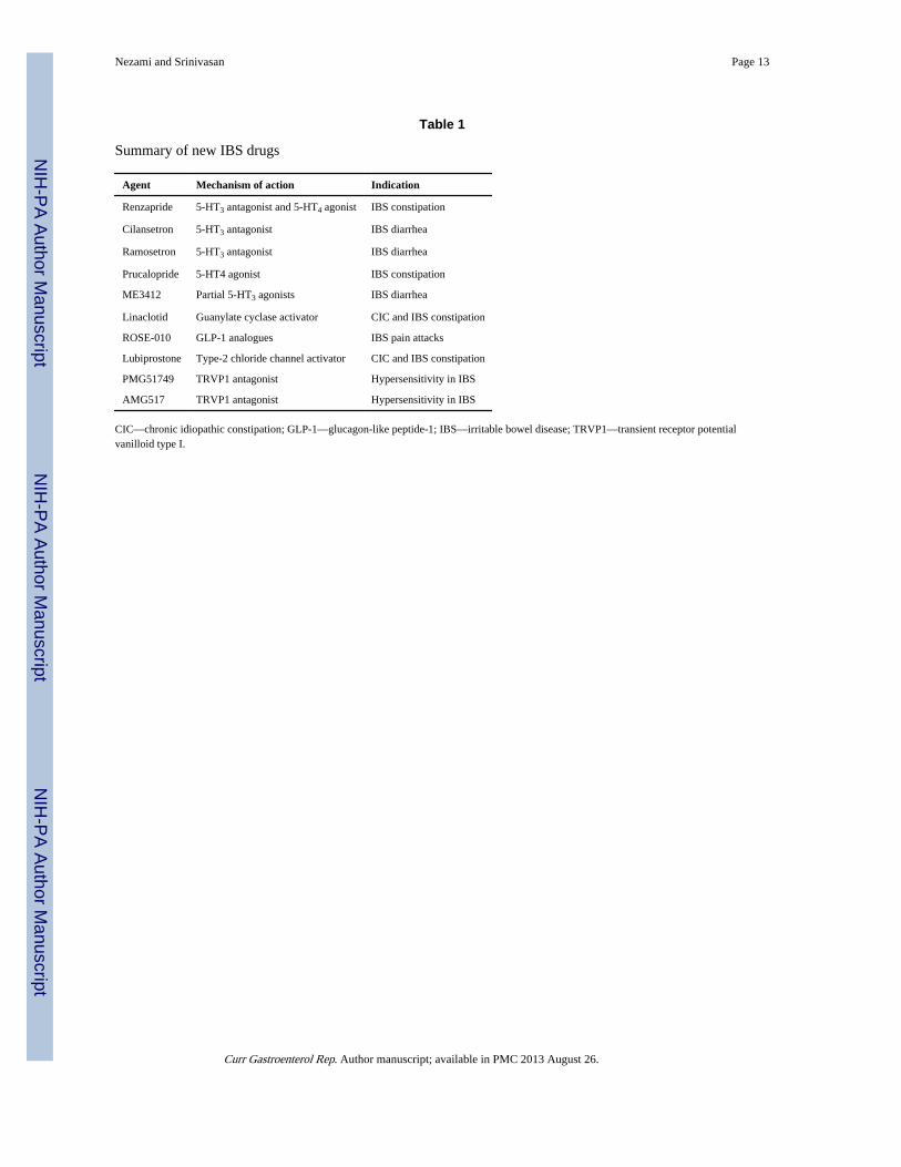
Irritable bowel syndromeIBS is a disorder of the brain-gut link associated with exaggerated response to stress;characteristic symptoms include abdominal pain/discomfort and concurrent disturbance indefecation. Because IBS dominantly involves mothers and their children, researchers haveattempted to find the genetic inheritance pattern of the disease. Mitochondrial DNA is acircular ring of DNA found in mitochondria, which is inherited only from the mother.Camilleri et al. [36•] studied the possibility that mitochondrial DNA single nucleotidepolymorphisms could confer risk to the development of IBS, and suggested that variation inmitochondrial DNA may be associated with satiation, gastric emptying, and possibly pain.Recent clinical investigations of IBS have identified novel therapeutic approaches thatmodulate the serotonin-mediated control of the ENS. Generally, activation of 5-HT2B, 5-HT3, or 5-HT4 receptors on enteric cholinergic neurons results in acetylcholine release andsmooth muscle contraction. On the other hand, serotonin stimulates 5-HT4, 5-HT1A, or 5-HT1D receptors on inhibitory enteric or nitrergic neurons to release NO and subsequentlycause smooth muscle relaxation [37]. Table 1 summarizes new drugs used for IBS treatment[38•, 39–41, 42•, 43•]. Transient receptor potential vanilloid type I (TRPV1 or VR1)receptor has gained attention regarding its prominent role in inflammation-induced pain andvisceral hypersensitivity. However, there are concerns with its antagonists PMG51749 andAMG517 hyperthermia because of their effects on thermoregulation [43•].
Hydrogen sulfide (H2S) is an endogenous gasotransmitter (gaseous neurotransmitter), whichexerts different effects in the digestive system, including relaxation of ileal smooth muscleand stimulation of intestinal secretion and nociceptive processes [44]. By far, H2S isimplicated as an agent both preventing and causing tissue damage and inflammation. Thelatter effect appears to be mediated via activation of KATP channels and to be NO dependent[45•].
The ENS and Secretion in the Small IntestineThe main physiological task of enteric secretion is the continuous hydration of luminalcontents to ensure appropriate mixing and absorption of nutrients and effective protectionagainst potentially harmful pathogens or enterotoxins. The intestinal crypt cells secretchloride into the lumen, resulting in accumulation of fluid. The regulation of chloridesecretion occurs by neural reflex pathways within the ENS.
Nezami and Srinivasan Page 5
Curr Gastroenterol Rep. Author manuscript; available in PMC 2013 August 26.
NIH
-PA Author Manuscript
NIH
-PA Author Manuscript
NIH
-PA Author Manuscript

Cells and neurotransmittersMultiple cell surface receptors are involved in intestinal secretion [46]. The intestinalsecretory reflexes arise from complex interactions between excitatory and inhibitoryneurotransmitters, released by both the ENS and afferent neurons (Table 2). However,intestinal secretion is also modulated by a variety of messengers released from epithelial,endocrine, and immune cells.
Firing of secretomotor neurons releases acetylcholine or VIP, which stimulates secretion ofCl− and HCO3−, as well as fluid secretion from epithelial cells. It is now apparent thatmechanical stimulation releases 5-HT from enterochromaffin cells with subsequentactivation of cholinergic and VIP secretomotor neurons. Pathophysiological changes in thesesignaling mechanisms contribute to disorders of motility and secretion. 5-HT is alsoinvolved in carcinoid diarrhea, cholera toxin, and bile salt-induced fluid and electrolytesecretion by activating the ENS.
DiarrheaDiarrhea is characterized by frequent loose or liquid bowel movements. It can represent asymptom of diseases such as inflammatory bowel disease (IBD), intestinal ischemia, celiacdisease, infection, or functional bowel disorders. Understanding secretomotor neurons isbasic to understanding neurogenic secretory diarrhea, constipation, and therapeuticstrategies. Hyperactivity of secretomotor neuronal activity elevates Cl− secretion andinduces neurogenic secretory diarrhea. Secretory diarrhea results from increased chloridesecretion, decreased sodium absorption, or increased mucosal permeability. Cholera, theprototype of secretory diarrhea, is caused by the enterotoxin of Vibrio cholerae (choleratoxin). Cholera toxin strongly activates adenylyl cyclase, causing a prolonged increase inintracellular concentration of cyclic adenosine monophosphate (AMP) within cryptenterocytes. Continuous cAMP production activates chloride channels, resulting in unabatedwater and electrolyte secretion that leads to voluminous watery diarrhea. Additionally,cholera toxin affects the ENS, resulting in an independent stimulus of secretion. Gwynne etal. [47] recently showed that cholera toxin induces specific and sustained hyperexcitabilityof secretomotor neurons in enteric pathways. This effect of cholera toxin depends on 5-HT3,nicotinic, and neurokinin 1 receptors. Cholera toxin is thought to activate neural pathwaysvia release of 5-HT from enterochromaffin cells, which depends on 5-HT3 receptors.Hyperactivity of secretomotor neuronal activity also elevates Cl− secretion and inducesneurogenic secretory diarrhea. In food allergies and inflammatory states, mast cellmediators, including histamine, serotonin, and prostaglandins, elevate secretomotor firing,which in turn stimulates the secretion of NaCl and large volumes of H2O.
The ENS and Inflammation in the Small IntestineInflammation causes significant changes in intestinal functions including motility, secretion,and sensation [48]. The interplay between ENS and inflammation highlights the existence ofclose interactions between ENS and enteric immune cells. In this scenario, EGCs play animportant role in enteric permeability, because extreme cases of inflammation and necrosisoccur in the absence of glial cell function. Patients with chronic IBD display varying levelsof enteric inflammation, and enteric ganglionitis is reported in some patients with severeIBS. Elevated intestinal permeability is apparent in patients with Crohn’s disease,necrotizing enterocolitis, diabetes, and IBS. This is in accordance with the fact that IBSsymptoms are more frequent in IBD patients than in the general population [49••].
Nezami and Srinivasan Page 6
Curr Gastroenterol Rep. Author manuscript; available in PMC 2013 August 26.
NIH
-PA Author Manuscript
NIH
-PA Author Manuscript
NIH
-PA Author Manuscript

Cells and neurotransmittersAmple evidence exists that gastrointestinal inflammation is related to an imbalance in thefunction of peptidergic neurons, including SP, VIP, and NPY [50]. EGCs increase GDNFsecretion during intestinal inflammation, which could act to protect intestinal epithelial cellsfrom cytokine-induced apoptosis. Glucagon-like peptide-2 (GLP-2) is an important regulatorof nutritional absorptive capacity with cell differentiation properties and anti-inflammatoryactions, which is produced by various ENS neurons. In animal models of IBD, GLP-2significantly improves mucosal inflammation indices, reduces levels of inflammatorycytokines (interferon-γ, tumor necrosis factor-α, interleukin [IL]-1β) and inducible NOS,and increases levels of IL-10 [51]. GLP-2 probably reduces intestinal mucosal inflammationby activation of VIP neurons of the submucosal plexus.
IBD and neurogenic inflammationPathologic changes of the ENS in IBD include hypertrophy, hyperplasia, and axonal damageto nerve fibers and neuronal cell bodies, and hyperplasia of EGCs [52].
Enteric neurons can directly secrete inflammatory mediators (eg, IL-8). A similar role couldbe played by EGCs as they respond to intestinal inflammation by proliferating andproducing inflammatory cytokines (eg, IL-6). Conversely, EGCs could inhibit inflammationas they secrete mediators (eg, nerve growth factor and neurotrophin-3) that have anti-inflammatory properties in animal models of colitis. EGCs seem to be active elements of theENS during intestinal inflammatory and immune responses by acting as antigen-presentingcells and interacting with the mucosal immune system via the expression of cytokines andcytokine receptors. Specific ablation of EGCs leads to a breakdown of the epitheliumbarrier, suggesting a role of EGCs in maintaining the integrity or permeability of the mucosa[53]. Neurogenic inflammation refers to an inflammatory reflex arc by sensory neurons,which transmits noxious stimulus centrally and results in both pain perception and an intenselocal inflammatory reaction. Inflammation affects neuronal function and survival;conversely, neurogenic inflammation has been suggested to play a key role in thepathogenesis of IBD. Porcher et al. [54] described the almost complete abolition of ICCswithin the longitudinal and circular muscle layers in Crohn’s disease, and a significantreduction in numbers within the myenteric and deep muscular plexuses. These changes mayexplain the development of dysmotility in some patients. In an interesting study, Takami etal. [55•] showed that surgical denervation performed before chemical induction of IBDsuppresses the score in all of the inflammatory indices such that almost no sign ofinflammation is observed in histological evaluation.
NPY is widely expressed in the central and peripheral nervous system and is involved in theregulation of several physiological processes, including energy balance, food intake, andnociception. In an experimental model of acute colitis, Hassani et al. [56] showed that lackof or inhibition of the NPY Y1 receptor makes the animals less susceptible to damagecaused by colonic inflammation. Other studies of inflammatory processes in IBD highlightthe role of neuronal NPY as a mediator of increased neuronal NOS (nNOS) and subsequentinflammation [57].
ConclusionsThe enteric nervous system has a complex role in the small intestine, interacting with all celltypes to modulate motility, secretion, pain perception, and inflammation (Fig. 1).Understanding the receptors and signal transduction pathways involved in mediating thesefunctions can aid in the development of new therapeutic targets.
Nezami and Srinivasan Page 7
Curr Gastroenterol Rep. Author manuscript; available in PMC 2013 August 26.
NIH
-PA Author Manuscript
NIH
-PA Author Manuscript
NIH
-PA Author Manuscript

AcknowledgmentsThis work was supported by the following grants: National Institutes of Health DK080684 (Dr. Srinivasan) andVeterans Administration Merit award (Dr. Srinivasan).
References and Recommended ReadingPapers of particular interest, published recently, have been highlighted as:
• Of importance
•• Of major importance
1. Gershon, MD. A Groundbreaking New Understanding of Nervous Disorders of the Stomach andIntestine. New York: HarperCollins; 1999. The Second Brain.
2••. Liu MT, Kuan YH, Wang J, et al. 5-HT4 receptor-mediated neuroprotection and neurogenesis inthe enteric nervous system of adult mice. J Neurosci. 2009; 29:9683–9699. This article reportsthat stimulation of the 5-HT4 receptor is required postnatally for ENS growth and maintenance,and demonstrates for the first time the adult enteric neurogenesis in mice. [PubMed: 19657021]
3. Burns AJ, Thapar N. Advances in ontogeny of the enteric nervous system. NeurogastroenterolMotil. 2006; 18:876–887. [PubMed: 16961690]
4. Gianino S, Grider JR, Cresswell J, et al. GDNF availability determines enteric neuron number bycontrolling precursor proliferation. Development. 2003; 130:2187–2198. [PubMed: 12668632]
5. Srinivasan S, Anitha M, Mwangi S, Heuckeroth RO. Enteric neuroblasts require thephosphatidylinositol 3-kinase/Akt/Forkhead pathway for GDNF-stimulated survival. Mol CellNeurosci. 2005; 29:107–119. [PubMed: 15866051]
6. Phillips RJ, Kieffer EJ, Powley TL. Aging of the myenteric plexus: neuronal loss is specific tocholinergic neurons. Auton Neurosci. 2003; 106:69–83. [PubMed: 12878075]
7. Johnson RJ, Schemann M, Santer RM, Cowen T. The effects of age on the overall population andon sub-populations of myenteric neurons in the rat small intestine. J Anat. 1998; 192 (Pt 4):479–488. [PubMed: 9723975]
8. Kasparek MS, Fatima J, Iqbal CW, et al. Age-related changes in functional NANC innervation withVIP and substance P in the jejunum of Lewis rats. Auton Neurosci. 2009; 151:127–134. [PubMed:19734110]
9. Saffrey MJ. Ageing of the enteric nervous system. Mech Ageing Dev. 2004; 125:899–906.[PubMed: 15563936]
10. Penagini R, Spiller RC, Misiewicz JJ, Frost PG. Effect of ileal infusion of glycochenodeoxycholicacid on segmental transit, motility, and flow in the human jejunum and ileum. Gut. 1989; 30:609–617. [PubMed: 2731753]
11••. Poole DP, Godfrey C, Cattaruzza F, et al. Expression and function of the bile acid receptorGpBAR1 (TGR5) in the murine enteric nervous system. Neurogastroenterol Motil. 2010;22:814–825. e227–818. This article studies the mechanism of action of bile acids on entericmotility and secretion. The authors found that human bile acid receptor 1 (GpBAR1) is expressedin enteric neurons, and its activation on inhibitory motor neurons releases nitric oxide andsuppresses motility. [PubMed: 20236244]
12. Pham T, Guerrini S, Wong H, et al. Distribution of galanin receptor 1 immunoreactivity in the ratstomach and small intestine. J Comp Neurol. 2002; 450:292–302. [PubMed: 12209857]
13. Simpson J, Sundler F, Humes DJ, et al. Prolonged elevation of galanin and tachykinin expressionin mucosal and myenteric enteric nerves in trinitrobenzene sulphonic acid colitis.Neurogastroenterol Motil. 2008; 20:392–406. [PubMed: 18208479]
14. Kalff JC, Carlos TM, Schraut WH, et al. Surgically induced leukocytic infiltrates within the ratintestinal muscularis mediate postoperative ileus. Gastroenterology. 1999; 117:378–387.[PubMed: 10419919]
15. Schwarz NT, Kalff JC, Turler A, et al. Prostanoid production via COX-2 as a causative mechanismof rodent postoperative ileus. Gastroenterology. 2001; 121:1354–1371. [PubMed: 11729115]
Nezami and Srinivasan Page 8
Curr Gastroenterol Rep. Author manuscript; available in PMC 2013 August 26.
NIH
-PA Author Manuscript
NIH
-PA Author Manuscript
NIH
-PA Author Manuscript

16•. Wattchow DA, De Fontgalland D, Bampton PA, et al. Clinical trial: the impact of cyclooxygenaseinhibitors on gastrointestinal recovery after major surgery—a randomized double blind controlledtrial of celecoxib or diclofenac vs. placebo. Aliment Pharmacol Ther. 2009; 30:987–998. Thisrecent clinical trial demonstrates that celecoxib, but not diclofenac, reduced the development ofileus, but neither agent accelerated early recovery of normal bowel motility. [PubMed:19694636]
17. Kadowaki M, Nagakura Y, Tokita K, et al. Adenosine A1 receptor blockade reverses experimentalpostoperative ileus in rat colon. Eur J Pharmacol. 2003; 458:197–200. [PubMed: 12498926]
18. Antonioli L, Fornai M, Colucci R, et al. Regulation of enteric functions by adenosine:pathophysiological and pharmacological implications. Pharmacol Ther. 2008; 120:233–253.[PubMed: 18848843]
19. Anitha M, Gondha C, Sutliff R, et al. GDNF rescues hyperglycemia-induced diabetic entericneuropathy through activation of the PI3K/Akt pathway. J Clin Invest. 2006; 116:344–356.[PubMed: 16453021]
20. Ordog T. Interstitial cells of Cajal in diabetic gastroenteropathy. Neurogastroenterol Motil. 2008;20:8–18. [PubMed: 18173559]
21. Russell JW, Sullivan KA, Windebank AJ, et al. Neurons undergo apoptosis in animal and cellculture models of diabetes. Neurobiol Dis. 1999; 6:347–363. [PubMed: 10527803]
22. Moriyama R, Tsukamura H, Kinoshita M, et al. In vitro increase in intracellular calciumconcentrations induced by low or high extracellular glucose levels in ependymocytes andserotonergic neurons of the rat lower brainstem. Endocrinology. 2004; 145:2507–2515. [PubMed:14962992]
23•. Izbeki F, Wittman T, Rosztoczy A, et al. Immediate insulin treatment prevents gut motilityalterations and loss of nitrergic neurons in the ileum and colon of rats with streptozotocin-induced diabetes. Diabetes Res Clin Pract. 2008; 80:192–198. This article shows that thenitrergic neurons located in different intestinal segments exhibit different susceptibilities to adiabetic state, and that early insulin treatment prevented the nitrergic cell loss in the ileum andcolon. [PubMed: 18242757]
24. Anitha M, Chandrasekharan B, Salgado JR, et al. Glial-derived neurotrophic factor modulatesenteric neuronal survival and proliferation through neuropeptide Y. Gastroenterology. 2006;131:1164–1178. [PubMed: 17030186]
25. Adeghate E, Ponery AS, Sharma AK, et al. Diabetes mellitus is associated with a decrease invasoactive intestinal polypeptide content of gastrointestinal tract of rat. Arch Physiol Biochem.2001; 109:246–251. [PubMed: 11880929]
26. Pfeiffer RF. Gastrointestinal dysfunction in Parkinson’s disease. Lancet Neurol. 2003; 2:107–116.[PubMed: 12849267]
27. Braak H, de Vos RA, Bohl J, Del Tredici K. Gastric alpha-synuclein immunoreactive inclusions inMeissner’s and Auerbach’s plexuses in cases staged for Parkinson’s disease-related brainpathology. Neurosci Lett. 2006; 396:67–72. [PubMed: 16330147]
28. Anderson G, Noorian AR, Taylor G, et al. Loss of enteric dopaminergic neurons and associatedchanges in colon motility in an MPTP mouse model of Parkinson’s disease. Exp Neurol. 2007;207:4–12. [PubMed: 17586496]
29. Greene JG, Noorian AR, Srinivasan S. Delayed gastric emptying and enteric nervous systemdysfunction in the rotenone model of Parkinson’s disease. Exp Neurol. 2009; 218:154–161.[PubMed: 19409896]
30•. Bielefeldt K, Davis B, Binion DG. Pain and inflammatory bowel disease. Inflamm Bowel Dis.2009; 15:778–788. This recent article extensively reviews the current knowledge aboutmechanisms of abdominal pain in IBD. [PubMed: 19130619]
31. Wright K, Rooney N, Feeney M, et al. Differential expression of cannabinoid receptors in thehuman colon: cannabinoids promote epithelial wound healing. Gastroenterology. 2005; 129:437–453. [PubMed: 16083701]
32. McLean PG, Borman RA, Lee K. 5-HT in the enteric nervous system: gut function andneuropharmacology. Trends Neurosci. 2007; 30:9–13. [PubMed: 17126921]
Nezami and Srinivasan Page 9
Curr Gastroenterol Rep. Author manuscript; available in PMC 2013 August 26.
NIH
-PA Author Manuscript
NIH
-PA Author Manuscript
NIH
-PA Author Manuscript

33. Sawynok J, Liu XJ. Adenosine in the spinal cord and periphery: release and regulation of pain.Prog Neurobiol. 2003; 69:313–340. [PubMed: 12787573]
34. Delafoy L, Gelot A, Ardid D, et al. Interactive involvement of brain derived neurotrophic factor,nerve growth factor, and calcitonin gene related peptide in colonic hypersensitivity in the rat. Gut.2006; 55:940–945. [PubMed: 16401692]
35. Buhner S, Li Q, Vignali S, et al. Activation of human enteric neurons by supernatants of colonicbiopsy specimens from patients with irritable bowel syndrome. Gastroenterology. 2009;137:1425–1434. [PubMed: 19596012]
36•. Camilleri M, Carlson P, Zinsmeister AR, et al. Mitochondrial DNA and gastrointestinal motor andsensory functions in health and functional gastrointestinal disorders. Am J Physiol GastrointestLiver Physiol. 2009; 296:G510–G516. This recent article describes the role of mitochondrialDNA polymorphisms in development of IBS. The authors suggest a possible association betweenmtDNA variation and some IBS-related symptoms. [PubMed: 19147801]
37. McLean PG, Borman RA, Lee K. 5-HT in the enteric nervous system: gut function andneuropharmacology. Trends Neurosci. 2007; 30:9–13. [PubMed: 17126921]
38•. Anderson JL, May HT, Bair TL, et al. Lack of association of tegaserod with adversecardiovascular outcomes in a matched case-control study. J Cardiovasc Pharmacol Ther. 2009;14:170–175. The authors performed a large-scale study on adverse effects of tegaserod, andfound no increase in cardiovascular events in 2.5 years of follow-up compared to untreatedpatients. These results question the previous observations that led to withdrawal of this drug in2007. [PubMed: 19602672]
39. Camilleri M, McKinzie S, Fox J, et al. Effect of renzapride on transit in constipation-predominantirritable bowel syndrome. Clin Gastroenterol Hepatol. 2004; 2:895–904. [PubMed: 15476153]
40. Kawano K, Mori T, Fu L, et al. Comparison between partial agonist (ME3412) and antagonist(alosetron) of 5-hydroxytryptamine 3 receptor on gastrointestinal function. NeurogastroenterolMotil. 2005; 17:290–301. [PubMed: 15787949]
41. Bryant AP, Busby RW, Bartolini WP, et al. Linaclotide is a potent and selective guanylate cyclaseC agonist that elicits pharmacological effects locally in the gastrointestinal tract. Life Sci. 86:760–765. [PubMed: 20307554]
42•. Hellström PM. GLP-1 playing the role of a gut regulatory compound. Acta Physiol (Oxf). 2010(Epub ahead of print). This randomized, double-blind clinical trial investigates the effect ofGLP-1 analogue ROSE-010 in humans, and shows that ROSE-010 is effective in pain reliefresponse in IBS patients affected by pain attacks.
43•. Akbar A, Walters JR, Ghosh S. Review article: visceral hypersensitivity in irritable bowelsyndrome: molecular mechanisms and therapeutic agents. Aliment Pharmacol Ther. 2009;30:423–435. This review provides a useful insight into the new molecular concepts emerging inthe field of visceral hypersensitivity. [PubMed: 19493256]
44. Xu GY, Winston JH, Shenoy M, et al. The endogenous hydrogen sulfide producing enzymecystathionine-beta synthase contributes to visceral hypersensitivity in a rat model of irritablebowel syndrome. Mol Pain. 2009; 5:44. [PubMed: 19660142]
45•. Gallego D, Clave P, Donovan J, et al. The gaseous mediator, hydrogen sulphide, inhibits in vitromotor patterns in the human, rat and mouse colon and jejunum. Neurogastroenterol Motil. 2008;20:1306–1316. This article describes the emerging gastric motility properties of the gaseousmediator H2S. H2S is implicated as an agent both preventing and causing tissue damage andinflammation. [PubMed: 19019033]
46. Cooke HJ. Neurotransmitters in neuronal reflexes regulating intestinal secretion. Ann N Y AcadSci. 2000; 915:77–80. [PubMed: 11193603]
47. Gwynne RM, Ellis M, Sjovall H, Bornstein JC. Cholera toxin induces sustained hyperexcitabilityin submucosal secretomotor neurons in guinea pig jejunum. Gastroenterology. 2009; 136:299–308.e294. [PubMed: 19026646]
48. Vasina V, Barbara G, Talamonti L, et al. Enteric neuroplasticity evoked by inflammation. AutonNeurosci. 2006; 126–127:264–272.
49••. Keohane J, O’Mahony C, O’Mahony L, et al. Irritable bowel syndrome-type symptoms inpatients with inflammatory bowel disease: a real association or reflection of occult inflammation?
Nezami and Srinivasan Page 10
Curr Gastroenterol Rep. Author manuscript; available in PMC 2013 August 26.
NIH
-PA Author Manuscript
NIH
-PA Author Manuscript
NIH
-PA Author Manuscript

Am J Gastroenterol. 2010 (Epub ahead of print). This article suggests that although IBS-likesymptoms are common in patients with IBD, the mechanism in most cases is likely to be occultinflammation rather than coexistent IBS.
50. Okajima K, Harada N. Regulation of inflammatory responses by sensory neurons: molecularmechanism(s) and possible therapeutic applications. Curr Med Chem. 2006; 13:2241–2251.[PubMed: 16918352]
51. Alavi K, Schwartz MZ, Palazzo JP, Prasad R. Treatment of inflammatory bowel disease in a rodentmodel with the intestinal growth factor glucagon-like peptide-2. J Pediatr Surg. 2000; 35:847–851.[PubMed: 10873024]
52. Villanacci V, Bassotti G, Nascimbeni R, et al. Enteric nervous system abnormalities ininflammatory bowel diseases. Neurogastroenterol Motil. 2008; 20:1009–1016. [PubMed:18492026]
53. Cabarrocas J, Savidge TC, Liblau RS. Role of enteric glial cells in inflammatory bowel disease.Glia. 2003; 41:81–93. [PubMed: 12465048]
54. Porcher C, Baldo M, Henry M, et al. Deficiency of interstitial cells of Cajal in the small intestine ofpatients with Crohn’s disease. Am J Gastroenterol. 2002; 97:118–125. [PubMed: 11808934]
55•. Takami Y, Mantyh CR, Pappas TN, et al. Extrinsic surgical denervation ameliorates TNBS-induced colitis in rats. Hepatogastroenterology. 2009; 56:682–686. This interesting articleextensively challenges the neurogenic inflammation concept by performing surgical denervationof specific sensorimotor neurons in an animal model of colitis. The authors report that surgicallydenervated animals showed almost no sign of inflammation after inducing colitis. [PubMed:19621680]
56. Hassani H, Lucas G, Rozell B, Ernfors P. Attenuation of acute experimental colitis by preventingNPY Y1 receptor signaling. Am J Physiol Gastrointest Liver Physiol. 2005; 288:G550–G556.[PubMed: 15499082]
57. Chandrasekharan B, Bala V, Kolachala VL, et al. Targeted deletion of neuropeptide Y (NPY)modulates experimental colitis. PLoS One. 2008; 3:e3304. [PubMed: 18836554]
Nezami and Srinivasan Page 11
Curr Gastroenterol Rep. Author manuscript; available in PMC 2013 August 26.
NIH
-PA Author Manuscript
NIH
-PA Author Manuscript
NIH
-PA Author Manuscript

Figure 1.Role of enteric neurons in the small intestine: summary of the mechanisms of entericneuronal modulation of peristalsis, secretion, pain perception, and inflammation in the smallintestine. A—adenosine; Ach—acetylcholine; CB—cannabinoid; COX-2—cyclo-oxygenase-2; ECC—enterochromaffin cell; GLP—glucagon-like peptide-1; H2S—hydrogensulfide; NO—nitric oxide; NPY—neuropeptide Y; SP—substance P; TRPV1—transientreceptor potential vanilloid type I; VIP—vasoactive intestinal peptide.
Nezami and Srinivasan Page 12
Curr Gastroenterol Rep. Author manuscript; available in PMC 2013 August 26.
NIH
-PA Author Manuscript
NIH
-PA Author Manuscript
NIH
-PA Author Manuscript
NIH
-PA Author Manuscript
NIH
-PA Author Manuscript
NIH
-PA Author Manuscript
Nezami and Srinivasan Page 13
Table 1
Summary of new IBS drugs
Agent Mechanism of action Indication
Renzapride 5-HT3 antagonist and 5-HT4 agonist IBS constipation
Cilansetron 5-HT3 antagonist IBS diarrhea
Ramosetron 5-HT3 antagonist IBS diarrhea
Prucalopride 5-HT4 agonist IBS constipation
ME3412 Partial 5-HT3 agonists IBS diarrhea
Linaclotid Guanylate cyclase activator CIC and IBS constipation
ROSE-010 GLP-1 analogues IBS pain attacks
Lubiprostone Type-2 chloride channel activator CIC and IBS constipation
PMG51749 TRVP1 antagonist Hypersensitivity in IBS
AMG517 TRVP1 antagonist Hypersensitivity in IBS
CIC—chronic idiopathic constipation; GLP-1—glucagon-like peptide-1; IBS—irritable bowel disease; TRVP1—transient receptor potentialvanilloid type I.
Curr Gastroenterol Rep. Author manuscript; available in PMC 2013 August 26.

NIH
-PA Author Manuscript
NIH
-PA Author Manuscript
NIH
-PA Author Manuscript
Nezami and Srinivasan Page 14
Table 2
Effects of ENS neurotransmitters on secretion
Inhibitory Stimulatory
Adrenergic α2 Adrenergic β
5-HT4 5-HT3
Purinergic P2y Nicotinic
Adenosine A Muscarinic M3
GABA
Histamine H3
VIP
Somatostatin
GABA—γ-aminobutyric acid; VIP—vasoactive intestinal peptide.
Curr Gastroenterol Rep. Author manuscript; available in PMC 2013 August 26.