Engineering An ER-Targeted Dynein-Anchoring Fusion Protein ...
47
Dartmouth College Dartmouth College Dartmouth Digital Commons Dartmouth Digital Commons ENGS 88 Honors Thesis (AB Students) Other Engineering Materials Spring 6-10-2020 Engineering An ER-Targeted Dynein-Anchoring Fusion Protein to Engineering An ER-Targeted Dynein-Anchoring Fusion Protein to Mediate Spindle and Nuclear Positioning in Budding Yeast Mediate Spindle and Nuclear Positioning in Budding Yeast Dayle Wang [email protected] Follow this and additional works at: https://digitalcommons.dartmouth.edu/engs88 Part of the Engineering Commons Dartmouth Digital Commons Citation Dartmouth Digital Commons Citation Wang, Dayle, "Engineering An ER-Targeted Dynein-Anchoring Fusion Protein to Mediate Spindle and Nuclear Positioning in Budding Yeast" (2020). ENGS 88 Honors Thesis (AB Students). 20. https://digitalcommons.dartmouth.edu/engs88/20 This Thesis (Senior Honors) is brought to you for free and open access by the Other Engineering Materials at Dartmouth Digital Commons. It has been accepted for inclusion in ENGS 88 Honors Thesis (AB Students) by an authorized administrator of Dartmouth Digital Commons. For more information, please contact [email protected].
Transcript of Engineering An ER-Targeted Dynein-Anchoring Fusion Protein ...
Dartmouth College Dartmouth College
Dartmouth Digital Commons Dartmouth Digital Commons
ENGS 88 Honors Thesis (AB Students) Other Engineering Materials
Spring 6-10-2020
Engineering An ER-Targeted Dynein-Anchoring Fusion Protein to Engineering An ER-Targeted Dynein-Anchoring Fusion Protein to
Mediate Spindle and Nuclear Positioning in Budding Yeast Mediate Spindle and Nuclear Positioning in Budding Yeast
Dayle Wang [email protected]
Follow this and additional works at: https://digitalcommons.dartmouth.edu/engs88
Part of the Engineering Commons
Dartmouth Digital Commons Citation Dartmouth Digital Commons Citation Wang, Dayle, "Engineering An ER-Targeted Dynein-Anchoring Fusion Protein to Mediate Spindle and Nuclear Positioning in Budding Yeast" (2020). ENGS 88 Honors Thesis (AB Students). 20. https://digitalcommons.dartmouth.edu/engs88/20
This Thesis (Senior Honors) is brought to you for free and open access by the Other Engineering Materials at Dartmouth Digital Commons. It has been accepted for inclusion in ENGS 88 Honors Thesis (AB Students) by an authorized administrator of Dartmouth Digital Commons. For more information, please contact [email protected].

ENGINEERING AN ER-TARGETED DYNEIN-ANCHORING FUSION PROTEIN TO
MEDIATE SPINDLE AND NUCLEAR POSITIONING IN BUDDING YEAST
by
DAYLE WANG
Bachelor of Arts Honors Thesis
Thayer School of Engineering Dartmouth College
Hanover, New Hampshire
Date______________________
Approved: __________________ Advisor’s Signature
Advisor’s Signature
__________________ Author’s Signature
June 10, 2020


ii
Abstract
Current evidence suggests that, in budding yeast (Saccharomyces cerevisiae),
nuclear migration protein 1 (Num1) binds and anchors cytoplasmic dynein, a
microtubule-based motor protein, to the plasma membrane, where dynein pulls on
cytoplasmic microtubules attached to the nucleus to move the nucleus to the cell division
site; however, the exact location where Num1 enables dynein to exert pulling forces at
the cell cortex remains unclear. To assess the ability of the cortical endoplasmic
reticulum (ER) associated with the plasma membrane to function as a platform for dynein
anchoring by Num1, I genetically engineered Num1 fusion to a resident ER protein of the
tricalbin (TCB) protein family. Through quantification and assessment of live-cell
fluorescence microscopy images, I found that a fusion protein consisting of Tcb3 and the
coiled-coil (CC) dynein-anchoring domain of Num1 can serve as a platform for dynein
attachment and function in nuclear positioning, rescuing the mitotic spindle alignment
defect in a background lacking Num1 (num1Δ). Therefore, the cortical ER appears to be
able to support Num1 function in dynein anchoring.

iii
Preface
While I began Dartmouth planning to major in biology, I became interested in
engineering sciences due to the interdisciplinary opportunities and focus on improving
lives and society at Thayer School of Engineering. This honors thesis project combines
my interest in biological sciences and engineering sciences, and I am grateful to both the
Department of Biological Sciences as well as Thayer School of Engineering for this
opportunity. This project would not be possible without my advisor, Dr. Wei-Lih Lee,
who provided invaluable advice and guidance for my project, and my Thayer co-advisor,
Dr. Margaret Ackerman; I thank them for their support and time. I am especially thankful
for Dr. Lee’s encouragement, scientific mentorship, and high expectations. I would like
to give immense thanks to Safia Omer for her patience with all my questions and her
mentorship, as well as her scientific expertise. I would also like to thank Mary Horner-
Richardson. I am grateful to the Lee lab and their companionship, support, and patience.
Thank you to Jenna Wheeler and Dr. Douglas Van Citters for their support and guidance
both for this thesis and during my time at Thayer.
Last (but certainly not least), thank you to my family and friends.

iv
Table of Contents
Abstract…………………………………………………………………………….……...i
Preface……………………………………………………………………………………iii
Table of Contents…………………………………………………………………………iv
List of Figures…………………………………………………………………………….vi
List of Tables………………………………………………………………………….... vii
Chapter 1. Introduction………………...………………………………………..…...……1
1.1 Background…………………………………………………………………....1
1.2 Current Research Questions…………………………………………………...7
1.3 Thesis Purpose……………………………………………………………….12
Chapter 2. Materials and Methods...……………………………………………...……...14
2.1 Construction of Tcb3-CC strains…………………………………………….14
2.2 Construction of Tcb3-CC dyn1∆ strains………………………………….….18
2.3 Construction of Tcb3-CC-GFP strains……………...…………………..........18
2.4 Imaging………………………………………………………………………20
2.5 Image Analysis……………………………………………………………….20
Chapter 3. Results………………………………………………...………………...……23
3.1 Misaligned anaphase spindle defect of num1Δ is rescued by Tcb3-CC…......23
3.2 Tcb3-CC rescue of misaligned anaphase defect is dynein-dependent……….24
v
3.3 Tcb3-CC shows different localization compared to Tcb3 and Num1...…......26
Chapter 4. Discussion………………………………………………...……………...…..27
Chapter 5. Future Work…………...……………………………………...……...………30
Chapter 6. Conclusion…………………………………………………...……………….34
References…………………………………………………...…………………………...35

vi
List of Figures
Figure 1. Mitotic Spindle Positioning Pathways in Budding Yeast………………...….....3
Figure 2. Current Model for Dynein Function in Spindle Positioning……………………4
Figure 3. Diagram of the Num1 Protein………………………………………...……...…5
Figure 4. Deletion of ER-PM tethering proteins Scs2 and Scs22 disrupts Num1 distribution and localization……………………………………………………………...11 Figure 5. Project Summary………………………………………………………………12
Figure 6. Tcb3 and Fusion Proteins Diagram……………………………………………13
Figure 7. Tcb3-CC Strain Construction………………………………………………….16
Figure 8. Tcb3-CC-GFP Strain Construction……………………………………………19
Figure 9. Misaligned versus Aligned Anaphase Spindles……………………………….21
Figure 10. Tcb3-CC Misaligned Anaphase Spindle Assay……………………………...24
Figure 11. Misaligned Anaphase Spindle Assay for dyn1Δ num1Δ TCB3-CC cells…………………………………………………………………………………...….25 Figure 12. Localization and fluorescence intensity of Tcb3-CC-GFP………………......26

vii
List of Tables
Table 1. Comparison of Num1 and its functional homolog, NuMA……………………...7
Table 2. Key Resources………………………………………………………………….17

1
Chapter 1. Introduction
1.1 Background
Cytoplasmic dynein is a motor protein that functions in the transport of cargo,
organelles, and RNA during cellular interphase (Ananthanarayanan, 2016; Reck-Peterson
et al., 2018). During cell division events in mitosis and meiosis, cytoplasmic dynein is the
primary motor responsible for the correct distribution of chromosomes to daughter cells
(Ananthanarayanan, 2016). Given dynein’s central role in cellular transport, including
axonal transport in neuronal cells, and in proper separation of genetic material, mutations
affecting cytoplasmic dynein have been implicated in various neurodegenerative and
neurodevelopmental diseases as well as in improper division of genetic material in
meiosis and mitosis resulting in aneuploidy and often leading to disease states and
malfunctioning cells (Chen et al., 2014; Greenberg et al., 2018). Dynein functions as a
transport motor through attachment to the microtubule cytoskeletal network found within
cells (Reck-Peterson et al., 2018; Vale, 2003). These microtubules are tubular structures
made up of tubulin, consisting of a minus end that originates from the microtubule
organizing center (MTOC) as well as a plus end that usually extends towards the cell
periphery (Reck-Peterson et al., 2018). Motors that associate and use these microtubule
networks are thereby classified as ‘plus-end-directed’ or ‘minus-end-directed’; dynein is
a minus-end directed motor. Microtubules are also important in mitosis and meiosis for
building the mitotic spindle, which is composed of astral microtubules that emanate
outwards to the cell periphery and interpolar microtubules that extend towards the
chromosomes in the middle. During cellular division, dynein is anchored near the cell
periphery; as the anchored dynein walks toward the minus end of the attached astral

2
microtubule, it generates a pulling force on the mitotic spindle, thereby leading to
movement of the associated genetic material towards the cell periphery. Currently, in the
general model for dynein activation and attachment to its cargoes, dynein must first form
a complex with dynactin, a high-molecular weight, multi-subunit complex (Reck-
Peterson et al., 2018; Canty and Yildiz, 2020). This dynein-dynactin complex then seems
to require “activating adaptors” that serve to activate dynein’s motor activity and to
attach dynein to its cargoes both during interphase and during cellular division processes
(Reck-Peterson et al., 2018). In this project, the function of cytoplasmic dynein and its
‘activating adaptor’ during mitosis—specifically, the nuclear migration protein 1 (Num1)
in budding yeast—are the focus.
Saccharomyces cerevisiae, also known as budding yeast, is a useful and powerful
model organism that has been used in the scientific community since the 18th century
(Duina et al., 2014). Key features such as a well-characterized genome, facile genetic
manipulation through homologous recombination, and the availability of standardized
auxotrophic genetic markers make S. cerevisiae an ideal organism for biological research
(Duina et al., 2014). In yeast, cell division happens at a ‘predetermined’ site. As cells
prepare to divide a bud is formed; this bud later becomes the daughter cell after the
completion of cell division during cytokinesis. The mitotic spindle must thus be
positioned across the bud neck to facilitate transfer of the genetic material into the new
daughter cell. In yeast, two main pathways are involved in proper spindle positioning: the
Kar9 pathway and the dynein pathway (Figure 1) (Farkasovsky and Küntzel, 2001;
Markus et al., 2012a). The Kar9 pathway is dynein-independent and functions to orient
the mitotic spindle early in the cell cycle; briefly, in this mechanism, a myosin motor

3
(Myo2) binds a complex consisting of the Kar9 protein and a microtubule plus-end
binding protein, Bim1 (Figure 1B) (Markus et al., 2012a; Bloom, 2001). Myo2 then
moves along cortical actin cables in the cell to move the Kar9-Bim1 complex and the
astral microtubule plus end as cargo across the bud neck (Markus et al., 2012a).
Figure 1. Mitotic Spindle Positioning Pathways in Budding Yeast: A) Microscopy image of metaphase spindle in S. cerevisiae with spindle pole bodies marked by Spc42-CFP and microtubules marked by GFP-Tub1. B) The Kar9 pathway operates early in the cell cycle and is independent of dynein. Instead, it uses the Myo2 motor and actin cables along the cell cortex. C) The dynein pathway operates later in the cell cycle and depends on dynein-dynactin’s interaction with the cortical dynein-anchoring protein Num1. (Markus et al., 2012a)

4
The dynein-dependent pathway for budding yeast spindle orientation takes place
later in the cell cycle and follows the general pathway for dynein outlined earlier (Figure
1C, Figure 2) (Markus et al., 2012a). It is currently believed that dynein first forms a
complex with the Pac1 protein before interacting with another protein, Bik1 (Markus et
al., 2012a). The dynein-Pac1-Bik1 complex then binds the plus end of an astral
microtubule and recruits the dynactin complex; the microtubule’s growth allows for the
bound dynein-dynactin complex to find cortical patches of the protein Num1 at the cell
periphery, where dynein is “offloaded” from the microtubule plus end to Num1 patches
(Markus et al., 2012a; Markus and Lee, 2011). Num1 serves to anchor the dynein-
dynactin complex which then binds to a new astral microtubule. Num1’s anchoring of the
dynein-dynactin complex allows for the movement of the dynein motor towards the
minus end of the associated microtubule to pull the astral microtubule along the cell
cortex, thereby positioning the mitotic spindle correctly across the bud neck (Markus and
Lee, 2011).
Figure 2. Current Model for Dynein Function in Spindle Positioning: First, Pac1 interacts with cytoplasmic dynein (1). This dynein-Pac1 complex then interacts with the plus end of astral microtubules emerging from the nucleus (2). Dynactin is then recruited to the dynein-Pac1-microtubule-plus-end complex (3). Pac1 dissociates and the dynein-dynactin complex finds a Num1 cortical patch where dynein-dynactin is offloaded and anchored (4). As the anchored dynein-dynactin binds to a microtubule and moves towards the minus end of the microtubule, the spindle is pulled into the bud (5). (Markus and Lee, 2011)
5
Num1 is a 313 kDa protein first discovered to control nuclear migration in
budding yeast in the 1990s (Kormanec et al., 1991). Specifically, early characterizations
of the Num1 protein found that Num1 localizes along the cell cortex and is expressed
during the S and G2 phases of the cell cycle and that cells with disrupted Num1 function
often contain two nuclei in the mothers of budded cells, similar to yeast dynein mutants,
and have abnormal microtubule morphology (Kormanec et al., 1991; Farkasovsky and
Küntzel, 1995). Shortly after this initial research into dynein and Num1’s involvement in
spindle orientation, Num1 was then discovered to function as a dynein anchor given its
interactions with microtubules as well as findings showing that Num1 is required for
dynein-dynactin dependent microtubule sliding along the cell cortex (Farkasovsky and
Küntzel, 2001; Adames and Cooper, 2000; Heil-Chapdelaine et al., 2000). The Num1
protein contains a predicted N-terminal coiled-coil domain (CC), a predicted calcium
binding EF hand motif, followed by a highly repetitive central region containing thirteen
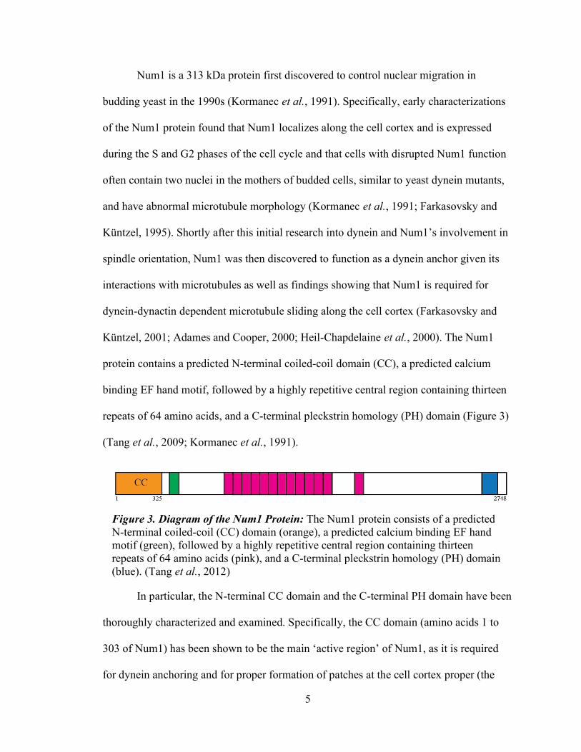
repeats of 64 amino acids, and a C-terminal pleckstrin homology (PH) domain (Figure 3)
(Tang et al., 2009; Kormanec et al., 1991).
In particular, the N-terminal CC domain and the C-terminal PH domain have been
thoroughly characterized and examined. Specifically, the CC domain (amino acids 1 to
303 of Num1) has been shown to be the main ‘active region’ of Num1, as it is required
for dynein anchoring and for proper formation of patches at the cell cortex proper (the
Figure 3. Diagram of the Num1 Protein: The Num1 protein consists of a predicted N-terminal coiled-coil (CC) domain (orange), a predicted calcium binding EF hand motif (green), followed by a highly repetitive central region containing thirteen repeats of 64 amino acids (pink), and a C-terminal pleckstrin homology (PH) domain (blue). (Tang et al., 2012)

6
domain has also been termed the patch assembly domain); domain analysis involving
expressing altered proteins in yeast cells and examining misaligned anaphase spindle
phenotypes as well as in vitro pull-down assays in which the purified CC domain (amino
acids 1 to 325 of Num1) was able to pull down components of the dynein heavy and
intermediate chains have verified that the CC domain is a dynein-anchoring domain of
Num1 (Tang et al., 2012). Furthermore, deletion of dynactin components eliminated the
pull-down behavior of the CC domain, thereby showing that the dynein-anchoring
activity of Num1 operates in a dynactin-dependent manner. The PH domain, on the other
hand, is responsible for the targeting of the Num1 protein to the cell cortex. This
targeting activity was reported in early studies of the Num1 protein and was fully
established through construction of fluorescently-labelled truncated Num1-constructs
(Farkasovsky and Küntzel, 1995); although the PH domain was not sufficient for the
formation of cortical patches matching those of full length-Num1, all constructs
contained deleted PH regions were found to localize to diffusely cytoplasm, thereby
indicating that the PH domain is required for the proper targeting of Num1 to the cell
cortex (Tang et al., 2009). Num1 constructs with the PH domain replaced with a
membrane-targeting CAAX motif were also able to serve as cortical dynein anchors,
therefore further confirming the dynein anchoring region of Num1 to be CC and that the
PH domain is not necessary for dynein association with Num1 (Tang et al., 2009).
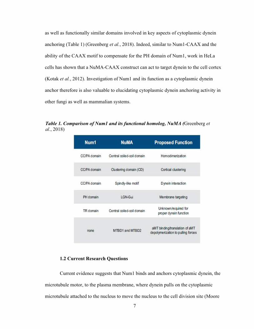
Beyond its importance in the dynein pathway in S. cerevisiae, Num1 is also
functionally similar to proteins found in other fungi such as Mcp5 in fission yeast and the
NuMA protein found in mammalian cells (Ananthanarayanan, 2016; Greenberg et al.,
2018). Num1 and NuMA share several features including cortical clustering morphology
7
as well as functionally similar domains involved in key aspects of cytoplasmic dynein
anchoring (Table 1) (Greenberg et al., 2018). Indeed, similar to Num1-CAAX and the
ability of the CAAX motif to compensate for the PH domain of Num1, work in HeLa
cells has shown that a NuMA-CAAX construct can act to target dynein to the cell cortex
(Kotak et al., 2012). Investigation of Num1 and its function as a cytoplasmic dynein
anchor therefore is also valuable to elucidating cytoplasmic dynein anchoring activity in
other fungi as well as mammalian systems.
1.2 Current Research Questions
Current evidence suggests that Num1 binds and anchors cytoplasmic dynein, the
microtubule motor, to the plasma membrane, where dynein pulls on the cytoplasmic
microtubule attached to the nucleus to move the nucleus to the cell division site (Moore
Table 1. Comparison of Num1 and its functional homolog, NuMA (Greenberg et al., 2018)

8
et al., 2009; Xiang, 2018). However, there remains several key questions concerning how
dynein is regulated and activated as well as how cortical pulling forces are generated and
how dynein is anchored to the diffusion-prone cell membrane.
One major question regarding dynein function in the spindle positioning pathway
is how dynein, a minus-end directed motor, localizes to the plus end of the astral
microtubules and how dynein is regulated to begin moving towards the minus end of the
microtubule after attachment to the cell cortex. In the general study of dynein, domain
analysis examining potential regulatory ATPase sites within the dynein motor itself have
been conducted (Cho et al., 2008). Other regulators of dynein particularly in the spindle
positioning pathway are also of interest. Although it is known that Pac1 forms a complex
with dynein during the early steps of dynein localization and anchoring, Pac1 (and the
mammalian analog, LIS1) have unclear roles in regulation of dynein activity. Current
research suggests that the offloading of the dynein-dynactin complex to the cortical
anchor itself may activate the complex through the dissociation of the associated Pac1
protein (Ananthanarayanan, 2016). Similarly, in mammalian cells, dynein appears to exist
in an autoinhibited ‘phi’ formation and the dynein-dynactin complex then is activated by
a cargo adaptor (such as NuMA, the functional homolog of Num1 in mammalian cells)
(Canty and Yildiz, 2020). Though this seems to indicate that Pac1/LIS1 acts to inhibit the
activity of dynein until it is bound to the cargo adaptor or cortical anchor, Pac1/LIS1 has
also been recently suggested to be an ‘activator’ of dynein as it appears to have
preferential binding to an ‘open’ conformation of dynein which would then promote the
formation of dynein-dynactin complexes and the subsequent binding to Num1/NuMA
and activation of the dynein-dynactin complex (Canty and Yildiz, 2020).

9
Besides Pac1/LIS1, the microtubule-associated protein (MAP) She1 has also been
shown to affect dynein motility and its function in spindle positioning in S. cerevisiae.
The motility of dynein as well as related characteristics of dynein including its ATPase
activity and binding affinity towards microtubules have been examined with and without
She1 in vitro (Ecklund et al., 2017; Markus et al., 2012b). While Markus et al. (2012b)
demonstrated She1’s specificity towards the dynein motor and characterized She1’s
behavior with respect to microtubules, Ecklund et al. (2017) further characterized the
stepping behavior of dynein, the kinetics for the interaction of dynein and microtubules,
and the ATPase activity of dynein with and without She1. They determined that She1
increases dynein’s binding affinity to microtubules and decreases the stepping frequency
of dynein (Ecklund et al., 2017). Using a combination of in vitro and in vivo yeast two-
hybrid assays, they also determined that She1 interacts with dynein at dynein’s
microtubule binding domain and that dynein-She1 regulation requires binding of
microtubules by She1 (Ecklund et al., 2017). Other studies have also performed domain
analysis of the She1 protein to investigate the dynein-regulating domain of She1 (Zhu et
al., 2017). Interestingly, She1 has also been found to stabilize metaphase spindles, with
she1Δ cells showing increased percentages of bent or collapsed spindles (Zhu et al.,
2017). These studies indicate the complexity of dynein regulation and overall
coordination and regulation of cell division in S. cerevisiae.
The complex regulation and activation of dynein is not fully understood, but in
vitro studies of dynein and studies of dynein activity in S. cerevisiae continue to elucidate
the cellular components required for the localization of dynein to the plus end of astral

10
microtubules, followed by the anchoring of dynein at the cortical anchoring site and the
subsequent generation of pulling forces on the astral microtubules and mitotic spindle.
Recent research has also implicated the involvement of other organelles in Num1
localization and function, raising questions about the exact location of dynein anchoring
and about how dynein can generate pulling forces when anchored to Num1. For example,
the cortical endoplasmic reticulum (ER) also seems to be involved in proper function and
localization of Num1 (Omer et al., 2018; Lackner et al., 2013). In yeast, the cortical ER
covers about 40% of the plasma membrane and these ER-plasma membrane (ER-PM)
contacts are maintained by three groups of proteins: the tricalbins, Ist2, and Scs2 and
Scs22 (Manford et al., 2012) (Figure 5, left).The deletion of two of these ER-PM
tethering proteins, Scs2 and Scs22, in budding yeast leads to the disruption of Num1
localization with a change in the distribution of Num1 patches and with overall fewer
Num1 patches found in scs2/22Δ cells (Figure 4) (Omer et al., 2018). Additionally,
although spindle orientation assays showed that these Num1 patches observed in
scs2/22Δ were sufficient for dynein pathway function, the spindle movement in the
scs2/22Δ dynein pathway occurred via a mechanism different from that of the traditional
sliding of microtubules along the cell cortex by Num1-anchored dynein (Omer et al.,
2018). Additionally, plasma membrane directed Num1-GFP-CAAX fusion constructs
restored the microtubule sliding mechanism for dynein-mediated spindle positioning
(Omer et al., 2018). These findings indicate that the cortical ER is somehow important to
the localization and distribution of Num1 and indicate that the Num1 patches lost by
disruption of the cortical ER are responsible for the microtubule sliding along the cell
cortex during spindle positioning in the dynein pathway. In support of these findings in

11
scs2/22Δ cells and the importance of the ER in proper Num1 localization, Num1 has also
been found to coimmunoprecipitate with many ER proteins (Lackner et al., 2013). These
studies therefore point to the importance of the cortical ER in Num1 localization,
distribution, and function, although the cortical ER’s exact role in Num1’s function and
distribution remains unclear. Additionally, these studies contribute to a large area of cell
biological research that has revealed the importance of membrane contact sites including
the ER-PM junction in cellular dynamics and function (Prinz, 2014).
Figure 4. Deletion of ER-PM tethering proteins Scs2 and Scs22 disrupts Num1 distribution and localization: 2D projections of 3D confocal stack images demonstrate the dramatic change in Num1 distribution and localization upon the disruption of ER-PM tethering. (Tang et al., 2012)

12
1.3 Thesis Purpose
Given the current scientific paradigm and questions surrounding dynein and its
cortical anchors, this project investigated whether dynein attachment to the plasma
membrane is strictly required for dynein function in spindle and nuclear positioning and
aims to learn more about how dynein is anchored at the cell cortex. To query the
necessity of the plasma membrane and the importance of the ER in Num1 function, I
tested the hypothesis that cortical ER associated with the plasma membrane can function
as a platform for dynein attachment by Num1. Specifically, I genetically engineered a
fusion protein made up of the dynein-binding CC domain of Num1 and a resident ER
protein, Tcb3, in a yeast strain lacking Num1 (num1Δ) (Figure 5). I then assayed this
Tcb3-CC fusion protein for function as a dynein anchor through a fluorescence
microscopy-based spindle orientation assay. Additionally, I generated a GFP-labeled
Figure 5. Project Summary: While wild-type Num1 is believed to localize to the cell cortex via its PH domain, it is unclear how Num1 anchors dynein by solely localizing to the diffusive plasma membrane. This project tested the ability of the ER to function as an anchoring “platform” by fusing the dynein-binding CC domain of Num1 to the C-terminus of the Tcb3 protein in num1Δ cells. Note that there are three protein families involved in ER-PM tethering: the tricalbin (Tcb) proteins, Ist2, and Scs2 and Scs22. (Figure adapted from Manford et al., 2012)

13
fusion protein to begin preliminary examination of the localization of the engineered
construct, and experiments were also conducted to assess the dynein-dependent nature of
the observed data. Figure 6 details the fusion proteins created for this project.
The Tcb3 protein was chosen to generate this ER targeted dynein anchor (Figure
6). Tcb3 is a protein of the tricalbin family in yeast; the TCB1, TCB2, and TCB3 genes
were first identified because they encoded C2 domains which are important in plants and
animals for calcium regulation and membrane interactions (Creutz et al., 2004). The
exact function of the tricalbins in the cell is unclear, but their localization and function as
ER-PM tethering proteins have been described (Manford et al., 2012; Toulmay and Prinz,
2012). Toulmay and Prinz (2012) identified the tricalbins as containing a proposed
membrane contact site targeting domain and as localizing primarily to the cortical ER.
Though Toulmay and Prinz (2012) found that strains lacking all tricalbins still maintained
similar ER-PM contacts to wild-type strains, Manford et al. (2012) demonstrated that the
tricalbins are sufficient for cortical ER tethering even in the absence of other ER-PM
tethering proteins. Tcb3 is thus an ER-PM tethering protein which localizes to the cortical
ER and serves as the ER-targeting portion of my engineered dynein-anchoring protein.
Figure 6. Tcb3 and Fusion Proteins Diagram: Tcb3 is a 1545 amino acid protein with a transmembrane domain near its N-terminus. The fusion protein created for this project consisted of a Tcb3 protein linked to Num1’s CC domain, (amino acids 1 to 325 of the Num1 protein). The CC domain was linked to the C-terminus of the Tcb3 protein using a flexible Gly-Ala-Gly-Ala (GAGA) linker sequence (brown). Additionally, a GFP-labelled variation of Tcb3-CC was also created. The GFP is linked to the C-terminus of the Tcb3-CC protein via a GAGA linker.

14
Chapter 2. Materials and Methods
2.1 Construction of Tcb3-CC Strains
Figure 7 details the process for the creation of the Tcb3-CC yeast strains. To
genetically modify a num1Δ yeast strain (yWL5875) to express Tcb3-CC fusion proteins,
I designed primers capable of producing fusion between the TCB3 gene at its native
chromosomal locus and DNA encoding the CC domain. A plasmid (bWL961) consisting
of the region encoding the CC domain (amino acids 1 to 325) of the NUM1 gene as well
as a HIS3 selectable marker had previously been constructed. To modify yWL5875 and
to take advantage of homologous recombination in budding yeast, the CC domain and
HIS3 selectable marker had to be amplified via polymerase chain reaction (PCR) from
the plasmid bWL961 using primers with the appropriate homology targeting the genomic
area of interest. This method is commonly known as PCR product-mediated homologous
recombination (Longtine et al., 1998). This project thus commenced with the design of
primers capable of generating the fusion protein of interest. The forward primer was
engineered to contain a 58 base-pair (bp) sequence homologous to the TCB3 gene locus
prior to the stop codon followed by the optimized codons for budding yeast that encoded
a flexible Glycine-Alanine-Glycine-Alanine (GAGA) linker and a 20 bp sequence that
corresponds to the N-terminus of the CC domain. The reverse primer was designed to
contain a 60 bp reverse complement homology to the TCB3 gene locus after the stop
codon followed by a 21 bp sequence matching the HIS3 selectable marker of the plasmid.
The sequences of these primers, Lee-2708 and Lee-2709, are included in Table 2. All
primers were ordered from Integrated DNA Technologies (IDT) (Coralville, IA) and PCR
reagents and buffers were from New England Biolabs (NEB) (Ipswich, MA). Each PCR

15
reaction intended for use in the subsequent yeast transformation step was set up with Q5
reaction buffer supplemented with deoxyribonucleotides (dNTPS), respective forward
and reverse primers, plasmid template DNA (bWL961), and Q5 high-fidelity DNA
polymerase.
Following successful PCR of this “TCB3-targeted” PCR product as demonstrated
by gel electrophoresis in a 1% agarose gel, I performed gel purification of the PCR
product with Qiagen’s QIAquick Gel Extraction Kit (Germantown, MD) to remove DNA
contaminants. I then transformed competent cells of the yWL5875 yeast strain with the
purified PCR product using the lithium acetate protocol (Knop et al., 1999). All yeast
media for growing yeast was obtained from Sunrise Science Products (San Diego, CA).
Successful transformants were selected using selective media plates (-HIS) as successful
transformants would have integrated the HIS3 selectable marker and thus be able to
survive on these plates. These transformed colonies were then streaked to single colonies
two times. Next, I confirmed the insertion of the CC domain at the TCB3 locus by
diagnostic PCR. Briefly, PCR primers which respectively matched a region within the
native TCB3 gene outside the PCR product (Lee-2716) and a region within the CC
domain sequence (Lee-2717) were designed and used to amplify DNA from the genomic
DNA of transformants in question. Diagnostic PCR reactions were run with Taq reaction
buffer supplemented with dNTPs, respective forward and reverse primers, Taq DNA
polymerase, and yeast cells from colonies in question to provide the necessary template
DNA. A PCR gel showing a band of the expected size confirms successful integration.
Two successful colonies from each transformation were kept and stored as glycerol stock
for this project (yWL5916 and yWL5917).

16
Figure 7. Tcb3-CC Strain Construction.

17
Table 2. Key Resources Table
RESOURCE IDENTIFIER GENOTYPE/SEQUENCE
S. cerevisiae
Strains
yWL706 Num1-yEGFP::spHIS5 ura3-52 lys2-801 leu2-∆1 his3-∆200 trp1-∆63
yWL4429 TUB1::HIS3p::mRuby2-Tub1::URA3 ura3-52 lys2-801 leu2-∆1 his3-∆200 trp1-∆63
yWL5875 num1∆::TRP1 HIS3p:mRuby2-Tub1+3'UTR ::HPH ura3-52 lys2-801 leu2-∆1 his3-∆200 trp1-∆63
yWL5876 num1∆::TRP1 HIS3p:mRuby2-Tub1+3'UTR ::HPH ura3-52 lys2-801 leu2-∆1 his3-∆200 trp1-∆63
yWL5916 num1∆::TRP1 HIS3p:mRuby2-Tub1+3'UTR ::HPH TCB3-CC(1-325aa)::HIS3 ura3-52 lys2-801 leu2-∆1 his3-∆200 trp1-∆63
yWL5917 num1∆::TRP1 HIS3p:mRuby2-Tub1+3'UTR ::HPH TCB3-CC(1-325aa)::HIS3 ura3-52 lys2-801 leu2-∆1 his3-∆200 trp1-∆63
yWL6054 num1∆::TRP1 HIS3p:mRuby2-Tub1+3'UTR ::HPH TCB3-Num1CC(1-325aa)-GFP::KAN ura3-52 lys2-801 leu2-∆1 his3-∆200 trp1-∆63
yWL6055 num1∆::TRP1 HIS3p:mRuby2-Tub1+3'UTR ::HPH TCB3-Num1CC(1-325aa)-GFP::KAN ura3-52 lys2-801 leu2-∆1 his3-∆200 trp1-∆63
yWL6100 dyn1∆::KAN num1∆::TRP1 HIS3p:mRuby2-Tub1+3'UTR ::HPH TCB3-CC(1-325aa)::HIS3 ura3-52 lys2-801 leu2-∆1 his3-∆200 trp1-∆63
yWL6101 dyn1∆::KAN num1∆::TRP1 HIS3p:mRuby2-Tub1+3'UTR ::HPH TCB3-CC(1-325aa)::HIS3 ura3-52 lys2-801 leu2-∆1 his3-∆200 trp1-∆63
yWL6102 dyn1∆::KAN num1∆::TRP1 HIS3p:mRuby2-Tub1+3'UTR ::HPH TCB3-CC(1-325aa)::HIS3 ura3-52 lys2-801 leu2-∆1 his3-∆200 trp1-∆63
Primers Lee-186 CCCACAACAACAATCTTTAGG
Lee-285 AGGGCAATAATACTCGTTCAGAGCTTAAATTGGAAAGTACGTCAAAACGTTTTTTAGGCACGGATCCCCGGGTTAATTAA
Lee-286 AATAGGCACGTCCACTGAGAAAACCGCGGACAAGCAAGACACGCGTACCTGAAAAGGGAATTCGAGCTCGTTTAAAC
Lee-2515 GCCTCGAAACGTGAGTCTTT
Lee-2708 TAAAGACAAGAATGGTCAGGTACCTCCCGTGCCAGAAGTTCCTCAAGAATACACGCAGGGTGCTGGTGCTATGTCCCACAACAACAGGCA
Lee-2709 CCTGTTAACACACCAAATGTGCCCTTATTGAGCGTATAAAAGAATAGTTTTCACTGTTTAAGATTGTACTGAGAGTGCACC
Lee-2716 GCAAGAGCAGCAATGGAAATG
Lee-2717 GCTTGAGAGACTTGATTTGCTC
Lee-2725 GAGGGGACCACTAGTGAAGAAGACATTTTTGATATAGTGATCGAAATTGACCACGGTGCTGGTGCTATGTCTAAAGGTGAAGAATTATTC
Lee-2726 CCTGTTAACACACCAAATGTGCCCTTATTGAGCGTATAAAAGAATAGTTTTCACTGTTTAAACAAGAATCTTTTTATTGTCAGTAC
Lee-2745 CTTCCGGAATTTGCAGGCAG
Lee-2775 CACCATAAGTTAAAGTAGTGACTAAGG
18
2.2 Construction of Tcb3-CC dyn1∆ strains
To confirm that the observed rescue of nuclear positioning by the Tcb3-CC
construct was dynein-dependent, I created yeast strains lacking dynein pathway
components. For these experiments, I disrupted the gene encoding the protein Dyn1 with
a KAN selectable marker in the Tcb3-CC num1Δ yeast strain that I previously created.
Dyn1 is the heavy chain of the cytoplasmic dynein motor protein complex and, thus,
disruption of this gene should result in complete disruption of dynein function (Moore et
al., 2009). These dyn1Δ strains were created via PCR product-mediated homologous
recombination with primers Lee-285 and Lee-286 and the plasmid template pBJ1153
(Longtine et al., 1998). Successful transformants were selected for on selective media
plates containing the antibiotic G418 (to which the KAN selectable marker confers
resistance) and streaked to single colonies two times. These colonies were then confirmed
to have successful knockout of the dynein gene via diagnostic PCR using primers Lee-
186 and Lee-2515. A total of 3 independent strains were isolated and kept as glycerol
stock for this DYN1 deletion (yWL6100, yWL6101, and yWL6102).
2.3 Construction of Tcb3-CC-GFP strains
Figure 8 details the process I used to create fluorescently labeled versions of
Tcb3-CC for localization studies of the Tcb3-CC construct. For these yeast strains, I
designed primers Lee-2725 and Lee-2726 to fuse green fluorescent protein (GFP) to the
Tcb3-CC fusion gene in the yeast strains previously created. Again, these strains were
created via PCR product-mediated homologous recombination; successful transformants
were selected for on selective media plates (+G418) and streaked to single colonies two
times (Longtine et al., 1998). These colonies were then confirmed to have successful

19
fusion with GFP via diagnostic PCR using primers Lee-2745 and Lee-2775. A total of 2
independent strains were kept as glycerol stock from this experiment (yWL6054, and
yWL6055). Imaging was performed on yWL6054.
Figure 8. Tcb3-CC-GFP Strain Construction.

20
2.4 Imaging
For live-cell imaging, cultures were grown in 3 mL yeast extract-peptone-
dextrose (YPD) media overnight then diluted into 3 mL non-fluorescent synthetic-defined
(SD) media. These cultures were once more diluted into SD culture and grown to the
mid-log phase. All growth occurred at 30°C. Once grown to mid-log, 1 to 3 mL of the
cultures were centrifuged at 5000 RPM for 1 minute. The supernatant was discarded from
the cell pellets. Next, 1 to 2 μL of the cells was mounted on a 1.7% agarose pad
containing SD for imaging at room temperature using a 1.49 NA 100x oil immersion
objective on a Nikon TiE inverted microscope equipped with a Nikon LUN4 laser unit
(405 nm, 488 nm, 561 nm, and 640 nm), an EMCCD camera (iXon 888; Andor), and a
sCMOS camera (Zyla 4.2; Andor). For GFP and mRuby2 fluorescence (many yeast
strains used in this study contained tubulin fluorescently tagged with mRuby2 and thus
have fluorescently marked mitotic spindles), a multi-pass quad filter cube set (C-TIRF;
Chroma) was used. This microscope was controlled by NIS-Elements Software (Nikon).
Z-stacks of 3 μm with step size of 0.5 μm were captured with 200 ms exposure for the
spindle orientation assay images. For GFP images, Z-stacks of 25 μm with step size of
0.3 μm were captured with 150 ms exposure.
2.5 Image Analysis
After imaging, I analyzed and quantified the images using ImageJ/FIJI software
(NIH; Bethesda, MD). A common way to assay for dynein function in S.cerevisiae is to
assess the percentage of misaligned anaphase spindles; misaligned and aligned spindles
are distinguished by the orientation of the spindle relative to the mother-bud axis (Figure

21
9). For the spindle alignment assay, images were maximum intensity projected and cells
showing anaphase spindles were scored as aligned or misaligned. Images were collected
from the control num1Δ strains (yWL5875, yWL5876) and a control wild-type strain
(yWL4429) as well as from two experimental num1Δ TCB3-CC strains (yWL5916,
yWL5917) for the Tcb3-CC rescue analysis. Images were collected from num1Δ
(yWL5875) and three dyn1Δ num1Δ TCB3-CC strains (yWL6100, yWL6101, and
yWL6102) for the dynein-dependent rescue analysis.
Figure 9. Misaligned versus Aligned Anaphase Spindles: Wild-type cells show aligned anaphase spindles that have traversed the bud neck. On the other hand, num1Δ cells typically show misaligned anaphase spindle defects in which the anaphase spindle is not properly oriented along the mother-bud axis. Representative maximum intensity projections of wide-field stack images of mRuby2-Tub1 misaligned and aligned anaphase spindles in wild-type and num1Δ yeast cells are shown.
22
For patch intensity determination, I used Fiji to draw circles encompassing 3x3
pixels to measure the integrated intensity of GFP foci and a nearby cytoplasmic area (as
background) within the same cell. Cytoplasmic background intensity was then subtracted
from the GFP foci intensity.
After imaging and analysis, GraphPad PRISM software and Excel were used to
compile data, perform the statistical analysis, and generate the graphs included in this
report. Unpaired two-tail Student’s t tests assuming equal variance were used to
determine statistical significance.

23
Chapter 3. Results
3.1 Misaligned anaphase spindle defect of num1Δ is rescued by Tcb3-CC
Previously, Num1 has been shown to be required for proper nuclear positioning;
num1Δ cells must rely solely on the Kar9 pathway for movement of the spindle into the
daughter cell (Heil-Chapdelaine et al., 2000). Previous work with plasma membrane
targeted CC fusion constructs has demonstrated complete rescue of the misaligned
anaphase spindle defect and the binucleate phenotype in num1Δ cells (Tang et al., 2009;
Tang et al., 2012; Schmit et al., 2018). Since the cortical ER has been implicated in
Num1 function and the exact requirement for Num1’s role in dynein anchoring remains
unknown, I quantified the ability of the ER-directed Tcb3-CC to mediate spindle
positioning function compared to the wild-type and num1Δ control strains
In cells containing the Tcb3-CC fusion protein, anaphase spindle alignment
appeared to be rescued when compared to num1Δ cells (Figure 10). While the num1Δ
strains examined in this study showed 31.1% of cells exhibiting a misaligned anaphase
spindle phenotype, the num1Δ TCB3-CC strains showed 15.6% of cells exhibiting a
misaligned anaphase spindle phenotype. The observed decrease was statistically
significant as determined by an unpaired t-test (p<0.0001), indicating restoration of
anaphase spindle alignment function. Additionally, the observed percentage for the
num1Δ TCB3-CC strains was statistically indistinguishable from that exhibited by a wild-
type strain harboring the complete function of Num1 (14.5% misaligned anaphase
spindles).

24
3.2 Tcb3-CC rescue of misaligned anaphase defect is dynein-dependent
Next, to verify that the rescue of the misaligned anaphase spindle defect in num1Δ
was dynein-dependent, I deleted the gene encoding the heavy chain of the cytoplasmic
dynein motor protein complex (DYN1) from the engineered num1Δ TCB3-CC strains.
Knocking out this essential dynein gene assessed whether the engineered Tcb3-CC fusion
protein functions in the same dynein-dependent manner as wild-type Num1 for the
observed rescue. The dyn1Δ num1Δ TCB3-CC cells analyzed indicated that the rescue
mediated by Tcb3-CC was indeed dynein dependent. Deleting the DYN1 gene resulted in
a return to the num1Δ phenotype with 23.8% of anaphase spindles being misaligned
compared to 26.4% in a num1Δ strain imaged side-by-side (Figure 11). The dyn1Δ
num1Δ TCB3-CC misaligned anaphase spindle percentage was not statistically different
Figure 10. Tcb3-CC Misaligned Anaphase Spindle Assay: A) Maximum intensity projections of wide-field stack images of mRuby2-Tub1 misaligned and aligned anaphase spindles in wild-type, num1Δ, and num1Δ TCB3-CC cells. B) Percent of misaligned anaphase spindles in wild-type, num1Δ, and num1Δ TCB3-CC yeast strains. Data from two num1Δ strains and two num1Δ TCB3-CC strains were analyzed and combined for the associated percentages. Error bars represent the standard error of proportion (SEP) (n=55 for wildtype, 235 for num1Δ, and 295 for num1Δ TCB3-CC).

25
from the misaligned anaphase spindle percentage for num1Δ cells, thus indicating that the
rescue of anaphase spindle alignment supplied by the engineered Tcb3-CC is abolished
by the deletion of dynein.
Figure 11. Misaligned Anaphase Spindle Assay for dyn1Δ num1Δ TCB3-CC cells: Percent of misaligned anaphase spindles in num1Δ, and dyn1Δ TCB3-CC yeast strains. Data from three dyn1Δ TCB3-CC strains were imaged, analyzed, and combined for the associated percentages. Error bars represent the SEP (n=72 for num1Δ and 332 for dyn1Δ num1Δ TCB3-CC).
26
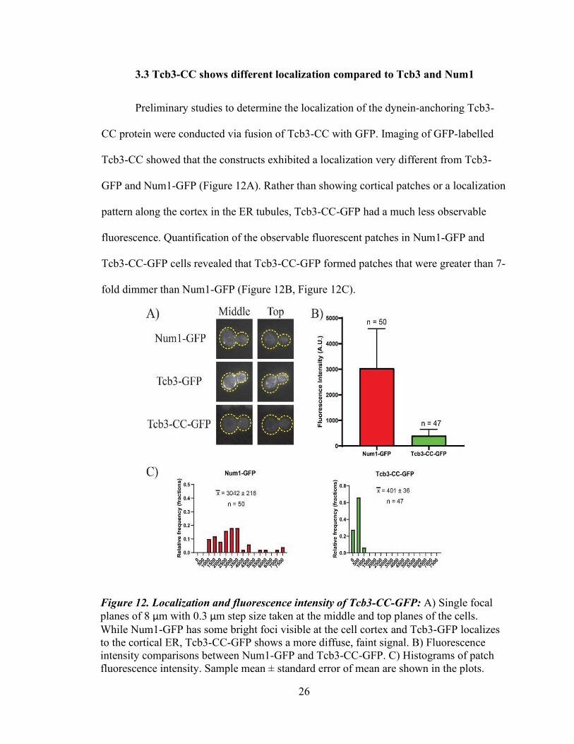
3.3 Tcb3-CC shows different localization compared to Tcb3 and Num1
Preliminary studies to determine the localization of the dynein-anchoring Tcb3-
CC protein were conducted via fusion of Tcb3-CC with GFP. Imaging of GFP-labelled
Tcb3-CC showed that the constructs exhibited a localization very different from Tcb3-
GFP and Num1-GFP (Figure 12A). Rather than showing cortical patches or a localization
pattern along the cortex in the ER tubules, Tcb3-CC-GFP had a much less observable
fluorescence. Quantification of the observable fluorescent patches in Num1-GFP and
Tcb3-CC-GFP cells revealed that Tcb3-CC-GFP formed patches that were greater than 7-
fold dimmer than Num1-GFP (Figure 12B, Figure 12C).
Figure 12. Localization and fluorescence intensity of Tcb3-CC-GFP: A) Single focal planes of 8 μm with 0.3 μm step size taken at the middle and top planes of the cells. While Num1-GFP has some bright foci visible at the cell cortex and Tcb3-GFP localizes to the cortical ER, Tcb3-CC-GFP shows a more diffuse, faint signal. B) Fluorescence intensity comparisons between Num1-GFP and Tcb3-CC-GFP. C) Histograms of patch fluorescence intensity. Sample mean ± standard error of mean are shown in the plots.

27
Chapter 4. Discussion
In this study, I have engineered an ER-directed dynein-anchoring protein through
genetic fusion of a resident ER protein, Tcb3, to the dynein-binding CC domain of the
Num1 protein. While previous work has indicated that plasma-membrane targeted Num1
can successfully facilitate nuclear positioning in budding yeast (Tang et al., 2009; Tang
et al., 2012; Schmit et al., 2018), the requirements for dynein anchoring and the strict
necessity of the plasma membrane for cortical anchoring of dynein and anaphase spindle
alignment are still unknown. Additionally, it is unlikely that the Num1 localizes solely to
the plasma membrane, as the plasma membrane is a diffusive surface that does not seem
conducive to providing an anchoring platform for dynein. Therefore, I hypothesize that
ER-PM junctions are locations where Num1 anchors dynein and that the cortical ER can
support dynein function and force production.
My data show that Tcb3-CC can rescue anaphase spindle alignment in a num1Δ
cell background. Though the rescue observed was not statistically different from wild-
type, num1Δ TCB3-CC cells still showed a slightly higher percentage of misaligned
anaphase spindles compared to wild-type cells (Figure 10). This difference could be a
result of differing protein expression levels between wild-type Num1 protein and the
Tcb3-CC fusion protein. The preliminary patch brightness data and the GFP localization
images seem to indicate that the Tcb3-CC fusion protein is more diffuse and does not
form the bright cortical patches characteristic of wild-type Num1, therefore hinting that
the protein levels of Tcb3-CC differ from full-length Num1. The existence of less
anchors in the cell and/or a lack of the stationary multi-molecule Num1 patches thought
to anchor dynein could explain why there is still 15.6% misaligned anaphase spindles
28
observed in the num1Δ TCB3-CC yeast (Tang et al., 2012). Further experiments to
determine more fully the patch formation and protein expression levels of Tcb3-CC are
needed to clarify the differences between Num1 and the engineered Tcb3-CC construct.
Another possibility for the partial rather than full rescue of the misaligned
anaphase spindle defect back to wild-type levels concerns the Tcb3 protein chosen as the
ER-targeting vehicle. The tricalbins are not fully understood with respect to their function
and behavior within the cell. One previous study characterizing the tricalbin proteins
found that the C-terminal portion of Tcb2 interacts with Tcb1 and Tcb3 in a two-hybrid
interaction assay and suggested that the tricalbin proteins form heterotrimeric complexes
(Creutz et al., 2004). The formation of a heterotrimeric complex or the interaction
between Tcb1, Tcb2, and the engineered Tcb3-CC protein could physically block
dynein’s access to the CC domain or otherwise interfere with appropriate dynein
anchoring and force generation, thereby yielding a misaligned anaphase spindle defect
slightly higher than that exhibited by the wild-type cells. Additionally, a domain of Tcb3
near its C-terminus (the third C2 domain of the Tcb3 protein) believed to mediate binding
to the plasma membrane has been shown to demonstrate calcium-sensitive binding to
lipids in vitro (Schulz and Creutz, 2004). This calcium sensitivity could also play an role
in the dynein-anchoring properties of Tcb3-CC as tighter binding of the Tcb3 to the
plasma membrane could result in a more stable dynein-anchoring platform whereas lesser
binding might yield lesser rescue of the misaligned spindle defect.
Although Tcb3-CC appears to facilitate proper nuclear positioning to a lesser
extent than the native Num1 protein, the dynein-dependent rescue suggests that the ER
membrane can indeed serve as an anchoring platform for dynein. This result indicates

29
that the plasma membrane is not the sole membrane able to act as an anchoring platform
for dynein during cell division in S. cerevisiae. My data further complements previous
findings demonstrating that disrupting cortical ER affects Num1 localization (Omer et
al., 2018).
Interestingly, preliminary work into discovering how and where Tcb3-CC
facilitates dynein function reveals that Tcb3-CC-GFP localizes differently than Tcb3
despite being expressed from the same genetic locus by the same promoter element.
These data suggest that the fusion of the CC changes protein stability or somehow
induces a change in the localization of the Tcb3 protein. Additionally, it is surprising that
these dimmer patches — presumably made up of less dynein-anchoring proteins than the
bright patches of Num1-GFP found in wild-type cells — can still anchor dynein and
facilitate nuclear and spindle positioning. These data seem to go against the hypothesis
that brighter Num1 patches can anchor more dynein and are thus better able to orient the
spindle and suggest instead that even “dim” patches suffice for dynein function. Further
studies of protein expression levels and localization are needed to determine if these dim
patches are responsible for the dynein function I have observed or if these dim patches
can instead explain why there is a slightly incomplete rescue of dynein function in
spindle positioning from a biological standpoint.

30
Chapter 5. Future Work
An additional test for dynein function in budding yeast involves the deletion of
Kar9, the main protein component of the dynein-independent pathway for nuclear
migration in budding yeast during mitosis. Genetic assays involving crosses between
kar9Δ and num1Δ yeast were used in early studies of Num1 to support the idea that
Num1 functions in a pathway parallel to the Kar9 pathway (Heil-Chapdelaine et al.,
2000). Therefore, Num1 constructs can be tested for function via a genetic cross with a
kar9Δ mutant. For example, a Num1 fusion protein targeted to the plasma membrane
using a CAAX motif demonstrated function comparable to wild-type cells in the dynein
pathway both in a binucleate assay (analogous to the spindle alignment assay in this
project) and in viability of double mutants in a genetic cross with kar9Δ cells (Tang et al.,
2009; Tang et al., 2012). Thus, for future work, kar9Δ cells can be crossed with cells
containing the Tcb3-CC fusion proteins to assay for viability of double mutant yeast
(containing both the Kar9 deletion as well as the Tcb3-CC fusion gene). This genetic
assay allows for an alternative, stricter test for dynein function in spindle orientation than
the spindle alignment assay as it examines if the engineered dynein anchor is sufficient
for sole handling of spindle orientation and positioning without the parallel Kar9
pathway. In other words, does the Tcb3-CC fusion protein rescue the growth defect of
kar9Δ num1Δ yeast in addition to rescuing the num1Δ misaligned anaphase spindle defect
caused by num1Δ, and can this engineered cytoplasmic dynein anchor function as well as
Num1 and other cortically-anchored variations of Num1?
Two key questions that can be examined using the engineered yeast strains
created in this thesis project are (1) whether the engineered dynein anchor protein

31
localizes differently than the unmodified Tcb3 protein or the native Num1 protein and (2)
whether the levels of Tcb3-CC protein differ from the levels of Num1 or Tcb3 in wild-
type yeast. Further localization studies of the Tcb3-CC proteins could provide insights
into the reason for the observed rescue of dynein function as demonstrated in this project.
One possibility is to test whether dynein localizes to the dim patches composed of Tcb3-
CC. Such colocalization studies could also help reveal the mechanism through which
dynein contacts the engineered anchor at the ER-mediated platform (Tang et al., 2009;
Tang et al., 2012).
In addition to its function in the dynein pathway and cellular division, Num1 is
required for proper yeast mitochondrial division and distribution (Cerveny et al., 2007;
Lackner et al., 2013; Klecker et al., 2013). Indeed, the CC domain has been shown as
necessary for cortical mitochondrial attachment, and the ER has been proposed as a
component of a complex that maintains proper mitochondrial distribution in yeast (Tang
et al., 2012; Lackner et al., 2013). Whether dynein function and mitochondrial division
involves different populations of Num1 protein and whether dynein function and
mitochondrial attachment are independent of one another remains an area of active
research and debate (Tang et al., 2012; Kraft and Lackner, 2019; Omer et al., 2018; Omer
et al., 2020). Previous work has examined the ability of artificially anchored and
differentially targeted Num1 variants as well as the ability of chimeric proteins
containing the Num1 PH domain to tether mitochondria to the cell cortex (Kraft and
Lackner, 2019; Tang et al., 2012; Klecker et al., 2013; Lackner et al., 2013). Many of
these studies used plasma membrane or mitochondria tethered variants of Num1.
Therefore, it would be interesting to see whether or how an ER-directed Num1 functions

32
in the mitochondrial tethering role of Num1 and to study the mitochondrial phenotype in
the num1Δ TCB3-CC yeast generated in this project.
Although I focused on directing Num1 to the ER using Tcb3, the other proteins in
the tricalbin family (Tcb1 and Tcb2) could be tested to see whether they produce
differential rescue of the num1Δ misaligned anaphase spindle defect and other num1Δ
phenotypes. In particular, the tricalbins vary in their size and features such as calcium
sensitivity (Schulz and Creutz, 2004). Thus, they may provide different strength
anchoring platforms if fused with CC. Additionally, recent studies have used domain
analysis paired with fluorescent protein tagging to show that the domains of Tcb2 are
differentially required for its localization in the cell; for example, GFP-labelled constructs
consisting only of Tcb2’s N-terminus and TMD appeared to localize throughout the ER
whereas full-length Tcb2 localized to the cortical ER and truncated version of Tcb2
missing the N-terminus and TMD appeared to localize mostly to the plasma membrane
and cytosol (Toulmay and Prinz, 2012). Similar domain analysis with truncated version
of Tcb3 or other tricalbin proteins fused to CC could help clarify the targeting behavior
of the CC domain and the exact requirements for dynein anchoring. Spindle alignment
assays and genetic assays as discussed in previous paragraphs paired with localization
experiments could prove valuable to understanding how ER-targeted dynein-anchoring
proteins behave, thereby providing insights into the requirements for the anchoring
platform of dynein.
Though the localization of the tricalbins to the cortical ER is well characterized,
the exact function of the tricalbins is poorly understood. Recent studies have implicated
the tricalbins in cellular lipid flux between different organelles and in the formation of

33
specific organelle contact sites, and tricalbins have been identified in a proteomic screen
for proteins enriched specifically in tubular ER (Hoffmann et al., 2019; Wang et al.,
2017). Additionally, the tricalbins have been suggested to help maintain plasma
membrane integrity through facilitation of cortical ER and plasma membrane lipid
exchange (Collado et al., 2019). Interestingly, Tcb3 has also been found to be important
in cellular aging through an algorithm-based analysis, which showed that a tcb3Δ yeast
strain has a longer life span than wild-type yeast (Borklu Yucel and Ulgen, 2011).
Another study involving genetic engineering of S. cerevisiae for biofuel production has
discovered that mutations in the tricalbin proteins — specifically, a mutation involving a
truncation of the Tcb3 protein — improve tolerance of yeast towards jet fuel (Brennan et
al., 2015). Therefore, tricalbin-CC fusions could also be assessed with the goal of better
understanding tricalbin function and behavior, and further research into tricalbin function
could reveal more about the behavior of the Tcb3-CC fusion I created.
34
Chapter 6. Conclusion
Through the design of a fusion protein consisting of a resident ER protein, Tcb3,
and the key CC domain of Num1 involved in dynein, this project assessed the ability of
the ER membrane to act as an anchoring platform for force production by dynein as well
as the necessity of the plasma membrane in the successful interaction of Num1 and
dynein for spindle positioning in budding yeast. Analysis of live-cell microscopy images
revealed a rescue of the num1Δ phenotype of misaligned anaphase spindles by the Tcb3-
CC construct. Importantly, this project demonstrates statistically-significant and dynein-
dependent rescue of nuclear positioning in the num1Δ strain expressing Tcb3-CC,
indicating that dynein attachment at the plasma membrane is not a requirement for
Num1-mediated dynein function and that the ER can function as an anchoring platform
for dynein function in spindle positioning in S. cerevisiae, further supporting previous
reports that point to the importance of the ER in Num1 function and morphology (Omer
et al., 2018; Lackner et al., 2013).
Future studies are required to learn more about the requirements for Num1’s
anchoring of cytoplasmic dynein during mitosis as well as Num1’s function in tethering
and distribution of mitochondria and the exact nature of the Tcb3 protein chosen as an
ER-directing agent here. This study and future studies in S. cerevisiae concerning Num1
and spindle positioning will elucidate more generally the requirements for cytoplasmic
dynein anchoring and the behavior of dynein and cortical anchoring proteins in the
distribution of genetic material and other cellular processes.
35
References
Adames, N. R. and Cooper, J. A. (2000) 'Microtubule interactions with the cell cortex causing nuclear movements in Saccharomyces cerevisiae', J Cell Biol, 149(4), pp. 863-74.
Ananthanarayanan, V. (2016) 'Activation of the motor protein upon attachment: Anchors weigh in on cytoplasmic dynein regulation', Bioessays, 38(6), pp. 514-25.
Bloom, K. (2001) 'Nuclear migration: cortical anchors for cytoplasmic dynein', Curr Biol, 11(8), pp. R326-9.
Borklu Yucel, E. and Ulgen, K. O. (2011) 'A network-based approach on elucidating the multi-faceted nature of chronological aging in S. cerevisiae', PLoS One, 6(12), pp. e29284.
Brennan, T. C., Williams, T. C., Schulz, B. L., Palfreyman, R. W., Krömer, J. O. and Nielsen, L. K. (2015) 'Evolutionary Engineering Improves Tolerance for Replacement Jet Fuels in Saccharomyces cerevisiae', Appl Environ Microbiol, 81(10), pp. 3316-25.
Canty, J. T. and Yildiz, A. (2020) 'Activation and Regulation of Cytoplasmic Dynein', Trends Biochem Sci, 45(5), pp. 440-453.
Cerveny, K. L., Studer, S. L., Jensen, R. E. and Sesaki, H. (2007) 'Yeast mitochondrial division and distribution require the cortical num1 protein', Dev Cell, 12(3), pp. 363-75.
Chen, X. J., Xu, H., Cooper, H. M. and Liu, Y. (2014) 'Cytoplasmic dynein: a key player in neurodegenerative and neurodevelopmental diseases', Sci China Life Sci, 57(4), pp. 372-7.
Cho, C., Reck-Peterson, S. L. and Vale, R. D. (2008) 'Regulatory ATPase sites of cytoplasmic dynein affect processivity and force generation', J Biol Chem, 283(38), pp. 25839-45.
Collado, J., Kalemanov, M., Campelo, F., Bourgoint, C., Thomas, F., Loewith, R., Martínez-Sánchez, A., Baumeister, W., Stefan, C. J. and Fernández-Busnadiego, R. (2019) 'Tricalbin-Mediated Contact Sites Control ER Curvature to Maintain Plasma Membrane Integrity', Dev Cell, 51(4), pp. 476-487.e7.
Creutz, C. E., Snyder, S. L. and Schulz, T. A. (2004) 'Characterization of the yeast tricalbins: membrane-bound multi-C2-domain proteins that form complexes involved in membrane trafficking', Cell Mol Life Sci, 61(10), pp. 1208-20.

36
Duina, A. A., Miller, M. E. and Keeney, J. B. (2014) 'Budding yeast for budding geneticists: a primer on the Saccharomyces cerevisiae model system', Genetics, 197(1), pp. 33-48.
Ecklund, K. H., Morisaki, T., Lammers, L. G., Marzo, M. G., Stasevich, T. J. and Markus, S. M. (2017) 'She1 affects dynein through direct interactions with the microtubule and the dynein microtubule-binding domain', Nat Commun, 8(1), pp. 2151.
Farkasovsky, M. and Küntzel, H. (1995) 'Yeast Num1p associates with the mother cell cortex during S/G2 phase and affects microtubular functions', J Cell Biol, 131(4), pp. 1003-14.
Farkasovsky, M. and Küntzel, H. (2001) 'Cortical Num1p interacts with the dynein intermediate chain Pac11p and cytoplasmic microtubules in budding yeast', J Cell Biol, 152(2), pp. 251-62.
Greenberg, S. R., Tan, W. and Lee, W. L. (2018) 'Num1 versus NuMA: insights from two functionally homologous proteins', Biophys Rev, 10(6), pp. 1631-1636.
Heil-Chapdelaine, R. A., Oberle, J. R. and Cooper, J. A. (2000) 'The cortical protein Num1p is essential for dynein-dependent interactions of microtubules with the cortex', J Cell Biol, 151(6), pp. 1337-44.
Hoffmann, P. C., Bharat, T. A. M., Wozny, M. R., Boulanger, J., Miller, E. A. and Kukulski, W. (2019) 'Tricalbins Contribute to Cellular Lipid Flux and Form Curved ER-PM Contacts that Are Bridged by Rod-Shaped Structures', Dev Cell, 51(4), pp. 488-502.e8.
Klecker, T., Scholz, D., Förtsch, J. and Westermann, B. (2013) 'The yeast cell cortical protein Num1 integrates mitochondrial dynamics into cellular architecture', J Cell Sci, 126(Pt 13), pp. 2924-30.
Knop, M., Siegers, K., Pereira, G., Zachariae, W., Winsor, B., Nasmyth, K. and Schiebel, E. (1999) 'Epitope tagging of yeast genes using a PCR-based strategy: more tags and improved practical routines', Yeast, 15(10B), pp. 963-72.
Kormanec, J., Schaaff-Gerstenschläger, I., Zimmermann, F. K., Perecko, D. and Küntzel, H. (1991) 'Nuclear migration in Saccharomyces cerevisiae is controlled by the highly repetitive 313 kDa NUM1 protein', Mol Gen Genet, 230(1-2), pp. 277-87.
Kotak, S., Busso, C. and Gönczy, P. (2012) 'Cortical dynein is critical for proper spindle positioning in human cells', J Cell Biol, 199(1), pp. 97-110.
Kraft, L. M. and Lackner, L. L. (2019) 'A conserved mechanism for mitochondria-dependent dynein anchoring', Mol Biol Cell, 30(5), pp. 691-702.

37
Lackner, L. L., Ping, H., Graef, M., Murley, A. and Nunnari, J. (2013) 'Endoplasmic reticulum-associated mitochondria-cortex tether functions in the distribution and inheritance of mitochondria', Proc Natl Acad Sci U S A, 110(6), pp. E458-67.
Longtine, M. S., McKenzie, A., Demarini, D. J., Shah, N. G., Wach, A., Brachat, A., Philippsen, P. and Pringle, J. R. (1998) 'Additional modules for versatile and economical PCR-based gene deletion and modification in Saccharomyces cerevisiae', Yeast, 14(10), pp. 953-61.
Manford, A. G., Stefan, C. J., Yuan, H. L., Macgurn, J. A. and Emr, S. D. (2012) 'ER-to-plasma membrane tethering proteins regulate cell signaling and ER morphology', Dev Cell, 23(6), pp. 1129-40.
Markus, S. M., Kalutkiewicz, K. A. and Lee, W. L. (2012a) 'Astral microtubule asymmetry provides directional cues for spindle positioning in budding yeast', Exp Cell Res, 318(12), pp. 1400-6.
Markus, S. M., Kalutkiewicz, K. A. and Lee, W. L. (2012b) 'She1-mediated inhibition of dynein motility along astral microtubules promotes polarized spindle movements', Curr Biol, 22(23), pp. 2221-30.
Markus, S. M. and Lee, W. L. (2011) 'Microtubule-dependent path to the cell cortex for cytoplasmic dynein in mitotic spindle orientation', Bioarchitecture, 1(5), pp. 209-215.
Moore, J. K., Stuchell-Brereton, M. D. and Cooper, J. A. (2009) 'Function of dynein in budding yeast: mitotic spindle positioning in a polarized cell', Cell Motil Cytoskeleton, 66(8), pp. 546-55.
Omer, S., Brock, K., Beckford, J. and Lee, W.-L. 2020. Distinct Num1 Populations Mediate Spindle Positioning and Mitochondrial Tethering in Budding Yeast.
Omer, S., Greenberg, S. R. and Lee, W. L. (2018) 'Cortical dynein pulling mechanism is regulated by differentially targeted attachment molecule Num1', Elife, 7.
Prinz, W. A. (2014) 'Bridging the gap: membrane contact sites in signaling, metabolism, and organelle dynamics', J Cell Biol, 205(6), pp. 759-69.
Reck-Peterson, S. L., Redwine, W. B., Vale, R. D. and Carter, A. P. (2018) 'The cytoplasmic dynein transport machinery and its many cargoes', Nat Rev Mol Cell Biol, 19(6), pp. 382-398.
Schmit, H. L., Kraft, L. M., Lee-Smith, C. F. and Lackner, L. L. (2018) 'The role of mitochondria in anchoring dynein to the cell cortex extends beyond clustering the anchor protein', Cell Cycle, 17(11), pp. 1345-1357.

38
Schulz, T. A. and Creutz, C. E. (2004) 'The tricalbin C2 domains: lipid-binding properties of a novel, synaptotagmin-like yeast protein family', Biochemistry, 43(13), pp. 3987-95.
Tang, X., Germain, B. S. and Lee, W. L. (2012) 'A novel patch assembly domain in Num1 mediates dynein anchoring at the cortex during spindle positioning', J Cell Biol, 196(6), pp. 743-56.
Tang, X., Punch, J. J. and Lee, W. L. (2009) 'A CAAX motif can compensate for the PH domain of Num1 for cortical dynein attachment', Cell Cycle, 8(19), pp. 3182-90.
Toulmay, A. and Prinz, W. A. (2012) 'A conserved membrane-binding domain targets proteins to organelle contact sites', J Cell Sci, 125(Pt 1), pp. 49-58.
Vale, R. D. (2003) 'The molecular motor toolbox for intracellular transport', Cell, 112(4), pp. 467-80.
Wang, X., Li, S., Wang, H., Shui, W. and Hu, J. (2017) 'Quantitative proteomics reveal proteins enriched in tubular endoplasmic reticulum of', Elife, 6.
Xiang, X. (2018) 'Nuclear movement in fungi', Semin Cell Dev Biol, 82, pp. 3-16.
Zhu, Y., An, X., Tomaszewski, A., Hepler, P. K. and Lee, W. L. (2017) 'Microtubule cross-linking activity of She1 ensures spindle stability for spindle positioning', J Cell Biol, 216(9), pp. 2759-2775.


![Crystal clear insights into how the dynein motor moves · 2013. 4. 10. · 2010)]. In dynein, four of the AAA+ domains bind nucleotides. The size of the dynein motor domain, the presence](https://static.fdocuments.us/doc/165x107/60ed0c0f1235ef420447d9e4/crystal-clear-insights-into-how-the-dynein-motor-moves-2013-4-10-2010-in.jpg)