Energy Taxis toward Host-Derived Nitrate Supports a ... · rium in the mouse typhoid model...
11
Energy Taxis toward Host-Derived Nitrate Supports a Salmonella Pathogenicity Island 1-Independent Mechanism of Invasion Fabian Rivera-Chávez, a Christopher A. Lopez, a Lillian F. Zhang, a Lucía García-Pastor, b Alfredo Chávez-Arroyo, a Kristen L. Lokken, a Renée M. Tsolis, a Sebastian E. Winter, c Andreas J. Bäumler a Department of Medical Microbiology and Immunology, School of Medicine, University of California at Davis, Davis, California, USA a ; Departamento de Genética, Universidad de Sevilla, Seville, Spain b ; Department of Microbiology, University of Texas Southwestern Medical Center, Dallas, Texas, USA c ABSTRACT Salmonella enterica serovar Typhimurium can cross the epithelial barrier using either the invasion-associated type III secretion system (T3SS-1) or a T3SS-1-independent mechanism that remains poorly characterized. Here we show that flagellum-mediated motility supported a T3SS-1-independent pathway for entering ileal Peyer’s patches in the mouse model. Flagellum-dependent invasion of Peyer’s patches required energy taxis toward nitrate, which was mediated by the methyl- accepting chemotaxis protein (MCP) Tsr. Generation of nitrate in the intestinal lumen required inducible nitric oxide synthase (iNOS), which was synthesized constitutively in the mucosa of the terminal ileum but not in the jejunum, duodenum, or cecum. Tsr-mediated invasion of ileal Peyer’s patches was abrogated in mice deficient for Nos2, the gene encoding iNOS. We conclude that Tsr-mediated energy taxis enables S. Typhimurium to migrate toward the intestinal epithelium by sensing host-derived ni- trate, thereby contributing to invasion of Peyer’s patches. IMPORTANCE Nontyphoidal Salmonella serovars, such as S. enterica serovar Typhimurium, are a common cause of gastroen- teritis in immunocompetent individuals but can also cause bacteremia in immunocompromised individuals. While the invasion-associated type III secretion system (T3SS-1) is important for entry, S. Typhimurium strains lacking a functional T3SS-1 can still cross the intestinal epithelium and cause a disseminated lethal infection in mice. Here we observed that flagellum-mediated motility and chemotaxis contributed to a T3SS-1-independent pathway for invasion and systemic dissemi- nation to the spleen. This pathway required the methyl-accepting chemotaxis protein (MCP) Tsr and energy taxis toward host- derived nitrate, which we found to be generated by inducible nitric oxide synthase (iNOS) in the ileal mucosa prior to infection. Collectively, our data suggest that S. Typhimurium enhances invasion by actively migrating toward the intestinal epithelium along a gradient of host-derived nitrate emanating from the mucosal surface of the ileum. Received 27 May 2016 Accepted 19 June 2016 Published 19 July 2016 Citation Rivera-Chávez F, Lopez CA, Zhang LF, García-Pastor L, Chávez-Arroyo A, Lokken KL, Tsolis RM, Winter SE, Bäumler AJ. 2016. Energy taxis toward host-derived nitrate supports a Salmonella pathogenicity island 1-independent mechanism of invasion. mBio 7(4):e00960-16. doi:10.1128/mBio.00960-16. Editor Larry S. McDaniel, University of Mississippi Medical Center Copyright © 2016 Rivera-Chávez et al. This is an open-access article distributed under the terms of the Creative Commons Attribution 4.0 International license. Address correspondence to Andreas J. Bäumler, [email protected]. This article is a direct contribution from a Fellow of the American Academy of Microbiology. External solicited reviewers: Andres Vazquez-Torres, University of Colorado School of Medicine; John Gunn, The Ohio State University. S almonella enterica serovar Typhimurium causes a dissemi- nated infection in genetically susceptible mice, which is com- monly used to study the pathogenesis of typhoid fever (mouse typhoid model) (summarized in reference 1). S. Typhimurium initiates infection of mice by preferentially invading the intestinal epithelium of Peyer’s patches in the terminal ileum (2), followed by dissemination of the pathogen to internal organs, such as the spleen, where it replicates within macrophages (3). Modeling of epithelial invasion in the 1980s using cultured epithelial cell lines identified flagellum-mediated motility (4) and the invasion-associated type III secretion system (T3SS-1) (5) en- coded by Salmonella pathogenicity island 1 (SPI1) (6) as potential virulence factors contributing to epithelial entry. An initial char- acterization of these potential virulence factors in the mouse ty- phoid model suggested that inactivation of flagellum biosynthesis genes causes less attenuation (2- to 9-fold) (7) than inactivation of T3SS-1 biosynthesis genes (16- to 60-fold) (5, 8, 9). This early work helped erect the concept that T3SS-1 represents the main virulence factor for mucosal invasion, while flagella are not a ma- jor contributor to Salmonella pathogenesis. Consequently, subse- quent work focused largely on elucidating the mechanism under- lying T3SS-1-mediated epithelial entry. However, recent studies suggest that flagella play a more sig- nificant role during the interaction of S. Typhimurium with its vertebrate host than previously appreciated. The development of models for S. Typhimurium-induced gastroenteritis revealed that flagellum-mediated motility contributes to the development of intestinal inflammation in calves (10) and in streptomycin- pretreated mice (mouse colitis model) (11). Furthermore, flagellum-mediated motility is required for driving a luminal ex- pansion of S. Typhimurium during colitis (12, 13). These obser- vations suggest that it might be warranted to reconsider the con- tribution of flagella to Salmonella pathogenesis. There is also reason to believe that T3SS-1-mediated invasion is not the sole pathway for S. Typhimurium to cross the epithelial barrier in the ileum. That is, studies on the spread of S. Typhimu- RESEARCH ARTICLE crossmark July/August 2016 Volume 7 Issue 4 e00960-16 ® mbio.asm.org 1 on April 3, 2019 by guest http://mbio.asm.org/ Downloaded from
Transcript of Energy Taxis toward Host-Derived Nitrate Supports a ... · rium in the mouse typhoid model...

Energy Taxis toward Host-Derived Nitrate Supports a SalmonellaPathogenicity Island 1-Independent Mechanism of Invasion
Fabian Rivera-Chávez,a Christopher A. Lopez,a Lillian F. Zhang,a Lucía García-Pastor,b Alfredo Chávez-Arroyo,a Kristen L. Lokken,a
Renée M. Tsolis,a Sebastian E. Winter,c Andreas J. Bäumlera
Department of Medical Microbiology and Immunology, School of Medicine, University of California at Davis, Davis, California, USAa; Departamento de Genética,Universidad de Sevilla, Seville, Spainb; Department of Microbiology, University of Texas Southwestern Medical Center, Dallas, Texas, USAc
ABSTRACT Salmonella enterica serovar Typhimurium can cross the epithelial barrier using either the invasion-associated typeIII secretion system (T3SS-1) or a T3SS-1-independent mechanism that remains poorly characterized. Here we show thatflagellum-mediated motility supported a T3SS-1-independent pathway for entering ileal Peyer’s patches in the mouse model.Flagellum-dependent invasion of Peyer’s patches required energy taxis toward nitrate, which was mediated by the methyl-accepting chemotaxis protein (MCP) Tsr. Generation of nitrate in the intestinal lumen required inducible nitric oxide synthase(iNOS), which was synthesized constitutively in the mucosa of the terminal ileum but not in the jejunum, duodenum, or cecum.Tsr-mediated invasion of ileal Peyer’s patches was abrogated in mice deficient for Nos2, the gene encoding iNOS. We concludethat Tsr-mediated energy taxis enables S. Typhimurium to migrate toward the intestinal epithelium by sensing host-derived ni-trate, thereby contributing to invasion of Peyer’s patches.
IMPORTANCE Nontyphoidal Salmonella serovars, such as S. enterica serovar Typhimurium, are a common cause of gastroen-teritis in immunocompetent individuals but can also cause bacteremia in immunocompromised individuals. While theinvasion-associated type III secretion system (T3SS-1) is important for entry, S. Typhimurium strains lacking a functionalT3SS-1 can still cross the intestinal epithelium and cause a disseminated lethal infection in mice. Here we observed thatflagellum-mediated motility and chemotaxis contributed to a T3SS-1-independent pathway for invasion and systemic dissemi-nation to the spleen. This pathway required the methyl-accepting chemotaxis protein (MCP) Tsr and energy taxis toward host-derived nitrate, which we found to be generated by inducible nitric oxide synthase (iNOS) in the ileal mucosa prior to infection.Collectively, our data suggest that S. Typhimurium enhances invasion by actively migrating toward the intestinal epitheliumalong a gradient of host-derived nitrate emanating from the mucosal surface of the ileum.
Received 27 May 2016 Accepted 19 June 2016 Published 19 July 2016
Citation Rivera-Chávez F, Lopez CA, Zhang LF, García-Pastor L, Chávez-Arroyo A, Lokken KL, Tsolis RM, Winter SE, Bäumler AJ. 2016. Energy taxis toward host-derived nitratesupports a Salmonella pathogenicity island 1-independent mechanism of invasion. mBio 7(4):e00960-16. doi:10.1128/mBio.00960-16.
Editor Larry S. McDaniel, University of Mississippi Medical Center
Copyright © 2016 Rivera-Chávez et al. This is an open-access article distributed under the terms of the Creative Commons Attribution 4.0 International license.
Address correspondence to Andreas J. Bäumler, [email protected].
This article is a direct contribution from a Fellow of the American Academy of Microbiology. External solicited reviewers: Andres Vazquez-Torres, University of Colorado Schoolof Medicine; John Gunn, The Ohio State University.
Salmonella enterica serovar Typhimurium causes a dissemi-nated infection in genetically susceptible mice, which is com-
monly used to study the pathogenesis of typhoid fever (mousetyphoid model) (summarized in reference 1). S. Typhimuriuminitiates infection of mice by preferentially invading the intestinalepithelium of Peyer’s patches in the terminal ileum (2), followedby dissemination of the pathogen to internal organs, such as thespleen, where it replicates within macrophages (3).
Modeling of epithelial invasion in the 1980s using culturedepithelial cell lines identified flagellum-mediated motility (4) andthe invasion-associated type III secretion system (T3SS-1) (5) en-coded by Salmonella pathogenicity island 1 (SPI1) (6) as potentialvirulence factors contributing to epithelial entry. An initial char-acterization of these potential virulence factors in the mouse ty-phoid model suggested that inactivation of flagellum biosynthesisgenes causes less attenuation (2- to 9-fold) (7) than inactivation ofT3SS-1 biosynthesis genes (16- to 60-fold) (5, 8, 9). This earlywork helped erect the concept that T3SS-1 represents the main
virulence factor for mucosal invasion, while flagella are not a ma-jor contributor to Salmonella pathogenesis. Consequently, subse-quent work focused largely on elucidating the mechanism under-lying T3SS-1-mediated epithelial entry.
However, recent studies suggest that flagella play a more sig-nificant role during the interaction of S. Typhimurium with itsvertebrate host than previously appreciated. The development ofmodels for S. Typhimurium-induced gastroenteritis revealed thatflagellum-mediated motility contributes to the development ofintestinal inflammation in calves (10) and in streptomycin-pretreated mice (mouse colitis model) (11). Furthermore,flagellum-mediated motility is required for driving a luminal ex-pansion of S. Typhimurium during colitis (12, 13). These obser-vations suggest that it might be warranted to reconsider the con-tribution of flagella to Salmonella pathogenesis.
There is also reason to believe that T3SS-1-mediated invasionis not the sole pathway for S. Typhimurium to cross the epithelialbarrier in the ileum. That is, studies on the spread of S. Typhimu-
RESEARCH ARTICLE
crossmark
July/August 2016 Volume 7 Issue 4 e00960-16 ® mbio.asm.org 1
on April 3, 2019 by guest
http://mbio.asm
.org/D
ownloaded from
rium in the mouse typhoid model identified a T3SS-1-independent pathway contributing to early dissemination of thepathogen from the intestinal lumen to the spleen (14). However,the S. Typhimurium virulence factors contributing to this T3SS-1-independent route of entry remain to be identified.
Here we investigated whether flagellum-mediated motilitycontributes to a T3SS-1-independent pathway of Peyer’s patchinvasion and studied the underlying mechanism.
RESULTSFlagella and T3SS-1 independently contribute to Peyer’s patchinvasion and dissemination to the spleen. To investigate the con-tribution of flagella and T3SS-1 to invasion and dissemination inthe mouse typhoid model, groups of mice were infected intragas-trically with an S. Typhimurium wild-type strain (IR715), a non-motile mutant lacking both flagellin genes of S. Typhimurium(fliC fljB mutant [SW473]), a T3SS-1-deficient mutant (invA mu-tant [SW399]), and a strain lacking both flagella and T3SS-1 (invAfliC fljB mutant [FR90]). We reasoned that if flagella and T3SS-1contribute to the same invasion pathway, the invasion defect ob-served for an invA fliC fljB mutant should be similar to that of aninvA mutant, because the mutation in invA would epistaticallymask the phenotype caused by mutations in fliC and fljB. Alterna-tively flagella and T3SS-1 might each contribute to a differentinvasion pathway, which would predict that combining the invA,fliC, and fljB mutations would yield a stronger phenotype thanproduced by either an invA mutant or an fliC fljB mutant.
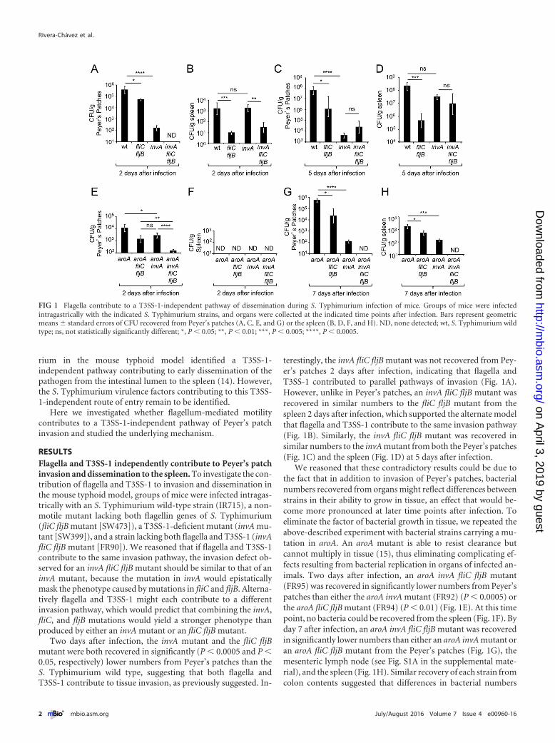
Two days after infection, the invA mutant and the fliC fljBmutant were both recovered in significantly (P � 0.0005 and P �0.05, respectively) lower numbers from Peyer’s patches than theS. Typhimurium wild type, suggesting that both flagella andT3SS-1 contribute to tissue invasion, as previously suggested. In-
terestingly, the invA fliC fljB mutant was not recovered from Pey-er’s patches 2 days after infection, indicating that flagella andT3SS-1 contributed to parallel pathways of invasion (Fig. 1A).However, unlike in Peyer’s patches, an invA fliC fljB mutant wasrecovered in similar numbers to the fliC fljB mutant from thespleen 2 days after infection, which supported the alternate modelthat flagella and T3SS-1 contribute to the same invasion pathway(Fig. 1B). Similarly, the invA fliC fljB mutant was recovered insimilar numbers to the invA mutant from both the Peyer’s patches(Fig. 1C) and the spleen (Fig. 1D) at 5 days after infection.
We reasoned that these contradictory results could be due tothe fact that in addition to invasion of Peyer’s patches, bacterialnumbers recovered from organs might reflect differences betweenstrains in their ability to grow in tissue, an effect that would be-come more pronounced at later time points after infection. Toeliminate the factor of bacterial growth in tissue, we repeated theabove-described experiment with bacterial strains carrying a mu-tation in aroA. An aroA mutant is able to resist clearance butcannot multiply in tissue (15), thus eliminating complicating ef-fects resulting from bacterial replication in organs of infected an-imals. Two days after infection, an aroA invA fliC fljB mutant(FR95) was recovered in significantly lower numbers from Peyer’spatches than either the aroA invA mutant (FR92) (P � 0.0005) orthe aroA fliC fljB mutant (FR94) (P � 0.01) (Fig. 1E). At this timepoint, no bacteria could be recovered from the spleen (Fig. 1F). Byday 7 after infection, an aroA invA fliC fljB mutant was recoveredin significantly lower numbers than either an aroA invA mutant oran aroA fliC fljB mutant from the Peyer’s patches (Fig. 1G), themesenteric lymph node (see Fig. S1A in the supplemental mate-rial), and the spleen (Fig. 1H). Similar recovery of each strain fromcolon contents suggested that differences in bacterial numbers
FIG 1 Flagella contribute to a T3SS-1-independent pathway of dissemination during S. Typhimurium infection of mice. Groups of mice were infectedintragastrically with the indicated S. Typhimurium strains, and organs were collected at the indicated time points after infection. Bars represent geometricmeans � standard errors of CFU recovered from Peyer’s patches (A, C, E, and G) or the spleen (B, D, F, and H). ND, none detected; wt, S. Typhimurium wildtype; ns, not statistically significantly different; *, P � 0.05; **, P � 0.01; ***, P � 0.005; ****, P � 0.0005.
Rivera-Chávez et al.
2 ® mbio.asm.org July/August 2016 Volume 7 Issue 4 e00960-16
on April 3, 2019 by guest
http://mbio.asm
.org/D
ownloaded from

recovered from tissue were not due to altered growth of bacteria inthe intestinal lumen (see Fig. S1B). In summary, when bacterialgrowth in tissue was prevented by a mutation in aroA, recovery ofbacteria from the Peyer’s patches and the spleen consistently sup-ported the idea that flagellum-mediated invasion was indepen-dent of invasion mediated by T3SS-1, which suggested that thesetwo virulence factors acted in two parallel pathways of invasionand dissemination.
Next, we wanted to determine whether an early time point afterinfection would enable us to study invasion by virulent S. Typhi-murium before growth in tissue could take place. A previous studysuggested that bacteria could be recovered from Peyer’s patch tis-sue as early as 1 h after intragastric inoculation with S. entericaserovar Enteritidis (2). To determine whether invasion was detect-able at this early time point, mice were infected with the S. Typhi-murium wild type, and the ileum and jejunum were collected 1 hlater and treated with gentamicin to kill extracellular bacteria re-siding in the intestinal lumen (gentamicin protection assay). Re-covery of tissue-associated S. Typhimurium revealed marked in-vasion in the ileum but not in the jejunum (Fig. 2A), which wasconsistent with observations made with S. Enteritidis-infectedmice (2).
To further investigate the contribution of S. Typhimurium vir-
ulence factors to invasion, mice were infected with a 1:1 mixture ofthe S. Typhimurium wild type and either an fliC fljB mutant, aninvA mutant, or an invA fliC fljB mutant. One hour after infection,ileal Peyer’s patches were collected and incubated with gentamicinto kill extracellular bacteria. The wild type was recovered in highernumbers from ileal Peyer’s patches than either an fliC fljB mutantor an invA mutant (Fig. 2B and C), suggesting that both flagellaand T3SS-1 contributed to invasion. However, the fitness advan-tage of the wild type over an invA fliC fljB mutant was significantlylarger than that over an fliC fljB mutant (P � 0.05) or an invAmutant (P � 0.0005). These results provided further support forthe hypothesis that flagella and T3SS-1 contributed to parallelpathways of ileal Peyer’s patch invasion.
We then wanted to investigate the mechanism by which flagellacontributed to invasion of ileal Peyer’s patches at 1 h after infec-tion. Flagellum-mediated motility is required for chemotaxis to-ward chemoattractants (reviewed in reference 16). We thus hy-pothesized that flagellum-mediated motility might be required byS. Typhimurium in vivo for locating the mucosal surface throughchemotaxis. To test this idea, mice were infected with a 1:1 mix-ture of the S. Typhimurium wild type and a chemotaxis-deficientmutant (cheB mutant [AT349]), and bacteria were recovered fromileal Peyer’s patches 1 h after infection using a gentamicin protec-
FIG 2 Flagella and chemotaxis contribute to epithelial invasion through a T3SS-1-independent pathway. (A to C) Groups of mice were infected intragastricallywith the indicated S. Typhimurium strains or strain mixtures, and organs were collected at the indicated time points after infection. Bars represent geometricmeans � standard errors of CFU (A and C) or the competitive index (CI) (B) recovered from Peyer’s patches. (D and E) The indicated S. Typhimurium strains(D) or strain mixtures (E) were allowed to invade model epithelia (polarized Caco-2 cells) for 30 min in an anaerobe chamber before extracellular bacteria werekilled by treatment with gentamicin under aerobic conditions. Bars represent geometric means � standard errors of gentamicin-resistant CFU recovered frommodel epithelia. *, P � 0.05; **, P � 0.01; ***, P � 0.005; ****, P � 0.0005; wt, S. Typhimurium wild type.
Energy Taxis Contributes to Salmonella Invasion
July/August 2016 Volume 7 Issue 4 e00960-16 ® mbio.asm.org 3
on April 3, 2019 by guest
http://mbio.asm
.org/D
ownloaded from

tion assay. The S. Typhimurium wild type was recovered in ap-proximately 8-fold-higher numbers than the cheB mutant fromileal Peyer’s patches (P � 0.05) (Fig. 2A and B), suggesting thatchemotaxis was required for invasion.
We next sought to investigate whether the contribution of mo-tility and chemotaxis to invasion could be observed in a tissueculture model of infection. Polarized human colonic epithelial(Caco-2) cells were transferred into an anaerobe chamber tomimic the limited oxygen availability characteristic of the intesti-nal lumen (��7.6 mm Hg PO2 or ��1% O2) (17), and bacterialinvasion was allowed to proceed for 30 min before killing of ex-tracellular bacteria with gentamicin. We recovered significantlyhigher numbers of bacteria from model epithelia infected with theS. Typhimurium wild type compared to model epithelia infectedwith either an invA mutant (P � 0.01) or an fliC fljB mutant (P �0.01) (Fig. 2D). However, both an invA mutant (P � 0.0005) anda fliC fljB mutant (P � 0.005) were recovered in significantlyhigher numbers from Caco-2 cells than an invA fliC fljB mutant(Fig. 2D), suggesting that in this model, flagella and T3SS-1 alsocontributed to parallel pathways of invasion, which was similar towhat was observed in mice.
Next, Caco-2 cells were infected with a 1:1 mixture of the S. Ty-phimurium wild type and either an fliC fljB mutant, an invA mu-tant, or an invA fliC fljB mutant. Similar to what we observed atearly time points (i.e., 1 h) after infection of mice (Fig. 2B), recov-ery of intracellular bacteria from Caco-2 cells revealed that an invAfliC fljB mutant exhibited a significantly (P � 0.005) larger inva-sion defect than either an fliC fljB mutant or an invA mutant(Fig. 2E). Furthermore, an invA mutant was recovered in approx-imately 10-fold-higher numbers than an invA fliC fljB mutantwhen cells were infected with a 1:1 mixture of both strains (P �0.005), showing that flagella contributed to invasion of Caco-2cells independently of T3SS-1 (Fig. 2E). Finally, the S. Typhimu-rium wild type was recovered in approximately 10-fold-highernumbers than a chemotaxis-deficient cheB mutant when cellswere infected with a 1:1 mixture of both strains (P � 0.005), sug-gesting that chemotaxis contributed to invasion in this model(Fig. 2E).
Constitutive iNOS synthesis in the ileal mucosa is a source ofluminal nitrate. The results presented above sparked our interestin identifying possible signals that could direct chemotaxis ofS. Typhimurium toward the mucosal surface of the ileum. A pre-vious report suggests that constitutive inducible nitric oxide syn-thase (iNOS) synthesis is observed in the ileal mucosa of untreatedmice but not in other parts of the murine gastrointestinal tract(18). We therefore investigated iNOS synthesis in different partsof the gastrointestinal tract of untreated mice using Western blot-ting. While iNOS synthesis was not detected in the duodenum,jejunum, or cecum of mice, we detected constitutive iNOS syn-thesis in the ileal mucosa (Fig. 3A and B). These data suggestedthat constitutive iNOS synthesis was a feature that differentiatedthe ileal mucosa from that in other parts of the murine gastroin-testinal tract.
Next, we used immunohistochemistry to investigate the distri-bution and localization of iNOS synthesis in the ileal mucosa. Noimmunohistochemical signal was detected in mice deficient forNos2, the gene encoding iNOS. In contrast, immunohistochemi-cal detection of iNOS synthesis in wild-type (C57BL/6) mice gen-erated a strong signal in phagocytes located in the lamina propria
and a weaker signal at an apical location in epithelial cells liningthe ileal mucosa (Fig. 3C).
We previously showed that nitric oxide (NO) generated byiNOS can react to form nitrate in the intestinal lumen (19, 20). Toinvestigate whether constitutive iNOS synthesis in the ileal mu-cosa is a source of nitrate in the intestinal lumen, we measurednitrate concentrations in luminal contents from different parts ofthe gastrointestinal tract. Ileal contents contained on average6 mM nitrate, which was significantly higher than the nitrate con-centrations detected in contents from the jejunum (P � 0.005),duodenum (P � 0.01), or cecum (P � 0.005) of wild-type(C57BL/6) mice (Fig. 3D). In contrast, ileal contents from Nos2-deficient mice contained little nitrate (Fig. 3E), suggesting thatelevated concentrations of nitrate in the ileal contents of wild-typemice were iNOS derived.
Invasion of ileal Peyers’s patches requires Tsr-mediated che-motaxis toward nitrate. Respiratory electron acceptors, such asnitrate, serve as chemoattractants for S. Typhimurium (21, 22).The methyl-accepting chemotaxis protein (MCP) involved insensing nitrate in the murine intestine is tsr (taxis to serine andrepellents) (13). To investigate whether Tsr was required for inva-sion of the ileum, mice were infected with a 1:1 mixture of theS. Typhimurium wild type and a tsr mutant (FR4), and bacteriawere recovered from ileal Peyer’s patches 1 h after infection usinga gentamicin protection assay. The S. Typhimurium wild type wasrecovered in approximately 8-fold-higher numbers than the tsrmutant (P � 0.05) (Fig. 4A and B), suggesting that Tsr was re-quired for invasion. When the experiment was repeated in theabsence of gentamicin treatment, bacteria were recovered fromPeyer’s patch homogenates at approximately 10-fold-highernumbers (Fig. 4B), implying that approximately 90% of bacteriawere gentamicin sensitive (i.e., extracellular), presumably residingin the mucus layer associated with the luminal surface of Peyer’spatches. Importantly, in the absence of gentamicin treatment, theS. Typhimurium wild type was recovered in similar numbers tothe tsr mutant (Fig. 4A), illustrating that Tsr did not confer ageneral fitness advantage in the lumen of the ileum. Finally, intro-duction of the cloned tsr gene on a plasmid (pFR6) into the tsrmutant (FR4) restored the defect in Peyer’s patch invasion 1 hafter infection, as shown using a gentamicin protection assay(Fig. 4A and B). Collectively, these results support the idea thatTsr-mediated chemotaxis contributed to Peyer’s patch invasion.
Tsr mediates chemotaxis toward respiratory electron acceptorsindirectly, by sensing the proton motive force, a process termedenergy taxis (23–26). The S. Typhimurium genome encodes threenitrate reductases, encoded by the napABC, narZYV, and narGHIgenes, which can use nitrate as a respiratory electron acceptor andgenerate proton motive force (19). Inactivation of napA, narZ,and narG abrogates nitrate reductase activity in S. Typhimurium,thereby preventing Tsr-mediated energy taxis toward nitrate (13).Thus, we wanted to determine whether inactivation of genes en-coding nitrate reductases in S. Typhimurium could abrogate Tsr-mediated invasion of ileal Peyer’s patches. Mice were infected witha 1:1 mixture of a nitrate respiration-deficient mutant (napA narZnarG mutant [CAL50]) and a nitrate respiration-deficient mutantlacking Tsr (napA narZ narG tsr mutant [FR46]). Bacteria wererecovered from ileal Peyer’s patches 1 h after infection using agentamicin protection assay. Remarkably, in the absence of nitraterespiration, Tsr did not contribute to invasion of Peyer’s patches(Fig. 4A). Furthermore, inactivation of nitrate respiration caused
Rivera-Chávez et al.
4 ® mbio.asm.org July/August 2016 Volume 7 Issue 4 e00960-16
on April 3, 2019 by guest
http://mbio.asm
.org/D
ownloaded from

a marked reduction in bacterial numbers recovered from Peyer’spatches (Fig. 4B). These data suggested that Tsr-mediated energytaxis toward nitrate contributed to invasion of ileal Peyer’s patchesby S. Typhimurium.
Tsr-mediated invasion of ileal Peyer’s patches is Nos2 depen-dent. We next wanted to determine whether iNOS was requiredfor Tsr-mediated invasion of ileal Peyer’s patches. To this end,wild-type (C57BL/6) mice and congenic Nos2-deficient mice wereinfected with a 1:1 mixture of the S. Typhimurium wild type and a tsrmutant, and bacteria were recovered from ileal Peyer’s patches 1 hafter infection using a gentamicin protection assay. The S. Typhimu-rium wild type was recovered in higher numbers (P � 0.05) than thetsr mutant from Peyer’s patches of wild-type (C57BL/6) mice but notfrom Peyer’s patches of Nos2-deficient mice (Fig. 5A). Furthermore,Nos2 deficiency caused a marked reduction in bacterial numbers re-covered from Peyer’s patches (Fig. 5B).
To further characterize the role of iNOS in directing invasion,wild-type (C57BL/6) mice and Nos2-deficient mice were infectedwith a single S. Typhimurium strain, and bacteria were recoveredfrom Peyer’s patches and the spleen 2 days later. A tsr mutant was
recovered in significantly lower numbers (P � 0.05) from Peyer’spatches (Fig. 5C) and the spleen (Fig. 5D) of wild-type (C57BL/6)mice than from the S. Typhimurium wild type. Remarkably, recoveryof the S. Typhimurium wild type from Nos2-deficient mice pheno-copied numbers of the tsr mutant recovered from wild-type(C57BL/6) mice (Fig. 5C and D). Collectively, these data supportedthe idea that Nos2 was required for Tsr-mediated invasion of Peyer’spatches.
Tsr-mediated chemotaxis contributes to invasion of cul-tured model epithelia. We next sought to investigate whether Tsr-mediated invasion could be observed in a tissue culture model ofinfection. Human intestinal epithelial Caco-2 cells synthesize lowlevels of iNOS constitutively, and iNOS synthesis further increasesduring infection (27). We thus wanted to determine whether acontribution to invasion of Tsr-mediated energy taxis toward ni-trate could be detected using polarized Caco-2 cell monolayers(model epithelia) infected with S. Typhimurium in an anaerobechamber as described above. Nitrate was detectable in the apicalcompartment of polarized Caco-2 cell cultures under these con-ditions (Fig. 5E). The S. Typhimurium wild type was recovered in
FIG 3 Constitutive iNOS synthesis is a source of host-derived nitrate in the murine ileum. (A) Image of the murine intestinal tract, indicating the origin of tissuesamples analyzed in panel B. (B) Synthesis of iNOS in protein samples extracted from the indicated regions of the untreated murine gastrointestinal tract ofwild-type (C57BL/6) mice or Nos2-deficient mice (n � 4) was detected by Western blotting using anti-mouse iNOS antibody (top panel). Detection of tubulin(right panel) or actin (left panel) by Western blotting served as a loading control. Each panel represents data from one representative animal. Molecular massesof standard proteins are indicated on the right. (C) Synthesis of iNOS in Peyer’s patches of the ileal mucosa from wild-type (C57BL/6) mice (left panels) orNos2-deficient mice (right panels) was detected by immunohistochemistry (brown precipitate) in histological sections counterstained with hematoxylin (bluestain). Representative images captured with a 10� (top panels) or 40� (center panel) objective are shown. The bottom panel shows enlarged images of epithelialcells (E) and phagocytes (P). (D and E) The concentration of nitrate (NO3
�) in luminal contents of the indicated regions of the gastrointestinal tract of wild-type(C57BL/6) mice (D) or Nos2-deficient mice (E) was detected was detected by a modified Griess assay. Bars represent geometric means, and each dot representsdata from one animal. **, P � 0.01; ***, P � 0.005; ns, not statistically significantly different.
Energy Taxis Contributes to Salmonella Invasion
July/August 2016 Volume 7 Issue 4 e00960-16 ® mbio.asm.org 5
on April 3, 2019 by guest
http://mbio.asm
.org/D
ownloaded from

significantly (P � 0.01) higher numbers than a tsr mutant whenCaco-2 cells were infected with a 1:1 mixture of both strains(Fig. 5F). In the presence of the iNOS inhibitor aminoguanidinehydrochloride (AG), we detected significantly (P � 0.0005) lowernitrate levels from Caco-2 cell cultures compared to mock-treatedCaco-2 cells (Fig. 5E), indicating that nitrate production was de-pendent on iNOS activity. Interestingly, a contribution of tsr toinvasion was no longer observed when the invasion assay wasperformed in the presence of the iNOS inhibitor aminoguanidinehydrochloride (AG), suggesting that iNOS activity in Caco-2 cellswas required for Tsr-dependent invasion.
Since inactivation of napA, narZ, and narG prevents Tsr-mediated energy taxis toward nitrate (13), we predicted that Tsr-mediated invasion would be abrogated by a genetic ablation ofnitrate reductase activity in S. Typhimurium. Consistent with thisidea, an napA narZ narG mutant was not recovered in highernumbers than an napA narZ narG tsr mutant when Caco-2 cellswere infected with a 1:1 mixture of both strains (Fig. 5F). Thesedata suggested that Tsr-mediated energy taxis toward nitrate con-tributed to invasion of Caco-2 cells in a low-oxygen environment.
In an aerobic environment, the proton motive force generated byoxygen respiration would be predicted to inhibit energy taxis towardnitrate (21, 23, 24). We thus reasoned that performing an invasionassay in a conventional 95% air (79% N2, 21% O2) atmosphere sup-plemented with 5% carbon dioxide (CO2) (19.95% O2 or 152 mm HgPO2) would prevent Tsr-mediated invasion. Consistent with this pre-diction, a contribution of tsr to invasion was no longer observed afterincreasing oxygenation of the medium by performing the invasionassay outside an anaerobe chamber (Fig. 5F).
DISCUSSION
Flagellum-mediated motility contributes markedly to S. Typhi-murium invasion of epithelial cells in vitro (28–30). However,inactivation of flagellum biosynthesis genes causes little attenua-tion of S. Typhimurium in the mouse (7), while inactivation ofT3SS-1 biosynthesis genes leads to a significant reduction inmouse virulence (5). These initial observations have focused sub-sequent research on studying T3SS-1-dependent invasion of epi-thelial cells by S. Typhimurium, which has provided numerous
important molecular insights into the underlying mechanism(31–42). In contrast, the mechanism underlying flagellum-mediated invasion remains poorly characterized.
Based on in vitro studies, it has been proposed that flagellum-mediated motility merely increases the frequency of contact be-tween pathogen and host cells to enhance T3SS-1-mediated invasion(43, 44). This model is not consistent with the observation thatflagellum-mediated invasion was T3SS-1 independent, suggestingthat it does not fully capture the sophisticated role motility plays dur-ing the interaction of S. Typhimurium with its host. Our results indi-cated that flagellum-mediated invasion of Peyer’s patches proceededthrough a mechanism that was T3SS-1 independent. Previous worksuggested that T3SS-1-independent mechanisms contribute to S. Ty-phimurium invasion in the mouse model (14); however, our report isthe first to identify an S. Typhimurium virulence factor that partici-pated in a T3SS-1-independent route of entry into Peyer’s patches.Flagellum-mediated invasion can be observed using cultured epithe-lial cells (4, 28–30), pointing to this cell type as a potential target forTsr-mediated invasion in vivo.
Flagella enabled S. Typhimurium to migrate toward nitrateusing energy taxis both in vivo and in vitro, but the latter requiredassay conditions that mimicked the limited oxygen availability inthe gut. We found that nitrate needed for Tsr-mediated energytaxis was generated by iNOS synthesized by the host, suggestingthat this mechanism likely enhanced invasion by enabling thepathogen to locate the mucosal surface in vivo. Dendritic cells canshuttle S. Typhimurium across the mucosal surface in a processthat is T3SS-1 independent (14, 45), and it is possible that iNOSexpressed in these phagocytes attracts the pathogen by energytaxis. Thus, additional work is needed to determine whether epi-thelial cells, phagocytes, or both cell types contribute to theflagellum-dependent, T3SS-1-independent pathway of Peyer’spatch invasion.
Energy taxis toward nitrate might contribute to a preference ofS. Typhimurium for invading the ileal mucosa, because iNOS isexpressed constitutively at this site, but not in other parts of thegastrointestinal tract (18). However, there are likely additionalmechanisms contributing to the reported preferential invasion ofileal Peyer’s patches (2), such as a predilection of S. Typhimurium
FIG 4 Energy taxis toward nitrate drives S. Typhimurium invasion of Peyer’s patches. Groups of mice were infected intragastrically with the indicatedS. Typhimurium strain mixtures, and Peyer’s patches were collected at 1 h after infection. Bars represent geometric means � standard errors of the competitiveindex (CI) (A) or CFU (B) recovered from Peyer’s patches. *, P � 0.05; **, P � 0.01; ns, not statistically significantly different; wt, S. Typhimurium wild type; tsr(c), S. Typhimurium tsr mutant (FR4) complemented with a plasmid (pFR6) carrying the intact tsr gene.
Rivera-Chávez et al.
6 ® mbio.asm.org July/August 2016 Volume 7 Issue 4 e00960-16
on April 3, 2019 by guest
http://mbio.asm
.org/D
ownloaded from

for invading M cells located in the follicle-associated epithelium ofPeyer’s patches (46), the endocytic nature of M cells (47), and athinner mucus layer overlaying Peyer’s patches (48, 49). Further-more, there is a suppression of T3SS-1 synthesis in the cecum by agradient of bile salts (50) and microbiota-derived propionate andbutyrate (51). Nonetheless, our data suggest that energy taxis con-tributes to invasion of epithelial cells both in vivo and in vitrothrough a mechanism that is T3SS-1 independent, which illus-trates that flagella play an important role in the pathogenic life-style of S. Typhimurium.
MATERIALS AND METHODSBacterial strains. The plasmids and bacterial strains used in this study arelisted in Tables 1 and 2, respectively. Unless indicated otherwise, bacteriawere routinely cultured aerobically at 37°C in LB broth or on LB agarplates (BD Biosciences). Antibiotics were added to the media at the fol-lowing concentrations: 0.03 mg/ml chloramphenicol, 0.1 mg/ml carben-
icillin, 0.05 mg/ml kanamycin, 0.05 mg/ml nalidixic acid, and 0.01 mg/mltetracycline (Tet).
Generalized phage transduction. Phage P22 HT int-105 was used forgeneralized transduction. Transductants were routinely purified fromphage contamination on Evans blue-uranine agar and then cross-streakedagainst P22 H5 to confirm phage sensitivity.
Using phage transduction, the aroA554::Tn10 mutation from S. Ty-phimurium strain SL1346 was introduced into S. Typhimurium strainsIR715 and SW473, generating strains AJB131 and FR94, respectively. TheinvA::pSW127 mutation from S. Typhimurium strain SW399 was intro-duced into S. Typhimurium strains SW473, AJB131, FR91, and FR94 toyield strains FR90, FR92, FR93, and FR95, respectively.
Animal experiments. All experiments in this study were approved bythe Institutional Animal Care and Use Committee at the University ofCalifornia at Davis.
(i) Mouse typhoid model. Female C57BL/6J mice or Nos2-deficientmice (B6.129P2-Nos2tm1Lau/J), aged 8 to 12 weeks, were obtained fromThe Jackson Laboratory (Bar Harbor, ME). For infections with individual
FIG 5 Host-derived nitrate drives a Tsr-dependent epithelial invasion of S. Typhimurium. (A to D) Groups of mice were infected intragastrically with theindicated S. Typhimurium strain mixtures (A and B) or strains (D and E), and organs were collected at the indicated time points after infection. Bars representgeometric means � standard errors of the competitive index (CI) (A) or CFU (B to D) recovered from Peyer’s patches. (C and D) CFU of the S. Typhimuriumwild type (wt) recovered from Peyer’s patches and spleen of C56BL/6 mice were determined in figure panels Fig. 1A and B, respectively, and are shown forcomparison. (E) Nitrate concentration measured in the apical compartment of Transwell plates in the presence or absence of Caco-2 cells. (F) Model epithelia(polarized Caco-2 cells) were treated with the iNOS inhibitor aminoguanidine (AG) (�) or remained untreated (�) prior to S. Typhimurium infection. Theindicated S. Typhimurium strain mixtures were allowed to invade model epithelia for 30 min in an anaerobe chamber (O2 �) or aerobically (O2 �) beforeextracellular bacteria were killed by treatment with gentamicin. Bars represent geometric means � standard errors of gentamicin-resistant CFU recovered frommodel epithelia. *, P � 0.05; ***, P � 0.005; ****, P � 0.0005; ns, not statistically significantly different.
Energy Taxis Contributes to Salmonella Invasion
July/August 2016 Volume 7 Issue 4 e00960-16 ® mbio.asm.org 7
on April 3, 2019 by guest
http://mbio.asm
.org/D
ownloaded from

S. Typhimurium strains or strain mixtures, groups (n � 4) of mice wereinoculated with approximately 1 � 109 CFU per animal. Animals wereeuthanized at the indicated time points to collect organs.
(ii) One-hour infections. For 1-h infections, groups (n � 4) of femaleC57BL/6J mice (Taconic Farms, Hudson, NY, or The Jackson Laboratory)or Nos2-deficient mice were inoculated with 0.1 ml of a suspension con-taining a 1:1 mixture containing 5 � 108 CFU of each bacterial strain in LBbroth. Approximately 1 h after infection, mice were euthanized, and aminimum of three Peyer’s patches were collected and incubated for90 min in phosphate-buffered saline (PBS) containing gentamicin(100 �M). The ratio of both strains recovered from tissue (output ratio)was divided by the ratio present in the inoculum (input ratio) to deter-mine the competitive index (CI).
Caco-2 invasion assays. Caco-2 cells were maintained on minimumessential medium (MEM) containing 10% fetal bovine serum (FBS). Topolarize Caco-2 cells, 0.5 ml of medium containing approximately 1 � 105
cells was seeded apically in 0.4-�m 12-mm Transwell plates (polycarbon-ate membrane [Corning-Costar]), and 1.0 ml of medium was added to thebasolateral compartment. The medium was changed every 2 days, and thetransepithelial electrical resistance was measured after a week and the daybefore the experiment using the Millicell-ERS electrical resistance system.Invasion assays were performed 2 weeks after seeding of Transwell plateswith Caco-2 cells. The day before the experiment, bacterial cells weregrown overnight under aerobic conditions and resuspended in PBS at adensity of 109 CFU/ml. Caco-2 cells were washed 2 times with PBS, 10 �lof bacterial cell culture was added to the apical compartment of Transwellplates in an anaerobe chamber, and the mixture was incubated for 30 minat 37°C. For aminoguanadine (AG) treatment, cells were incubated in PBScontaining 1 mg/ml AG for 1 h before transfer into the anaerobe chamber
and addition of bacterial cells. After 30 min of incubation, Transwellplates were removed from the anaerobe chamber, and cells were incu-bated in medium containing gentamicin (100 �M) for 90 min. Cells werewashed three times with PBS, and intracellular bacteria were enumeratedby spreading serial 10-fold dilutions of Caco-2 cell lysates (1% TritonX-100) on LB plates containing the appropriate antibiotics to determineCFU. Each assay was repeated at least three times independently.
Nitrate measurements. Intestinal nitrate measurements were per-formed as described previously with some modifications (52). Briefly,uninfected mice were euthanized, and the intestine was removed anddivided along its sagittal plane. The mucus layer was gently scraped fromthe tissue and homogenized in 200 �l PBS and then placed on ice. Sampleswere centrifuged at 5,000 � g for 10 min at 4°C to remove the remaining solidparticles. The supernatant was then filter sterilized (0.2-�m Acrodisc syringefilter [Pall Life Sciences, Port Washington, NY]). Measurement of intestinalnitrate followed an adaptation of the Griess assay. In this assay, nitrate wasfirst reduced to nitrite by combining 50 �l of each sample with 50 �l of Griessreagent 1 containing vanadium(III) chloride (0.5 M HCl, 0.2 mM VCl3, 1%sulfanilamide) (53), and then the mixture was incubated at room tempera-ture for 10 min. Next, 50 �l of Griess reagent 2 [0.1% (1-naphthyl)ethylenediamine dichloride] was added to each sample. Absor-bance at 540 nm was measured immediately after the addition of Griessreagent 2 to detect any nitrite present in the samples. The samples were thenincubated for 8 h at room temperature (to allow for reduction of nitrate tonitrite), and the absorbance at 540 nm was measured again. The initial absor-bance was subtracted from the absorbance after 8 h to determine nitrateconcentrations in the cecal mucus layer. Samples were tested in duplicate, andall measurements were standardized to the initial sample weight.
To measure nitrate in tissue culture assays, the day before experiment,
TABLE 1 Bacterial strains used in this study
Strain Genotypea Source or reference
S. TyphimuriumIR715 Nalidixic acid-resistant derivative of ATCC 14028 54SL1346 aroA554::Tn10 55SPN452 IR715 �invA::tetRA �spiB::KSAC (Tetr Kanr) 56SW399 IR715 invA::pSW127 (Carbr) 30SW473 IR715 �fliC(�25 to �1494) fljB5001::MudJ 30AT349 cheB::Tn10 (Tetr) 57AJB131 IR715 aroA::Tn10 (Tetr) This studyCAL50 IR715 �napA �narZ narG::pCAL5 19FR4 IR715 tsr::pFR3 (Cmr) 13FR46 IR715 tsr::pFR3 (Cmr) �napA �narZ narG::pCAL5 13FR90 IR715 �fliC(�25 to �1494) fljB5001::MudJ
invA::pSW127This study
FR91 IR715 aroA::Tn10 (Tetr) TSR::pFR3 (Cmr) This studyFR92 IR715 aroA::Tn10 (Tetr) invA::pSW127 (Carbr) This studyFR93 IR715 aroA::Tn10 (Tetr) TSR::pFR3 (Cmr) invA::pSW127 (Carbr) This studyFR94 IR715 aroA::Tn10 (Tetr) �fliC(�25 to �1494) fljB5001::MudJ This studyFR95 IR715 aroA::Tn10 (Tetr) �fliC(�25 to �1494) fljB5001::MudJ invA::pSW127 This study
E. coliDH5� �pir F� endA1 hsdR17 (r� m�) supE44 thi-1 recA1 gyrA96 relA1 �(lacZYA-argF)U169 deoR nupG �80lacZ�M15 �pir 58S17-1 �pir zxx::RP4-2-Tetr::Mu-Kanr::Tn7 recA1 thi pro hsdR (r� m�) �pir 59TOP10 F� mcrA �(mrr-hsdRMS-mcrBC) 80lacZ�M15 lacX74 recA1 araD139 �(ara-leu)7697 galE15 galK rpsL endA1 nupG Invitrogen
a Cmr, chloramphenicol resistance; Carbr, carbenicillin resistance; Kanr, kanamycin resistance.
TABLE 2 Plasmids used in this study
Designation Relevant characteristic(s) Source or reference
pCR2.1 Cloning vector InvitrogenpGP704 ori(R6K) mobRP4 (Carbr) 60pEP185.2 ori(R6K) mobRP4 (Cmr) 61pFR6 tsr open reading frame and promoter cloned into pWSK29 13pSW127 =invA= gene fragment cloned into pGP704, insertion of pSW127 into IR715 chromosome invA gene 30
Rivera-Chávez et al.
8 ® mbio.asm.org July/August 2016 Volume 7 Issue 4 e00960-16
on April 3, 2019 by guest
http://mbio.asm
.org/D
ownloaded from

polarized Caco-2 cell medium was changed to MEM containing 10% fetalbovine serum (FBS) medium without phenol. Supernatant was collected,and nitrate was measured using the Griess assay as described above.
Western blotting. The concentration of mouse protein from intesti-nal tissue was measured using a Pierce Micro bicinchoninic acid (BCA)protein assay kit in accordance with the manufacturer’s instructions. Foreach lane, a 0.02-mg sample of protein was boiled for 3 min and resolvedby SDS-PAGE and then transferred from gels onto a polyvinylidene fluoridemembrane (Millipore) by using a semidry transfer method (Bio-Rad Labo-ratories). Nonfat dried milk (2.5%) and Tween 20 (0.1% [Bio-Rad]) in a PBSsolution were used as blocking agents. To detect iNOS, a 1:1,500 dilution ofthe primary antibody (mouse anti-mouse iNOS [BD Transduction Labora-tories]) in blocking buffer was added to the membrane. As a loading control,tubulin or actin was detected with a 1:5,000 dilution of the primary antibody(rabbit anti-mouse �/�-tubulin and rabbit anti-mouse pan-actin [Cell Sig-naling]) in blocking buffer. A horseradish peroxidase-conjugated goat anti-mouse or goat anti-rabbit antibody (Bio-Rad) diluted 1:5,000 in blockingbuffer was used as the secondary antibody. Chemiluminescence (WesternLightning Plus ECL chemiluminescent substrate [PerkinElmer]) was visual-ized by using a BioSpectrum (UVP) or G:Box (Syngene) imaging system. Rawimages were processed with Photoshop CS2 (Adobe Systems) to uniformlyadjust the brightness levels of the entire image.
Immunohistochemistry. To detect iNOS, a 1:1,500 dilution of theprimary antibody (mouse anti-mouse iNOS [BD Transduction Laborato-ries]) in blocking buffer (2.5% dry milk, 0.1% Tween 20) was added to thetissue, and the mixture was incubated in a humidified chamber overnight at4°C. Slides were then washed in phosphate-buffered saline (PBS) and thenincubated with biotinylated secondary antibody for 20 min at room temper-ature, washed in PBS, and incubated with streptavidin-peroxidase complex(LSAB1 kit [Dako, Carpinteria, CA]) for 20 min at room temperature. Thereaction was developed with a 0.024% diaminobenzidine solution (Dako)and counterstained with Mayer’s hematoxylin for 60 s.
Statistical analysis. Fold changes of ratios (bacterial numbers ormRNA levels) were transformed logarithmically prior to statistical analy-sis. An unpaired Student’s t test was used to determine whether differ-ences in fold changes between groups were statistically significant (P �0.05). A paired Student’s t test was used to determine whether differencesin the recovery of two bacterial strains from the same animal (competitiveinfection) were statistically significant.
SUPPLEMENTAL MATERIALSupplemental material for this article may be found at http://mbio.asm.org/lookup/suppl/doi:10.1128/mBio.00960-16/-/DCSupplemental.
Figure S1, PDF file, 0.03 MB.
ACKNOWLEDGMENTS
This work was supported by Public Health Service grants AI096528(A.J.B.), AI112949 (A.J.B.), AI103248 (S.E.W.), AI112241 (C.A.L.), andAI060555 (F.R.-C.).
FUNDING INFORMATIONThis work, including the efforts of Andreas J Baumler, was funded by HHS| NIH | National Institute of Allergy and Infectious Diseases (NIAID)(AI096528). This work, including the efforts of Andreas J Baumler, wasfunded by HHS | NIH | National Institute of Allergy and Infectious Dis-eases (NIAID) (AI112949). This work, including the efforts of Sebastian EWinter, was funded by HHS | NIH | National Institute of Allergy andInfectious Diseases (NIAID) (AI103248). This work, including the effortsof Christopher A. Lopez, was funded by HHS | NIH | National Institute ofAllergy and Infectious Diseases (NIAID) (AI112241). This work, includ-ing the efforts of Fabian Rivera-Chávez, was funded by HHS | NIH |National Institute of Allergy and Infectious Diseases (NIAID)(AI060555).
REFERENCES1. Tsolis RM, Xavier MN, Santos RL, Bäumler AJ. 2011. How to become a
top model: impact of animal experimentation on human Salmonella dis-
ease research. Infect Immun 79:1806 –1814. http://dx.doi.org/10.1128/IAI.01369-10.
2. Carter PB, Collins FM. 1974. The route of enteric infection in normalmice. J Exp Med 139:1189 –1203. http://dx.doi.org/10.1084/jem.139.5.1189.
3. Fields PI, Swanson RV, Haidaris CG, Heffron F. 1986. Mutants ofSalmonella typhimurium that cannot survive within the macrophage areavirulent. Proc Natl Acad Sci U S A 83:5189 –5193. http://dx.doi.org/10.1073/pnas.83.14.5189.
4. Liu SL, Ezaki T, Miura H, Matsui K, Yabuuchi E. 1988. Intact motilityas a Salmonella typhi invasion-related factor. Infect Immun 56:1967–1973.
5. Galán JE, Curtiss R, III. 1989. Cloning and molecular characterization ofgenes whose products allow Salmonella typhimurium to penetrate tissueculture cells. Proc Natl Acad Sci U S A 86:6383– 6387. http://dx.doi.org/10.1073/pnas.86.16.6383.
6. Mills DM, Bajaj V, Lee CA. 1995. A 40-kb chromosomal fragment en-coding Salmonella typhimurium invasion genes is absent from the corre-sponding region of the Escherichia coli K-12 chromosome. Mol Microbiol15:749 –759.
7. Lockman HA, Curtiss R, III. 1990. Salmonella typhimurium mutantslacking flagella or motility remain virulent in BALB/c mice. Infect Immun58:137–143.
8. Jones BD, Falkow S. 1994. Identification and characterization of a Sal-monella typhimurium oxygen-regulated gene required for bacterial inter-nalization. Infect Immun 62:3745–3752.
9. Bäumler AJ, Tsolis RM, Valentine PJ, Ficht TA, Heffron F. 1997.Synergistic effect of mutations in invA and lpfC on the ability of Salmonellatyphimurium to cause murine typhoid. Infect Immun 65:2254 –2259.
10. Schmitt CK, Ikeda JS, Darnell SC, Watson PR, Bispham J, Wallis TS,Weinstein DL, Metcalf ES, O’Brien AD. 2001. Absence of all componentsof the flagellar export and synthesis machinery differentially alters viru-lence of Salmonella enterica serovar Typhimurium in models of typhoidfever, survival in macrophages, tissue culture invasiveness, and calf en-terocolitis. Infect Immun 69:5619 –5625. http://dx.doi.org/10.1128/IAI.69.9.5619-5625.2001.
11. Stecher B, Hapfelmeier S, Müller C, Kremer M, Stallmach T, HardtWD. 2004. Flagella and chemotaxis are required for efficient induction ofSalmonella enterica serovar Typhimurium colitis in streptomycin-pretreated mice. Infect Immun 72:4138 – 4150. http://dx.doi.org/10.1128/IAI.72.7.4138-4150.2004.
12. Stecher B, Barthel M, Schlumberger MC, Haberli L, Rabsch W, KremerM, Hardt WD. 2008. Motility allows S. Typhimurium to benefit from themucosal defence. Cell Microbiol 10:1166 –1180. http://dx.doi.org/10.1111/j.1462-5822.2008.01118.x.
13. Rivera-Chávez F, Winter SE, Lopez CA, Xavier MN, Winter MG,Nuccio SP, Russell JM, Laughlin RC, Lawhon SD, Sterzenbach T,Bevins CL, Tsolis RM, Harshey R, Adams LG, Bäumler AJ. 2013.Salmonella uses energy taxis to benefit from intestinal inflammation. PLoSPathog 9:e1003267. http://dx.doi.org/10.1371/journal.ppat.1003267.
14. Vazquez-Torres A, Jones-Carson J, Bäumler AJ, Falkow S, Valdivia R,Brown W, Le M, Berggren R, Parks WT, Fang FC. 1999. Extraintestinaldissemination of Salmonella by CD18-expressing phagocytes. Nature 401:804 – 808. http://dx.doi.org/10.1038/44593.
15. Grant AJ, Morgan FJ, McKinley TJ, Foster GL, Maskell DJ, MastroeniP. 2012. Attenuated Salmonella Typhimurium lacking the pathogenicityisland-2 type 3 secretion system grow to high bacterial numbers insidephagocytes in mice. PLoS Pathog 8:e1003070. http://dx.doi.org/10.1371/journal.ppat.1003070.
16. Wadhams GH, Armitage JP. 2004. Making sense of it all: bacterial che-motaxis. Nat Rev Mol Cell Biol 5:1024 –1037. http://dx.doi.org/10.1038/nrm1524.
17. Carreau A, El Hafny-Rahbi B, Matejuk A, Grillon C, Kieda C. 2011.Why is the partial oxygen pressure of human tissues a crucial parameter?Small molecules and hypoxia. J Cell Mol Med 15:1239 –1253. http://dx.doi.org/10.1111/j.1582-4934.2011.01258.x.
18. Hoffman RA, Zhang G, Nüssler NC, Gleixner SL, Ford HR, SimmonsRL, Watkins SC. 1997. Constitutive expression of inducible nitric oxidesynthase in the mouse ileal mucosa. Am J Physiol 272:G383–G392.
19. Lopez CA, Winter SE, Rivera-Chávez F, Xavier MN, Poon V, NuccioSP, Tsolis RM, Bäumler AJ. 2012. Phage-mediated acquisition of a typeIII secreted effector protein boosts growth of Salmonella by nitrate respi-ration. mBio 3:e00143-12. http://dx.doi.org/10.1128/mBio.00143-12.
20. Winter SE, Winter MG, Xavier MN, Thiennimitr P, Poon V, Keestra
Energy Taxis Contributes to Salmonella Invasion
July/August 2016 Volume 7 Issue 4 e00960-16 ® mbio.asm.org 9
on April 3, 2019 by guest
http://mbio.asm
.org/D
ownloaded from

AM, Laughlin RC, Gomez G, Wu J, Lawhon SD, Popova IE, Parikh SJ,Adams LG, Tsolis RM, Stewart VJ, Bäumler AJ. 2013. Host-derivednitrate boosts growth of E. coli in the inflamed gut. Science 339:708 –711.http://dx.doi.org/10.1126/science.1232467.
21. Taylor BL, Miller JB, Warrick HM, Koshland DE, Jr. 1979. Electronacceptor taxis and blue light effect on bacterial chemotaxis. J Bacteriol140:567–573.
22. Niwano M, Taylor BL. 1982. Novel sensory adaptation mechanism inbacterial chemotaxis to oxygen and phosphotransferase substrates. ProcNatl Acad Sci U S A 79:11–15. http://dx.doi.org/10.1073/pnas.79.1.11.
23. Laszlo DJ, Taylor BL. 1981. Aerotaxis in Salmonella typhimurium: role ofelectron transport. J Bacteriol 145:990 –1001.
24. Rebbapragada A, Johnson MS, Harding GP, Zuccarelli AJ, FletcherHM, Zhulin IB, Taylor BL. 1997. The Aer protein and the serine chemo-receptor Tsr independently sense intracellular energy levels and transduceoxygen, redox, and energy signals for Escherichia coli behavior. Proc NatlAcad Sci U S A 94:10541–10546. http://dx.doi.org/10.1073/pnas.94.20.10541.
25. Greer-Phillips SE, Alexandre G, Taylor BL, Zhulin IB. 2003. Aer and Tsrguide Escherichia coli in spatial gradients of oxidizable substrates. Micro-biology 149:2661–2667. http://dx.doi.org/10.1099/mic.0.26304-0.
26. Edwards JC, Johnson MS, Taylor BL. 2006. Differentiation betweenelectron transport sensing and proton motive force sensing by the Aer andTsr receptors for aerotaxis. Mol Microbiol 62:823– 837. http://dx.doi.org/10.1111/j.1365-2958.2006.05411.x.
27. Witthöft T, Eckmann L, Kim JM, Kagnoff MF. 1998. Enteroinvasivebacteria directly activate expression of iNOS and NO production in hu-man colon epithelial cells. Am J Physiol 275:G564 –G571.
28. Jones BD, Lee CA, Falkow S. 1992. Invasion of Salmonella typhimuriumis affected by the direction of flagellar rotation. Infect Immun 60:2475–2480.
29. Van Asten FJ, Hendriks HG, Koninkx JF, Van der Zeijst BA, Gaastra W.2000. Inactivation of the flagellin gene of Salmonella enterica serotypeEnteritidis strongly reduces invasion into differentiated Caco-2 cells.FEMS Microbiol Lett 185:175–179. http://dx.doi.org/10.1016/S0378-1097(00)00098-7.
30. Winter SE, Thiennimitr P, Nuccio SP, Haneda T, Winter MG, WilsonRP, Russell JM, Henry T, Tran QT, Lawhon SD, Gomez G, Bevins CL,Rüssmann H, Monack DM, Adams LG, Bäumler AJ. 2009. Contributionof flagellin pattern recognition to intestinal inflammation during Salmo-nella enterica serotype Typhimurium infection. Infect Immun 77:1904 –1916. http://dx.doi.org/10.1128/IAI.01341-08.
31. Francis CL, Ryan TA, Jones BD, Smith SJ, Falkow S. 1993. Rufflesinduced by Salmonella and other stimuli direct macropinocytosis of bac-teria. Nature 364:639 – 642. http://dx.doi.org/10.1038/364639a0.
32. Hardt WD, Chen LM, Schuebel KE, Bustelo XR, Galan JE. 1998. S.typhimurium encodes an activator of Rho GTPases that induces mem-brane ruffling and nuclear responses in host cells. Cell 93:815– 826.
33. Norris FA, Wilson MP, Wallis TS, Galyov EE, Majerus PW. 1998. SopB,a protein required for virulence of Salmonella Dublin, is an inositol phos-phate phosphatase. Proc Natl Acad Sci U S A 95:14057–14059. http://dx.doi.org/10.1073/pnas.95.24.14057.
34. Zhou D, Mooseker MS, Galán JE. 1999. Role of the S. typhimuriumactin-binding protein SipA in bacterial internalization. Science 283:2092–2095. http://dx.doi.org/10.1126/science.283.5410.2092.
35. Hayward RD, Koronakis V. 1999. Direct nucleation and bundling ofactin by the SipC protein of invasive Salmonella. EMBO J 18:4926 – 4934.http://dx.doi.org/10.1093/emboj/18.18.4926.
36. Steele-Mortimer O, Knodler LA, Marcus SL, Scheid MP, Goh B, PfeiferCG, Duronio V, Finlay BB. 2000. Activation of Akt/protein kinase B inepithelial cells by the Salmonella typhimurium effector sigD. J Biol Chem275:37718 –37724. http://dx.doi.org/10.1074/jbc.M008187200.
37. Jepson MA, Kenny B, Leard AD. 2001. Role of sipA in the early stages ofSalmonella typhimurium entry into epithelial cells. Cell Microbiol3:417– 426. http://dx.doi.org/10.1046/j.1462-5822.2001.00124.x.
38. McGhie EJ, Hayward RD, Koronakis V. 2001. Cooperation betweenaction-binding proteins of invasive Salmonella: SipA potentiates SipC nu-cleation and bundling of actin. EMBO J 20:2131–2139. http://dx.doi.org/10.1093/emboj/20.9.2131.
39. Friebel A, Ilchmann H, Aepfelbacher M, Ehrbar K, Machleidt W, HardtWD. 2001. SopE and SopE2 from Salmonella typhimurium activate differ-ent sets of RhoGTPases of the host cell. J Biol Chem 276:34035–34040.http://dx.doi.org/10.1074/jbc.M100609200.
40. Criss AK, Ahlgren DM, Jou TS, McCormick BA, Casanova JE. 2001. TheGTPase Rac1 selectively regulates Salmonella invasion at the apical plasmamembrane of polarized epithelial cells. J Cell Sci 114:1331–1341.
41. Terebiznik MR, Vieira OV, Marcus SL, Slade A, Yip CM, Trimble WS,Meyer T, Finlay BB, Grinstein S. 2002. Elimination of host cellPtdIns(4,5)P(2) by bacterial SigD promotes membrane fission during in-vasion by Salmonella. Nat Cell Biol 4:766 –773. http://dx.doi.org/10.1038/ncb854.
42. Raffatellu M, Wilson RP, Chessa D, Andrews-Polymenis H, Tran QT,Lawhon S, Khare S, Adams LG, Bäumler AJ. 2005. SipA, SopA, SopB,SopD and SopE2 contribute to Salmonella enterica serotype Typhimuriuminvasion of epithelial cells. Infect Immun 73:146 –154. http://dx.doi.org/10.1128/IAI.73.1.146-154.2005.
43. Khoramian-Falsafi T, Harayama S, Kutsukake K, Pechère JC. 1990.Effect of motility and chemotaxis on the invasion of Salmonella typhimu-rium into HeLa cells. Microb Pathog 9:47–53. http://dx.doi.org/10.1016/0882-4010(90)90039-S.
44. Misselwitz B, Barrett N, Kreibich S, Vonaesch P, Andritschke D, RoutS, Weidner K, Sormaz M, Songhet P, Horvath P, Chabria M, Vogel V,Spori DM, Jenny P, Hardt WD. 2012. Near surface swimming of Salmo-nella Typhimurium explains target-site selection and cooperative inva-s ion. PLoS Pathog 8:e1002810. http: / /dx.doi .org/10.1371/journal.ppat.1002810.
45. Rescigno M, Rotta G, Valzasina B, Ricciardi-Castagnoli P. 2001. Den-dritic cells shuttle microbes across gut epithelial monolayers. Immunobi-ology 204:572–581. http://dx.doi.org/10.1078/0171-2985-00094.
46. Jones BD, Ghori N, Falkow S. 1994. Salmonella typhimurium initiatesmurine infection by penetrating and destroying the specialized epithelialM cells of the Peyer’s patches. J Exp Med 180:15–23. http://dx.doi.org/10.1084/jem.180.1.15.
47. Neutra MR, Frey A, Kraehenbuhl J. 1996. Epithelial M cells: gateways formucosal infection and immunization. Cell 86:345–348. http://dx.doi.org/10.1016/S0092-8674(00)80106-3.
48. Pelaseyed T, Bergström JH, Gustafsson JK, Ermund A, BirchenoughGM, Schütte A, van der Post S, Svensson F, Rodríguez-Piñeiro AM,Nyström EE, Wising C, Johansson ME, Hansson GC. 2014. The mucusand mucins of the goblet cells and enterocytes provide the first defense lineof the gastrointestinal tract and interact with the immune system. Immu-nol Rev 260:8 –20. http://dx.doi.org/10.1111/imr.12182.
49. Ermund A, Gustafsson JK, Hansson GC, Keita AV. 2013. Mucus prop-erties and goblet cell quantification in mouse, rat and human ileal Peyer’sp a t c h e s . P L o S O n e 8 : e 8 3 6 8 8 . h t t p : / / d x . d o i . o r g / 1 0 . 1 3 7 1 /journal.pone.0083688.
50. Prouty AM, Gunn JS. 2000. Salmonella enterica serovar Typhimuriuminvasion is repressed in the presence of bile. Infect Immun 68:6763– 6769.http://dx.doi.org/10.1128/IAI.68.12.6763-6769.2000.
51. Lawhon SD, Maurer R, Suyemoto M, Altier C. 2002. Intestinal short-chain fatty acids alter Salmonella typhimurium invasion gene expressionand virulence through BarA/SirA. Mol Microbiol 46:1451–1464. http://dx.doi.org/10.1046/j.1365-2958.2002.03268.x.
52. Lopez CA, Rivera-Chávez F, Byndloss MX, Bäumler AJ. 2015. Theperiplasmic nitrate reductase NapABC supports luminal growth of Salmo-nella enterica serovar Typhimurium during colitis. Infect Immun 83:3470 –3478. http://dx.doi.org/10.1128/IAI.00351-15.
53. Doane TA, Horwáth WR. 2003. Spectrophotometric determination ofnitrate with a single reagent. Anal Lett 36:2713–2722. http://dx.doi.org/10.1081/AL-120024647.
54. Stojiljkovic I, Bäumler AJ, Heffron F. 1995. Ethanolamine utilization inSalmonella typhimurium: nucleotide sequence, protein expression, andmutational analysis of the cchA cchB eutE eutJ eutG eutH gene cluster. JBacteriol 177:1357–1366.
55. Hoiseth SK, Stocker BAD. 1981. Aromatic-dependent Salmonella typhi-murium are non-virulent and effective as live vaccines. Nature 291:238 –239. http://dx.doi.org/10.1038/291238a0.
56. Raffatellu M, George MD, Akiyama Y, Hornsby MJ, Nuccio SP, PaixaoTA, Butler BP, Chu H, Santos RL, Berger T, Mak TW, Tsolis RM,Bevins CL, Solnick JV, Dandekar S, Bäumler AJ. 2009. Lipocalin-2resistance confers an advantage to Salmonella enterica serotype Typhimu-rium for growth and survival in the inflamed intestine. Cell Host Microbe5:476 – 486. http://dx.doi.org/10.1016/j.chom.2009.03.011.
57. Mariconda S, Wang Q, Harshey RM. 2006. A mechanical role for thechemotaxis system in swarming motility. Mol Microbiol 60:1590 –1602.http://dx.doi.org/10.1111/j.1365-2958.2006.05208.x.
Rivera-Chávez et al.
10 ® mbio.asm.org July/August 2016 Volume 7 Issue 4 e00960-16
on April 3, 2019 by guest
http://mbio.asm
.org/D
ownloaded from

58. Grant SG, Jessee J, Bloom FR, Hanahan D. 1990. Differential plasmidrescue from transgenic mouse DNAs into Escherichia coli methylation-restriction mutants. Proc Natl Acad Sci U S A 87:4645– 4649. http://dx.doi.org/10.1073/pnas.87.12.4645.
59. Simon R, Priefer U, Pühler A. 1983. A broad host range mobilization systemfor in vivo genetic engineering: transposon mutagenesis in Gram-negative bacte-ria. Biotechnology 1:784–791. http://dx.doi.org/10.1038/nbt1183-784.
60. Miller VL, Mekalanos JJ. 1988. A novel suicide vector and its use in
construction of insertion mutations: osmoregulation of outer membraneproteins and virulence determinants in Vibrio cholerae requires toxR. JBacteriol 170:2575–2583.
61. Kinder SA, Badger JL, Bryant GO, Pepe JC, Miller VL. 1993. Cloning ofthe YenI restriction endonuclease and methyltransferase from Yersiniaenterocolitica serotype O:8 and construction of a transformable R� M�
mutant. Gene 136:271–275. http://dx.doi.org/10.1016/0378-1119(93)90478-L.
Energy Taxis Contributes to Salmonella Invasion
July/August 2016 Volume 7 Issue 4 e00960-16 ® mbio.asm.org 11
on April 3, 2019 by guest
http://mbio.asm
.org/D
ownloaded from