ENDOVASCULAR TREATMENT OF AVMS - Jefferson TREATMENT OF AVMS Rohan Chitale, PGY III Department of...
66
ENDOVASCULAR TREATMENT OF AVMS Rohan Chitale, PGY III Department of Neurosurgery, TJUH Grand Rounds April 2, 2010
Transcript of ENDOVASCULAR TREATMENT OF AVMS - Jefferson TREATMENT OF AVMS Rohan Chitale, PGY III Department of...

ENDOVASCULAR TREATMENT OF AVMSRohan Chitale, PGY III
Department of Neurosurgery, TJUH
Grand Rounds April 2, 2010

Arteriovenous Malformations
Congenital abnormal collection of blood vessels wherein blood flows from arterial circulation directly into venous circulation, bypassing capillary bedsIncidence/Prevalence not definitively studied, but estimated incidence is 1.1/100,000 in population-based studiesSlight male preponderanceAverage age diagnosed ~33 years old

Arteriovenous Malformations
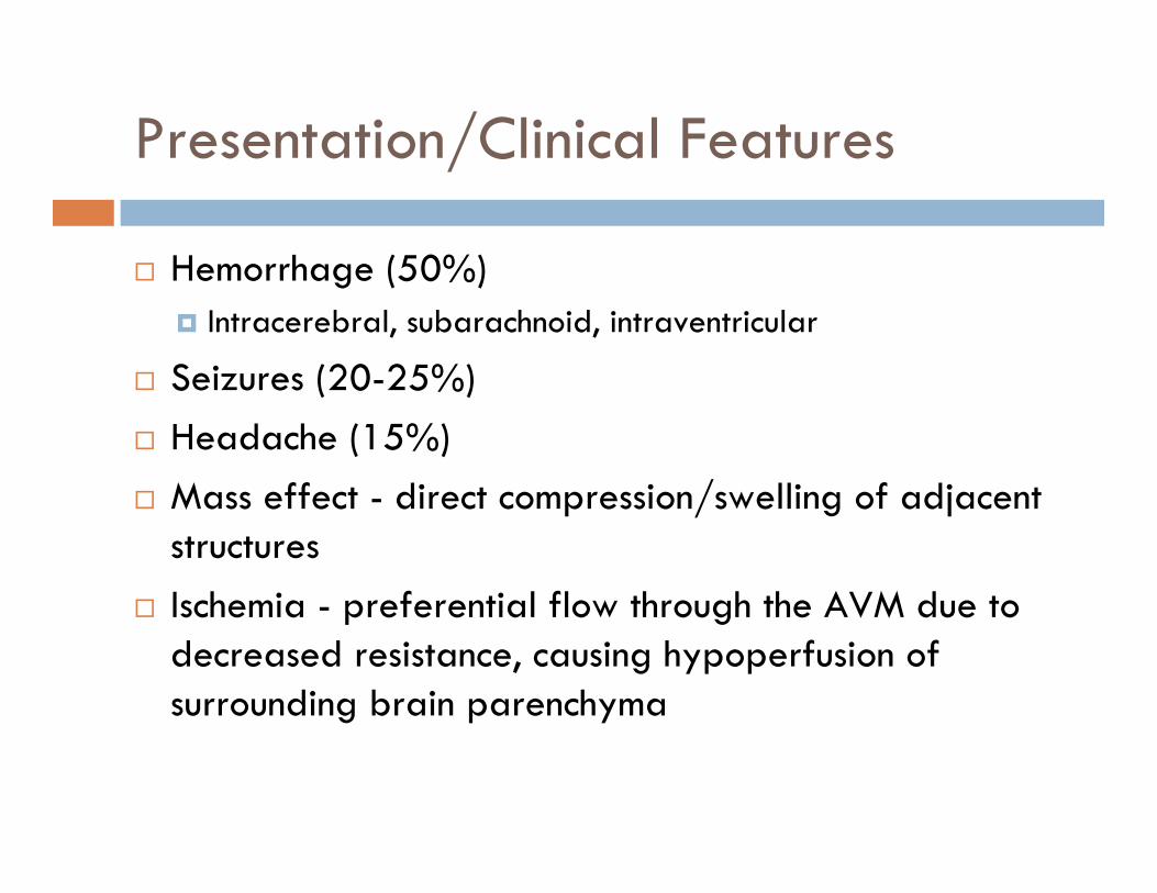
Presentation/Clinical Features
Hemorrhage (50%)Intracerebral, subarachnoid, intraventricular
Seizures (20-25%)Headache (15%)Mass effect - direct compression/swelling of adjacent structuresIschemia - preferential flow through the AVM due to decreased resistance, causing hypoperfusion of surrounding brain parenchyma

Why Treat AVMs
2-4% annual risk of ruptureIndependent risk factors for rupture:
Associated aneurysm (feeding artery, intranidal)Previous ruptureSmall AVM sizeDeep venous drainage
Goal is to determine risk of conservative treatment vs risk of intervention
ARUBA trial – A Randomized Trial of Unruptured Brain AVMs

Spetzler-Martin Classification
SizeSmall (< 3cm) 1Medium (3-6 cm) 2Large (> 6 cm) 3
Eloquence of adjacent brainNon-eloquent 0Eloquent 1
Pattern of venous drainageSuperficial only 0Deep 1

Pretherapeutic Evaluation
Noninvasive TechniquesCT/CTAMRI/MRAfMRI
Local increase in cerebral blood flow and volume after stimulation of eloquent cortex
Diffusion-Tensor ImagingVisualization of white matter pathways and their relationship to AVM

Imaging

Imaging

Pretherapeutic Evaluation
Invasive TechniquesProvocative Injection Testing(Wada’s Test)
Amobarbital sodium injection of selective feeding vessels to predict/avoid post-embolization deficits

Pretherapeutic Evaluation
AngiographyArterial feeders, size of AVM, nidus characteristics, drainage patternOther anatomical characteristics
deep location, deep venous drainage pattern, presence of a single draining vein, venous stenosis, eloquent location and diameter of AVM
Associated aneursyms(feeding artery, intranidal, circle of Willis, venous)PseudoaneurysmsAVM-associated AV Fistulas

Pretherapeutic Evaluation
AngiographyAngiogram following hemorrhage can result in false negative secondary to nidus compressionGold standard is follow-up angiogram 3 months after hemorrhage for detection of AVM

Perioperative/Anesthetic Considerations
Continuous arterial transductionsDeliberate systemic hypotensionGeneral anesthesiaadenosine-induced cardiac pause – slow flow through AVM for controlled deposition of embolic material
Pulse Oximeter Ipsilateral to femoral sheathVessel obstruction, thromboemboli, over-compression following sheath removal
Foley catheter- fluid management(Supplemental O2 // Nasopharyngeal airways for those under sedative-hypnotic agent)

Anatomy-Based Management
Neuroanatomy to understand at-risk territoriesDone under general anesthesia
Patient comfortEnsures motionless patient
Theoretically, because AVM has no functional intervening tissue, embolization of appropriate vessels should not cause functional deficitNeurophysiologic monitoring
SSEP/EEG

Physiology-Based Management
Evaluation of functional anatomy during procedureRequires intravenous anesthesia with short-acting agents(propofol, midazolam)Wide variability and cortical reorganization described in AVM patients

Endovascular Technique
Preoperative discussionVascular access with No. 7 Fr gauge sheath into femoral artery via Seldinger techniqueAnticoagulation algorithms to prevent thromboembolic complications
JHN-continuous heparinized flush, no bolusNo. 6 Fr gauge guiding catheter introduced into ICA or verterbral arteryFlow-directed/flow-assisted microcatheters to reach intranidal target
Variability in flexibility, torque, maneuverability, and responsiveness.

Identification of Embolization Point
Tip of catheter directly into nidusTip of catheter into feeder that does not share supply with adjacent normal brain
If normal brain at risk, pretherapeutic/intraoperative functional evaluation can be performed to avoid neurologic deficit from embolization

Injection of Embolic Material
Polyvinyl alcoholn-butyl cyanoacrylateOnyxEthiblocSilkMicrocoilsCombination

Polyvinyl Alcohol (PVA)
Available in different sizes based on size of target vesselsSlower occlusion than liquid embolic agentsOcclude low-pressure shunts first
Results in increase in intranidal vessel pressure hemorrhage risk prior to nidal obliteration
High recanalization ratesDevelopment of collateral feeders secondary to proximal occlusion of AVM with large size particles may contribute to this high rate
Used with success preoperatively to reduce flow prior to open or radiosurgical treatment

n-BCA
Liquid monomer that permanently polymerizes to a solid compound after contact with anionsOcclusion by initiation of inflammatory endothelial responseCombined with Ethiodal (iodine-based oil) to make lesion opaque on X-rays
Tantalum powder added to increase opacity(currently less common)

n-BCA
Viscosity and Polymerization timeIncreasing Ethiodal concentration increases viscosity and delays polymerization
Delayed polymerization helpful in lesions that exhibit slow flowIncreased viscosity delays transit within lesion
Temperature, homogeneity of mixture, and changes in pH of solution also influence polymerization time

n-BCA
Acetic acid and glaciar acid to decrease pHDelays polymerization without compromising viscosity
Dextrose 5%Delays polymerization time by delaying contact of blood with n-BCA

n-BCA
Factors that influence choice of viscosity and polymerization time:
Distance between microcatheter and nidusTortuosity of vesselsIntranidal flow

n-BCA
1:1 dilution(nBCA:oil) used when flow is very fast and catheter is close to nidus

n-BCA
“Flow Control” – Microcatheter occludes distal blood flow as it reaches smaller vessels, thus preventing blood from contacting glue, allowing for distal penetrationInjection stops when glue reaches venous system through nidus or there is backflow towards the catheterRemove catheter quickly to prevent catheter retention
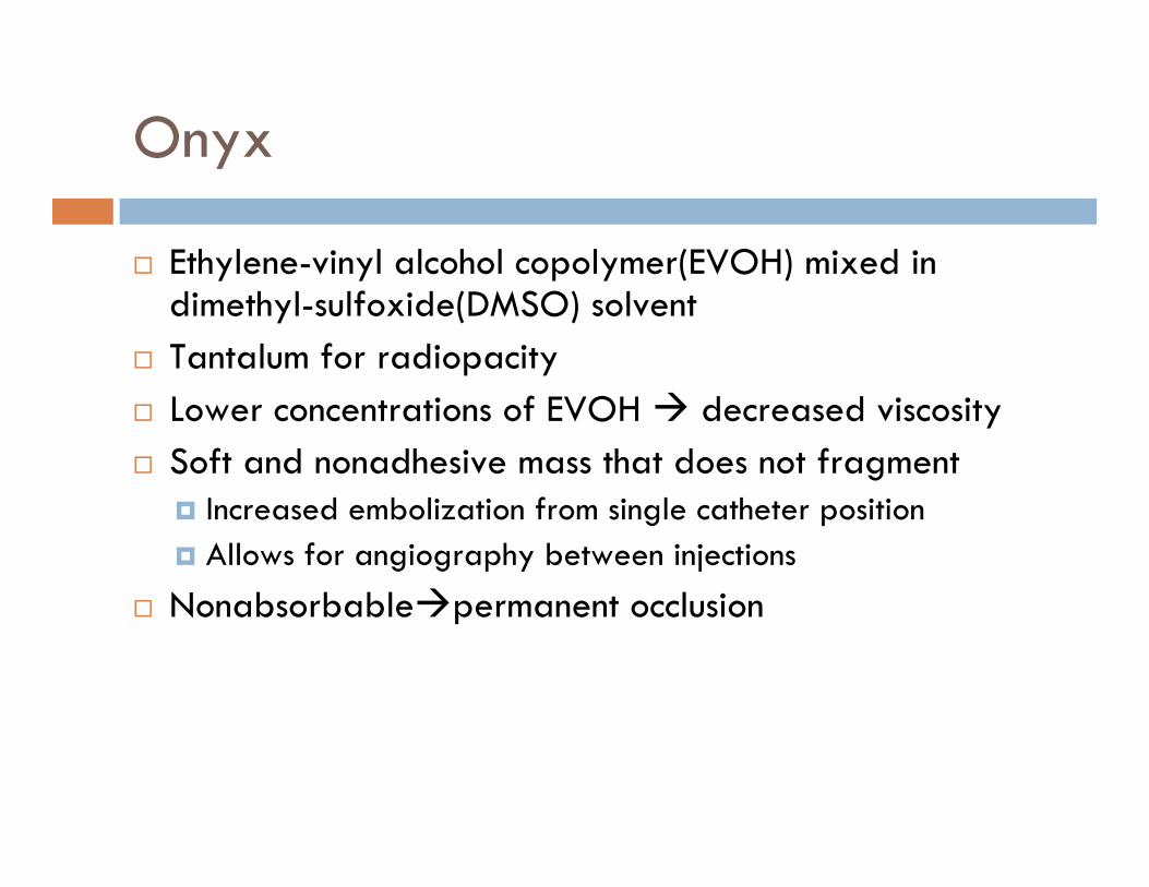
Onyx
Ethylene-vinyl alcohol copolymer(EVOH) mixed in dimethyl-sulfoxide(DMSO) solventTantalum for radiopacityLower concentrations of EVOH decreased viscositySoft and nonadhesive mass that does not fragment
Increased embolization from single catheter positionAllows for angiography between injections
Nonabsorbable permanent occlusion

Onyx
PitfallsAngiotoxicity related to DMSO
swine rete mirabilis embolization model report risk of mild transient acute vasospasm
Separation of tantalum from OnyxIncreases difficulty of viewing smaller feeding pedicles

Treatment Objectives
Embolization for definitive cureEmbolization as a precursor for definitive operative resectionEmbolization as a precursor for radiosurgeryEmbolization as palliative treatment for progressive debilitating symptomsTarget embolization of high risk lesions

Treatment Objectives
Embolization for definitive cureN-BCA complete obliteration rates(~10%)
Deruty, et al – 5% but primarily used in high-grade AVMVinuela, et al – 9.9% in small to medium AVM with less than 4 pedicles(405 pts)Lundqvist, et al – 13% Valavanis and Yasargil – 40% in 387 consecutive patientsGobin – 11.2% in patients after embolization prior to radiosurgery (125 pts who were poor surgical candidates)

Treatment Objectives
Embolization for definitive cureOnyx complete obliteration rates
Perez-Higueras, et al – 22% in series of 45 patientsPierot, et al – 4.2% in series of 48 patientsLeonardi, et al – 5.9% among 34 pts SM III-IVvan Rooij, et al – 15.9% among 44 pts SM I-IIWeber, et al – 20% among 93 patientsMounayer, et al – 27.7% among 94 patients treated with Onyx, n-BCA, or combinationPanagiotopoulos – 24.4% among 82 pts
Reconstitution of flow to AVM nidus can occur through dilation of pre-existent collateralization
Rare cases reported for n-BCARecent reports of reperfusion for Onyx(Perez-Higuera, Weber, Panagiotopoulos)

Treatment Objectives
Anatomic features predictive of angiographic cure with n-BCA (Valavanis and Christoforidis)
Direct or dominant feeding arteriesMonocompartmental nidusDominant fistulous component of nidus without perinidal angiogenesis
Anatomic features predictive of angiographic cure with Onyx
Supratentorial and cortical locationCompact and plexiform nidusSmall number of supplying(direct) feeders1 superficial draining vein

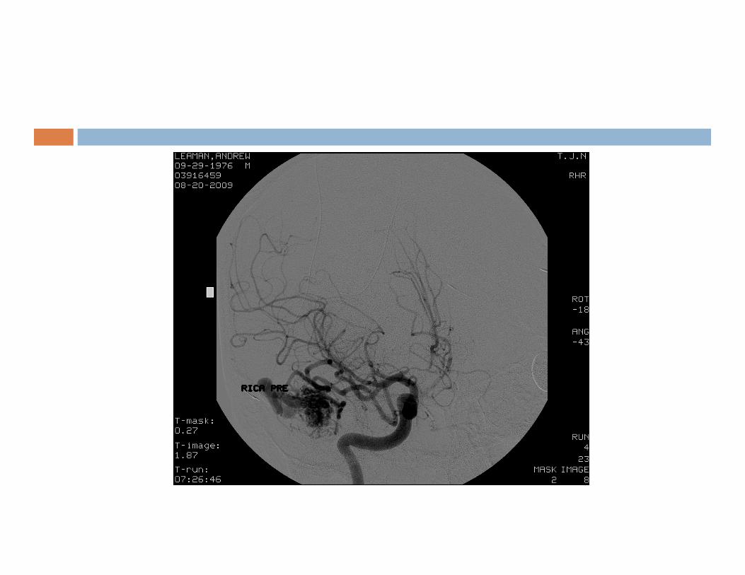
Case 1
26 yo female PMH migraines who presented with a seizure two years ago. Imaging revealed R temporoparietal AVM fed by MCA and PCA branches, with superficial drainage veins

Case 1

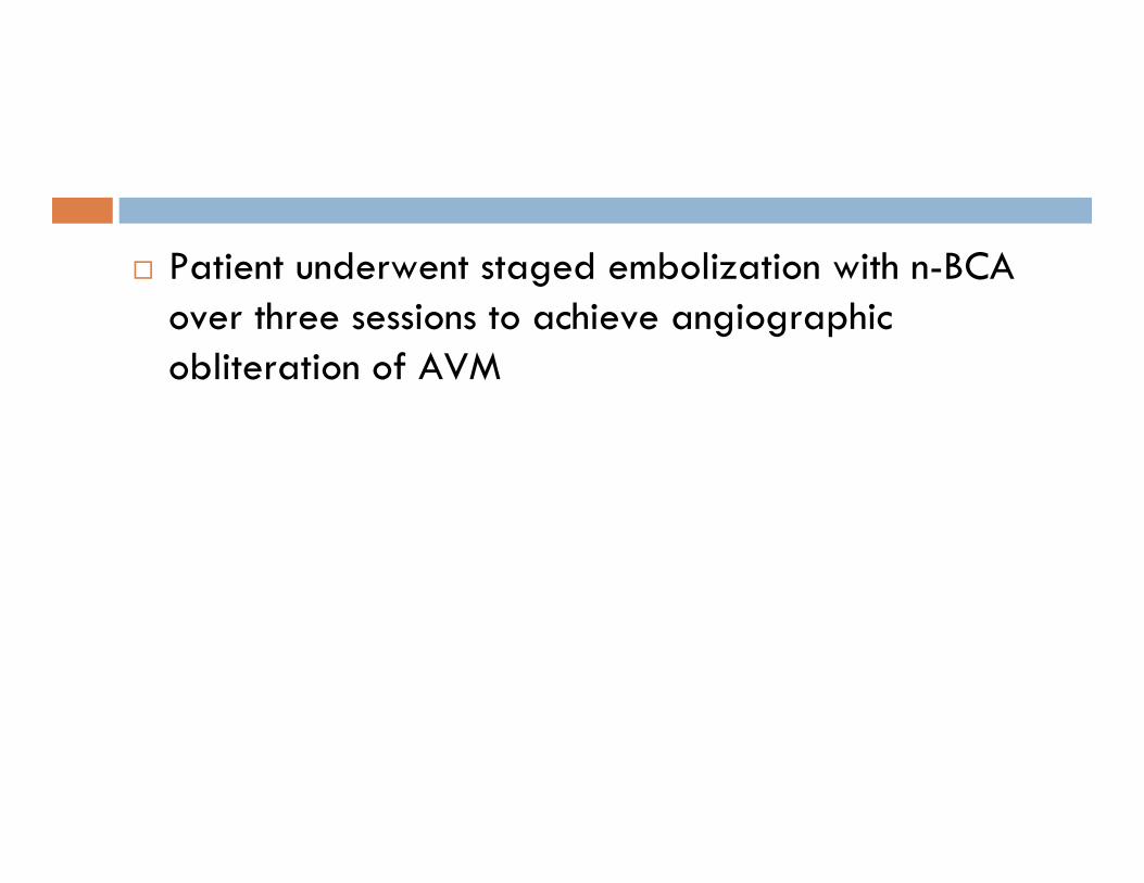
Patient underwent staged embolization with n-BCA over three sessions to achieve angiographic obliteration of AVM


Treatement Objectives
Embolization as a precursor for definitive operative resection
Goals:Reduction of size of nidusOcclusion of deep arterial feeding vessels which may be surgically inaccessibleTreatment of intranidal aneurysms and high-flow fistulas to promote progressive nidus thrombosisReduce blood loss, improve operative time, convert high Spetzler-Martin AVMs to lower grade lesions reduce morbidity and mortality

Treatment Objectives
Embolization as a precursor for definitive operative resection
Avoid embolizing draining veinsRestricting venous outflow increases hemorrhage risk
Proximal embolization collateral development treacherous open surgical resection

Case 2
32 yo male with no PMH presented with seizure. Imaging revealed R temporal AVM being fed by the MCA with superficial drainage.


Embolization of M4 pedicle and Anterior Temporal pedicle resulted in 95% treatment of AVM.


Craniotomy for complete excision of remaining AVM was performed.Intraoperatively, lesions embolized with n-BCA exhibit a harder, less compliant mass in contrast to lesions embolized with Onyx, which is a softer agent


Treatment Objectives
Embolization as a precursor for radiosurgeryGoals:
Decrease target size of AVMAllows higher dose of radiation to smaller volume
Reduce weakness in AVM angioarchitecture by eliminating intranidal/venous aneursyms
Reduces hemorrhage risk in latency period
Flow reduction alone without reduction in AVM volume does not improve radiosurgical success

Case 3
40 x 40 mm L parietal AVM fed by L MCA and PCA

Staged embolization over four sessions with n-BCA enabled reduction of AVM volume prior to radiosurgery. Patient now undergoing LINAC treatmetn for residual AVM.


Note residual AVM with diffuse pattern and persistence of early venous drainage

Treatment Objectives
Embolization as palliation for progressive debilitating symptoms
Alternative for large, non-resectable AVMsProgressive neurologic deficits secondary to arterial stealMedically intractible seizuresVenous hypertension causing HA or local mass effect from engorged veins
Target embolization of high-risk lesionsAssociated aneursymsIncreased pressure in veins that have outflow restriction
Rapidly forming collaterals reduces long-term effectiveness

Complications
Hemorrhage/EdemaArterial perforation, intranidal aneurysm rupture, draining vein occlusionOcclusive Hyperemia—passive engorgement of vessels and arterial stagnation in adjacent brain following obliteration of AVM resulting in edema/hemorrhageNormal Perfusion Pressure Breakthrough Theory—loss of autoregulation in surrounding ischemic tissue following embolization, resulting in disruption of capillary bedsMural necrosis induced by embolic material

Complications
Treatment of HemorrhageProtamine for reversal of anticoagulation
Treatment of EdemaSteroids

Complications
IschemiaInadvertent embolization of normal blood vesselsThrombotic emboliRetention of microcatheter
TreatmentDeliberate hypertensionPlatelet glycoprotein IIb/IIIa inhibitors

Morbidity and Mortality
10% morbidity regarding temporary neurological deficit8% morbidity regarding permanent deficit~1% mortalityComplications tend to occur in higher grade AVMs

Risk of Embolization by SM Grade—Kim, et al Neurosurgery 2006
Retrospective review of 153 patients, 508 vessels, 203 sessionsAge, sex, AVM grade, location of lesion, number and location of embolized arteries, and number of embolization sessions reviewed with respect to neurologic/vascular complicationsNumber of branches embolized was only variable related to neurologic deficit (p=0.017)

Risk of Embolization—Kim, et al Neurosurgery 2006
Immediate/Long term neurologic deficit(selection bias-most ii-iii not usually embolized; represent more challenging subset of lesions requiring multimodality tx)
Number of avm per grade was too small for meaningful analysis
Spetzler-Martin Grade
Immediate neurologic deficit (p=0.103)
Follow-up neurologic deficit
I 0 % 0 %
II 8 % 5%
III 12 % 7 %
IV 15 % 10%
V 18 % 18%

Conclusion
Pre-operative evaluationNoninvasive/Invasive ImagingAnesthetic/Perioperative ConsiderationsEndovascular TechniqueEmbolization MaterialsTreatment Objectives of Endovascular ApproachOutcomes/ComplicationsFuture

Bibliography1. Ogilvy, C.S., et al., Recommendations for the management of intracranial arteriovenous malformations: a statement for healthcare professionals from a special writing group of the Stroke Council, American Stroke
Association. Circulation, 2001. 103(21): p. 2644-57.
2. Friedlander, R.M., Clinical practice. Arteriovenous malformations of the brain. N Engl J Med, 2007. 356(26): p. 2704-12.
3. Latchaw, R.E., et al., Functional magnetic resonance imaging as a management tool for cerebral arteriovenous malformations. Neurosurgery, 1995. 37(4): p. 619-25; discussion 625-6.
4. Maldjian, J., et al., Functional magnetic resonance imaging of regional brain activity in patients with intracerebral arteriovenous malformations before surgical or endovascular therapy. J Neurosurg, 1996. 84(3): p. 477-83.
5. Berube, J., et al., Diffusion tensor imaging analysis of long association bundles in the presence of an arteriovenous malformation. J Neurosurg, 2007. 107(3): p. 509-14.
6. Feliciano, C.E., et al., Provocative Test with Propofol: Experience in Patients with Cerebral Arteriovenous Malformations Who Underwent Neuroendovascular Procedures. AJNR Am J Neuroradiol, 2009.
7. Lazar, R.M., et al., Anterior translocation of language in patients with left cerebral arteriovenous malformation. Neurology, 1997. 49(3): p. 802-8.
8. Moo, L.R., et al., Tailored cognitive testing with provocative amobarbital injection preceding AVM embolization. AJNR Am J Neuroradiol, 2002. 23(3): p. 416-21.
9. Muller-Forell, W. and A. Valavanis, How angioarchitecture of cerebral arteriovenous malformations should influence the therapeutic considerations. Minim Invasive Neurosurg, 1995. 38(1): p. 32-40.
10. Brown, R.D., Jr., et al., The natural history of unruptured intracranial arteriovenous malformations. J Neurosurg, 1988. 68(3): p. 352-7.
11. Brown, R.D., Jr., D.O. Wiebers, and G.S. Forbes, Unruptured intracranial aneurysms and arteriovenous malformations: frequency of intracranial hemorrhage and relationship of lesions. J Neurosurg, 1990. 73(6): p. 859-63.
12. Cunha e Sa, M.J., et al., The treatment of associated intracranial aneurysms and arteriovenous malformations. J Neurosurg, 1992. 77(6): p. 853-9.
13. Hartmann, A., et al., Risk of endovascular treatment of brain arteriovenous malformations. Stroke, 2002. 33(7): p. 1816-20.
14. Starke, R.M., et al., Treatment guidelines for cerebral arteriovenous malformation microsurgery. Br J Neurosurg, 2009. 23(4): p. 376-86.
15. Turjman, F., et al., Aneurysms related to cerebral arteriovenous malformations: superselective angiographic assessment in 58 patients. AJNR Am J Neuroradiol, 1994. 15(9): p. 1601-5.
16. Hartmann, A., et al., Treatment of arteriovenous malformations of the brain. Curr Neurol Neurosci Rep, 2007. 7(1): p. 28-34.
17. Stapf, C., et al., Predictors of hemorrhage in patients with untreated brain arteriovenous malformation. Neurology, 2006. 66(9): p. 1350-5.
18. Redekop, G., et al., Arterial aneurysms associated with cerebral arteriovenous malformations: classification, incidence, and risk of hemorrhage. J Neurosurg, 1998. 89(4): p. 539-46.
19. Pritz, M.B., Ruptured supratentorial arteriovenous malformations associated with venous aneurysms. Acta Neurochir (Wien), 1994. 128(1-4): p. 150-62.
20. Al-Shahi, R. and C. Warlow, A systematic review of the frequency and prognosis of arteriovenous malformations of the brain in adults. Brain, 2001. 124(Pt 10): p. 1900-26.
21. Spetzler, R.F., et al., Relationship of perfusion pressure and size to risk of hemorrhage from arteriovenous malformations. J Neurosurg, 1992. 76(6): p. 918-23.
22. Forster, D.M., L. Steiner, and S. Hakanson, Arteriovenous malformations of the brain. A long-term clinical study. J Neurosurg, 1972. 37(5): p. 562-70.
23. Pollock, B.E., et al., Factors that predict the bleeding risk of cerebral arteriovenous malformations. Stroke, 1996. 27(1): p. 1-6.
24. Lasjaunias, P., et al., Cerebral arteriovenous malformations (C. AVM) and associated arterial aneurysms (AA). Analysis of 101 C. AVM cases, with 37 AA in 23 patients. Acta Neurochir (Wien), 1988. 91(1-2): p. 29-36.
25. Marks, M.P., et al., Intranidal aneurysms in cerebral arteriovenous malformations: evaluation and endovascular treatment. Radiology, 1992. 183(2): p. 355-60.

Bibliography26. Crawford, P.M., et al., Arteriovenous malformations of the brain: natural history in unoperated patients. J Neurol Neurosurg Psychiatry, 1986. 49(1): p. 1-10.
27. Graf, C.J., G.E. Perret, and J.C. Torner, Bleeding from cerebral arteriovenous malformations as part of their natural history. J Neurosurg, 1983. 58(3): p. 331-7.
28. Hartmann, A., et al., Determinants of staged endovascular and surgical treatment outcome of brain arteriovenous malformations. Stroke, 2005. 36(11): p. 2431-5.
29. Schaller, C. and J. Schramm, Microsurgical results for small arteriovenous malformations accessible for radiosurgical or embolization treatment. Neurosurgery, 1997. 40(4): p. 664-72; discussion 672-4.
30. Spears, J., et al., A discriminative prediction model of neurological outcome for patients undergoing surgery of brain arteriovenous malformations. Stroke, 2006. 37(6): p. 1457-64.
31. Khayata, M.H., et al., False aneurysm associated with rupture of an arteriovenous malformation--implications for treatment: case report. Neurosurgery, 1993. 33(4): p. 753-6.
32. Haw, C.S., et al., Complications of embolization of arteriovenous malformations of the brain. J Neurosurg, 2006. 104(2): p. 226-32.
33. Yuki, I., et al., Treatment of brain arteriovenous malformations with high-flow arteriovenous fistulas: risk and complications associated with endovascular embolization in multimodality treatment. J Neurosurg, 2009.
34. Schmidek, H.H. and D.W. Roberts, Schmidek & Sweet operative neurosurgical techniques : indications, methods, and results. 5th ed. 2006, Philadelphia: Saunders Elsevier. 2 v. (xxxix, 2337, 67 p.).
35. Soderman, M., et al., Management of patients with brain arteriovenous malformations. Eur J Radiol, 2003. 46(3): p. 195-205.
36. Willinsky, R.A., et al., Delayed angiography in the investigation of intracerebral hematomas caused by small arteriovenous malformations. Neuroradiology, 1993. 35(4): p. 307-11.
37. Griffiths, P.D., C.J. Beveridge, and A. Gholkar, Angiography in non-traumatic brain haematoma. An analysis of 100 cases. Acta Radiol, 1997. 38(5): p. 797-802.
38. Hino, A., et al., Value of repeat angiography in patients with spontaneous subcortical hemorrhage. Stroke, 1998. 29(12): p. 2517-21.
39. Eskridge, J.M., Interventional neuroradiology. Radiology, 1989. 172(3 Pt 2): p. 991-1006.
40. Purdy, P.D., H.H. Batjer, and D. Samson, Management of hemorrhagic complications from preoperative embolization of arteriovenous malformations. J Neurosurg, 1991. 74(2): p. 205-11.
41. Vinuela F, H.V., Dion JE, Interventional Neuroradiology: Endovascular Therapy of the Central Nervous System. 1992, New York, NY: Raven Press.
42. Pile-Spellman, J., et al., Adenosine-induced cardiac pause for endovascular embolization of cerebral arteriovenous malformations: technical case report. Neurosurgery, 1999. 44(4): p. 881-6; discussion 886-7.
43. Pikus, H.J., M.L. Beach, and R.E. Harbaugh, Microsurgical treatment of arteriovenous malformations: analysis and comparison with stereotactic radiosurgery. J Neurosurg, 1998. 88(4): p. 641-6.
44. Pik, J.H. and M.K. Morgan, Microsurgery for small arteriovenous malformations of the brain: results in 110 consecutive patients. Neurosurgery, 2000. 47(3): p. 571-5; discussion 575-7.
45. Heros, R.C., J. Morcos, and K. Korosue, Arteriovenous malformations of the brain. Surgical management. Clin Neurosurg, 1993. 40: p. 139-73.
46. Davidson, G.S. and K.G. Terbrugge, Histologic long-term follow-up after embolization with polyvinyl alcohol particles. AJNR Am J Neuroradiol, 1995. 16(4 Suppl): p. 843-6.
47. Purdy, P.D., et al., Preoperative embolization of cerebral arteriovenous malformations with polyvinyl alcohol particles: experience in 51 adults. AJNR Am J Neuroradiol, 1990. 11(3): p. 501-10.
48. Wallace, R.C., et al., The safety and effectiveness of brain arteriovenous malformation embolization using acrylic and particles: the experiences of a single institution. Neurosurgery, 1995. 37(4): p. 606-15; discussion 615-8.
49. N-butyl cyanoacrylate embolization of cerebral arteriovenous malformations: results of a prospective, randomized, multi-center trial. AJNR Am J Neuroradiol, 2002. 23(5): p. 748-55.
50. Linfante, I. and A.K. Wakhloo, Brain aneurysms and arteriovenous malformations: advancements and emerging treatments in endovascular embolization. Stroke, 2007. 38(4): p. 1411-7.

Bibliography51. Sorimachi, T., et al., Embolization of cerebral arteriovenous malformations achieved with polyvinyl alcohol particles: angiographic reappearance and complications. AJNR Am J Neuroradiol, 1999. 20(7): p. 1323-8.
52. Mathis, J.A., et al., The efficacy of particulate embolization combined with stereotactic radiosurgery for treatment of large arteriovenous malformations of the brain. AJNR Am J Neuroradiol, 1995. 16(2): p. 299-306.
53. Debrun, G.M., et al., Embolization of the nidus of brain arteriovenous malformations with n-butyl cyanoacrylate. Neurosurgery, 1997. 40(1): p. 112-20; discussion 120-1.
54. Wikholm, G., C. Lundqvist, and P. Svendsen, The Goteborg cohort of embolized cerebral arteriovenous malformations: a 6-year follow-up. Neurosurgery, 2001. 49(4): p. 799-805; discussion 805-6.
55. Wakhloo, A.K., et al., Transvenous n-butyl-cyanoacrylate infusion for complex dural carotid cavernous fistulas: technical considerations and clinical outcome. AJNR Am J Neuroradiol, 2005. 26(8): p. 1888-97.
56. Lieber, B.B., et al., Acute and chronic swine rete arteriovenous malformation models: effect of ethiodol and glacial acetic acid on penetration, dispersion, and injection force of N-butyl 2-cyanoacrylate. AJNR Am J Neuroradiol, 2005. 26(7): p. 1707-14.
57. Molyneux, A.J. and S.C. Coley, Embolization of spinal cord arteriovenous malformations with an ethylene vinyl alcohol copolymer dissolved in dimethyl sulfoxide (Onyx liquid embolic system). Report of two cases. J Neurosurg, 2000. 93(2 Suppl): p. 304-8.
58. Weber, W., et al., Endovascular treatment of intracranial arteriovenous malformations with onyx: technical aspects. AJNR Am J Neuroradiol, 2007. 28(2): p. 371-7.
59. Murayama, Y., et al., Nonadhesive liquid embolic agent for cerebral arteriovenous malformations: preliminary histopathological studies in swine rete mirabile. Neurosurgery, 1998. 43(5): p. 1164-75.
60. Valavanis, A. and M.G. Yasargil, The endovascular treatment of brain arteriovenous malformations. Adv Tech Stand Neurosurg, 1998. 24: p. 131-214.
61. Jahan, R., et al., Embolization of arteriovenous malformations with Onyx: clinicopathological experience in 23 patients. Neurosurgery, 2001. 48(5): p. 984-95; discussion 995-7.
62. Chaloupka, J.C., et al., A reexamination of the angiotoxicity of superselective injection of DMSO in the swine rete embolization model. AJNR Am J Neuroradiol, 1999. 20(3): p. 401-10.
63. Taki, W., et al., A new liquid material for embolization of arteriovenous malformations. AJNR Am J Neuroradiol, 1990. 11(1): p. 163-8.
64. Deruty, R., et al., The combined management of cerebral arteriovenous malformations. Experience with 100 cases and review of the literature. Acta Neurochir (Wien), 1993. 123(3-4): p. 101-12.
65. Vinuela, F., G. Duckwiler, and G. Guglielmi, Contribution of interventional neuroradiology in the therapeutic management of brain arteriovenous malformations. J Stroke Cerebrovasc Dis, 1997. 6(4): p. 268-71.
66. Lundqvist, C., G. Wikholm, and P. Svendsen, Embolization of cerebral arteriovenous malformations: Part II--Aspects of complications and late outcome. Neurosurgery, 1996. 39(3): p. 460-7; discussion 467-9.
67. Gobin, Y.P., et al., Treatment of brain arteriovenous malformations by embolization and radiosurgery. J Neurosurg, 1996. 85(1): p. 19-28.
68. Pierot, L., et al., Endovascular treatment of brain arteriovenous malformations using onyx: results of a prospective, multicenter study. J Neuroradiol, 2009. 36(3): p. 147-52.
69. Perez-Higueras A, R.L.R., Quinones Taria D, Endovascular treatment of cerebral AVM: our experience with Onyx. Interventional Neuroradiology, 2005(11): p. 141-157.
70. Leonardi M, S.L., Cenni P, et al., Brain AVM embolization with Onyx: analysis of treatment in 34 patients. Interventional Neuroradiology, 2005(11): p. 185-204.
71. Tevah J, H.I., Endovascular treatment of cerebral AVMs with a new material: Onyx. Interventional Neuroradiology, 2005(11): p. 165-170.
72. Joseph S, C.H., Murali K., Endovascular treatment of cerebral AVMs with Onyx: initial experience. Interventional Neuroradiology, 2005(11): p. 171-178.
73. Florio, F., et al., Endovascular treatment of intracranial arterio-venous malformations with Onyx embolization: preliminary experience. Radiol Med, 2003. 106(5-6): p. 512-20.
74. van Rooij, W.J., M. Sluzewski, and G.N. Beute, Brain AVM embolization with Onyx. AJNR Am J Neuroradiol, 2007. 28(1): p. 172-7; discussion 178.
75. Song, D.L., et al., [Clinical experience of 70 cases of cerebral arteriovenous malformations embolization with Onyx, a novel liquid embolic agent]. Zhonghua Wai Ke Za Zhi, 2007. 45(4): p. 223-5.

Bibliography76. Mounayer, C., et al., Nidal embolization of brain arteriovenous malformations using Onyx in 94 patients. AJNR Am J Neuroradiol, 2007. 28(3): p. 518-23.
77. Wikholm, G., C. Lundqvist, and P. Svendsen, Embolization of cerebral arteriovenous malformations: Part I--Technique, morphology, and complications. Neurosurgery, 1996. 39(3): p. 448-57; discussion 457-9.
78. Valavanis A, C.G., Endovascular management of cerebral arteriovenous malformations. Neurointerventionist, 1999(1): p. 34-40.
79. Fournier, D., et al., Revascularization of brain arteriovenous malformations after embolization with bucrylate. Neuroradiology, 1990. 32(6): p. 497-501.
80. Katsaridis, V., C. Papagiannaki, and E. Aimar, Curative embolization of cerebral arteriovenous malformations (AVMs) with Onyx in 101 patients. Neuroradiology, 2008. 50(7): p. 589-97.
81. Panagiotopoulos, V., et al., Embolization of intracranial arteriovenous malformations with ethylene-vinyl alcohol copolymer (Onyx). AJNR Am J Neuroradiol, 2009. 30(1): p. 99-106.
82. Yu, S.C., et al., Complete obliteration of intracranial arteriovenous malformation with endovascular cyanoacrylate embolization: initial success and rate of permanent cure. AJNR Am J Neuroradiol, 2004. 25(7): p. 1139-43.
83. Oran, I., M. Parildar, and A. Derbent, Ventricular/paraventricular small arteriovenous malformations: role of embolisation with cyanoacrylate. Neuroradiology, 2005. 47(4): p. 287-94.
84. Paulsen, R.D., et al., Embolization of basal ganglia and thalamic arteriovenous malformations. Neurosurgery, 1999. 44(5): p. 991-6; discussion 996-7.
85. Hamada, J., et al., A mixture of ethylene vinyl alcohol copolymer and ethanol yielding a nonadhesive liquid embolic agent to treat cerebral arteriovenous malformations: initial clinical experience. J Neurosurg, 2002. 97(4): p. 881-8.
86. Spetzler, R.F., et al., Surgical management of large AVM's by staged embolization and operative excision. J Neurosurg, 1987. 67(1): p. 17-28.
87. Jafar, J.J., et al., The effect of embolization with N-butyl cyanoacrylate prior to surgical resection of cerebral arteriovenous malformations. J Neurosurg, 1993. 78(1): p. 60-9.
88. DeMeritt, J.S., et al., Outcome analysis of preoperative embolization with N-butyl cyanoacrylate in cerebral arteriovenous malformations. AJNR Am J Neuroradiol, 1995. 16(9): p. 1801-7.
89. Cromwell, L.D. and A.B. Harris, Treatment of cerebral arteriovenous malformations: a combined neurosurgical and neuroradiological approach. J Neurosurg, 1980. 52(5): p. 705-8.
90. Debrun, G., et al., Embolization of cerebral arteriovenous malformations with bucrylate. J Neurosurg, 1982. 56(5): p. 615-27.
91. Liebman KM, R.R., The hemodynamic changes measured in cerebral arterio-venous malformations following endovascular treatment. J Neurovasc Dis, 1997(2): p. 112-116.
92. Dawson, R.C., 3rd, et al., Treatment of arteriovenous malformations of the brain with combined embolization and stereotactic radiosurgery: results after 1 and 2 years. AJNR Am J Neuroradiol, 1990. 11(5): p. 857-64.
93. Dion, J.E. and J.M. Mathis, Cranial arteriovenous malformations. The role of embolization and stereotactic surgery. Neurosurg Clin N Am, 1994. 5(3): p. 459-74.
94. Rao, V.R., et al., Dissolution of isobutyl 2-cyanoacrylate on long-term follow-up. AJNR Am J Neuroradiol, 1989. 10(1): p. 135-41.
95. Pollock, B.E., et al., Repeat stereotactic radiosurgery of arteriovenous malformations: factors associated with incomplete obliteration. Neurosurgery, 1996. 38(2): p. 318-24.
96. Berenstein A, L.P., Surgical Neuroangiography. Vol. 4. 1987, New York, NY: Springer-Verlag.
97. Fox, A.J., et al., Rolandic arteriovenous malformations: improvement in limb function by IBC embolization. AJNR Am J Neuroradiol, 1985. 6(4): p. 575-82.
98. Vinuela, F.V., et al., Dominant-hemisphere arteriovenous malformations: therapeutic embolization with isobutyl-2-cyanoacrylate. AJNR Am J Neuroradiol, 1983. 4(4): p. 959-66.
99. Grzyska, U. and J. Fiehler, Pathophysiology and treatment of brain AVMs. Klin Neuroradiol, 2009. 19(1): p. 82-90.
100. Meisel, H.J., et al., Effect of partial targeted N-butyl-cyano-acrylate embolization in brain AVM. Acta Neurochir (Wien), 2002. 144(9): p. 879-87; discussion 888.

Bibliography101. Rosenwasser RH, T.J., Gannon PM, et al., Current Strategies for the Management of Cerebral Arteriovenous Malformations. 1998, Rolling Meadows, Illinois: American Association of Neurological Surgeons.
102. Purdy, P.D., et al., Intraarterial sodium amytal administration to guide preoperative embolization of cerebral arteriovenous malformations. J Neurosurg Anesthesiol, 1991. 3(2): p. 103-6.
103. Purdy, P.D., et al., Arteriovenous malformations of the brain: choosing embolic materials to enhance safety and ease of excision. J Neurosurg, 1992. 77(2): p. 217-22.
104. Wilson, C.B. and G. Hieshima, Occlusive hyperemia: a new way to think about an old problem. J Neurosurg, 1993. 78(2): p. 165-6.
105. al-Rodhan, N.R., et al., Occlusive hyperemia: a theory for the hemodynamic complications following resection of intracerebral arteriovenous malformations. J Neurosurg, 1993. 78(2): p. 167-75.
106. Guglielmi, G., Analysis of the hemodynamic characteristics of brain arteriovenous malformations using electrical models: baseline settings, surgical extirpation, endovascular embolization, and surgical bypass. Neurosurgery, 2008. 63(1): p. 1-10; discussion 11.
107. Spetzler RF, W.C., Weinstein PH, et al., Normal perfusion pressure breakthrough theory. Clin Neurosurg, 1977. 25: p. 651-672.
108. Picard, L., et al., Acute spontaneous hemorrhage after embolization of brain arteriovenous malformation with N-butyl cyanoacrylate. J Neuroradiol, 2001. 28(3): p. 147-65.
109. Young, W.L. and J. Pile-Spellman, Anesthetic considerations for interventional neuroradiology. Anesthesiology, 1994. 80(2): p. 427-56.
110. Frizzel, R.T. and W.S. Fisher, 3rd, Cure, morbidity, and mortality associated with embolization of brain arteriovenous malformations: a review of 1246 patients in 32 series over a 35-year period. Neurosurgery, 1995. 37(6): p. 1031-9; discussion 1039-40.
111. Luessenhop, A.J. and W.T. Spence, Artificial embolization of cerebral arteries. Report of use in a case of arteriovenous malformation. J Am Med Assoc, 1960. 172: p. 1153-5.
112. Fiorella, D., et al., The role of neuroendovascular therapy for the treatment of brain arteriovenous malformations. Neurosurgery, 2006. 59(5 Suppl 3): p. S163-77; discussion S3-13.
113. Taylor, C.L., et al., Complications of preoperative embolization of cerebral arteriovenous malformations. J Neurosurg, 2004. 100(5): p. 810-2.
114. Vinuela, F., et al., Combined endovascular embolization and surgery in the management of cerebral arteriovenous malformations: experience with 101 cases. J Neurosurg, 1991. 75(6): p. 856-64.
115. Kim, L.J., et al., Postembolization neurological deficits in cerebral arteriovenous malformations: stratification by arteriovenous malformation grade. Neurosurgery, 2006. 59(1): p. 53-9; discussion 53-9.
116. Ledezma, C.J., et al., Complications of cerebral arteriovenous malformation embolization: multivariate analysis of predictive factors. Neurosurgery, 2006. 58(4): p. 602-11; discussion 602-11.
117. Jayaraman, M.V., et al., Neurologic complications of arteriovenous malformation embolization using liquid embolic agents. AJNR Am J Neuroradiol, 2008. 29(2): p. 242-6.
118. Pierot L, J.A., Herbreteau D, et al., Endovascular treatment of brain arteriovenous malformations using Onyx: preliminary results of a prospective multicenter study. Interventional Neuroradiology, 2005. 11: p. 159-164.
119. Leonardi M, S.L., Cenni P, et al., Brain AVM embolization with Onyx: analysis of treatment in 34 patients. Interventional Neuroradiology, 2005. 11: p. 185-204.
120. Tevah J, H.I., Endovascular treatment of cerebral AVMs with a new material: Onyx. Interventional Neuroradiology, 2005. 11: p. 165-170.
121. Weber, W., et al., Preoperative embolization of intracranial arteriovenous malformations with Onyx. Neurosurgery, 2007. 61(2): p. 244-52; discussion 252-4.