Endothelial Cell Tube Formation Assay · 2. Thaw basement membrane extract and prepare 96-well...
11
Application Note Background New vessels in the body are formed by three processes: vasculogenesis, arte- riogenesis and angiogenesis. Vasculo- genesis only occurs at an early stage of development, giving rise to the primitive circulatory system, while angio- and arte- riogenesis (also) take place in adulthood. Arteriogenesis involves the remodeling and maturation of existing vessels to yield fully developed, functional arteries, usually when larger arteries are occluded. Angiogenesis is the physiological forma- tion of new blood vessels from existing ones, a process that is essential for em- bryonic and fetal development and organ growth, supports the healing of wounds and skeletal growth, and is also an inte- gral part of pregnancy and the female reproductive cycle [1; 2]. It is triggered by tissue hypoxia or insufficient oxygen tension [3]. Newly formed blood ves- sels lined with endothelial cells supply oxygen and nutrients to tissues, promote immune surveillance by hematopoietic cells, and remove waste products [2]. Angiogenesis is a tightly regulated pro- cess that is balanced by pro- and anti- angiogenic signals including integrins, chemokines, angiopoietins, oxygen sens- ing agents, junctional molecules and en- dogenous inhibitors [4]. It is a hallmark of over 50 different disease states, and its dysfunction is implicated in cancer, pso- riasis, various eye diseases, rheumatoid arthritis, asthma and other autoimmune diseases, infectious diseases, coronary arterial diseases, stroke, atherosclerosis and impaired wound healing, among others [1, 5]. Physiological angiogenesis is a highly organized sequence of cellular events (see Fig. 1) comprising vascular initia- tion, sprouting, formation, maturation, remodeling and regression, which are controlled and modulated to meet tissue requirements. Pathological angiogenesis, by contrast, is less well controlled, with vessels rarely maturing, remodeling or regressing in response to disease [6]. Endothelial Cell Tube Formation Assay (Angiogenesis Assay) A B C D Endothelial cell (EC) Pericyte Figure 1: Angiogenesis is a highly organized sequence of cellular events. (A) Angiogenic stimuli give rise to a cytokine gradient and activate endothelial cells. (B) Endothelial cells break down the basement membrane, sprout toward the external gradient and begin to migrate toward the angiogenic stimulus. Pericytes detach from the blood vessel. (C) Reassembly of endothelial cells and formation of new cell-cell contacts and vessel lumina. (D) Vessel stabilization by pericyte recruitment and initiation of blood flow. Initiation Angiogenic stimuli from external sources built up a cytokine gradient (e.g. FGF, VEGF, etc.) and activate endothelial cells Sprouting g Proliferation g Migration Upregulation of matrix metalloproteases by endothelial cells and breakdown of the underlying basement membrane Pericyte detachment and first EC sprouting towards the external gradient EC proliferation and migration towards the angiogenic stimulus Tube Formation ECs reassemble and establish new cell-cell contacts Formation of new vessel lumen Vessel Maturation Stabilization and structural support of newly formed vessel by pericytes and SMCs Initiation of blood flow
Transcript of Endothelial Cell Tube Formation Assay · 2. Thaw basement membrane extract and prepare 96-well...

Application Note
Background
New vessels in the body are formed by three processes: vasculogenesis, arte-riogenesis and angiogenesis. Vasculo-genesis only occurs at an early stage of development, giving rise to the primitive circulatory system, while angio- and arte-riogenesis (also) take place in adulthood. Arteriogenesis involves the remodeling and maturation of existing vessels to yield fully developed, functional arteries, usually when larger arteries are occluded. Angiogenesis is the physiological forma-tion of new blood vessels from existing ones, a process that is essential for em-bryonic and fetal development and organ growth, supports the healing of wounds
and skeletal growth, and is also an inte-gral part of pregnancy and the female reproductive cycle [1; 2]. It is triggered by tissue hypoxia or insufficient oxygen tension [3]. Newly formed blood ves-sels lined with endothelial cells supply oxygen and nutrients to tissues, promote immune surveillance by hematopoietic cells, and remove waste products [2]. Angiogenesis is a tightly regulated pro-cess that is balanced by pro- and anti-angiogenic signals including integrins, chemokines, angiopoietins, oxygen sens-ing agents, junctional molecules and en-dogenous inhibitors [4]. It is a hallmark of over 50 different disease states, and its dysfunction is implicated in cancer, pso-riasis, various eye diseases, rheumatoid
arthritis, asthma and other autoimmune diseases, infectious diseases, coronary arterial diseases, stroke, atherosclerosis and impaired wound healing, among others [1, 5].
Physiological angiogenesis is a highly organized sequence of cellular events (see Fig. 1) comprising vascular initia-tion, sprouting, formation, maturation, remodeling and regression, which are controlled and modulated to meet tissue requirements. Pathological angiogenesis, by contrast, is less well controlled, with vessels rarely maturing, remodeling or regressing in response to disease [6].
Endothelial Cell Tube Formation Assay (Angiogenesis Assay)
A
B
C
D
Endothelial cell (EC)
Pericyte
Figure 1: Angiogenesis is a highly organized sequence of cellular events. (A) Angiogenic stimuli give rise to a cytokine gradient and activate endothelial cells. (B) Endothelial cells break down the basement membrane, sprout toward the external gradient and begin to migrate toward the angiogenic stimulus. Pericytes detach from the blood vessel. (C) Reassembly of endothelial cells and formation of new cell-cell contacts and vessel lumina. (D) Vessel stabilization by pericyte recruitment and initiation of blood flow.
Initiation
Angiogenic stimuli from external sources built up a cytokine gradient (e.g. FGF, VEGF, etc.) and activate endothelial cells
Sprouting g Proliferation g Migration
Upregulation of matrix metalloproteases by endothelial cells and breakdown of the underlying basement membrane Pericyte detachment and first EC sprouting towards the
external gradient EC proliferation and migration towards the angiogenic stimulus
Tube Formation
ECs reassemble and establish new cell-cell contacts Formation of new vessel lumen
Vessel Maturation
Stabilization and structural support of newly formed vessel by pericytes and SMCs Initiation of blood flow
2 Application Note – Endothelial Cell Tube Formation Assay
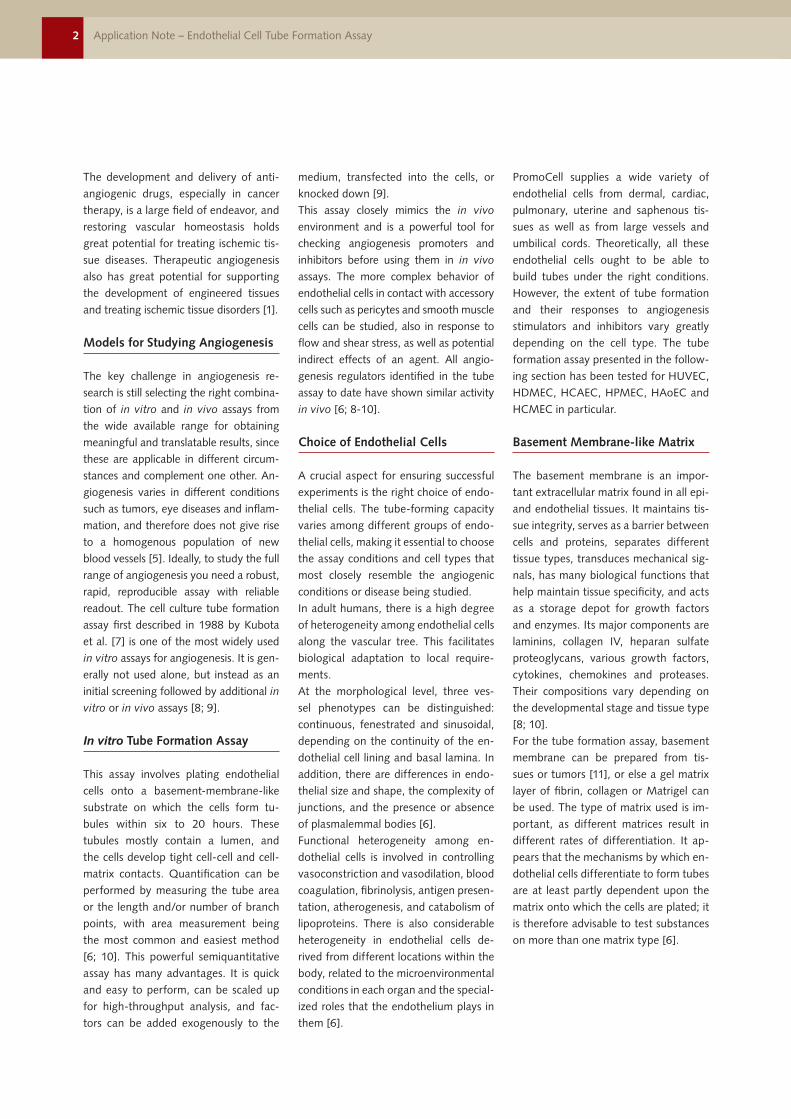
The development and delivery of anti-angiogenic drugs, especially in cancer therapy, is a large field of endeavor, and restoring vascular homeostasis holds great potential for treating ischemic tis-sue diseases. Therapeutic angiogenesis also has great potential for supporting the development of engineered tissues and treating ischemic tissue disorders [1].
Models for Studying Angiogenesis
The key challenge in angiogenesis re-search is still selecting the right combina-tion of in vitro and in vivo assays from the wide available range for obtaining meaningful and translatable results, since these are applicable in different circum-stances and complement one other. An-giogenesis varies in different conditions such as tumors, eye diseases and inflam-mation, and therefore does not give rise to a homogenous population of new blood vessels [5]. Ideally, to study the full range of angiogenesis you need a robust, rapid, reproducible assay with reliable readout. The cell culture tube formation assay first described in 1988 by Kubota et al. [7] is one of the most widely used in vitro assays for angiogenesis. It is gen-erally not used alone, but instead as an initial screening followed by additional in
vitro or in vivo assays [8; 9].
In vitro Tube Formation Assay
This assay involves plating endothelial cells onto a basement-membrane-like substrate on which the cells form tu-bules within six to 20 hours. These tubules mostly contain a lumen, and the cells develop tight cell-cell and cell-matrix contacts. Quantification can be performed by measuring the tube area or the length and/or number of branch points, with area measurement being the most common and easiest method [6; 10]. This powerful semiquantitative assay has many advantages. It is quick and easy to perform, can be scaled up for high-throughput analysis, and fac-tors can be added exogenously to the
medium, transfected into the cells, or knocked down [9]. This assay closely mimics the in vivo environment and is a powerful tool for checking angiogenesis promoters and inhibitors before using them in in vivo assays. The more complex behavior of endothelial cells in contact with accessory cells such as pericytes and smooth muscle cells can be studied, also in response to flow and shear stress, as well as potential indirect effects of an agent. All angio-genesis regulators identified in the tube assay to date have shown similar activity in vivo [6; 8-10].
Choice of Endothelial Cells
A crucial aspect for ensuring successful experiments is the right choice of endo-thelial cells. The tube-forming capacity varies among different groups of endo-thelial cells, making it essential to choose the assay conditions and cell types that most closely resemble the angiogenic conditions or disease being studied. In adult humans, there is a high degree of heterogeneity among endothelial cells along the vascular tree. This facilitates biological adaptation to local require-ments. At the morphological level, three ves-sel phenotypes can be distinguished: continuous, fenestrated and sinusoidal, depending on the continuity of the en-dothelial cell lining and basal lamina. In addition, there are differences in endo-thelial size and shape, the complexity of junctions, and the presence or absence of plasmalemmal bodies [6]. Functional heterogeneity among en-dothelial cells is involved in controlling vasoconstriction and vasodilation, blood coagulation, fibrinolysis, antigen presen-tation, atherogenesis, and catabolism of lipoproteins. There is also considerable heterogeneity in endothelial cells de-rived from different locations within the body, related to the microenvironmental conditions in each organ and the special-ized roles that the endothelium plays in them [6].
PromoCell supplies a wide variety of endothelial cells from dermal, cardiac, pulmonary, uterine and saphenous tis-sues as well as from large vessels and umbilical cords. Theoretically, all these endothelial cells ought to be able to build tubes under the right conditions. However, the extent of tube formation and their responses to angiogenesis stimulators and inhibitors vary greatly depending on the cell type. The tube formation assay presented in the follow-ing section has been tested for HUVEC, HDMEC, HCAEC, HPMEC, HAoEC and HCMEC in particular.
Basement Membrane-like Matrix
The basement membrane is an impor-tant extracellular matrix found in all epi- and endothelial tissues. It maintains tis-sue integrity, serves as a barrier between cells and proteins, separates different tissue types, transduces mechanical sig-nals, has many biological functions that help maintain tissue specificity, and acts as a storage depot for growth factors and enzymes. Its major components are laminins, collagen IV, heparan sulfate proteoglycans, various growth factors, cytokines, chemokines and proteases. Their compositions vary depending on the developmental stage and tissue type [8; 10]. For the tube formation assay, basement membrane can be prepared from tis-sues or tumors [11], or else a gel matrix layer of fibrin, collagen or Matrigel can be used. The type of matrix used is im-portant, as different matrices result in different rates of differentiation. It ap-pears that the mechanisms by which en-dothelial cells differentiate to form tubes are at least partly dependent upon the matrix onto which the cells are plated; it is therefore advisable to test substances on more than one matrix type [6].

3Application Note – Endothelial Cell Tube Formation Assay
Use aseptic techniques and a laminar flow bench.
Assay Materials
I. Materials
PromoCell Endothelial Cells (see page 8) PromoCell Endothelial Cell Growth Medium (see page 9) Cell culture dishes or flasks 96-well, 48-well, or 24-well cell culture plates Reduced growth factor basement membrane extract, BME (e.g. PK-CA577-
K519) Phosphate-Buffered Saline w/o Ca2+/Mg2+ (C-40232) or HEPES-BSS (C-40000) Accutase (C-41310)
Note: For best results we recommend using Accutase for cell detachment. The PromoCell DetachKit (C-41200 or C-41202) might be used as an alternative, but
is not recommended due to more aggressive nature of trypsin-based cell detachment. Sterile 15 ml centrifuge tubes Test substances with potential angiogenic or anti-angiogenic ability. As positive control hFGF-2 (Recombinant Human Fibroblast Growth Factor 2, e.g.
C-60240) or hVEGF-165 (Recombinant Human Vascular Endothelial Cell Growth Factor 165, e.g. C-64420) can be used. As negative control Suramine or Sulfora-phane are suitable.
Recommended:
PromoKine Angiogenesis Assay Kit (PK-CA577-K905) Includes extracellular matrix, staining dye solution, and inhibitor control
Optional for fixation and staining:
Methanol (e.g. Roth, Product No. 4627.4) Sterile water Calcein AM solution (e.g. PK-CA707-80011-1) Crystal Violet solution (e.g. Sigma, Product No. C3886)

4 Application Note – Endothelial Cell Tube Formation Assay
Use aseptic techniques and a laminar flow bench.
Assay Protocol
II. Tube Formation Assay Protocol
1. Seed PromoCell endothelial cells and allow them to grow
Plate endothelial cells in an appropriate culture vessel using the recommended PromoCell growth medium. Use seeding densities between 5.000 cells/cm2 and 20.000 cells/cm2 as recommended in the respective product manual. Replace culture medium every 2 – 3 days. Allow the cells to reach 70 – 90% confluency.
Note: For Tube Formation Assay Endothelial Cells should be used at early pas-sages (e.g. P2 – P5). Cells should be passaged at least twice after thawing from liquid nitrogen. For best results, passage cells one day before the experiment using a higher seeding density than usual of at least 20.000 cells/cm2.
Day 0: Day before assay starts
2. Thaw basement membrane extract and prepare 96-well plate
Remove Basement Membrane Extract (BME) from the freezer and place it in a refrig-erator on ice. Thawing process will be completed after overnight-incubation at 4°C. Label wells of the 96-well plate according to your experimental approach and pre-cool it in the refrigerator overnight.For 48-well plates or 24-well plates all volumes, cell numbers and seeding densities have to be scaled up dependent on the corresponding growth area.
Note: After thawing, BME can be aliquoted and further stored at -20°C. For that purpose prepare appropriate storage tubes and cool them down at 4°C. Pi-pette BME into the tubes and put them in the freezer, immediately. Pipetting should be performed quickly because BME gels rapidly at temperatures above 15°C. Ideally, keep BME on ice while pipetting. Before aliquoting carefully pipet up and down a few times. If BME is too viscous, it was not thawed completely or it started to gel. In this case, put it on ice again until BME solution is completely liquid.
Day 1: Start of Tube Formation Assay
3. Adjust media and reagents to room temperature
Place PBS, Accutase, recommended Endothelial Cell Basal Medium and recom-mended growth medium at room temperature for at least 1 hour.
4. Coat 96-well plate with Basement Membrane Extract
Place a tube of fully thawed BME on ice. Invert the tube for a few times. Load 50-80 µl of BME per well of the pre-cooled 96-well plate. BME should be evenly distributed across each well. Incubate the 96-well plate in a humidified incubator (37°C, 5% CO2) for 30 min – 1 hour. Proceed with step 5 – 8 during this incuba-tion process.
Note: Pipetting should be performed quickly because BME gels rapidly at temperatures above 15°C. Basement membrane extract should not contain any precipitates or be partially polymerized. The formation of air bubbles in the BME must be avoided. Vibration should be avoided during incubation at 37°C, be-cause this will result in uneven surfaces on the gel.

5Application Note – Endothelial Cell Tube Formation Assay
Use aseptic techniques and a laminar flow bench.
Assay Protocol5. Prepare test media
Dissolve test substances in recommended Basal Medium. 1 ml of each test me-dium is needed to resuspend 100.000 – 150.000 endothelial cells.
Note: For testing substances of unknown activity, we recommend to analyze multiple concentrations. It is important to include positive and negative controls, accompanied with inhibitors or stimulators of angiogenesis. As positive control we suggest a test medium with 50 ng/ml FGF (PromoKine, C-60240). Also fully supplemented PromoCell Endothelial Cell Growth Medium can be used. As negative controls media containing 30 µM Suramin or 1 – 10 µM Sulforaphane are suitable.If the test substance is dissolved in DMSO or alcohol, make sure that the final DMSO or alcohol concentration in the test medium is below 1% (v/v).
6. Detach endothelial cells
Endothelial Cells should be 70 – 90% confluent. Remove medium from the culture vessel and wash the cells twice with PBS or HEPES-BSS. Remove the washing solution and add 50 µl Accutase solution per cm2 of vessel surface. Close the vessel and incu-bate at 37°C for 3 – 5 min. Examine cells under a microscope. Gently tap the side of the vessel to accelerate cell detachment. When about 80% of the cells have detached, add 100 µl of growth medium per cm2 of vessel surface and gently pipet up and down to generate a single cell suspension.
7. Count cells
Transfer the cell suspension into an appropriate centrifuge tube and rinse the vessel surface again with 100 µl Endothelial Cell Growth Medium per cm2 of vessel surface to collect remaining cells. Determine cell number according to your standard procedure.
8. Prepare cells for Tube Formation Assay
Prepare 15 ml centrifuge tubes (one tube per test condition) and transfer 100.000 – 150.000 endothelial cells in each tube. Centrifuge the tubes at 300 g for 3 min and resuspend each pellet in 1 ml of the corresponding test or control medium.
Note: Resuspend the cells thoroughly to generate a homogenous single cell suspension. Incomplete resuspension will otherwise lead to different cell densi-ties in the wells of the 96-well plate which is a critical point during the assay.
9. Prepare cells for Tube Formation Assay
Add 100 µl (= 10.000 – 15.000 cells) of each single cell suspension per well on top of the gelled BME. Be careful not to touch the surface of the gel.
Note: Seeding density is critical. About 30.000 – 45.000 cells/cm2 (e.g. 96-well plate: 10.000 – 15.000 cells per well) are suitable for PromoCell primary endothelial cells but might vary dependent on cell type. Too few cells will result in incomplete tube formation. Too many cells will lead to large cell clusters and monolayers.

6 Application Note – Endothelial Cell Tube Formation Assay
Use aseptic techniques and a laminar flow bench.
Assay Protocol10. Incubate at 37°C
Incubate the 96-well plate in a humidified incubator (37°C, 5% CO2) for 4 to 24 hours. Cells can be monitored at desired time points using an inverted microscope.
Note: Incubation times are highly cell type dependent. HUVECs, for example, develop nice tubes 4 – 6 hours after seeding. After 24 hours endothelial cells typically undergo apoptosis under this conditions. We recommend to check for tube formation periodically during the experiment.
III. Trouble Shooting
If cells do not adhere to the BME, this might be due to the following reasons: Used BME lot is suboptimal. In general, BME composition is undefined and
varies significantly from lot to lot. We recommend to test different BME lots and store a stock of successfully used lots or use a pre-screened matrix (e.g. PromoKine PK-CA577-K905). Quality of the cell preparation is poor: Prepare new cells and do not stress
them by forceful pipetting or too long incubation in basal medium without supplementation.
If cells adhere to the BME but no fully formed tubes can be observed, the following might be the reason: Concentration of test factors is too low or factors are inactive. In this case titra-
te the concentration of the test substance and use freshly made stock solutions. Incubation time is not optimal: Monitor tube formation under a microscope
periodically. Verify that protein concentration in the BME is at least 10 mg/ml
If tube formation is observed in negative controls, the following might be the reason: Cell number per well is too high for the specific endothelial cell type Used BME lot contains a too high concentration of growth factors. In general,
BME composition is undefined and varies significantly from lot to lot. We re-commend to test different BME lots and store a stock of successfully used lots or use a pre-screened matrix (e.g. PromoKine PK-CA577-K905).
Trouble Shooting

7Application Note – Endothelial Cell Tube Formation Assay
Use aseptic techniques and a laminar flow bench.
Analysis of Tube Formation
IV. Image Analysis of Tube Formation
(A) By light microscopy
The tubular network in the wells can be imaged without fixation or labelling us-ing an inverted microscope (see Fig. 2 A and C).
Note: Phenol red in the medium might decrease picture quality. Either use spe-cific PromoCell media without phenol red or replace culture medium with 100 µl of pre-warmed PBS. In this case, remove the culture medium carefully without touching tubes on the surface. Cell tubes are very fragile and easily detach from the surface. Be very careful when pipetting and aspirating solutions. Add and remove solutions slowly to minimize shearing forces.
(B) By Calcein AM labelling
Prepare 6 µM of Calcein AM solution in recommended basal medium. Add 50 µl of the solution per well without aspirating the medium. Incubate the plate in a humidified incubator (37°C, 5% CO2) for 30 min. Calcein AM-labelled cells can be observed immediately using an inverted fluorescence microscope with 485 nm excitation or 520 nm emission filter.If this protocol is not applicable (maybe because of high background staining) you can use the following alternative:Prepare 2 µM of Calcein AM solution in PBS or PromoCell phenol red free basal medium. Carefully remove the medium from the wells and add 100 µl of the solution per well. Incubate the plate in a humidified incubator (37°C, 5% CO2) for 15 – 30 min and observe cells immediately using an inverted fluorescence microscope with 485 nm excitation or 520 nm emission filter (see Fig. 2 B).
(C) By fixation and Crystal Violet staining of endothelial cells on top of the
gelled BME
Prepare 0,1% Crystal Violet staining solution in 1% methanol in distilled water (w/v). Carefully remove the medium from the wells and wash the cells with 100 µl PBS per well.
Note: Medium should be removed very carefully without touching the tubular network. Cell tubes are very fragile and easily detach from the surface. Be very careful when pipetting and aspirating solutions. Slowly add and remove solu-tions to minimize shearing forces.Remove PBS and add 100 µl of -20°C cold methanol per well. Incubate the plate for 30 sec – 1 min at room temperature.
Note: Incubation time should not exceed 1 min, because this will lead to precipitates of BME proteins. These will interfere with subsequent imaging pro-cesses.Carefully remove the methanol and wash the tubes twice using sterile water. Remove the water and add 100 µl of Crystal Violet solution per well. Incubate the plate for 15 – 30 min at room temperature. Wash the cells twice using sterile water and image cells using an inverted microscope (see Fig. 2 D).

8 Application Note – Endothelial Cell Tube Formation Assay
Use aseptic techniques and a laminar flow bench.
Analysis of Tube Formation
A B
C D
Figure 2: Image analysis of endothelial cell tube formation. (A) Light microscopy image of PromoCell HUVECs cultured on Basement Membrane Extract for 17 hours. (B) Calcein staining of PromoCell HUVECs cultured on Basement Membrane Extract for 17 hours using PromoKine’s Angiogenesis Assay Kit. (C) Light microscopy image of PromoCell HCAECs cultured on Basement Membrane Extract and stimulated with 50 ng/ml FGF for 16 hours. (D) Crystal Violet staining of PromoCell HCAECs cultured on BME for 16 hours.
The following endothelial cell types have been successfully tested by PromoCell for in vitro tube formation:
Endothelial Cell Type Size Catalog Number
Human Umbilical Vein Endothelial Cells (HUVEC)
500.000 cryopreserved cells
C-12200
Human Dermal Microvascular Endothelial Cells (HDMEC), juvenile forskin
500.000 cryopreserved cells
C-12210
Human Dermal Microvascular Endothelial Cells (HDMEC), adult donor
500.000 cryopreserved cells
C-12212
Human Coronary Artery Endothelial Cells (HCAEC)
500.000 cryopreserved cells
C-12221
Human Aortic Endothelial Cells (HAoEC) 500.000 cryopreserved cells
C-12271
Human Pulmonary Microvascular Endo-thelial Cells (HPMEC)
500.000 cryopreserved cells
C-12281
Human Cardiac Microvascular Endothelial Cells (HCMEC)
500.000 cryopreserved cells
C-12285
Products

9Application Note – Endothelial Cell Tube Formation Assay
Choice of Endothelial Cell Growth Medium
PromoCell Endothelial Cell Growth Media are available with and without ECGS (Endothelial Cell Growth Supplement, bovine hypothalamic extract) and VEGF (Vascular Endothelial Growth Factor) for large vessel (e.g. HUVEC) and microvascular endothelial cells (e.g. HDMEC). Both media variants are suitable for performing tube formation assays.Endothelial Cell Growth Medium (MV) 2 lacks ECGS, but contains Insulin-like Growth Factor (Long R3 IGF) and VEGF. Generally, VEGF leads to higher endothelial cell proliferation in culture. But because of its multiple effects on cell metabolism, it may also interfere with certain experimental setups. In these cases, we recommend the use of Endothelial Cell Growth Medium which does not contain VEGF.
Endothelial Cell Origin Growth Medium Size Catalog Number Supplementation
Large Vessels Endothelial Cell Growth Medium
500 ml C-22110 Contains ECGS/Heparin*
Endothelial Cell Growth Medium 2
500 ml C-22011 Contains VEGF, IGF*
Microvascular Vessels,Coronary Artery, Aorta
Endothelial Cell Growth Medium MV
500 ml C-22020 Contains ECGS/Heparin*
Endothelial Cell Growth Medium MV 2
500 ml C-22022 Contains VEGF, IGF, FGF*
* full supplementation details are available at www.promocell.com
Related Products
Cell Types and Media Size Catalog Number
Human Umbilical Vein Endothelial Cells (HUVEC) single donor
500,000 cryopreserved cells 500,000 proliferating cells
C-12200 C-12250
Human Umbilical Vein Endothelial Cells (HUVEC) pooled
500,000 cryopreserved cells 500,000 proliferating cells
C-12203 C-12253
Human Umbilical Vein Endothelial Cells(HUVEC) isolated in Growth Medium 2,single donor
500,000 cryopreserved cells500,000 proliferating cells
C-12206C-12207
Human Umbilical Vein Endothelial Cells(HUVEC) isolated in Growth Medium 2,pooled
500,000 cryopreserved cells500,000 proliferating cells
C-12208C-12209
Human Umbilical Vein Endothelial Cells (HUVEC) pre-screened
500,000 cryopreserved cells 500,000 proliferating cells
C-12205 C-12255
Human Umbilical Artery Endothelial Cells (HUAEC)
500,000 cryopreserved cells 500,000 proliferating cells
C-12202 C-12252
Human Aortic Endothelial Cells (HAoEC)
500,000 cryopreserved cells 500,000 proliferating cells
C-12271 C-12272
Human Coronary Artery Endothelial Cells (HCAEC)
500,000 cryopreserved cells 500,000 proliferating cells
C-12221 C-12222
Human Pulmonary Artery Endothelial Cells (HPAEC)
500,000 cryopreserved cells 500,000 proliferating cells
C-12241 C-12242
Human Saphenous Vein Endothelial Cells (HSaVEC)
500,000 cryopreserved cells 500,000 proliferating cells
C-12231 C-12232
Human Dermal Microvascular Endothelial Cells (HDMEC) juvenile foreskin
500,000 cryopreserved cells 500,000 proliferating cells
C-12210 C-12260
Human Dermal Microvascular Endothelial Cells (HDMEC) adult donor
500,000 cryopreserved cells 500,000 proliferating cells
C-12212 C-12262
Human Dermal Microvascular Endothelial Cells (HDMEC) pre-screened
500,000 cryopreserved cells 500,000 proliferating cells
C-12215 C-12265
Human Dermal Blood Endothelial Cells (HDBEC) juvenile foreskin
500,000 cryopreserved cells 500,000 proliferating cells
C-12211 C-12214
Human Dermal Blood Endothelial Cells (HDBEC) adult donor
500,000 cryopreserved cells 500,000 proliferating cells
C-12225 C-12226

10 Application Note – Endothelial Cell Tube Formation Assay
Cell Types and Media Size Catalog Number
Human Dermal Lymphatic Endothelial Cells (HDLEC) juvenile foreskin
500,000 cryopreserved cells 500,000 proliferating cells
C-12216 C-12218
Human Dermal Lymphatic Endothelial Cells (HDLEC) adult donor
500,000 cryopreserved cells 500,000 proliferating cells
C-12217 C-12219
Human Cardiac Microvascular Endothelial Cells (HCMEC)
500,000 cryopreserved cells 500,000 proliferating cells
C-12285 C-12286
Human Pulmonary Microvascular Endothelial Cells (HPMEC)
500,000 cryopreserved cells 500,000 proliferating cells
C-12281 C-12282
Human Uterine Microvascular Endothelial Cells (HUtMEC)
500,000 cryopreserved cells 500,000 proliferating cells
C-12295 C-12296
Human Pericytes from Placenta (hPC-PL) 500,000 cryopreserved cells500,000 proliferating cells
C-12980C-12981
Endothelial Cell Growth Medium (Ready-to-use)
500 ml C-22010
Endothelial Cell Growth Medium 2 (Ready-to-use)
500 ml C-22011
Endothelial Cell Growth Medium MV (Ready-to-use)
500ml C-22020
Endothelial Cell Growth Medium MV 2 (Ready-to-use)
500ml C-22022
Endothelial Cell Growth Medium Kit 500 ml C-22110
Endothelial Cell Growth Medium 2 Kit 500 ml C-22111
Endothelial Cell Growth Medium MV Kit 500ml C-22120
Endothelial Cell Growth Medium MV 2 Kit 500ml C-22121
Endothelial Cell Basal Medium 500 ml C-22210
Endothelial Cell Basal Medium, phenol red-free
500 ml C-22215
Endothelial Cell Growth Medium SupplementMix
for 500 ml C-39215
Endothelial Cell Growth Medium 2 SupplementMix
for 500 ml C-39216
Endothelial Cell Growth Medium MV SupplementMix
for 500 ml C-39225
Endothelial Cell Growth Medium MV 2 SupplementMix
for 500 ml C-39226
Endothelial Cell Growth Medium SupplementPack
for 500 ml C-39210
Endothelial Cell Growth Medium 2 SupplementPack
for 500 ml C-39211
Endothelial Cell Growth Medium MV Supple-mentPack
for 500 ml C-39220
Endothelial Cell Growth Medium MV 2 SupplementPack
for 500 ml C-39221
DetachKit 30 ml125 ml250 ml
C-41200C-41210C-41220
Cryo-SFM 30 ml125 ml
C-29910C-29912
Angiogenesis Assay Kit 50 Assays PK-CA577-K905
Calcein AM solution, 4 mM in DMSO 100 µl PK-CA-707-80011-1
Cell Invasion Assay Kit (BME, 8 µm) 100 Assays PK-CA577K912
Cell Migration/Chemotaxis Assay Kit (8 µm) 100 Assays PK-CA577K906
DAPI 10 mg PK-CA707-40011
PromoFectin-HUVEC 0.1 ml0.5 ml
PK-CT-2000-HUV-10PK-CT-2000-HUV-50
Cell Biology Products Size Catalog Number

PromoCell GmbH
Sickingenstr. 63/6569126 HeidelbergGermany
Email: [email protected]
USA/CanadaPhone: 1 – 866 – 251 – 2860 (toll free)Fax: 1 – 866 – 827 – 9219 (toll free)
DeutschlandTelefon: 0800 – 776 66 23 (gebührenfrei)Fax: 0800 – 100 83 06 (gebührenfrei)
FranceTéléphone: 0800 – 90 93 32 (ligne verte)Téléfax: 0800 – 90 27 36 (ligne verte)
United KingdomPhone: 0800 – 96 03 33 (toll free)Fax: 0800 – 169 85 54 (toll free)
Other CountriesPhone: +49 6221 – 649 34 0Fax: +49 6221 – 649 34 40
© PromoCell GmbH 05/
2017
PromoCell GmbH
Sickingenstr. 63/6569126 HeidelbergGermany
Email: [email protected]
USA/CanadaPhone: 1 – 866 – 251 – 2860 (toll free)Fax: 1 – 866 – 827 – 9219 (toll free)
DeutschlandTelefon: 0800 – 776 66 23 (gebührenfrei)Fax: 0800 – 100 83 06 (gebührenfrei)
FranceTéléphone: 0800 – 90 93 32 (ligne verte)Téléfax: 0800 – 90 27 36 (ligne verte)
United KingdomPhone: 0800 – 96 03 33 (toll free)Fax: 0800 – 169 85 54 (toll free)
Other CountriesPhone: +49 6221 – 649 34 0Fax: +49 6221 – 649 34 40
© PromoCell GmbH
[1] Van Hove, A.H. and D.S. Benoit, Depot-Based Delivery Systems for Pro-Angiogenic Peptides: A Review. Front Bioeng Biotechnol,
2015. 3: p. 102.
[2] DeCicco-Skinner, K.L., et al., Endothelial cell tube formation assay for the in vitro study of angiogenesis. J Vis Exp, 2014(91):
p. e51312.
[3] Adams, R.H. and K. Alitalo, Molecular regulation of angiogenesis and lymphangiogenesis. Nat Rev Mol Cell Biol, 2007. 8(6):
p. 464-78.
[4] Bouis, D., et al., A review on pro- and anti-angiogenic factors as targets of clinical intervention. Pharmacol Res, 2006. 53(2):
p. 89-103.
[5] Carmeliet, P. and R.K. Jain, Angiogenesis in cancer and other diseases. Nature, 2000. 407(6801): p. 249-57.
[6] Staton, C.A., M.W. Reed, and N.J. Brown, A critical analysis of current in vitro and in vivo angiogenesis assays. Int J Exp Pathol,
2009. 90(3): p. 195-221.
[7] Kubota, Y., et al., Role of laminin and basement membrane in the morphological differentiation of human endothelial cells into
capillary-like structures. J Cell Biol, 1988. 107(4): p. 1589-98.
[8] Arnaoutova, I., et al., The endothelial cell tube formation assay on basement membrane turns 20: state of the science and the art.
Angiogenesis, 2009. 12(3): p. 267-74.
[9] Arnaoutova, I. and H.K. Kleinman, In vitro angiogenesis: endothelial cell tube formation on gelled basement membrane extract.
Nature protocols, 2010. 5(4): p. 628-635.
[10] Benton, G., et al., Matrigel: from discovery and ECM mimicry to assays and models for cancer research. Adv Drug Deliv Rev,
2014. 79-80: p. 3-18.
[11] Kleinman, H.K., et al., Basement membrane complexes with biological activity. Biochemistry, 1986. 25(2): p. 312-8.
References
Growth Factors Size Catalog Number
FGF-2, human, recombinant (E. coli) 50 µg C-60240
FGF-2, human, recombinant (HEK293) 10 µg C-60242
FGF-1, human, recombinant (E. coli) 50 µg C-60340
FGF-1, human, recombinant (Sf9) 10 µg C-60343
VEGF-165, human, recombinant (E. coli) 10 µg C-64420
VEGF-165, human, recombinant (HEK293) 10 µg C-64423
VEGF-121, human, recombinant (E. coli) 10 µg C-64410
VEGF-121, human, recombinant (Sf9) 10 µg C-64409
more products and information are available at www.promocell.com/cell-biology