Endothelial cell markers in vascular neoplasms: An immunohistochemical study comparing factor...
7
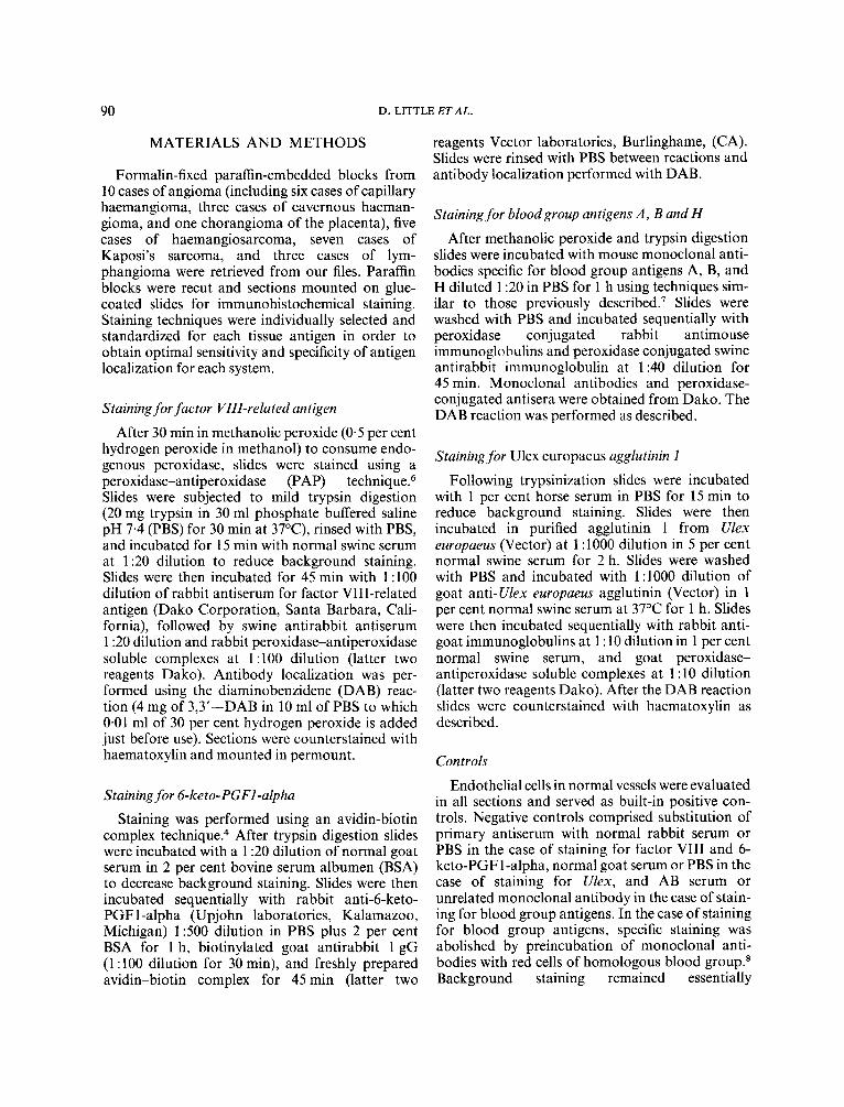
JOURNAL OF PATHOLOGY, VOL. 149: 89-95 (1986) ENDOTHELIAL CELL MARKERS IN VASCULAR NEOPLASMS : A N IMMUNOHISTOCHEMICAL ANTIGEN, BLOOD GROUP SPECIFIC EUROPAEUS 1 LECTIN STUDY COMPARING FACTOR VIII-RELATED ANTIGENS, 6-KETO-PGF1 ALPHA, AND ULEX DANIEL LITTLE JONATHAN W. SAID ROBERT J SIEGEL" MICHAEL FEALY AND MICHAEL C. FISHBEIN Divisions of Anatomic Pathology and Cardiology*, Cedars-Sinai Medical Center, 8700 Beverly Blvd., Los Angeles, CA. 90048, USA Received 2 September 1985 Accepted 20 January 1986 SUMMARY Markers for endothelial cells including Ulex europaeus 1 lectin, blood group A, B, and H, and the prostaglandin metabolite 6-keto-PGF1 alpha were evaluated in paraffin sections from formalin-fixedbenign and malignant vascular neoplasms using a variety of immunohistochemical techniques, and results compared with staining for factor VIII- related antigen. Staining for Ulex appeared more sensitive than factor VIII-related antigen in identifying poorly differentiated neoplasms including haemangiosarcomas and spindle cell proliferations in Kaposi's sarcoma. Staining for blood group related antigens correlated with blood group in all cases. Ulex europaeus 1 lectin was the only marker for endothelial cells in lymphangiomas. KEY woRDs-Endothelial cell, Ulex, factor VIII-related antigen, blood group antigen, Kaposi's sarcoma, angioma, angiosarcoma. INTRODUCTTON Immunohistochemical staining for factor VITI- related antigen has been suggested as a useful marker for endothelial cells in paraffin Variable staining for factor VIII in vascular neoplasms has led to questions regarding the sen- sitivity and specificity of the r e a ~ t i o n . ~ Staining for This work was supported in part by NIH SCOR in Ischemic Heart Disease Grant #HI 765 1 Addressee for correspondence: Michael C. Fishbein, Depart- ment of Pathology, Cedars-Sinai Medical Center, 8700 Beverly Blvd., Los Angeles, CA 90048, U.S.A. 0022-341 7/86/06008947%05.00 0 1986 by John Wiley & Sons, Ltd. factor VIII is generally strongest in small vessels ;2 poorly differentiated vascular tumours including angiosarcomas and proliferating spindle cells in Kaposi's sarcoma are frequently unstained.' Prob- lems with antibody specificity and cross reaction of factor VIII-related antibody with squamous cell carcinomas and cells with abundant cytoplasmic filaments have also been r e p ~ r t e d . ~ The aim of this study was to evaluate factor VIIT-related antigen in association with other endothelial cell markers including blood groups antigens, Ulex europaeus 1 lectin, and the prostaglandin metabolite 6-keto- PGF I -al~ha~,~ as immunohistochemical markers for vascular neoplasms.
-
Upload
daniel-little -
Category
Documents
-
view
213 -
download
1
Transcript of Endothelial cell markers in vascular neoplasms: An immunohistochemical study comparing factor...
JOURNAL OF PATHOLOGY, VOL. 149: 89-95 (1986)
ENDOTHELIAL CELL MARKERS IN VASCULAR NEOPLASMS : A N IMMUNOHISTOCHEMICAL
ANTIGEN, BLOOD GROUP SPECIFIC
EUROPAEUS 1 LECTIN
STUDY COMPARING FACTOR VIII-RELATED
ANTIGENS, 6-KETO-PGF1 ALPHA, AND ULEX
DANIEL LITTLE JONATHAN W. SAID ROBERT J SIEGEL"
MICHAEL FEALY AND
MICHAEL C. FISHBEIN
Divisions of Anatomic Pathology and Cardiology*, Cedars-Sinai Medical Center, 8700 Beverly Blvd., Los Angeles, CA. 90048, USA
Received 2 September 1985 Accepted 20 January 1986
SUMMARY Markers for endothelial cells including Ulex europaeus 1 lectin, blood group A, B, and H, and the prostaglandin
metabolite 6-keto-PGF1 alpha were evaluated in paraffin sections from formalin-fixed benign and malignant vascular neoplasms using a variety of immunohistochemical techniques, and results compared with staining for factor VIII- related antigen. Staining for Ulex appeared more sensitive than factor VIII-related antigen in identifying poorly differentiated neoplasms including haemangiosarcomas and spindle cell proliferations in Kaposi's sarcoma. Staining for blood group related antigens correlated with blood group in all cases. Ulex europaeus 1 lectin was the only marker for endothelial cells in lymphangiomas. KEY woRDs-Endothelial cell, Ulex, factor VIII-related antigen, blood group antigen, Kaposi's sarcoma, angioma, angiosarcoma.
INTRODUCTTON
Immunohistochemical staining for factor VITI- related antigen has been suggested as a useful marker for endothelial cells in paraffin Variable staining for factor VIII in vascular neoplasms has led to questions regarding the sen- sitivity and specificity of the r e a ~ t i o n . ~ Staining for
This work was supported in part by NIH SCOR in Ischemic Heart Disease Grant #HI 765 1
Addressee for correspondence: Michael C. Fishbein, Depart- ment of Pathology, Cedars-Sinai Medical Center, 8700 Beverly Blvd., Los Angeles, CA 90048, U.S.A.
0022-341 7/86/06008947%05.00 0 1986 by John Wiley & Sons, Ltd.
factor VIII is generally strongest in small vessels ;2
poorly differentiated vascular tumours including angiosarcomas and proliferating spindle cells in Kaposi's sarcoma are frequently unstained.' Prob- lems with antibody specificity and cross reaction of factor VIII-related antibody with squamous cell carcinomas and cells with abundant cytoplasmic filaments have also been r e p ~ r t e d . ~ The aim of this study was to evaluate factor VIIT-related antigen in association with other endothelial cell markers including blood groups antigens, Ulex europaeus 1 lectin, and the prostaglandin metabolite 6-keto- PGF I - a l ~ h a ~ , ~ as immunohistochemical markers for vascular neoplasms.
90 D. LITTLE ET AL.
MATERIALS AND METHODS
Formalin-fixed paraffin-embedded blocks from 10 cases of angioma (including six cases of capillary haemangioma, three cases of cavernous haeman- gioma, and one chorangioma of the placenta), five cases of haemangiosarcoma, seven cases of Kaposi's sarcoma, and three cases of lym- phangioma were retrieved from our files. Paraffin blocks were recut and sections mounted on glue- coated slides for immunohistochemical staining. Staining techniques were individually selected and standardized for each tissue antigen in order to obtain optimal sensitivity and specificity of antigen localization for each system.
Staining for factor VIII-related antigen
After 30 min in methanolic peroxide (0.5 per cent hydrogen peroxide in methanol) to consume endo- genous peroxidase, slides were stained using a peroxidase-antiperoxidase (PAP) technique.6 Slides were subjected to mild trypsin digestion (20 mg trypsin in 30 ml phosphate buffered saline pH 7.4 (PBS) for 30 rnin at 37"C), rinsed with PBS, and incubated for 15 min with normal swine serum at 1 :20 dilution to reduce background staining. Slides were then incubated for 45 min with 1 :lo0 dilution of rabbit antiserum for factor VIII-related antigen (Dako Corporation, Santa Barbara, Cali- fornia), followed by swine antirabbit antiserum 1 :20 dilution and rabbit peroxidase-antiperoxidase soluble complexes at 1 : 100 dilution (latter two reagents Dako). Antibody localization was per- formed using the diaminobenzidene (DAB) reac- tion (4 mg of 3,3'-DAB in 10 ml of PBS to which 0.01 ml of 30 per cent hydrogen peroxide is added just before use). Sections were counterstained with haematoxylin and mounted in permount.
Staining for 6-keto-PGFl-alpha
Staining was performed using an avidin-biotin complex technique? After trypsin digestion slides were incubated with a 1 :20 dilution of normal goat serum in 2 per cent bovine serum albumen (BSA) to decrease background staining. Slides were then incubated sequentially with rabbit anti-6-keto- PGFl -alpha (Upjohn laboratories, Kalamazoo, Michigan) 1500 dilution in PBS plus 2 per cent BSA for 1 h, biotinylated goat antirabbit 1 gG (1 :lo0 dilution for 30 min), and freshly prepared avidin-biotin complex for 45 min (latter two
reagents Vector laboratories, Burlinghame, (CA). Slides were rinsed with PBS between reactions and antibody localization performed with DAB.
Staining for bloodgroup antigens A , B and H
After methanolic peroxide and trypsin digestion slides were incubated with mouse monoclonal anti- bodies specific for blood group antigens A, B, and H diluted 1 :20 in PBS for 1 h using techniques sim- ilar to those previously de~cribed.~ Slides were washed with PBS and incubated sequentially with peroxidase conjugated rabbit antimouse immunoglobulins and peroxidase conjugated swine antirabbit immunoglobulin at 1 :40 dilution for 45 min. Monoclonal antibodies and peroxidase- conjugated antisera were obtained from Dako. The DAB reaction was performed as described.
Staining for Ulex europaeus agglutinin 1
Following trypsinization slides were incubated with 1 per cent horse serum in PBS for 15 rnin to reduce background staining. Slides were then incubated in purified agglutinin 1 from Ulex europaeus (Vector) at 1 : 1000 dilution in 5 per cent normal swine serum for 2 h. Slides were washed with PBS and incubated with 1 :I000 dilution of goat anti-Ulex europaeus agglutinin (Vector) in 1 per cent normal swine serum at 37°C for 1 h. Slides were then incubated sequentially with rabbit anti- goat immunoglobulins at 1 : 10 dilution in 1 per cent normal swine serum, and goat peroxidase- antiperoxidase soluble complexes at 1 : 10 dilution (latter two reagents Dako). After the DAB reaction slides were counterstained with haematoxylin as described.
Controls
Endothelial cells in normal vessels were evaluated in all sections and served as built-in positive con- trols. Negative controls comprised substitution of primary antiserum with normal rabbit serum or PBS in the case of staining for factor VIII and 6- keto-PGFl-alpha, normal goat serum or PBS in the case of staining for Ulex, and AB serum or unrelated monoclonal antibody in the case of stain- ing for blood group antigens. In the case of staining for blood group antigens, specific staining was abolished by preincubation of monoclonal anti- bodies with red cells of homologous blood group.8 Background staining remained essentially
ENDOTHELIAL CELL MARKERS IN VASCULAR NEOPLASMS 91
unchanged indicating non-specific attachment of antibody.
Grading system Sections were graded with regard to intensity of
specific staining, background staining, and artifac- tual staining on test slides and negative control slides. Intensity levels were labelled as 0-3 + : 1 + staining was faint, but diffuse, with a slight brown tint to the tissue; staining that was 3 + was as dark as the most intense staining observed, and 2 + stain- ing was intermediate. Barely detectable, focal stain- ing was assigned a grade of 0. Background staining, which could be non-specific due to diffusion of the antigen, refers to staining of tissues not known to contain the antigen to be localized. Non-specific staining refers to staining seen in negative controls in which the primary antiserum has been deleted.
RESULTS
Staining for factor VIII-related antigen (Table I ) (Fig. I )
The majority of cases of angiomas, endothelial cell sarcomas, and Kaposi’s sarcomas showed 2- 3 + specific staining of endothelial cell cytoplasm
with only two cases with 0 staining. In cases of Kaposi’s sarcoma both endothelial cells and proli- ferating spindle cells were stained, although in the latter staining was frequently focal and weak. Stain- ing was strongest in non-neoplastic endothelial cells of small capillaries, arterioles and veins, with less intense staining in neoplastic endothelial cells par- ticularly in poorly differentiated neoplasms. In cases of lymphangioma, endothelial cells of lymph channels showed no staining. In all cases negative controls ranged from 0-1 + . Background staining was usually absent or minimal with only one case staining 3 + and one staining 2 + .
Staining for 6-keto-PGFl-alpha (Table I )
All cases were negative for 6-keto-PGFl-alpha with the exception of weak (1 +) staining in one case of haemangiosarcoma. Neither normal nor neoplastic endothelial cells stained. Negative con- trols were graded 0 in all cases. Background staining was usually absent or mild.
Staining for blood group antigens A , B and H (Table I ) (Fig. 1)
In all cases positive staining for blood group anti- gens correlated with the blood group of the patient.
Table I-Immunohistochemical staining of vascular tumours
Factor VIII 6-Keto-PGF I-alpha Blood group Ulex
Angioma 3+ xxxx xxxxxxxxx xxxxxxxxxx 2+ xxxx
X I + 0 @ xxxxxxxxx
Sarcoma 3 + xxxx 2+ x 1 + X 0 xxxx
X xxxxx xx xx
Kaposi’s sarcoma 3 + xxxx xxxx xxxxxx 2+ xx xxx X
1 + 0 X xxxxxxx
Lymphangioma 3 + xxx 2+ X 1+ 0 xxx @ xx xx
Intensity of staining graded 0-3 + . x = a case, @ =case with 3 + background.
92 D. LITTLE ETAL.
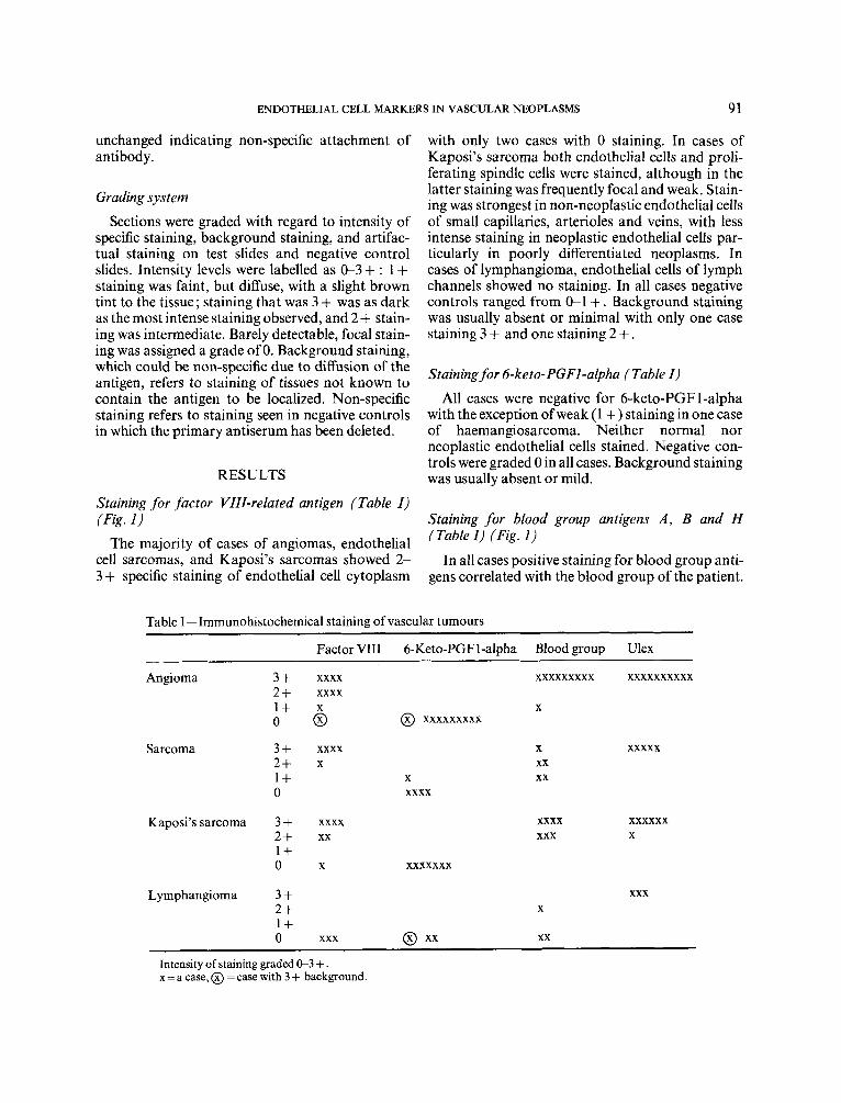
Fig. 1-Examples of staining for factor VIII-related antigen: (a) benign haemangioma of the skin; (b) low grade angiosarcoma of the breast, and (c) Kaposi’s sarcoma of skin. Neoplasms stained for blood group antigens are shown in (d) haemangioma, (e) angiosarcoma and ( f ) Kaposi’s sarcoma. Peroxidase stain, original magnification x 100. Insets in c and f show staining of spindle cells in Kaposi’s sarcoma cases (arrows). Original magnification x 252
The majority of cases demonstrated specific stain- or no specific staining. In Kaposi’s sarcoma, spindle ing of endothelial cells which was predominantly 2- cells stained less intensely than endothelial cells of 3 + (Fig. 1). The exceptions were one case of recognizable vascular channels. Background stain- angioma, two cases of haemangiosarcoma, and all ing was mainly 0-1 +, with three cases staining 2+ . three cases of lymphangioma which showed weak The negative controls were graded 0 in all cases.
ENDOTHELIAL CELL MARKERS IN VASCULAR NEOPLASMS 93
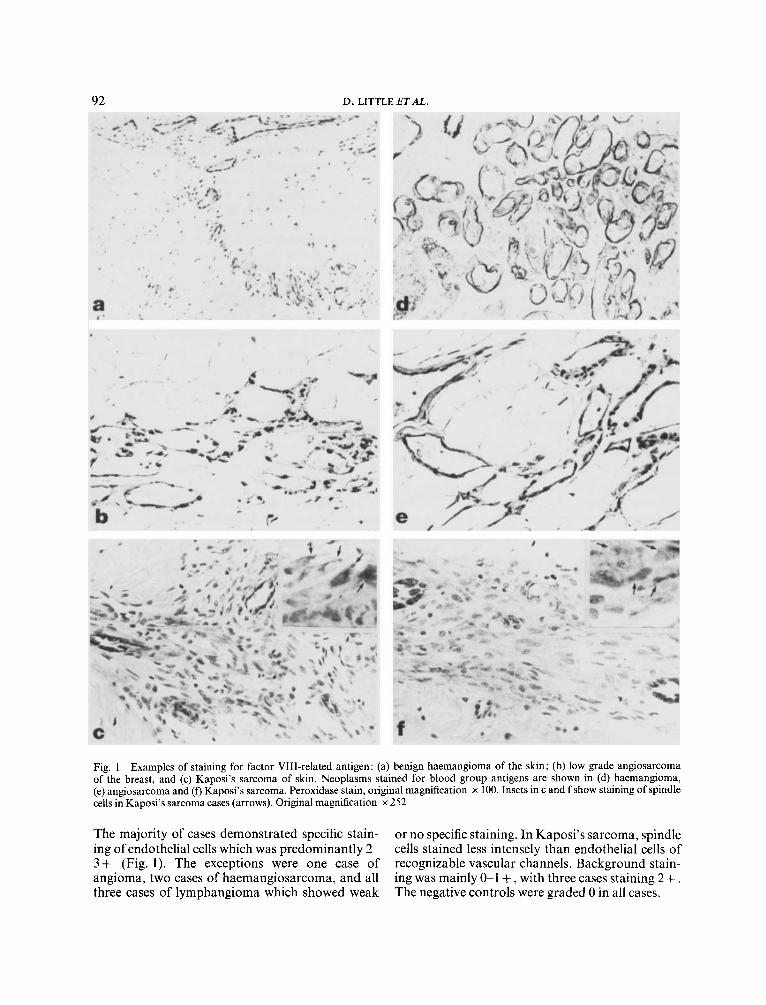
Fig. 2-Examples of staining for Ulex europaeus agglutinin 1 : (a) benign haemangioma; (b) lymphangioma; (c) low-grade angio- sarcoma, and (d) Kaposi's sarcoma. Note the granular staining of spindle cells in Kaposi's sarcoma (arrows) (d). Peroxidase stain, original magnification x 100 (a, b, c), x 160 (d)
Staining for Ulex europaeus agglutinin 1 (Table I ) (Fig. 2)
Strong granular staining of endothelial cells was seen in all angiomas and malignant vascular neoplasms. Staining was finely granular and limited to cell cytoplasm. Three cases of lymphangioma also stained strongly. Spindle cells as well as recognizable endothelial cells both stained in cases of Kaposi's sarcoma. Background staining and staining of negative controls ranged from Gl + .
DISCUSSION
Factor VIII-related antigen has been established as a marker of vascular endothelial cells in paraffin sections.lJ However, when applied as a marker for vascular tumours staining may be weak or absent, particularly in poorly differentiated neoplasms such
as angiosarcomas and the proliferating spindle cells of Kaposi's sarcoma.' Our results of staining for factor VIII in a variety of vascular neoplasms are similar to those of Burgdorf et al.' who found strongest staining in endothelial cells of small nor- mal cutaneous vessels. Weak staining for factor VIII may be due to differences in the amount of fac- tor VIII produced by different endothelia2s9 or may relate to the inability of dedifferentiated neoplastic endothelium to synthesize the antigen. In addition to lack of sensitivity when staining vascular tumours for factor VIII, cross reactivity with other antigens has been reported: with inappropriate staining of cells containing abundant cytoplasmic filaments including squamous and renal cell car- cinomas. This study was undertaken in order to evaluate other endothelial cell related antigens, namely the prostaglandin metabolite 6-keto-PGF 1 alpha, blood group related antigens and Ulex
94 D. LITTLE ETAL.
europaeus agglutinin 1 lectin as endothelial cell markers in vascular tumours.
Staining for Ulex europaeus agglutinin 1 was strongly positive in all neoplasms studied, and included poorly differentiated endothelial cell sarcomas, spindle cells in Kaposi's sarcoma, and lymphangiomas. These results are similar to those described by Holthofer et a1.9 and Miettinen et ~ 1 . ' ~ using immunofluorescence on frozen and paraffin- embedded tissues, and by Ordonez and Batsakis' using an immunoperoxidase technique. Staining was also noted in epithelial cells including epidermis as previously de~cribed.~ Staining for Ulex was the only marker for endothelial cells in cases of lym- phangioma. Beckstead et a1.'* also found normal lymphatic endothelial cells to be negative for vascular endothelial markers, with the exception of Ulex europaeus 1 lectin. These authors found spindle cells in Kaposi's sarcoma to be positive for Ulex lectin and devoid of factor VIII-related anti- gen and thus suggested that Kaposi's sarcoma may originate from lymphatic and not blood vessel endothelium. Our studies did detect staining for fac- tor VIII-related antigen as well as blood group anti- gens in some spindle cells from Kaposi's sarcoma, in addition to binding of Ulex lectin. Our findings are in accordance with those of other authorsl1J3J4 who describe staining for factor VIII in Kaposi's sarcoma cells. Other investigators have been unable to detect this antigen.'JO These inconsistent staining results may be due to differences in sensitivity of the reaction in paraffin sections, and/or the use of trypsin digestion in our studies, to enhance staining by exposure of antigenic sites.15 Also, the plastic embedding technique utilized by Beckstead et a1.I2 may be insensitive to small amounts of factor VIII- related antigen, as the authors suggest.
Also useful as endothelial markers in paraffin sec- tions were monoclonal antibodies directed at blood group antigens.16J7 These results are in accordance with those of Feigl et all8 who stained paraffin sec- tions of benign and malignant vascular tumours using the less sensitive mixed cell agglutination reac- tion.16 In all cases specific staining corresponded to the blood group of the patient. Correlation between positive staining and blood type raises the possi- bility that this technique may be useful in identify- ing the source of unmarked tissue samples, and in forensic applications. Weak or absence of staining in some cases of angiosarcoma and Kaposi's sarcoma however suggest that blood group antigens may occasionally be lost with neoplastic transform- ation. Similar loss of blood group antigens has
been described in neoplasms from other t i s s ~ e s . ~ J ~ , ~ ~
The prostaglandin metabolite 6-keto-PGF1- alpha has been localized in the intimal endothelial cells of canine coronary arteries and veins and human umbilical cord vessels using immuno- histochemical techniques on freshly harvested, formalin-fixed, paraffin-embedded In this study of human vascular tumours weak staining was found in only one case of haemangiosarcoma. All other neoplastic and normal endothelial cells failed to stain. This lack of staining may relate to low levels of 6-keto-PGFl-alpha in the tissues, or loss of antigen due to delay in fixation and processing.
In summary, factor VIII-related antigen, blood group antigens, and Ulex europaeus agglutinin 1 are variably effective as markers for endothelial cells in vascular neoplasms. Staining for Ulex appears to be the most sensitive reaction for identifying endo- thelial cell proliferations in paraffin embedded tissues.
REFERENCES
1. Burgdorf WHC, Mukai K, Rosai J. Immuno- histochemical identification of factor VIII-related antigen in endothelial cells of cutaneous lesions of alleged vascular nature. Am J Clin Path01 1981 ; 75: 1 67-1 7 1 .
2. Mukai K, Rosai J, Burgdorf WHC. Localization of factor VIII-related antigen in vascular endothelial cells using an immunoperoxidase method. Am J Clin
3. Wilson AJ. Factor VIII-related antigen staining by immunoperoxidase technic in smaller laboratories : a potential problem. Am J Clin Path01 1984; 81:
4. Harold JG, Siegel RJ, Edwalds GM, Satoh P, Fish- bein MC. Immunohistochemical localization of 6- keto-PGF 1 -alpha in canine coronary vasculature. Prostaglandins 1985; 29: 19-23.
5. Harold JG, Siegel RJ, Edwalds GM, Satoh P, Fish- bein MC. Immunohistochemical localization of pro- staglandin metabolites in canine and human vasculature. Circulation 1984; 70: 71.
6. Pinkus GS, Said JW. Specific identification of intra- cellular immunoglobulin in paraffin sections of multiple myeloma and macroglobulinemia using an immunoperoxidase technique. Am JPath011977; 87:
7. Pinkus GS, Said JW. Hodgkin's disease, lymphocyte predominance type, nodular-a distinct entity? Am J
8. Lee AK, DeLellis RA, Rosen P et al. ABH blood group isoantigen expression in breast carcinomas-
Path01 1980; 4: 273-276.
117-120.
47-58.
Path01 1985; 118: 1-6.
ENDOTHELIAL CELL MARKERS IN VASCULAR NEOPLASMS 95
an immunohistochemical evaluation using monoclonal antibodies. Am J Clin Pathol 1985; 83:
9. Holthofer H, Virtanen I, Kariniemi A-L, Hormia M, Linder E, Miettinen A. Ulex europaeus 1 lectin as a marker for vascular endothelium in human tissues. Lab Invest 1982; 47: 60-66.
10. Miettinen M, Holthofer H, Lehto V-P, Miettinen A, Virtanen I . Ulex europaeus 1 lectin as a marker for tumors derived from endothelial cells. Am J Clin
11. Ordonez NG, Batsakis JG. Comparison of Ulex europaeus 1 lectin and factor VIII-related antigen in vascular lesions. Arch Pathol Lab Med 1984; 108:
12. Beckstead JH, Wood GS, Fletcher V. Evidence for the origin of Kaposi’s sarcoma from lymphatic endo- thelium. Am J Patholl985; 119: 294-300.
13. Guarda LG, Silva EG, Ordonex NG, Smith JL Jr. Factor VIII in Kaposi’s sarcoma. Am J Clin Pathol
14. Nadji M, Morales AR, Ziegles-Weissman J, Penneys NS. Kaposi’s sarcoma. Immunohistologic evidence for an endothelial origin. Arch Pathol Lab Med 198 1 ; 105: 274-275.
308-3 19.
Path01 1983; 79: 32-36.
129-132.
1981 ; 76: 197-200.
15. Huang S-N, Minassian H, More JD. Application of immunofluorescent staining on paraffin sections improved by trypsin digestion. Lab Invest 1976; 35:
16. Coon JS, Weinstein RS. Detection of ABH tissue isoantigens by immunoperoxidase methods in nor- mal and neoplastic urothelium. Comparison with the erythrocyte adherence method. Am J Clin Pathol
17. Ernst C, Thurin J, Atkinson B etal. Monoclonal antibody localization of A and B isoantigens in nor- mal and malignant fixed human tissues. Am J Pathol 1984; 117: 451-461.
18. Feigl W, Denk H, Davidovits A, Holzner JH. Blood group isoantigens in human benign and malignant vascular tumors. Virchows Arch A [Pathol Anat Histol] 1976; 370: 323-332.
19. Limas C, Lange P, Fraley EE, Vessella RC. A, B, H, antigens in transitional cell tumors of the urinary bladder. Correlation with clinical course. Cancer
20. Walker PD, Karnik S, DeKernion JB, Pramberg JC. Cell surface blood group antigens in prostatic car- cinoma. Am J Clin Patholl984; 81 : 503-506.
383-390.
1981; 76: 163-171.
1979; 44: 2099-2107.