Endotélio Arterial e Aterotrombogénese I - Intervenção do ... · leucocitárias, inibe a...
23
Endotélio Arterial e Aterotrombogénese I - Intervenção do endotélio intacto na homeostasia vascular e sanguínea [80] J. MARTINS E SILVA, CARLOTA SALDANHA Unidade de Biopatologia Vascular do Instituto de Medicina Molecular e Instituto de Biopatologia Química da Faculdade de Medicina, Universidade de Lisboa, Lisboa, Portugal Rev Port Cardiol 2006; 25 (11): 1061-1083 1061 RESUMO O endotélio normal constitui uma barreira física e biológica entre o sangue e a parede vascular, que actua também como sensor e transductor de diversos factores endógenos e exógenos influentes na modulação da circulação sanguínea. A função endotelial em dado indivíduo e momento reflecte o equilíbrio entre factores de risco cardiovascular, predisposição genética e mecanismos vasculo-protectores. Nestes mecanismos tem acção predominante a disponibilidade e actividade do monóxido de óxido (NO) derivado do endotélio. Adicionalmente, outras substâncias vasoactivas sintetizadas na parede vascular e/ou pelos elementos celulares do sangue também afectam o comportamento da interface sangue- endotélio. A vasomotricidade depende do equilíbrio entre substâncias vasodilatoras (em que se destaca a prostaciclina) e vasoconstritores (principalmente a endotelina-1 e, também, a angiotensina II). O equilíbrio coagulação-anticoagulação/fibrinólise é também afectado por diversos tipos de proteínas específicas. São analisados os principais mecanismos de acção dos factores indicados, de que depende a fluidez sanguínea em condições normais e na presença de endotélio intacto. É feita referência a potenciais anomalias, a analisar na parte II desta revisão. Palavras-Chave Endotélio; Monóxido de azoto; Endotelina I, Prostaciclina; Vasomotricidade; Coagulação; Fibrinólise ABSTRACT Arterial Endothelium and Atherothrombogenesis I - Intact endothelium in vascular and blood homeostasis Normal endothelium constitutes a physical and biological barrier between the blood and the vascular wall, and also acts as a sensor and transducer of various endogenous and exogenous factors that modulate the blood circulation. Endothelial activity in a given individual at any particular moment reflects the balance between cardiovascular risk factors, genetic predisposition and vascular protection mechanisms. The availability and activity of endothelium-derived nitric oxide (NO) is a major factor in these mechanisms. Further, vasoactive substances synthesized by the vascular wall and/or by blood cells may affect the behavior of the blood-endothelium interface. Vasomotricity is dependent on the balance between vasodilator substances (particularly prostacylin) and vasoconstrictor products (mainly endothelin-1 and angiotensin II). The coagulation-anticoagulation or fibrinolysis balance is also affected by various different proteins. The mechanisms of these factors, on which blood fluidity depends under normal conditions and with intact endothelium, are discussed, along with mention of potential abnormalities, which will be examined in the second part of this review. Key words Endothelium; Nitric oxide; Endothelin-1; Prostacyclin; Vasomotricity; Coagulation; Fibrinolysis Recebido para publicação: Setembro de 2006 • Aceite para publicação: Outubro de 2006 Received for publication: September 2006 • Accepted for publication: October 2006
Transcript of Endotélio Arterial e Aterotrombogénese I - Intervenção do ... · leucocitárias, inibe a...

Endotélio Arterial e Aterotrombogénese I - Intervenção do endotélio intacto na homeostasia vascular e sanguínea [80]
J. MARTINS E SILVA, CARLOTA SALDANHA
Unidade de Biopatologia Vascular do Instituto de Medicina Molecular eInstituto de Biopatologia Química da Faculdade de Medicina, Universidade de Lisboa, Lisboa, Portugal
Rev Port Cardiol 2006; 25 (11): 1061-1083
1061
RESUMO
O endotélio normal constitui uma barreirafísica e biológica entre o sangue e a paredevascular, que actua também como sensor e
transductor de diversos factores endógenos eexógenos influentes na modulação da
circulação sanguínea. A função endotelial emdado indivíduo e momento reflecte o equilíbrio
entre factores de risco cardiovascular,predisposição genética e mecanismos
vasculo-protectores. Nestes mecanismos temacção predominante a disponibilidade e
actividade do monóxido de óxido (NO)derivado do endotélio.
Adicionalmente, outras substâncias vasoactivassintetizadas na parede vascular e/ou pelos
elementos celulares do sangue também afectamo comportamento da interface sangue-
endotélio. A vasomotricidade depende doequilíbrio entre substâncias vasodilatoras (em
que se destaca a prostaciclina) evasoconstritores (principalmente a endotelina-1
e, também, a angiotensina II). O equilíbriocoagulação-anticoagulação/fibrinólise étambém afectado por diversos tipos de
proteínas específicas. São analisados osprincipais mecanismos de acção dos factores
indicados, de que depende a fluidez sanguíneaem condições normais e na presença de
endotélio intacto. É feita referência apotenciais anomalias, a analisar na parte II
desta revisão.
Palavras-ChaveEndotélio; Monóxido de azoto; Endotelina I, Prostaciclina;
Vasomotricidade; Coagulação; Fibrinólise
ABSTRACT
Arterial Endothelium andAtherothrombogenesisI - Intact endothelium in vascular and bloodhomeostasis
Normal endothelium constitutes a physical andbiological barrier between the blood and thevascular wall, and also acts as a sensor andtransducer of various endogenous andexogenous factors that modulate the bloodcirculation. Endothelial activity in a givenindividual at any particular moment reflects thebalance between cardiovascular risk factors,genetic predisposition and vascular protectionmechanisms. The availability and activity ofendothelium-derived nitric oxide (NO) is amajor factor in these mechanisms.Further, vasoactive substances synthesized bythe vascular wall and/or by blood cells mayaffect the behavior of the blood-endotheliuminterface. Vasomotricity is dependent on thebalance between vasodilator substances(particularly prostacylin) and vasoconstrictorproducts (mainly endothelin-1 and angiotensinII). The coagulation-anticoagulation orfibrinolysis balance is also affected by variousdifferent proteins. The mechanisms of thesefactors, on which blood fluidity depends undernormal conditions and with intact endothelium,are discussed, along with mention of potentialabnormalities, which will be examined in thesecond part of this review.
Key wordsEndothelium; Nitric oxide; Endothelin-1; Prostacyclin;Vasomotricity; Coagulation; Fibrinolysis
Recebido para publicação: Setembro de 2006 • Aceite para publicação: Outubro de 2006Received for publication: September 2006 • Accepted for publication: October 2006

INTRODUÇÃO
Em editorial de 1995, no influente AmericanJournal of Cardiology, Franck Sacks (1)
manifestava a sua perplexidade por a cardiopatiaisquémica continuar a aumentar, não obstanteverificar-se franca melhoria nos valores dacolesterolemia total e da LDL-colesterol, assimcomo no controlo de outros reconhecidosfactores de risco cardiovascular.
Aquela observação sugeria duas possíveisexplicações genéricas para o aumento da
INTRODUCTION
In a 1995 editorial in the American Journal ofCardiology, Franck Sacks (1) questioned why the
incidence of ischemic heart disease was stillincreasing even though total and LDL cholesterollevels were clearly improving, as was control ofother known cardiovascular risk factors.
This fact suggested two possible explanationsfor the continuing increase in atheroscleroticcoronary disease: either the known risk factorswere still having harmful effects, despite having1062
Rev Port CardiolVol. 25 Novembro 06 / November 06
Abreviaturas (ou fórmulas químicas) utilizadasno texto (Nomenclatura anglo-saxónica):
• ACE: Angiotensin-converting enzyme• ADMA: Asymmetric dimethyl arginine• ADP: Adenosine 5'-diphophate• AMPc: Cyclic adenosine 3',5'- monophosphate• Ang II: Angiotensin II• AT-1: Angiotensin II type (receptors)• BH4: Tetrabiopterine• Ca2+: Calcium• CAT-1: cationic amino acid
transporter 1 (sistema y+)• ECE - Endothelin converting enzyme (type 1)• EDHF: Endothelium derived
hyperpolarizing factor• EDRF: Endothelium derived relaxing factor• ET-(1, 2 ou 3) (s): Endothelin-(1, 2 ou 3)• ETA, ETB: Endothelin receptor (type A or B)• FAD: Flavin adenine dinucleotide• FMN: Flavin mononucleotide• GMPc: Cyclic guanosine 3',5'-monophosphate• LDL (cholesterol) - Low density lipoproteins• NADP(H): Nicotinamide adenine dinucleotide
phosphate (reduced)• NO: Nitric oxide (monóxido de azoto);• N-OH arginine: N-hidroxil-L-arginine• NOS: Nitric oxide synthase (eNOS:
endothelial; nNOS: neuronal; iNOS: induced);• PAI-1: Plasminogen activator inhibitor-1• PDGF: Platelet derived growth factor;• PGH2: Prostaglandin H2• PGI2: Prostacyclin (Prostaglandin I2)• RNAm: Ribonucleic acid (messenger)• tPA: Tissue plasminogen activator• TXA2: Thromboxan A2• VEGF: Vascular endothelial growth factor
Abbreviations used in the text:
• ACE: Angiotensin-converting enzyme• ADMA: Asymmetric dimethylarginine• ADP: Adenosine 5'-diphosphate• Ang II: Angiotensin II• AT-1: Angiotensin II type 1(receptors)• BH4: Tetrahydrobiopterin• Ca2+: Calcium• cAMP: Cyclic adenosine
3',5'- monophosphate• CAT-1: cationic amino acid transporter 1
(y+ system)• cGMP: Cyclic guanosine 3',5'-monophosphate• ECE: Endothelin-converting enzyme (type 1)• EDHF: Endothelium-derived hyperpolarizing
factor• EDRF: Endothelium-derived relaxing factor• ET-(1, 2 or 3): Endothelin-(1, 2 or 3)• ETA, ETB: Endothelin receptor (type A or B)• FAD: Flavin adenine dinucleotide• FMN: Flavin mononucleotide• LDL (cholesterol): Low-density lipoprotein• NADP(H): Nicotinamide adenine dinucleotide
phosphate (reduced)• NO: Nitric oxide• N-OH arginine: N-hydroxyl-L-arginine• NOS: Nitric oxide synthase (eNOS:
endothelial; nNOS: neuronal; iNOS: induced)• PAI-1: Plasminogen activator inhibitor-1• PDGF: Platelet-derived growth factor• PGH2: Prostaglandin H2• PGI2: Prostacyclin (Prostaglandin I2)• mRNA: Messenger ribonucleic acid• tPA: Tissue plasminogen activator• TXA2: Thromboxane A2• VEGF: Vascular endothelial growth factor

coronariopatia aterosclerótica: (a) ou os factoresde risco identificados continuam a exercerefeitos deletérios apesar da sua aparentediminuição ou controlo ou, em alternativa, (b)existem outros mecanismos ou factores de riscoa serem valorizados ou a identificar no contexto.Aparentemente, ambas as hipóteses parecemcoexistir.
Por sua vez, o cálculo de risco cardiovascularpelo Framingham Coronary Risk Score não temrobustez suficiente para prever entre 30 a 40%de todos os eventos cardiovasculares, ao queacresce uma fracção importante de eventosassintomáticos que estão na posição de riscointermédio (2).
A par das observações que apoiam ainfluência da hiperlipidemia (secundária ahábitos dietéticos e ou de causa genética) comoprincipal factor patogénico determinante daslesões vasculares ateroscleróticas (3), têm sidoindicados outros resultados potencialmenterelevantes (4). Designadamente, tem sidodemonstrado que os factores de riscocardiovascular mais comuns actuam atravésda disfunção ou lesão do endotélio (5-7).
A presente revisão é subdividida em duaspartes. Nesta primeira parte será dado realceao endotélio intacto da vasculatura arterial.A estrutura e funcionalidades finais serãorelacionadas com a homeostasia vascular esanguínea, a par com indicações de causasou mecanismos potencialmente lesivos.
ESTRUTURA E FUNÇÕES GERAIS DOENDOTÉLIO
Disposição e particularidades estruturaisO sistema circulatório é delimitado
internamente por uma monocamada de célulasendoteliais que reveste todos os vasos sanguí-neos e estabelece a continuidade com oendocárdio. A superfície interna dos vasos eendocárdio apoia-se numa fina camada detecido conjuntivo, que forma a membrana basal.O endotélio e a membrana basal vascularconstituem a íntima vascular. Exceptuando oscapilares, formados quase unicamente porendotélio e alguns pericitos, todas as artériaspossuem uma camada média rica em fibraselásticas e células musculares lisas, e umacamada externa representada pelo tecido
apparently been reduced or controlled, or therewere other mechanisms or risk factors still to beidentified or recognized as such. It now appearsthat both hypotheses are true.
Furthermore, the Framingham Coronary RiskScore is not sufficiently reliable to predict30-40% of cardiovascular events, while there isalso a significant number of asymptomatic eventsthat have been categorized as intermediate risk (2).
Along with observations suggesting thathyperlipidemia secondary to diet and/or geneticpredisposition is the main factor leading toatherosclerotic vascular lesions (3), other factorshave also been shown to be potentiallyimportant (4). In particular, it has beendemonstrated that the main cardiovascularrisk factors are mediated by endothelialdysfunction or damage (5-7).
This review is divided into two parts, the firstof which focuses on intact endothelium of thearterial vasculature. Its structure and functionswill be described in relation to vascular and bloodhomeostasis, together with some indications ofpossible causes and mechanisms of damage.
GENERAL STRUCTURE ANDFUNCTIONS OF THE ENDOTHELIUM
Disposition and structureThe circulatory system is bounded internally
by a monolayer of endothelial cells that linesall blood vessels and the endocardium. Theinternal surface adheres to a thin layer ofconnective tissue, the basement membrane. Theendothelium and the basement membrane makeup the tunica intima of the vessel. With theexception of capillaries, which are composedalmost entirely of endothelium and somepericytes, all arteries also have a tunica mediarich in elastic fibers and smooth muscle cells andan external layer, the adventitia, consisting ofloose connective tissue.
Given that the vasa vasorum, which cross theadventitia and irrigate the media, also have alining of endothelium, it could be said that themedia, the middle layer of arteries and veins, liesbetween two active endothelial surfaces, thelumen and the vasa vasorum (8). It can thus beconcluded that the nourishment and oxygenationof the vessel wall depend on substances ofluminal and contraluminal origin (9). 1063
J. MARTINS E SILVA, CARLOTA SALDANHA
Rev Port Cardiol 2006; 25: 1061-1083
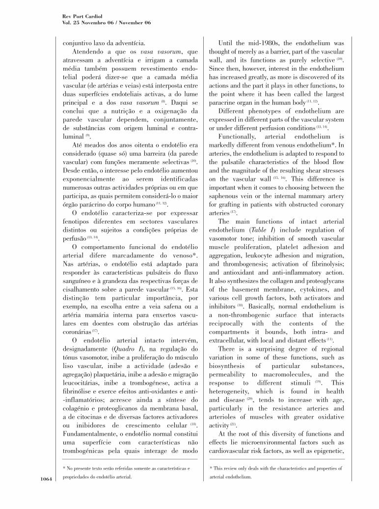
conjuntivo laxo da adventícia. Atendendo a que os vasa vasorum, que
atravessam a adventícia e irrigam a camadamédia também possuem revestimento endo-telial poderá dizer-se que a camada médiavascular (de artérias e veias) está interposta entreduas superfícies endoteliais activas, a do lumeprincipal e a dos vasa vasorum (8). Daqui seconclui que a nutrição e a oxigenação daparede vascular dependem, conjuntamente,de substâncias com origem luminal e contra-luminal (9).
Até meados dos anos oitenta o endotélio eraconsiderado (quase só) uma barreira (da paredevascular) com funções meramente selectivas (10).Desde então, o interesse pelo endotélio aumentouexponencialmente ao serem identificadasnumerosas outras actividades próprias ou em queparticipa, as quais permitem considerá-lo o maiorórgão parácrino do corpo humano (11, 12).
O endotélio caracteriza-se por expressarfenotipos diferentes em sectores vascularesdistintos ou sujeitos a condições próprias deperfusão (13, 14).
O comportamento funcional do endotélioarterial difere marcadamente do venoso*.Nas artérias, o endotélio está adaptado pararesponder às características pulsáteis do fluxosanguíneo e à grandeza das respectivas forças decisalhamento sobre a parede vascular (15, 16). Estadistinção tem particular importância, porexemplo, na escolha entre a veia safena ou aartéria mamária interna para enxertos vascu-lares em doentes com obstrução das artériascoronárias (17).
O endotélio arterial intacto intervém,designadamente (Quadro I), na regulação dotónus vasomotor, inibe a proliferação do músculoliso vascular, inibe a actividade (adesão eagregação) plaquetária, inibe a adesão e migraçãoleucocitárias, inibe a trombogénese, activa afibrinólise e exerce efeitos anti-oxidantes e anti--inflamatórios; acresce ainda a síntese docolagénio e proteoglicanos da membrana basal,a de citocinas e de diversas factores activadoresou inibidores de crescimento celular (18).Fundamentalmente, o endotélio normal constituiuma superfície com características nãotrombogénicas pela quais interage de modo
Until the mid-1980s, the endothelium wasthought of merely as a barrier, part of the vascularwall, and its functions as purely selective (10).Since then, however, interest in the endotheliumhas increased greatly, as more is discovered of itsactions and the part it plays in other functions, tothe point where it has been called the largestparacrine organ in the human body (11, 12).
Different phenotypes of endothelium areexpressed in different parts of the vascular systemor under different perfusion conditions (13, 14).
Functionally, arterial endothelium ismarkedly different from venous endothelium*. Inarteries, the endothelium is adapted to respond tothe pulsatile characteristics of the blood flowand the magnitude of the resulting shear stresseson the vascular wall (15, 16). This difference isimportant when it comes to choosing between thesaphenous vein or the internal mammary arteryfor grafting in patients with obstructed coronaryarteries (17).
The main functions of intact arterialendothelium (Table I) include regulation ofvasomotor tone; inhibition of smooth vascularmuscle proliferation, platelet adhesion andaggregation, leukocyte adhesion and migration,and thrombogenesis; activation of fibrinolysis;and antioxidant and anti-inflammatory action.It also synthesizes the collagen and proteoglycansof the basement membrane, cytokines, andvarious cell growth factors, both activators andinhibitors (18). Basically, normal endothelium isa non-thrombogenic surface that interactsreciprocally with the contents of thecompartments it bounds, both intra- andextracellular, with local and distant effects (11).
There is a surprising degree of regionalvariation in some of these functions, such asbiosynthesis of particular substances,permeability to macromolecules, and theresponse to different stimuli (19). Thisheterogeneity, which is found in healthand disease (20), tends to increase with age,particularly in the resistance arteries andarterioles of muscles with greater oxidativeactivity (21).
At the root of this diversity of functions andeffects lie microenvironmental factors such ascardiovascular risk factors, as well as epigenetic,
1064
Rev Port CardiolVol. 25 Novembro 06 / November 06
* No presente texto serão referidas somente as características e
propriedades do endotélio arterial.
* This review only deals with the characteristics and properties of
arterial endothelium.

recíproco com o conteúdo dos compartimentosque delimita (intra- e extracelulares), originandorepercussões locais e ou à distância (11).
Quadro I
Principais funções e ou efeitos do endotélio intacto enormal
É de notar a variação regional de algumasdaquelas funções, como a biossíntese desubstâncias específicas, a permeabilidade amacromoléculas e a resposta a diversos tiposde estímulo (19). Esta heterogeneidade, que severifica tanto na saúde como na doença (20), tendea acentuar-se com o envelhecimento, emparticular nas artérias de resistência e arteríolasde músculos com maior actividade oxidativa (21).
Na origem da referida diversidade endotelial(de funções e ou efeitos) intervêm factoresmicro-ambientais (p.ex., factores de riscocardiovascular), epigenéticos, bioquímicos ebiomecânicos, pelos quais as células endo-teliais detectam constantemente uma grandevariedade de sinais extracelulares, luminais(emanados do fluxo sanguíneo e seusconstituintes), provenientes da superfíciecontraluminal (e das restantes camadas daparede vascular e tecidos adjacentes) ou dosespaços inter-endoteliais (21, 22).
A transducção intracelular daqueles sinaisinclui receptores membranares, vias meta-bólicas próprias e o mecanismos de expressãogenética. O fenotipo da resposta aos estímulostraduz-se na modificação do comportamentocelular (p.ex., forma, proliferação, migração,equilíbrio vasomotor, equilíbrio hemos-tasiológico, apoptose, entre outros), que podevariar com o sector vascular afectado e o ritmo
biochemical and biomechanical elements, withthe result that the endothelium receives aconstant stream of signals, which can beextracellular, luminal (from the blood flow andits constituents), contraluminal, or from theother layers of the vessel wall and adjacenttissues, and from interendothelial spaces (21, 22).
Table I
Main functions and effects of normal intactendothelium
Intracellular transduction of these signalsinvolves membrane receptors, specific metabolicpathways and the mechanisms of geneexpression. The phenotype of the response tostimuli is reflected in modification of cellbehavior (such as shape, proliferation, migration,vasomotor balance, hemostasis, or apoptosis),which can vary according to the vascular sectoraffected and the organism's functional rhythm (22).
Aging and other pathophysiologic factorsalter endothelial morphology to a greater or lesserextent, and this affects vascular function.Age-related changes, which are independent ofknown risk factors, probably result fromchronobiologic deregulation of variousmechanisms that act on the endothelium (23).
The discovery that the endothelial surface,in both its normal and pathologic states, hasdirect effects on biochemical reactions in bloodconstituents (24), and the fact that theendothelium's functions are dynamicallymodulated by blood flow and the vascularwall (13, 25, 26), together led to a great advance inunderstanding, supporting the idea that theblood and the endothelium constitute an 1065
J. MARTINS E SILVA, CARLOTA SALDANHA
Rev Port Cardiol 2006; 25: 1061-1083
• Barreira física semipermeável.• Controlo do tónus arterial.• Efeitos anticoagulantes e profibrinolíticos.• Manutenção da fluidez sanguínea.• Inibição da adesão e agregação plaquetárias.• Inibição da adesão e migração leucocitárias.• Inibição da proliferação e migração do músculo
liso vascular.• Efeitos anti-inflamatórios.• Efeitos anti-oxidantes.• Formação e manutenção do colagénio e proteoglicanos
da membrana basal em que assenta a camada endotelial.• Formação e secreção de citocinas e de outras moléculas
inibidoras e estimuladoras do crescimento celular.
• Semipermeable physical barrier• Control of arterial tone• Anticoagulant and profibrinolytic effects• Maintenance of blood fluidity• Inhibition of platelet adhesion and aggregation• Inhibition of leukocyte adhesion and aggregation• Inhibition of vascular smooth muscle proliferation
and migration• Anti-inflammatory effects• Antioxidant effects• Formation and maintenance of collagen and
proteoglycans in the basement membrane to which the endothelium adheres
• Formation and secretion of cytokines and other molecules that inhibit or stimulate cell growth.

funcional do organismo (22).O envelhecimento e diversos factores
fisiopatológicos tendem a alterar a morfologiaendotelial de modo mais ou menos acentuado,com naturais repercussões nas funçõesvasculares. As modificações decorrentes doenvelhecimento são independentes dos factoresde risco conhecidos, resultando provavelmenteda desregulação cronobiológica de diversosmecanismos com acção no endotélio (23).
A demonstração de que a superfície endotelialintervém activamente (no estado normal oualterado) em reacções bioquímicas dosconstituintes sanguíneos (24), e o facto de que asfunções endoteliais estão sujeitas à modulaçãodinâmica emanada do fluxo de perfusão ou daparede vascular (13, 25, 26), representaram um grandeavanço conceptual, a justificar a proposta deque o sangue e o endotélio constituiriam umórgão interactivo (27). Por conseguinte, o endotélionormal actuará como indutor ou sensor deestímulos (locais e sistémicos) que possamalterar a sua funcionalidade, a da parede vasculare ou tecidos envolventes, e a dos constituintesdo sangue, a partir dos quais desencadearárespostas adaptativas que tendam a restabelecera normalidade.
No seu conjunto, a organização e disposiçãodo endotélio como interface sangue-paredevascular, assim como a sua intervenção dinâ-mica e especializada (sectorial), teriamimplicações manifestas quer no funcionamentonormal quer na resposta endotelial a estímulosfisiopatológicos (14, 20, 28).
O equilíbrio existente, em condições normais,entre os factores sintetizados pelo endotélio quemodulam a hemostasiologia (pró-trombogénicose anti-trombogénicos) e o tónus vascular(vasodilatadores e vasoconstrictores) (Quadro II)é determinante para a fluidez sanguínea eperfusão regular dos vasos (11).
Quadro II
Principais factores sintetizados no endotélio, queintervêm no tónus vascular e equilíbriohemostasiológico.
interactive organ (27). Thus, normal endotheliuminduces or senses local and systemic stimuli thatcan alter its functions and those of the vessel wall,the surrounding tissues, and blood constituents,triggering adaptive responses to restore normality.
Taken together, the organization anddisposition of the endothelium as the blood//vessel wall interface, as well as its dynamic andspecialized actions in different sectors, have clearimplications both for normal functioning andfor endothelial responses to pathophysiologicstimuli (14, 20, 28).
The balance under normal conditionsbetween endothelium-derived prothrombotic andantithrombotic factors that modulate hemostasisand vasodilators and vasoconstrictors thatregulate vascular tone (Table II) determines bloodfluidity and normal perfusion (11).
Table II
Main factors synthesized in the endothelium thataffect vascular tone and hemostasis.
Effects on hemostasisAs a result of this prothrombotic and
antithrombotic balance, the blood does notcoagulate inside vessels so long as theendothelium's integrity and function aremaintained. The lack of thrombogenicity is due tovarious factors synthesized by the vessel wall andparticularly by the endothelium. Among them areprostacyclin (PGI2), a metabolite of arachidonatefrom the endothelial cytoplasmic membrane. PGI2is a powerful inhibitor of platelet aggregation andalso has vasodilatory effects (29, 30). A significantnumber of anticoagulation mechanisms, such asthrombin-antithrombin complexes, protein C,thrombomodulin and tPA, are based in or1066
Rev Port CardiolVol. 25 Novembro 06 / November 06
Vasodilatadores
• Monóxido de azoto• Prostaciclina
Vasoconstrictores
• Endotelina-1• Angiotensina II • Factor de crescimento
derivado das plaquetas
Vasodilators
• Nitric oxide• Prostacyclin
Vasoconstrictors
• Endothelin-1• Angiotensin II • Platelet-derived
growth factor
Antithrombotic
• Prostacyclin• Thrombomodulin• Tissue plasminogen
activator • Urokinase • Heparin-type molecules
Prothrombotic
• Platelet activating factor• Tissue factor• von Willebrand factor• Plasminogen activator
inhibitor• Other coagulation factors
Efeitos na hemostasiologiaDevido aquele balanço protrombótico-
-antitrombótico o sangue não coagula no interiordos vasos desde que o endotélio permaneçaíntegro e funcional. Esta ausência detrombogenicidade resulta de diversos factoressintetizados pela parede vascular e, emespecial, pelo endotélio. Entre outros,destaca-se a prostaciclina (PGI2), que é ummetabolito derivado do araquidonato damembrana citoplásmica endotelial. A PGI2 éum potente inibidor da agregação plaquetáriae possui também acção vasodilatadora (29, 30).Uma parte importante dos mecanismos deanticoagulação (p.ex., os complexos trombina--antitrombina, proteína C, trombomodulina,tPA e outros), estão sedeados ou dependentesdo endotélio (24, 31).
Através daqueles processos o endotélioassegura que o sangue permaneça fluido e circuleem condições fisiológicas ou, em alternativa,tem a capacidade de intervir em situaçõespatológicas decorrentes com hemorragia outrombose, restabelecendo a normalidadequando possível. O mesmo tipo de respostacompensadora, mediada por transducçãomecânica endotelial, parece intervir napresença de alterações da viscosidadesanguínea e plasmática (32).
A contrabalançar os mecanismos anti-trombóticos, o endotélio sintetiza tambémfactores protrombóticos (33) além de participaractivamente nos mecanismos de coagulaçãosanguínea. Entre outros, destacam-se algunsfactores que promovem a agregação plaquetária(p.ex., factor von Willebrand, fibronectina,trombospondina) ou a coagulação (p.ex., ofactor V e o factor tecidual). Adicionalmente,o endotélio é um dos tipos celulares que sintetizao inibidor do activador do plasminogénio (PAI-1),o qual é um dos principais inibidores endógenos
dependent on the endothelium (24, 31).By means of these processes, the endothelium
is responsible for ensuring that the blood remainsfluid and circulates in physiologic conditions,and can also intervene in pathologic situationsarising from hemorrhage or thrombosis to restorenormality whenever possible. The same types ofcompensatory response mediated by endothelialmechanotransduction appear to occur in cases ofalterations in blood and plasma viscosity (32).
As a counterbalance to these antithromboticmechanisms, the endothelium also producesprothrombotic factors (33) and plays an active partin blood-clotting mechanisms. These factorsinclude those that promote platelet aggregation(such as von Willebrand factor, fibronectin, andthrombospondin) and coagulation (such as factorV and tissue factor). In addition, endothelial cellsare among those that synthesize plasminogenactivator inhibitor (PAI-1), which is one of themain endogenous inhibitors of fibrinolysis and isconsidered to be a cardiovascular risk factor (34).
Effects on vasomotricityVasomotor tone in resting animals (35) and
humans (36) depends on the partial vascularconstriction that is associated with sympatheticinnervation. Vascular smooth muscle is alsoaffected by myogenic, hormonal and metaboliccontrol mechanisms and various physical stimuli(37), but there is still debate as to whethervasodilatory innervation exists in primates (38, 39).While the presence of cholinergic sympatheticfibers has been histologically demonstrated inthe muscles of animals, this has not beenconfirmed in humans, although peripheralvasodilation has been observed following surgicalsympathectomy (40).
Vascular smooth muscle in resistance vesselsis particularly sensitive to vasodilation ofmetabolic origin induced by metabolitesproduced by the endothelium and leading toincreased compensatory local perfusion (37, 41, 42).Most of the vasodilatory response in humansattributed to sympathetic innervation may in factresult from secretion of nitric oxide (NO)stimulated by the endothelium through thelocal action of epinephrine or cholinergicmechanisms (40).
The discovery and characterization of the firstendothelial substances with vasodilatory action -endothelium-derived relaxing factor (EDRF) (38)
1067
J. MARTINS E SILVA, CARLOTA SALDANHA
Rev Port Cardiol 2006; 25: 1061-1083
Antitrombóticos
• Prostaciclina• Trombomodulina• Activador tecidual do
plasminogénio • Urocinase • Moléculas do
tipo - heparina
Protrombóticos
• Factor activador das plaquetas
• Factor tecidual• Factor von Willebrand• Inibidor do activador do
plasminogénio • Outros factores da
coagulação

da fibrinólise e, também, considerado umpotencial factor de risco cardiovascular (34).
Efeitos na vasomotricidadeO tónus vasomotor depende, em animais (35) e
humanos (36) em repouso, do estado de constricçãovascular parcial que se associa à inervaçãosimpática. Adicionalmente, o músculo lisovascular é também afectado por mecanismos decontrolo miogénico, hormonal, metabólico eestímulos físicos diversos (37), sendo controversaa existência de inervação vasodilatadora nosprimatas (38, 39). Enquanto nos músculos dosanimais foi demonstrada (histologicamente) aexistência de fibras simpáticas colinérgicascom acção vasodilatadora, o mesmo ainda nãofoi comprovado no homem, não obstanteverificar-se vasodilatação periférica pós-simpa-cectomia cirúrgica (40).
O músculo liso vascular dos vasos deresistência é particularmente sensível àvasodilatação de origem metabólica, induzida pormetabolitos sintetizados pelo endotélio, de quedepende o aumento da perfusão sanguínea localcompensadora (37, 41, 42). Eventualmente, ageneralidade da resposta vasodilatadoraatribuída à inervação simpática no homemresultará, na generalidade, da estimulação dasecreção de monóxido de azoto (NO) peloendotélio por acção local da epinefrina ou demecanismos colinérgicos (40).
A descoberta e caracterização das primeirassubstâncias endoteliais com acção vasodi-latadora - o factor relaxante derivado doendotélio (EDRF) (38) e o factor hiperpolarizantederivado do endotélio (EDHF) (43) - foi seguidapela identificação do vasoconstrictor endógenomais potente então conhecido, a endotelina-1(ET-1) (44).
A acetilcolina, a bradicinina e a serotoninaestão entre os activadores da vasodilatação cujaacção é mediada por receptores da membranacitoplasmática (Fig. 1) em artérias comendotélio intacto e funcionalmente normais (45). Aacetilcolina e a bradicina também exercemuma acção cardioprotectora, que simula o pré--condicionamento à isquémia e aumenta aresistência do miocárdio (46).
A acção moduladora do endotélio nosmecanismos da vasomotricidade constitui umadas grandes descobertas científicas que, comomuitas outras, se deveram ao acaso mas que
and endothelium-derived hyperpolarizing factor(EDHF) (43) - was followed by identification of themost powerful endogenous vasoconstrictor thenknown, endothelin-1 (ET-1) (44).
Acetylcholine, bradykinin and serotonin areexamples of vasodilatory activators whoseaction is mediated by receptors in thecytoplasmic membrane (Fig. 1) in arteries withintact endothelium and normal function (45).Acetylcholine and bradykinin also havecardioprotective properties, simulating ischemicpreconditioning and increasing myocardialresistance (46).
The discovery of the endothelium's role inmodulating vasomotricity, although (like manyother great scientific discoveries) the result ofchance, is still worth recalling. The powerfulvasodilator effect produced by acetylcholine invitro in preparations of rabbit thoracic aorta wasalready known and had been confirmed in othervessels. In one experiment, however, thisvasodilation was consistently replaced by intensevasoconstriction. The experiment was repeatedwith increasing concentrations, much higher thanthe levels that would have triggered vasodilation.The vasoconstriction was maintained, however,mediated by muscarinic receptors (43). Variouspossible explanations for this discrepancy wereexplored until it was shown that the loss ofvasorelaxation with acetylcholine was due to theunintentional removal of the internal surface ofthe vessel, which had been scraped away as partof the procedure for preparing the sample. Theresponse to acetylcholine was then systematicallystudied in vessels with and without endothelium.In those with intact endothelium, there wasvasorelaxation, while vasoconstriction occurredin vessels without endothelium.
Acetylcholine can thus induce relaxation orcontraction in vascular smooth muscle dependingon the presence or absence of endothelium, andon the type of signal transduction (43). Whilevasodilation is associated with NO formationfollowed by increased cGMP, vasoconstriction asinduced by serotonin, is triggered by activation ofcyclooxygenase, and subsequent production ofthromboxane A2 (TXA2) and prostaglandin H2(PGH2) (30). Binding of these two eicosanoidsto thromboxane-specific receptors triggerscontraction of vascular smooth muscle. Similarly,binding to the same type of receptors in plateletsincreases platelet adhesion and aggregation.1068
Rev Port CardiolVol. 25 Novembro 06 / November 06

continua a ser interessante recordar. Assim, erajá conhecido o forte efeito vasodilatador que aacetilcolina produzia in vitro em preparaçõesde aorta torácica de coelhos, e que já havia sidoverificada em outros vasos sanguíneos. Emdeterminada experiência foi constatado que aresposta vasodilatadora era sistematicamentesubstituída por intensa vasoconstricção. Oensaio foi repetido utilizando uma escalaprogressiva de concentrações muito superiores àsque deveriam desencadear a vasodilatação.Todavia a vasoconstricção manteve-se, mediadapelo receptor muscarínico (43). Perante aqueladiscrepância foram analisadas todas as hipóteses
Vasoconstrictors derived from membranearachidonic acid thus tend to oppose the effectsof NO and PGI-2 in both platelets andendothelium (41, 47).
It has now been confirmed that acetylcholine-induced vasorelaxation and vasoconstriction aremediated by five subtypes of muscarinicreceptors (M1-M5), the M3 subtype being themost common in human endothelial and vascularsmooth muscle cells, while M4 appears to existonly in other species (48).
1069
J. MARTINS E SILVA, CARLOTA SALDANHA
Rev Port Cardiol 2006; 25: 1061-1083
Estímulos extracelularesExtracellular stimuli
Relaxamento vascularVascular relaxation
(hiperpolarização)(hyperpolarization)
(hiperpolarização)(hyperpolarization)
AMPccAMP
GMPccGMP
EDHFPGI2NO
Lume vascularVessel lumen
Célula endotelialEndothelial cell
Célula muscular lisaSmooth muscle cell
Figura 1. Indução do relaxamento vascular por estimulação do endotélio intacto. Em condições basais de fluxo sanguíneo, o tónusvascular é modulado por diversos estímulos físicos (p.ex., tensão de cisalhamento laminar, fluxo pulsátil, hopóxia) e químicos (p.ex.,neurotransmissores, hormonas ou outros compostos em circulação), de que resulta habitualmente uma resposta vasodilatadora ou deconstrição atenuada. Aqueles estímulos, ao actuarem na superfície luminal das células endoteliais, induzem a secreção basal de trêstipos principais de substâncias: NO (monóxido de azoto). PGI2 (prostaciclina) e EDHF (factor hiperpolarizante). Este último pode sergerado, por vezes, por estímulos que também desencadeiam a síntese de NO. Além de provocar a hiperpolarização da membranacitoplasmática, activando o efluxo de K+ e a bomba Na+/K+, o EDHF também origina o relaxamento da camada do músculo liso vascular.O NO e a PGI2 são secretados na direcção luminal e contra-luminal pelo que, além de induzirem o relaxamento vascular, tambéminibem a activação e agregação plaquetárias. A acção vasodilatadora NO-dependente é potenciada pela acção inibidora que exercesobre a libertação da norepinefrina das extremidade nervosas na parede vascular, de que resulta a redução do tónus simpático vascular.A nível da camada do músculo liso vascular, o NO activa a guanilato ciclase, com o aumento da concentração local de GMPc, enquantoa PGI2, ao actuar sobre a adenilato ciclase, aumenta a formação de AMPc.
Figure 1. Induction of vascular relaxation through stimulation by intact endothelium. Under normal blood flow conditions, vascular toneis modulated by various stimuli, some physical (laminar shear stress, pulsatile flow, or hypoxia) and some chemical (neurotransmitters,hormones and other circulating compounds). These normally lead to vasodilation or slight vasoconstriction. Acting on the luminalsurface of endothelial cells, these stimuli induce baseline secretion of three main substances: NO, PGI2 and EDHF. The latter may beproduced by stimuli that also trigger NO synthesis. As well as causing hyperpolarization of the cytoplasmic membrane, activating K+
outflow and the Na+/K+ pump, EDHF also induces relaxation of vascular smooth muscle. NO and PGI2 are secreted both luminally andcontraluminally, so that they inhibit platelet activation and aggregation as well as causing vascular relaxation. NO-dependentvasodilation is strengthened by its inhibitory effect on norepinephrine release at nerve endings in the vessel wall, resulting in reducedvascular sympathetic tone. In vascular smooth muscle, NO activates guanylate cyclase, with increased local concentrations of cGMP,while PGI2, by acting on adenylate cyclase, increases cAMP formation.

que se afiguravam mais adequadas para umaexplicação. Por fim, ficou demonstrado que aperda de vasorrelaxamento à acetilcolinaresultara da remoção não intencional dasuperfície interna vascular que, durante oprocedimento técnico da preparação da amostra,havia sido raspada. A resposta à acetilcolinafoi então (intencionalmente) estudada em vasoscom e sem endotélio. Nos vasos com endotéliointacto havia vasorrelaxamento, nos vasos semendotélio observava-se vasoconstrição.
A acetilcolina pode induzir relaxamento oucontracção do músculo liso vascular consoanteexistir ou não endotélio, dependendo tambémdo tipo de transducção de sinal (43). Enquanto avasodilatação está associada à formação deNO e subsequente aumento do GMPc, avasoconstricção é accionada (tal como para aserotonina) pela activação da ciclooxigenase,com subsequente síntese de tromboxano A2(TXA2) e prostaglandina H2 (PGH2) (30). A uniãodestes dois eicosanoides aos receptoresespecíficos do tromboxano activa a contracçãodo músculo liso vascular. Igualmente, a suafixação ao mesmo tipo de receptores nasplaquetas aumenta a adesão e agregaçãoplaquetárias. Deste modo, os mediadores davasoconstricção derivados do ácido araquidónicomembranar tendem a opor-se ao efeito do NO ePGI-2, quer nas plaquetas quer no endotélio (41, 47).
Está actualmente confirmado que ovasorrelaxamento ou vasoconstrição, induzidospela acetilcolina, são mediados por cincosubtipos de receptores muscarínicos (M1-M5),sendo o subtipo M3 prevalente nas célulasendoteliais e do músculo liso vascular dohomem, enquanto o M2 parece existir somentenoutras espécies (48).
Substâncias vasodilatadoras produzidaspelo endotélio
Ao fim de algum tempo de polémica, veio aser confirmado que o EDRF era sinónimo deum nitrovasodilatador endógeno secretado pelascélulas endoteliais sob a forma de gás, omonóxido de azoto (NO), que actuaria por ummecanismo receptor-independente (49).
A síntese e secreção do NO ocorrem emcondições basais e em resposta a diversosestímulos vasoactivos, tais como a tensão decisalhamento de parede, factores derivadosdas plaquetas e hormonas. O NO, outras
Vasodilator substances produced by theendothelium
After a period of disagreement, it was finallyconfirmed that EDRF was in fact an endogenousnitrovasodilator secreted by endothelial cells inthe form of a gas, nitric oxide, which acts by areceptor-independent mechanism (49).
NO is synthesized and secreted under normalconditions and as a response to variousvasoactive stimuli, such as wall shear stress,platelet-derived factors and hormones. NO,related substances and PGI2 make up a group ofendogenous compounds with antihypertensiveaction arising from the peripheral vasodilationthat they induce (25, 26, 42).
As explained above, the endothelium has afundamental role in regulating vascular tone.Under normal conditions, it induces relaxation ofvascular smooth muscle being stimulated byfactors such as mechanical stress, hormones,neurotransmitters, platelets and platelet-derivedproducts, or coagulation proteins. This responseis mediated by NO (43, 49), which is synthesized bythe enzyme NO synthase (eNOS) from L-arginineby oxidation of the appropriate N-terminalguanidine group (Fig. 2a). eNOS is coded by theNOS S3 gene (50).
NO is the main endothelium-dependentvasodilation effector induced by shear stress inperfusion blood flow in small and large arteries (51).In intact endothelium, NO-induced relaxationdepends on the formation of a particularnucleotide, cyclic guanosine monophosphate(cGMP), and on reduced intracellular calciumlevels in vascular smooth muscle cells (52).
In addition, NO has a cytoprotective effect atthe vascular level (14, 53), by inhibiting plateletadhesion and aggregation (54), leukocyte adhesionand migration (55), expression of tissue factor (56),inflammation (57), and proliferation of vascularsmooth muscle (58). NO is also an importantmediator of blood and vascular homeostasis (59). Itsabsence leads to vasoconstriction, plateletaggregation, proliferation of vascular smoothmuscle and leukocyte adhesion (11, 60, 61), which inturn cause endothelial dysfunction and triggervarious cardiovascular diseases (5, 28, 62, 63).
NO synthesis is one of arginine's metabolicoptions (Fig. 2b); others include the ureacycle and conversion into proline or glutamine/glutamate or creatine and its derivatives (64, 65). TheNO synthesis pathway is complex and is1070
Rev Port CardiolVol. 25 Novembro 06 / November 06

substâncias afins e ainda a PGI2 constituemum grupo de compostos endógenos com acçãoanti-hipertensiva, pela vasodilatação periféricaque induzem (25, 26, 42).
De acordo com o exposto, o endotéliodesempenha uma função primordial na regu-lação do tónus vascular. No estado normal, oendotélio induz o relaxamento do músculo lisovascular ao ser estimulado p.ex., por forçasmecânicas, hormonas, neurotransmissores,plaquetas e produtos seus derivados, proteínas dacoagulação e outros. O mediador daquelaresposta é o NO (43, 49), sintetizado pela enzimaendotelial NO sintase (eNOS) a partir daL-arginina, por oxidação do respectivo grupoguanidino N-terminal (Fig. 2a) A eNOS écodificada pelo gene NOS S3 (50).
O NO é o principal efector da vasodilataçãoendotélio-dependente induzida pela tensão decisalhamento do fluxo sanguíneo de perfusão,nas pequenas e grandes artérias (51). No endo-télio intacto o relaxamento induzido pelo NOdepende da formação de um nucleótidoespecífico, o guanosina monofosfato cíclico(GMPc), e da diminuição do cálcio intracelularnas células do músculo liso vascular (52).
Adicionalmente, o NO exerce efeitocitoprotector a nível vascular (14, 53), ao inibir aadesão e agregação plaquetárias (54), a adesão emigração leucocitárias (55), a expressão do factortecidual (56), a inflamação (57) e a proliferação domúsculo liso vascular (58). Adicionalmente, o NOé um mediador importante da homeostasiasanguínea e vascular (59). Na sua ausência,prevalece a vasoconstricção, a agregaçãoplaquetária, a proliferação do músculo lisovascular e a adesão leucocitária (11, 60, 61), que estãona origem da disfunção endotelial e dodesencadeamento de diversas doençascardiovasculares (5, 28, 62, 63).
A síntese do NO é uma das possíveis opçõesmetabólicas da arginina (Fig. 2b), das quais sedestacam o ciclo da ureia, a conversão em prolinaou glutamina/glutamato e outros produtos (p.ex.creatina e derivados) (64, 65). A via para a síntese doNO é complexa e está sujeita a um mecanismoregulador multifactorial, de que serão referidossomente os aspectos mais genéricos (Fig. 3).
Características e mecanismos de acção da NOSsintase - Estão identificadas três isoformas daNOS, duas das quais são enzimas constitutivas
regulated by a multifactorial mechanism, whichwill be described in general terms only (Fig. 3).
Characteristics and mechanisms of NOsynthase - Three isoforms of NOS have beenidentified, two of which are constitutive (NOS-I ornNOS, for neural NOS, and NOS-III or eNOS, forendothelial NOS) and the other inducible (NOS-II or iNOS) (50). All are homodimeric, beingcomposed of two globular proteins (an oxidasedomain and a reductase domain) joined by aflexible protein linker. Their compositionincludes various coenzymes and cofactors: thenucleotides NADP, FAD and FMN,tetrahydrobiopterin (BH4) and the heme group (66).The reductase domain, on binding with NADPH,catalyzes a reaction that produces the electronsrequired for NO synthesis. These electrons aretransported along the protein linker to the oxidasedomain, which is the catalytic center of theenzyme and contains the heme group and BH4;this is where L-arginine is fixed. The heme groupis essential for catalysis to occur (50, 66).
The constitutive isoforms are activatedreversibly by the Ca2+/calmodulin complex,while the iNOS isoform binds to calmodulinbut is not dependent on calcium levels (67). Forpeak activity, the enzyme also requires thepresence of thiol compounds in reduced formsuch as glutathione. The constitutive forms ofthe enzyme intermittently produce smallquantities of NO, while iNOS synthesizes highconcentrations of NO in response to variousstimuli, with cytoprotective or cytotoxic effects (68).
All NOS isoforms catalyze the oxidation ofL-arginine to L-citrulline and NO in a two-stagereaction in which the intermediate metaboliteN-hydroxyl-L-arginine (N-OH-arginine) isformed. NADPH provides reducing equivalents,while the oxygen molecule gives up one of itsatoms to the citrulline molecule (the ureidegroup) and the other to form NO (as the radicalNOo) with one nitrogen atom from the guanidinegroup of arginine (Fig. 2b).
The concentration of arginine normally foundin endothelial cells saturates eNOS, and so intheory enzyme activity should not increase inresponse to raised arginine levels. However,NO synthesis is stimulated by the addition ofextracellular arginine (65, 69). Known as the“arginine paradox”, this response of eNOS toarginine may be due to the presence of some 1071
J. MARTINS E SILVA, CARLOTA SALDANHA
Rev Port Cardiol 2006; 25: 1061-1083

(NOS-I ou nNOS: neuronal; NOS-III ou eNOS:endotelial) sendo a outra inductível (NOS-II ouiNOS: induzida). (50) Todas as isoformas sãohomodiméricas, constituídas por duas proteínasglobulares (um domínio oxidase e um domínioredutase) unidas entre si por uma haste proteicaflexível. Na sua composição participamdiversas coenzimas e cofactores: os nucleótidosNADP, FAD e FMN, a tetrabiopterina (BH4) eo grupo heme (66). O domínio redutase, ao unir-seao NADPH, catalisa a reacção que produz oselectrões necessários para a síntese do NO. Esteselectrões são transportados ao longo da hasteproteica para o domínio oxidase, que é o centrocatalítico da enzima, onde se localizam o heme ea BH4 e é fixada a L-arginina. O grupo hemeé essencial para actividade catalítica daenzima (50, 66).
eNOS in the caveolae of the cytoplasmicmembrane, where the extracellular argininetransport protein CAT-1 is also found; this wouldbe the substrate for NO synthesis (70). Analternative explanation would be that it is causedby continual inhibition of NO by endogenousproducts such as asymmetric dimethylarginine(ADMA) produced by proteolysis of methylatedarginine (71, 72). Under normal conditions, theactivity of NOS isoforms would be reduced to afraction of their potential maximum, increasingwith the supplementary extracellular argininetaken up by the cells in exchange for itsmethylated derivatives that are then expelled (64).
Shear stress exerted by blood flow on theinternal surface of a normal vessel wall is acrucial factor in the peripheral vasodilatoryresponse to variations in perfusion (25, 26, 32). This1072
Rev Port CardiolVol. 25 Novembro 06 / November 06
NH2 |
C = O |
NH |
CH2 |
CH2 |
CH2 |
C - NH+3
|
COO-
+ •N = O
NH2 |
C = N - OH |
NH |
CH2 |
CH2 |
CH2 |
C - NH+3
|
COO-
NH2 |
C = NH+2
|
NH |
CH2 |
CH2 |
CH2 |
C - NH+3
|
COO-
L-ArgininaL-Arginine
(N-OH - L-Arginina)(N-OH - L-Arginine)
L-CitrulinaL-Citrulline
Monóxido de azoto (radical)Nitric oxide (radical)
O2
H2ONADPH
O2
H2O1/2 NADPH
ArgininaArginine
Arginina - succinatoArginine - succinate
DietaDiet
(Proteínas)(Proteins)
Proteínas teciduaisTissue proteins
CreatinaCreatine
UreiaUrea
OrnitinaOrnithineNO
PoliaminasPolyamines
Pirrolina-5'-carboxilatoPyrroline-5'-carboxylate
ProlinaProline
CitrulinaCitrulline
Glutamina-GlutamatoGlutamine-glutamate
Creatina-fosfatoCreatine phosphate
CreatininaCreatinine
Figura 2. (a) Biossíntese doradical monóxido de azoto (NO•) apartir da arginina, que é convertidaenzimaticamente em citrulina pelasintase do NO (NOS). A reacçãodecorre em duas etapas, em queo intermediário N-OH-L-Argininaé o produto da 1ª etapa e substractoda 2ª. A tracejado é demarcada atransformação do grupo guanidinada L-arginina em grupo ureído daL-citrulina.Além da arginina sãotambém substractos da reacção oO2 e a NADPH. A arginina e acitrulina são intermediários dociclo da ureia, pelo que a argininapode ser regenerada a partir dacitrulina. (b) Opções metabólicas daL-arginina.
Figure 2. (a) Biosynthesis of thenitric oxide radical (NO•) fromarginine, which is convertedenzymatically into citrulline byNOS in a two-stage process inwhich the intermediate N-OH-L-arginine is produced in the firststep and is the substrate for thesecond. The box demarcates thetransformation of the guanidinegroup of L-arginine into the ureidegroup of L-citrulline. Othersubstrates for the reaction besidesarginine are O2 and NADPH.Arginine and citrulline areintermediates in the urea cycleand thus arginine can beregenerated from citrulline.(b) Metabolic options forL-arginine.
a
b

As isoformas constitutivas são activadasreversivelmente pelo complexo Ca2+/calmodulina,enquanto a isoforma iNOS está unida àcalmodulina mas não depende dos níveis decálcio (67). Em actividade máxima a enzimarequer ainda a presença de compostos tiolna forma reduzida (p.ex., o glutatião). As formasconstitutivas da enzima produzem intermi-tentemente pequenas quantidades de NO,enquanto a iNOS sintetiza concentraçõeselevadas de NO em resposta a diversosestímulos, com efeitos citoprotectores oucitotóxicos (68).
Todas as isoformas de NOS catalisam aoxidação da L-arginina em L-citrulina e NO,numa reacção a duas etapas em que é formadoo metabolito intermediário N-hidroxilo-L--arginina (N-OH-arginina). O NADPH forneceequivalentes redutores, enquanto a molécula deoxigénio cede um dos seus átomos para a
response is modulated by the endothelium, bylocally secreting vasorelaxant substances, someof which also act on the blood constituents or areproduced by them (41, 42).
This vasodilatory response is an adaptiveresponse by the vasculature to stresses such asexercise hyperemia in normal individuals, and isinhibited by endogenous NOS inhibitors (73). As itis immediate, the response does not involvemRNA or NOS synthesis, but is attributed tomodulation of transmembrane ion transport (K+
and Cl-) in the affected endothelium (74). Changesin the rheological properties of blood lead tocompensatory alterations in microvasculardiameter and local perfusion followingproduction of endothelial NO mediated bymechanotransduction in the vessel wall (32).
In isolated arteries of both humans andexperimental animals, NOS inhibitors alsoinduce endothelium-dependent contraction (17, 75), 1073
J. MARTINS E SILVA, CARLOTA SALDANHA
Rev Port Cardiol 2006; 25: 1061-1083
Estímulos indutores da vasodilataçãoStimuli that induce vasodilation
Célula endotelialEndothelial cell
Célula do músculo liso vascularVasculkar smooth muscle cell
(receptores)(receptors)
NO + CitrulinaNO + Citrulline
ArgininaArginine (NOS)
NO
+(GC)
GAPGMPccGMP
Proteínas cinase GMPc dependentescGMP-dependent protein kinases
(fosforilação de proteínas-alvo)(phophorylation of target proteins)
Relaxamento muscularMuscle relaxation
Figura 3. Representação simpli-ficada da síntese do monóxido de carbono (NO) numa célula endotelial(e) a partir da arginina, poracção da enzima NO sintase tipo III (eNOS). O NO difunde para as células adjacentes do músculo liso vascular, onde estimula aguanilato ciclase (GC). Esta enzima catalisa a formação do guanosina mononucleótido cíclico (GMPc) a partir de uma molécula deguanosina trifosfato. O GMPc actua como 2º mensageiro na transducção do sinal, activando proteínas cinase GMPc dependentes,de que resulta o relaxamento muscular.
Figure 3. Simplified diagram of NO synthesis in an endothelial cell from arginine through the action of type III NO synthase (eNOS).NO diffuses into adjacent vascular smooth muscle cells, where it stimulates guanylate cyclase (GC). This enzyme catalyzes theformation of cyclic guanosine monophosphate (cGMP) from a molecule of guanosine triphosphate. cGMP acts as a second messengerin signal transduction, activating cGMP-dependent protein kinases and leading to muscle relaxation.
molécula da citrulina (grupo ureído) e outro paraformar NO (como radical NO•) com um átomo deazoto do grupo guanidino da arginina (Fig. 2b).
A concentração da arginina normalmenteexistente nas células endoteliais satura a eNOS,pelo que, em princípio, a actividade enzimáticanão deveria responder ao aumento daquelaconcentração. Porém, sucede que a síntese do NOé estimulada pelo acréscimo de argininaextracelular (65, 69). Este “paradoxo da arginina”(como é designada a resposta do eNOS àarginina), poderá resultar da coexistência departe da eNOS nas cavéolas da membranacitoplásmica, nas quais está também localizada aproteína de transporte CAT-1 para a argininaextracelular, que será o substracto para a síntesedo NO (70). Em alternativa, aquele efeito paradoxalresultaria da inibição corrente da NOS porprodutos endógenos (p.ex., a dimetilargininaassimétrica, ADMA), que derivam da proteóliseda arginina metilada (71, 72). No estado normal, aactividade das isoformas de NOS estaria reduzidaa uma fracção do seu valor máximo potencial,aumentando com os suplementos da argininaextracelular captada pelas células, por trocacom os seus derivados metilados, expelidos parao exterior (64).
A tensão de cisalhamento exercida pelo fluxosanguíneo sobre a superfície interna da paredevascular normal é um factor determinante daresposta vasodilatadora periférica às variações deperfusão (25, 26, 32). Essa resposta é modulada peloendotélio, ao secretar localmente substânciasvasorrelaxantes, algumas das quais actuamtambém nos constituintes do sangue ou emanamdestes (41, 42).
A resposta vasodilatadora induzida pelatensão de cisalhamento na parede vascularconstitui uma resposta adaptativa da vasculatura,p.ex., à hiperémia do exercício nos indivíduosnormais, sendo inibida por inibidores endógenosda NOS (73). Esta resposta, por ser imediata,exclui a participação do ácido ribonucleicomensageiro (RNAm) e do mecanismo de sínteseproteica para a NOS, sendo atribuída àmodulação do transporte iónico (K+ e Cl-)transmembranar no endotélio afectado (74). Asmodificações das propriedades reológicas dosangue induzem variações compensadoras dodiâmetro microvascular e da perfusão san-guínea local, na sequência da produção de NOendotelial mediada por transducção mecânica
and when infused in isolated organs or limbs,they reduce perfusion downstream (76).
Administration of NOS inhibitors in drinkingwater induced long-lasting hypertension in rats (75,
77). This effect was related to reduced intracellularcGMP levels (52). In NOS knockout mice, atendency was seen for hypertension and moreextensive cerebral infarction following occlusionof the middle cerebral artery (78). Inhibition ofNOS reduced cerebral irrigation and increasedtissue damage following local ischemia (78).Animals deficient in NOS have shorter lives,apparently due to higher incidence of damage tothe heart, kidneys and nervous system (56, 75, 77, 79).It has also been demonstrated that mice withthis deficiency have systemic arterial (80) andpulmonary (81) hypertension, diminishedendothelium-dependent vasorelaxation (80),decreased vascular remodeling followingreduction of blood flow and angiogenesis (82),increased intimal proliferation (83), and malformedheart valves (84).
NOS inhibition in animal models of acutemyocardial ischemia significantly reducedventricular mechanical efficiency (85).
Conversely, increased NO productionfollowing administration of NO donors or NOS-inducing substrates appears to protect againstpostischemic cerebral infarction independently ofcholesterolemia (86, 87).
A greater risk of myocardial ischemia is seenin humans with eNOS polymorphisms that reducethe enzyme's activity (88).
In turn, increased cGMP in platelets reducesplatelet adhesion and aggregation, whileadenosine diphosphate (ADP) and serotoninsecreted by platelets (17), as well as circulatingthrombin, immediately trigger NO and PGI2synthesis in adjacent endothelial cells in humansand animals (17). On the other hand, prolongedincubation of thrombin with endothelial cells ofthe human aorta suppresses expression of eNOSand upregulated endothelin-1 (ET-1) convertingenzyme (ECE) (90).
These interactions explain the simultaneousoccurrence in vessels with intact endothelium ofvasodilation and inhibition of platelet aggregationin sectors of the circulation in which activationof the coagulation and platelet cascade wouldlead to vasoconstriction and thrombosis. Giventhat an increase in thrombin is accompanied bymechanisms that induce atherogenesis, and as1074
Rev Port CardiolVol. 25 Novembro 06 / November 06

na parede vascular (32).Igualmente em artérias isoladas, de humanos
ou animais de experiência, os inibidores da NOSinduzem contracção endotélio-dependente (17, 75)
e, quando infundidos em órgãos isolados oumembros, diminuem a perfusão sanguínea ajuzante (76).
A administração de inibidores de NOS naágua de beber induzia hipertensão duradoura emratos (75, 77). Este efeito estava relacionado com adiminuição dos níveis intracelulares do GMPc
(52).Em murganhos deficientes no gene da NOS foiobservada tendência para hipertensão e enfartecerebral mais extenso, após oclusão da artériacerebral média (78). A inibição da NOS diminuiaa irrigação cerebral e promovia a lesãotecidual subsequente à isquémia local (78).Animais deficientes em NOS tinham menortempo de vida, aparentemente devido a umamaior incidência de lesões no coração, rins esistema nervoso (56, 75, 77, 79). Também foidemonstrado naquele tipo de deficiência emmurganhos hipertensão arterial sistémica (80)
e pulmonar (81), decréscimo da vasorrelaxa-mento endotélio-dependente (80), diminuição daremodelação vascular pós-redução do fluxo eda angiogénese (82), aumento de proliferação daíntima(83) e malformação das válvulas cardíacas (84).
A inibição do NOS em modelo animal deisquémia aguda do miocárdio deterioravasignificativamente a eficácia mecânica daenergética ventricular (85).
Pelo contrário, a produção aumentada de NO(pela administração de dadores de NO ousubstractos indutores de NOS) parecia conferirprotecção contra o enfarte cerebral pós-isquémia,independentemente da colesterolemia (86, 87).
Observou-se maior tendência para isquémiado miocárdio em humanos com polimorfismos deeNOS que diminuem a actividade enzimática (88).
Por sua vez, o aumento do GMPc nasplaquetas induz a diminuição da adesão eagregação plaquetárias (89), enquanto a adenosina-difosfato (ADP) e a serotonina secretados pelasplaquetas (17), assim como a trombina circulante,induzem imediatamente a síntese de NO e daPGI2 nas células endoteliais adjacentes, emanimais e humanos (17). Em contrapartida aincubação prolongada de trombina com célulasendoteliais da aorta humana reduziu a expressãoda eNOS e activou a da enzima conversorada endotelina-1 (ET-1) (90).
the endothelium is subject to prolonged exposureto this protease, it can be assumed that the effectof NOS inhibition would prevail, together withupregulation of ECE. The imbalance between NOand endothelin formed in the endotheliumthrough the action of thrombin could be behindthe mechanisms that influence the triggering ofacute coronary events (91).
The existence in the endothelium of aninducible form of NOS means that thevasodilatory response may be linked to themicrobicidal action seen in inflammationfollowing induction of local NO synthesis byvarious molecules that mediate septic shock,particularly cytokines, endotoxins, tumornecrosis factor and interleukin-1ß (92).
Formation and activity of PGI2Prostacyclin is secreted by endothelial cells
and is activated by stimuli that in most cases alsoinduce NO synthesis. Unlike NO, PGI2 inducesthe formation of cyclic adenosine 3',5'-monophosphate (cAMP) in vascular smoothmuscle cells and in platelets, thereby promotingvasodilation and, by inhibiting plateletaggregation, exerting an antithrombotic effect (30,
93). PGI2 has completely the opposite effect fromthromboxane, and hence the balance betweenthese two eicosanoids is important forhemorheological homeostasis.
Among other important effects, PGI2 (i) hasantioxidant properties, protecting themyocardium and blood vessels from significantlesions by binding to reactive forms of oxygenproduced by ischemia-reperfusion, (ii) increasesthe metabolism of cholesterol estersaccumulating in the vessel wall in smoothmuscle cells and macrophages, and (iii) inhibitsthe secretion of growth factors associated withwall thickening (93).
Vasoconstrictor substances produced by theendothelium
The tendency towards vasodilation iscountered by a group of endogenous substanceswith vasoconstrictor properties that are alsoproduced and secreted by the endothelium.Besides endothelin-1, particular mention shouldbe made of angiotensin II (Ang II), synthesized atthe luminal surface of endothelial cells byangiotensin-converting enzyme (ACE) (94), andplatelet-derived growth factor (PDGF) (95). 1075
J. MARTINS E SILVA, CARLOTA SALDANHA
Rev Port Cardiol 2006; 25: 1061-1083

Aquelas interacções justificam a ocorrênciasimultânea (em vasos com endotélio intacto) devasodilatação e inibição da agregação plaque-tária nos sectores da circulação em que, porhaver activação da cascata da coagulação edas plaquetas, poderia pressupor-se o desen-volvimento de vasoconstricção e trombose.Atendendo a que o aumento da trombina coexistecom os mecanismos indutores da aterogénese, eestando o endotélio sujeito a prolongadaexposição de níveis elevados daquela protease,seria de admitir que prevalecesse o efeito ini-bidor da NOS sintase, juntamente com a induçãoda enzima de conversão da endotelina-1 (ECE).O desequilíbrio entre o NO e a endotelinaformados no endotélio por acção da trombinapoderá estar na origem de mecanismos influentesno desencadeamento de eventos coronáriosagudos (91).
A existência (também no endotélio) de umaisoforma inductível de NOS justifica que aresposta vasodilatadora possa ocorrer a par com aacção microbicida observada em situaçõesinflamatórias, na sequência da indução da síntesedo NO local por diversas moléculas mediadorasdo choque séptico, designadamente citocinas,endotoxinas, factor da necrose tumoral einterleucina-1ß (92).
Formação e actividade da PGI2A prostaciclina é secretada por células endo-
teliais activada por estímulos que, nageneralidade, também induzem a síntese de NO.Ao contrário deste gás, a PGI2 induz a formaçãoda adenosina 3',5'-monofosfato cíclico (AMPc)nas células do músculo liso vascular e, também,nas plaquetas, pelo que promove a vasodilataçãoe, ao inibir a agregação plaquetária, exerceacção antitrombótica (30, 93). A PGI2 tem uma acçãodiametralmente oposta à do tromboxano, peloque o equilíbrio entre ambos os tipos deeicosanoides é importante para a homeostasiahemorreológica.
Entre outras acções importantes, a PGI2 (i)(actua como anti-oxidante, protegendo omiocárdio e os vasos de lesões significativas, aocaptar formas activas de oxigénio resultantes, emparticular, de estados de isquémia-reperfusão, (ii)aumenta a metabolização de ésteres decolesterol acumulados na parede vascular (emcélulas musculares lisas e macrófagos e (iii) inibea secreção de factores de crescimento associados
The signal transduction pathways mediated byAng II and NO are mutually regulated throughvarious mechanisms. NOS expression and NOproduction are affected by Ang II, while NOdepresses the AT1 receptor of Ang II.Additionally, the metabolic mediators of bothpathways induce antagonistic responses. Normalvascular structure and function depend onthe dynamic balance between Ang II and NO,while imbalance between them is implicatedin the pathogenesis of various cardiovasculardiseases (96).
Formation and activity of ET-1Among the most important of the
vasoconstrictors secreted by the endothelium isendothelin. There are at least three isoforms,ET-1, ET-2 and ET-3, type 1 apparently beingfound exclusively in the endothelium (44). Theactive forms are derived from the sameprecursor (preproendothelin), from which aninactivating segment is removed by endothelin-converting enzyme. This enzyme, which is foundin endothelial cells, is essential to the completeactivation of endothelin-1 and its vascularaction (97).
The genetic expression of ET-1 is induced byvarious factors, particularly thrombin,interleukin-1, epinephrine, angiotensin II,arginine vasopressin and hypoxia, among others(44, 98). ET-1 acts in both autocrine and paracrinefashion in that it is secreted by endothelial cellsat their contraluminal surface into smooth musclecells of the tunica media (99). The effect of ET-1effect depends on its concentration, since itinduces sustained vasoconstriction at high levelsand vasodilation at low levels (44, 98). Underphysiological conditions, ET-1 is found at verylow levels in the circulation and has a slightvasodilatory effect in most of the arterial territory.Vasoconstriction occurs following abnormalsituations such as myocardial ischemia,arrhythmia, or sudden death (98, 101). The endothelinsystem is involved in a wide range of otherpathophysiological situations (102), notably primarypulmonary hypertension (103). The latter is causedby increased expression of ECE, as has also beenshown in atherosclerosis and restenosis (104).
It has recently been suggested that there is alink between raised ET-1 and the action of leptinin obese hypertensive patients, particularly whenthe clinical setting develops into metabolic1076
Rev Port CardiolVol. 25 Novembro 06 / November 06

ao espessamento da parede vascular (93).
Substâncias vasoconstrictoras produzidaspelo endotélio
A tendência para a vasodilatação éequilibrada por uma classe de substânciasendógenas com acção vasoconstrictora, tambémgeradas e secretadas pelo endotélio. Além daendotelina-1, justificam referência especial aangiotensina II, (Ang II), sintetizada na superfícieluminal das células endoteliais pela enzimaconversora da angiotensina (ACE) (94), e o factor decrescimento derivado das plaquetas (PDGF) (95).
As vias de transducção de sinal mediadaspela Ang II e pelo NO são reguladas mutua-mente por diversos mecanismos. Deste modo, aexpressão da NOS e a produção de NO sãoafectadas pela Ang II, enquanto o NO deprime oreceptor AT1 da Ang II. Igualmente, osmediadores metabólicos de ambas as viasinduzem respostas antagónicas. Enquanto anormalidade da estrutura e funções vascularesdepende do equilíbrio dinâmico entre a Ang II eo NO, o seu desequilíbrio está implicado napatogénese de diversas doenças cardiovas-culares (96).
Formação e actividade da ET-1Relativamente aos vasoconstrictores secre-
tados pelo endotélio importa destacar aendotelina. Existem pelo menos três isoformas deendotelina (ET-1, ET-2 e ET-3), das quais o tipo1 parece ser exclusivo do endotélio (44). As formasactivas provêm de um precursor comum(preproendotelina), de que é removido umsegmento inactivador pela enzima conversorada endotelina. Esta enzima, presente nascélulas endoteliais, é indispensável para aactivação completa da endotelina-1 e respectivaacção vascular (97).
A expressão genética da ET-1 é induzida pordiversos factores, designadamente a trombina,interleucina-1, epinefrina, angiotensina II,vasopressina-arginina e hipóxia, entre outros (44, 98).A ET-1 actua de modo autócrino e parácrino,ao ser secretada das células endoteliais(pela superfície contra-luminal) para as célulasde músculo liso da camada vascular média (99).O efeito da ET-1 depende da sua concentração,pelo que induz vasoconstricção sustentadaa níveis elevados, e vasodilatação a níveisbaixos (44, 98). Em condições fisiológicas, a ET-1
syndrome with type II diabetes (105).Two receptors for endothelin-1 have been
identified in vascular smooth muscle cells, ETAand ETB, both associated with protein G,phospholipase C and protein kinase C (106). ETAreceptors predominate in arteries, while the ETBtype are found in veins and pulmonary vessels,although overall type A receptors are morenumerous in these vessels also (107).
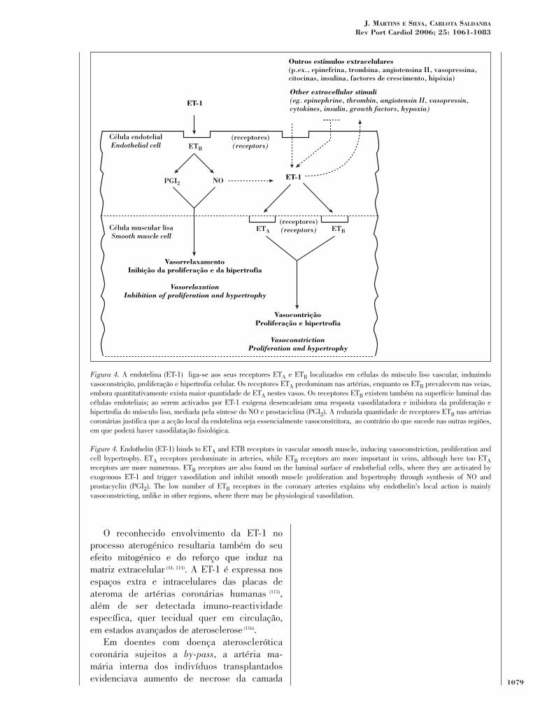
Although veins are more sensitive thanarteries, it is known that ET-1 has a significanteffect on regulation of arterial blood flow,particularly in the myocardium (108) due to thelow number of ETB receptors in the coronaryarteries (109).
Binding of ET-1 to either receptor triggerscontraction of vascular smooth muscle (leading toproliferation and hypertrophy) (Fig. 4). The ETBreceptor, being expressed in endothelial cellstogether with the formation of NO and PGI2, isbehind the episodes of vasodilation seen afterintravenous infusion of ET-1 in animals orisolated organs (110). Chronic antagonism of ET-1receptors in pigs with experimentally-inducedhypercholesterolemia preserved endothelialfunction and increased intracoronary NO activity(111) at the same time as it suppressed expressionof vascular endothelial growth factor (VEGF)and neovascularization of the vasa vasorum (112).Blocking ETA receptors also significantlyreduced atheromatous plaques in a mousemodel of atherosclerosis by inhibiting localneoangiogenesis (113).
The known involvement of ET-1 inatherogenesis also results from its mitogeniceffect and the way it strengthens the extracellularmatrix (44, 114). ET-1 is expressed in the extra- andintracellular spaces of atheromatous plaques inhuman coronary arteries (115), and specificimmunoreactivity has been detected in bothtissue and the circulation in advanced stages ofatherosclerosis (116).
In patients with atherosclerotic coronarydisease who have undergone bypass, the internalmammary artery used for grafting has shownevidence of increased necrosis of the tunicamedia, type 1 collagen and expression of ET-1and ETA e ETB receptors (117). These alterationsindicate endothelial dysfunction in the vesselsused for the grafts, although not as severe as inthe coronary lesions that were the reason for theintervention. 1077
J. MARTINS E SILVA, CARLOTA SALDANHA
Rev Port Cardiol 2006; 25: 1061-1083

existe em circulação a níveis muito baixos,exercendo uma acção vasodilatadora discreta emgrande parte do território arterial (100). A acçãovasoconstrictora sucede a situações anormais,estando associada, por exemplo, a isquémia domiocárdio, arritmia e morte súbita (98, 101). O sistemada endotelina está envolvido num vasto númerode outras condições fisiopatológicas (102), comdestaque para a hipertensão pulmonar pri-mária (103). Na origem daquela situação estará oaumento de expressão da enzima conversora daendotelina-1 (ECE-1), como foi demonstradona aterosclerose e re-estenose (104).
Foi recentemente sugerida a associação entreo aumento da ET-1 e a acção da leptina emdoentes hipertensos obesos, particularmentequando o quadro clínico evoluia para síndromametabólica com diabetes do tipo II (105).
Estão identificados dois receptores (nascélulas do músculo liso vascular) para aendotelina-1 (ETA e ETB), ambos associados àproteína G, fosfolipase C e proteína cinase C (106).Os receptores ETA predominam nas artérias,enquanto os do tipo ETB existem nas veias evasos pulmonares, embora no total continuema prevalecer nestes vasos os receptores do tipoA (107).
Não obstante as veias serem mais sensíveisdo que as artérias, é admissível que aET-1 intervenha significativamente na regulaçãodo fluxo no trajecto arterial, em particulardo miocárdio (108) devido ao reduzido númerode receptores ETB existentes nas artériascoronárias (109).
A fixação de ET-1 a ambos os receptoresinduz contracção (proliferação e hipertrofia) domúsculo liso vascular (Fig. 4). O receptorETB, por ser expresso também nas célulasendoteliais a par com a formação do NO e PGI2,está na origem dos episódios de vasodilataçãoobservados após infusão endovenosa de ET-1em animais ou órgãos isolados (110). A inibiçãocrónica dos receptores da ET-1 em porcos comhipercolesterolémia experimental preservava afunção endotelial, originando aumento deactividade do NO intracoronário (111), ao mesmotempo que reprimia a expressão da VEGF e aneovascularização dos vasa vasorum (112). Ainibição dos receptores ETA também reduziusignificativamente a placa de ateroma emmurganhos-modelo de aterosclerose, por inibiçãoda neo-angiogénese local (113).
By increasing oxidative stress on the vascularintima, ET-1 triggers an inflammatory responsethat may lead to wall remodeling and endothelialdysfunction, as found in ET-1-dependenthypertension (118). Rises in blood pressure andvascular remodeling are inhibited by theadministration of ET-1 receptor antagonists (102).
A new vasoconstrictor peptide has meanwhilebeen discovered in humans and in othervertebrates. At least ten times as potent as ET-1(119), this vasoconstrictor, known as urotensin II, isa cyclic peptide with a structure similar to that ofsomatostatin. It was first isolated in fish spinalcords (120) and subsequently cloned in humans (121).
ACKNOWLEDGEMENTS
Our thanks to Sra. D. Emília Alves for hercareful typing of the text.
1078
Rev Port CardiolVol. 25 Novembro 06 / November 06
1079
J. MARTINS E SILVA, CARLOTA SALDANHA
Rev Port Cardiol 2006; 25: 1061-1083
ET-1
ET-1
ETB
PGI2
ETA ETB
Célula endotelialEndothelial cell
Célula muscular lisaSmooth muscle cell
(receptores)(receptors)
(receptores)(receptors)
Other extracellular stimuli(eg. epinephrine, thrombin, angiotensin II, vasopressin, cytokines, insulin, growth factors, hypoxia)
Outros estímulos extracelulares(p.ex., epinefrina, trombina, angiotensina II, vasopressina, citocinas, insulina, factores de crescimento, hipóxia)
NO
VasorrelaxamentoInibição da proliferação e da hipertrofia
VasorelaxationInhibition of proliferation and hypertrophy
VasocontriçãoProliferação e hipertrofia
VasoconstrictionProliferation and hypertrophy
O reconhecido envolvimento da ET-1 noprocesso aterogénico resultaria também do seuefeito mitogénico e do reforço que induz namatriz extracelular (44, 114). A ET-1 é expressa nosespaços extra e intracelulares das placas deateroma de artérias coronárias humanas (115),além de ser detectada imuno-reactividadeespecífica, quer tecidual quer em circulação,em estados avançados de aterosclerose (116).
Em doentes com doença ateroscleróticacoronária sujeitos a by-pass, a artéria ma-mária interna dos indivíduos transplantadosevidenciava aumento de necrose da camada
Figura 4. A endotelina (ET-1) liga-se aos seus receptores ETA e ETB localizados em células do músculo liso vascular, induzindovasoconstrição, proliferação e hipertrofia celular. Os receptores ETA predominam nas artérias, enquanto os ETB prevalecem nas veias,embora quantitativamente exista maior quantidade de ETA nestes vasos. Os receptores ETB existem também na superfície luminal dascélulas endoteliais; ao serem activados por ET-1 exógena desencadeiam uma resposta vasodilatadora e inibidora da proliferação ehipertrofia do músculo liso, mediada pela síntese do NO e prostaciclina (PGI2). A reduzida quantidade de receptores ETB nas artériascoronárias justifica que a acção local da endotelina seja essencialmente vasoconstritora, ao contrário do que sucede nas outras regiões,em que poderá haver vasodilatação fisiológica.
Figure 4. Endothelin (ET-1) binds to ETA and ETB receptors in vascular smooth muscle, inducing vasoconstriction, proliferation andcell hypertrophy. ETA receptors predominate in arteries, while ETB receptors are more important in veins, although here too ETAreceptors are more numerous. ETB receptors are also found on the luminal surface of endothelial cells, where they are activated byexogenous ET-1 and trigger vasodilation and inhibit smooth muscle proliferation and hypertrophy through synthesis of NO andprostacyclin (PGI2). The low number of ETB receptors in the coronary arteries explains why endothelin's local action is mainlyvasoconstricting, unlike in other regions, where there may be physiological vasodilation.
1. Sacks F.M. - Is there anything to add to our lipid risk factors forcoronary heart disease? Am. J. Cardiol. 1995; 75:1263-1264.
2. Wilson P.W., D'Agostino R.B., Levy D., Belanger A.M.,Sibershatz H., Kannel W.B. - Prediction of coronary heart diseaseusing risk factor categories. Circulation. 1998; 97:1837-1847.
3. Keys S.A. - “Seven Countries: a Multivariate Analysis of Deathand Coronary Heart Disease”, Cambridge, M.A.: HarvardUniversity Press, 1980.
4. Rosenson R.S., Lowe G.D.O. - Effect of lipid and lipoproteinson thrombosis and rheology. Atherosclerosis 1988; 140:271-280.
5. Bonetti P.O., Lerman L.O., Lerman A. - Endothelialdysfunction: a marker of atherosclerostic risk. Arterioscler.Thromb. Vasc. Biol. 2003; 23:168-175.
6. Maas R., Boger R. - Old and new cardiovascular risk factors:from unresolved issues to new opportunities. Atherosclerosis2003; 4:5-17.
7. Davignon J., Ganz J. - Role of endothelial dysfunction inatherosclerosis. Circulation. 2004; 109:III27-III32.
8. Lerman A., Burnett J. - Intact and altered endothelium inregulation of vasomotion. Circulation 1992; 86 (Suppl. III):III.12-III.19.
9. Heistad D.D., Marcus M.L., Larsen G. E., Armstrong M.L. -Role of vasa vasorum in nourishment of the aortic wall. Am. J.Physiol. Heart. Circ. Physiol. 1981; 240:H781-H787.
10. Simionescu M., Simionescu N., Palade G.E. - Morphometricdata on the endothelium of blood capillaries. J. Cell Biol. 1974;60:128-150.
11. Gimbrone M.A.Jr. - Vascular endothelium in health anddisease. In: “Molecular Cardiovascular Medicine”, E. Haber (ed),New York: Scientific American Inc, 1995; pp. 49-61.
12. Aird W.C. - Endothelium as an organ system. Crit. Care Med.2004; 32:S271-S279.
13. Liepsch D.W. - Effect of blood flow parameters in flowpatterns at arterial bifurcations - studies in models. In: “BloodFlow in Large Arteries: Application to Atherosclerosis and ClinicalMedicine”, D.W. Liepsch (ed), Basel: Karger, 1990, pp. 63-76.
14. Behrendt D., Ganz P. - Endothelial function. From vascularbiology to clinical applications. Am. J. Cardiol. 2002; 90:40L-48L.
15. Ku D.D., Caulfield J.B., Kirklin J.K. - Endothelium-dependent responses in human coronary blood vessels. In:“Endothelium Regulation of Vascular Tone”, U.S. Ryan, G.M.,Rubanyi (eds), New York: Marcel Dekker, Inc, 1992; pp. 197-223.
16. Deng D., Tsalenko A., Vailaya A., Ben-Dor A., Kundu R.,Estay I., Tabibiazar R., Kincaid R., Yakhini Z., Bruhn L.,Quertermous T - Differences in vascular bed disease susceptibilityreflect differences in gene expression response to atherogenicstimuli. Circ. Res. 2006; 98:200-208.
17. Yang Z., Stulz P., Von D.L., Bauer E., Turina M., LüscherT.F. - Different interactions of platelets with arterial and venouscoronary bypass vessels. Lancet 1991; 337:939-943.
18. Georitsen M.E. - Physiologic function of normal endothelialcells. In: “Vascular Medicine: A textbook of vascular biology anddiseases”. J. Loscalzo, M.A. Creager, V.J. Dzau (eds), Boston:Little, Brown and Company, 1996, pp. 3-38.
média, colagénio do tipo 1 e expressão da ET-1e dos receptores ETA e ETB
(117). Estas alteraçõesindicavam disfunção endotelial nos vasos atransplantar, embora sem a gravidade aparentedas lesões coronárias que haviam justificado aintervenção cirúrgica.
A ET-1, ao aumentar a agressão oxidantesobre a íntima vascular, desencadeia umaresposta inflamatória que, eventualmente,justificará a remodelação da parede e a disfunçãoendotelial, verificadas na hipertensão arterialET-1 dependente (118). De facto, a tensão arterial ea remodelação vascular são bloqueadas pelaadministração de antagonistas de receptores deET-1 (102).
Foi entretanto identificado um novo péptidovasoconstrictor, existente no homem e outrosmamíferos, que é, pelo menos, cerca de dezvezes mais potente do que a ET-1 (119). Estevasoconstrictor, designado urotensina II, é umpéptido cíclico com estrutura semelhante àsomatostatina, tendo sido isolado inicialmenteda espinal medula de peixes (120) e depois clonadodo homem (121).
AGRADECIMENTOS
O nosso reconhecimento à Sra. D. EmíliaAlves pelo cuidadosa preparação dactilográficado texto.
Pedidos de separatas para:
Address for reprints:
J. MARTINS E SILVA
Conselho Directivo
Faculdade de Medicina de Lisboa
Av. Prof. Egas Moniz
1649-028 LISBOA
e-mail: [email protected]
1080
Rev Port CardiolVol. 25 Novembro 06 / November 06
BIBLIOGRAFIA / REFERENCES

19. Mehta D., Malik A.B. - Signaling mechanisms regulatingendothelial permeability. Physiol Rev. 2006; 86:279-367.
20. Aird W.A. - Mechanisms of endothelial cell heterogeneity inhealth and disease. Circ. Res. 2006; 98:159-162.21. Muller-Delp J.M. - Aging-induced adaptations ofmicrovascular reactivity. Microcirculation 2006; 13:301-314.
22. Brooks A.R., Leekes P.I., Rubanyi G.M. - Gene expressionprofiling of vascular endothelial cells exposed to fluid mechanicalforces: relevance for focal susceptibility to atherosclerosis.Endothelium 2004; 11:45-57.
23. Zeiher A.M., Drexler H., Saurbier B., Just H. - Endothelium-mediated coronary blood flow modulation in humans. Effects ofage, atherosclerosis, hypercholesterolemia, and hypertension. J.Clin. Invest. 1993; 92:652-662.
24. Pearson J.A. - The control of production and release ofhaemostatic factors in the endothelial cell. Baillière's Clin.Haematol. 1993; 6:629-651.
25. Nerem R.M. - Hemodynamics and the vascular endothelium.J. Biomech. Eng. 1993; 115:510-514.
26. Fisher A.B., Chien S., Barakat A.I., Nerem R.M. -Endothelial cellular response to altered shear stress. Am. J.Physiol. Lung .Cell Mol. Physiol. 2001; 281:L529-533.
27. Copley A.L. - Perihemorheology: the bridge between thevessel-blood organ and the organ it penetrates. Biorheology 1989;26:377-388.
28. Gimbrone M.A. Jr. - Vascular endothelium, hemodynamicforces and atherogenesis. Am. J. Pathol. 1999; 155:1-5.
29. Moncada S., Herman A.G., Higgs E.A., Vane J.R. -Differential formation of prostacyclin (PGX or PGI2) by layers ofthe arterial wall. Thromb. Res. 1977; 11:323-344.
30. Moncada S., Vane V.R. - Pharmacology and endogeneous rolesof prostaglandin endoperoxides, thromboxane A2 andprostacyclin. Pharmacol. Rev. 1979; 30:293-331.
31. Rosemberg R.D., Rosemberg J.S. - Natural anticoagulantmechanisms. J. Clin. Invest. 1984; 74:1-6.
32. Martini J., Cabrales P., Tsai A.G., Intaglietta M. -Mechanotransduction and the homeostatic significance ofmaintaining blood viscosity in hypotension, hypertension andhaemorrhage. J. Intern. Med. 2006; 259:364-372.
33. Kalafatis M., Egan J.O., Mann K.G. - Coagulation factors. In:“The Endothelium in Clinical Practice”, G.M. Rubanyi, V.J.Dzau (eds), New York: Marcel Dekker Inc., 1997, pp. 245-264.
34. Hoekstra T., Geleijnse J.M., Schouten E.G., Kluft C. -Plasminogen activator inhibitor-type 1: its plasma determinantsand relation with cardiovascular risk. Thromb. Haemost. 2004;91:861-872.
35. Janig W. - Organization of the lumbar sympathetic outflow toskeletal muscle and skin of the cat hindlimb. Rev. Physiol.Biochem. Pharmacol. 1985; 102:119-123.
36. Wallin B.G., Fagius J. - Peripheral sympathetic neural activityin conscious humans. Annu. Rev. Physiol. 1988; 50:565-576.
37. Brown M.D. - Metabolic control of blood flow with preferenceto heart, skeletal muscle and brain. In: “CardiovascularRegulation”, D. Jordan, J. Marchall (eds) London: PortlandPress 1975; pp. 113-126.
38. Bolton T.B., Lang R.J., Takewaki T. - Mechanism of action ofnoradrenaline and carbachol on smooth muscle of guinea-piganterior mesenteric artery. J. Physiol. 1984; 351:549-572.
39. Hainsworth R., Dinkhill M.J. - Regulation of blood volume.In: “Cardiovascular Regulation”, D. Jordan, J. Marchall (eds)London: Portland Press 1975; pp. 77-91.
40. Joyner M.J., Dietz N.M. - Sympathetic vasodilation in humanmuscle. Acta Physiol Scand. 2003; 177:329-336.
41. Bassenge E - Endothelial regulation of coronary tone. In:“Endothelial Regulation of Vascular Tone”, U.S. Ryan, G.M.Rubanyi (eds). New York: Marcel Dekker Inc., 1992, pp. 225-264.
42. Ralevic V., Loncoln J., Burnstock G. - Release of vasoactivesubstances from endothelial cells. In: “Endothelial Regulation ofVascular Tone”, U.S. Ryan, G.M. Rubanyi (eds). New York:Marcel Dekker Inc. 1992, pp. 297-338.
43. Furchgott R.F., Zawadzki J.V. - The obligatory role ofendotelial cells in the relaxation of arterial smooth muscle byacetylcholine. Nature 1980; 288:373-376.
44. Yanagisawa M., Kurihara H., Kimura S., Tomobe Y.,Kobayashi M., Mutsui Y., Yazaki Y., Goto K., Masaki T. - A novelpotent vasoconstrictor peptide produced by vascular endothelialcells. Nature 1988; 332:411-415.
45. Marcondes S., Antunes E. - The plasma and tissue kininogen-kallikrein-kinin system: role in the cardiovascular system. Curr.Med. Chem. Cardiovasc. Hematol. Agents. 2005; 3:33-44.
46. Critz S.D., Coheu M.V., Downey J.M. - Mechanisms ofacetylcholine- and bradykinin-induced preconditioning. Vascul.Pharmacol. 2005; 42:201-209.
47. Vanhoutte P.M., Perrault L.P., Vilaine J.P. - Endothelialdysfunction and vascular disease. In: “The Endothelium inClinical Pratice”, G.M. Rubanyi, V.J. Dzau (Eds), New York:Marcel Dekker Inc, 1997, pp. 265-289.
48. Walch L., Brink C., Norel X. - The muscarinic receptorsubtypes in human blood vessels. Therapie. 2001; 56:223-226.
49. Palmer R.M., Ferrige A.G., Moncada S. - Nitric oxide releaseaccounts for the biological activity of endothelium-derivedrelaxing factor. Nature 1987; 327:524-526.
50. MichelT., Feron O. - Nitric oxide synthases: which, where,how, and why? J. Clin. Invest. 1997; 100:2146-2152.
51. Goligorsky M.S., Li H., Brodsky S., Chen J. - Relationshipsbetween caveolae and eNOS: everything in proximity and theproximity of everything. Am. J. Physiol. Renal. Physiol.2002; 283:F1-F10.
52. Rapoport R.M., Draznin M.B., Murad F. - Endothelium-dependent relaxation in rat aorta may be mediated throughcyclic GMP-dependent protein phosphorylation. Nature1983; 306:174-176.
53. Kinlay S., Libby P., Ganz P. - Endothelial function andcoronary artery disease. Curr. Opin. Lipidol. 2001; 12:383-389.
54. Simon D.I., Stamler J.S., Jaraki O., Keaney J.F., OsborneJ.A., Francis S.A:, Singel D.J., Loscalzo J. - Antiplateletproperties of protein S-nitrosothiols derived from nitric oxide andendothelium-derived relaxing factor. Arterioscler. Thromb. 1993;13:791-799.
55. Kubes P., Suzuki M., Granger D.N. - Nitric oxide: anendogenous modulator of leukocyte adhesion. Proc. Natl. Acad.Sci. USA. 1991; 88:4651-4655.
56. Yang Y., Loscalzo J. - Regulation of tissue factor expression inhuman microvascular endothelial cells by nitric oxide.Circulation. 2000; 101:2144-2148.
57. Qian H.S., Neplioueva V., Shetty G.A:, Channon K.M.,George S.E. - Nitric oxide synthase gene therapy rapidlyreduces adhesion molecule expression and inflammatory cellinfiltration in carotid arteries of cholesterol-fed rabbits.Circulation 1999; 99:2979-2982.
58. von der Leyen H.E., Gibbores G.H., Morishita R., LewisN.P., Zhang L., Nakajima M., Kaneda Y., Cooke J.P., DzauV.J. - Gene therapy inhibiting neointimal vascular lesion: invivo transfer of endothelial cell nitric oxide synthase gene. Proc.Natl. Acad. Sci. USA 1995; 92:1137-1141.
59. Muller B., Kleschyov A.L., Alencar J.L., Vanin A., Stoclet J-C. - Nitric oxide transport and storage in the cardiovascular 1081
J. MARTINS E SILVA, CARLOTA SALDANHA
Rev Port Cardiol 2006; 25: 1061-1083

system. Ann. N. Y. Acad. Sci. 2002; 962:131-139.
60. Anderson T.J. - Assessment and treatment of endothelialdysfunction in humans. J. Am. Coll. Cardiol. 1999; 34:631-638.
61. Radomski M.W., Palmer R.M., Moncada S. - An L-arginine/nitric oxide pathway present in human platelets regulatesaggregation. Proc. Natl. Acad. Sci. (USA) 1990; 87:5193-5197.
62. Harrison D.G. - Cellular and molecular mechanisms ofendothelial cell dysfunction. J. Clin. Invest. 1997; 100:2153-2157.
63. Lerman A., Zeiher A.M. - Endothelial function: cardiacevents. Circulation 2005; 111:363-368.
64. Grillo M.A., Colombatto S. - Arginine revisited: minireviewarticle. Amino Acids. 2004; 26:345-351.
65. Huynh N.N., Chin-Dusting J. - Amino acids, arginine andnitric oxide in vascular health. Clin. Exp. Pharmacol. Physiol.2006;33:1-8.
66. Parkinson J.F., Phillips G.B. - Nitric oxide synthases;enzymology and mechanism-based inhibitors. In: “TheEndothelium in Clinical Practice”, G.M. Rubanyi, V.J. Dzan(eds), New York: Marcel Dekker Inc, 1997; pp. 95-123.
67. Bredt D.S., Snyder S.H. - Isolation of nitric oxide synthetase,a calmodulin-requiring enzyme. Proc. Natl. Acad. Sci. USA.1990; 87:681-685.
68. Wu K.K. - Regulation of endothelial nitric oxide synthaseactivity and gene expression. Ann. N. Y. Acad. Sci. 2002; 962:122-130.
69. Creager M.A., Gallagher S.J., Girerd X.J., Coleman S.M.,Dzan V.J., Cooke J.P. - L-arginine improves endothelium-dependent vasodilation in hypercholesterolemic humans. J. Clin.Invest. 1992; 90:1248-1253.
70. McDonald K.K., Zharikov S., Block E.R., Kilbery M. S. - Acaveolar complex between the cationic amino acid transporter 1and endothelial nitric-oxide synthase may explain the “arginineparadox”. J. Biol. Chem. 1997; 272:31213-31216.
71. Rees D.D., Palmer R.M., Schulz R., Hodson H.F., MoncadaS. - Characterization of three inhibitors of endothelial nitric oxidesynthase in vitro and in vivo. Br. J. Pharmacol. 1990; 101:746-752.
72. Achan V., Ho H.K., Heeschen C., Stuehlinger M., Jang J.J.,Kimoto M., Vallance P., Cooke J.P. - ADMA regulatesangiogenesis: genetic and metabolic evidence. Vasc. Med. 2005;10:7-14.
73. Joannides R., Haefeli W.E., Linder R., Richard V., BakkaliE.H., Thuillez C., Lüscher T.F. - Nitric oxide is responsible forflow-dependent dilatation of human peripheral conduit arteries invivo. Circulation. 1995; 91:1314-1319.
74. Lieu D.K., Papone P.A., Barakat A.I. - Differentialmembrane potential and ion current responses to different typesof shear stress in vascular endothelial cells. Am. J. Physiol. Cell.Physiol. 2004; 286:CI367-CI375.
75. Rees D.D., Palmer R.M.J., Moncada S. - Role of endothelium-derived nitric oxide in the regulation of blood pressure. Proc.Natl. Acad. Sci. (USA) 1989; 86:3375-3378.
76. Vallance P., Collier J., Moncada S. - Effects of endothelium-derived nitric oxide on peripheral arteriolar tone in man. Lancet1990; II:997-1000.
77. Moreau P., Takase H., Küng C.F., van Rouijen M-M.,Schaffner T., Lüscher T.F. - Structure and function of the ratbasilar artery during chronic nitric oxide synthase inhibition.Stroke 1995; 26:1922-1928.
78. Huang Z., Huang P.L., Ma J., Meng W., Ayata C., FishmanM.C., Moskowitz M.A. - Enlarged infarcts in endotelial nitricoxide synthase knockout mice are attenuated by nitro-L-arginine.J. Cereb. Blood Flow. Metab. 1996; 16:981-987.
79. Li W., Mital S., Ojaimi C., Csiszar A., Kaley G., Hintze T.H.- Premature death and age-related cardiac dysfunction in maleeNOS-knockout mice. J. Mol. Cell. Cardiol. 2004; 37:671-680.
80. Kojda G., Laursen J.B., Ramasamy S., Kent J.D., Kurz S.,Buchfield J., Shesely E.G., Harrison D.G. - Protein expression,vascular reactivity and soluble guanylate cyclase activity in micelacking the endothelial cell nitric oxide synthase: contributions ofNOS isoforms to blood pressure and heart rate control.Cardiovasc. Res. 1999; 42:206-213.
81. Fagan K.A., Fouty B.W., Tyler R.C., Morris Jr. K.G., KeplerL.K., Sato K., LeCras T.D., Abman S.H., Weinberger H.D.,Huang P.L., McMurtry I.F., Radman D.M. - The pulmonarycirculation of homozygous or heterozygous eNOS-null mice ishyperresponsive to mild hypoxia. J. Clin. Invest. 1999; 103:291-299.
82. Rudic R.D., Shesely E.G., Maeda N., Smithies O., Segal S.S.,Sessa W.C. - Direct evidence for the importance of endothelium-derived nitric oxide in vascular remodeling. J. Clin. Invest. 1998;101:731-736.
83. Moroi M., Zhang L., Yasuda T., Virmani R., Gold H.K.,Fishman M.C., Huang P.L. - Interaction of genetic deficiency ofendothelial nitric oxide, gender, and pregnancy in vascularresponse to injury in mice. J. Clin. Invest. 1998; 101:1225-1232.
84. Lee T.C., Zhao Y.D., Courtman D.W., Stewart D.J. -Abnormal aortic valve development in mice lacking endothelialnitric oxide synthase. Circulation. 2000; 101:2345-2348.
85. Nordhaug D., Steensrud T., Aghajani E., Korvald C., MyrmelT. - Nitric oxide synthase inhibition impairs myocardial efficiencyand ventriculo-arterial matching in acute ischemic heart failure.Eur. J. Heart. Fail. 2004; 6:705-713.
86. Zhang F., White J.C., Iadecola C. - Nitric oxide donorsincrease blood flow and reduce brain damage in focal ischemia:evidence that nitric oxide is beneficial in the early stages ofcerebral ischemia. J. Cereb. Blood Flow Metab. 1994; 14:217-226.
87. Moro M.A., Cardenas A., Hurtado O., Leza J.C., Lizasoain I.- Role of nitric oxide after brain ischaemia. Cell. Calcium. 2004;36:265-275.
88. Leeson C.P., Hingorani A.D:, Mullen M.J. Jeerooburkhan N.,Kattenhorn M., Cole T.J., Muller D.P., Lucas A., Humphries S.E.Deanfield J.E. - Glu298Asp endothelial nitric oxide synthase genepolymorphism interacts with environmental and dietary factors toinfluence endothelial function. Circ. Res. 2002; 90:1153-1158.
89. Radomski M.W., Palmer R.M., Moncada S. - Glucocorticoidsinhibit the expression of an inducible, but not the constitutive,nitric oxide synthase in vascular endotelial cells. Proc. Natl. Acad.Sci. (USA), 1990; 87:10043-10047.
90. Eto M., Barandier C., Rathgeb L., Kozai T., Joch H., WangZ., Luscher T.F. - Thrombin suppresses endothelial nitric oxidesynthase and upregulates endothelin-converting enzyme-1expression by distinct pathways: role of Rho/ROCK and mitogen-activated protein kinase. Circ. Res. 2001; 89:583-90.
91. Rentrop K.P. - Thrombi in acute coronary syndromes:revisited and revised. Circulation. 2000; 101:1619-1626.
92. Petros A., Bennett D., Vallance P. - Effect of nitric oxidesynthase inhibitors on hypotension in patients with septic shock.Lancet 1991; 338:1557-1558.
93. Vane J.R., Anggard E.E., Botting R.M. - Regulatory functionsof the vascular endothelium. N. Engl. J. Med. 1990; 323:27-36.
94. Ng K.K., Vane J.R. - Conversion of angiotensin I toangiotensin II. Nature 1967; 216:762-766.95. Berk B.C., Alexander R.W., Brock T.A., Gimbrone M.A. Jr.,Webb R.C. - Vasoconstriction: a new activity for platelets derivedgrowth factor. Science 1986; 232:87-90.
132. Yan C., Kim D., Aizawa T., Berk B.C. - Functional interplaybetween angiotensin II and nitric oxide. Cyclic GMP as a key1082
Rev Port CardiolVol. 25 Novembro 06 / November 06

mediator. Atheroscler. Thromb. Vasc. Biol. 2003; 23:26-36.
97. Turner A.J., Tanzawa K. - Mammalian membranemetallopeptidases: NEP, ECE, KELL, and PEX. FASEB J. 1997;11:355-364.
98. Kiowski W., Lüscher T.F., Linder L., Bühler F.R. -Endothelin-1-induced vasoconstriction in humans. Reversal bycalcium channel blockade but not by nitrovasodilators orendothelium-derived relaxing factor. Circulation 1991; 83:469-475.
99. Wagner O.F., Christ G., Wojta J., Vierhapper H., Parzer S.,Nowotny P.J., Schneider B., Waldhausl W., Binder B.R. - Polarsecretion of endothelin-1 by cultured endothelial cells. J. Biol.Chem. 1992; 267:16066-16068.
100. Verhaar M.C., Strachan F.E., Newby D.E., Cruden N.L.,Koomans H.A., Rabelink T.J., Webb D.J. - Endothelin-Areceptor antagonist-mediated vasodilatation is attenuated byinhibition of nitric oxide synthesis and by endothelin-B receptorblockade. Circulation. 1998; 97:752-756.
101. McCabe C., Hicks M.N., Kane K.A., Wainwright C.C. -Electrophysiological and haemodynamic effects of endothelin ETAand ETB receptors in normal and ischaemic working rabbithearts. Br. J. Pharmacol. 2005; 146:118-128.
102. Schiffrin E.L. - Vascular endothelin in hypertension. Vasc.Pharmacol. 2005; 413:19-29.
103. Goerre S., Wenk M., Bärtsch P., Lüscher T.F., NiroomandS., Hohernhaus H., Oelz O., Reinhart W.H. - Endothelin-1 inpulmonary hypertension associated with high altitude. Circulation1995; 91:359-364.
104. Minamino T., Kurihara H., Takahashi M., Shimada K.,Maemura K., Oda H., Ishikawa T., Uchiyama T., Tanzawa K.,Yasaki Y. - Endothelin-converting enzyme expression in the ratvascular injury model and human coronary atherosclerosis.Circulation. 1997; 95:221-230.
105. Quehenberger P., Exner M., Sunder-Plassmann R., RuzickaK., Bieglmayer C., Endler G., Muellner C., Speiser W., WagnerO. - Leptin induces endothelin-1 in endothelial cells in vitro. Circ.Res. 2002; 90:711-718.
106. Arai H., Hori S., Aramori I., Ohkubo H., Nakanishi S. -Cloning and expression of a cDNA encoding and endothelinreceptor. Nature 1990; 348:730-732.
107. Davie N., Haleen S.J., Upton P.D., Polak J.M., YaxoubM.H., Morrell N.W., Wharton J. - ET(A) and ET(B) receptorsmodulate the proliferation of human pulmonary artery smoothmuscle cells. Am. J. Respir. Crit. Care. Med. 2002; 165:398-405.
108. Lüscher T.F., Yang Z., Tschudi M., Von S.L., Stulz P.,Boulanger C., Siebenmann R., Turina M., Bühler F.R. -Interaction between endothelin-1 and endothelium-derivedrelaxing factor in human arteries and veins. Circ. Res. 1990;66:1088-1094.
109. Bacon C.R., Davenport A.P. - Endothelin receptors inhuman coronary artery and aorta. Br. J. Pharmacol. 1996;117:986-992.
110. Lüscher T.F. - Do we need endothelium antagonists?Cardiovasc. Res. 1993; 27:2089-2093.
111. Best P.J., McKenna C.J., Hasdai D., Holmes D.R.Jr.,Lerman A. - Chronic endothelin receptor antagonismpreserves coronary endothelial function in experimentalhypercholesterolemia. Circulation. 1999; 99:1747-1752.
112. Herrmann J., Best P.J., Ritman E.L., Holmes D.R., LermanL.O., Lerman A. - Chronic endothelin receptor antagonismprevents coronary vasa vasorum neovascularization inexperimental hypercholesterolemia. J. Am. Coll. Cardiol. 2002;39:1555-1561.
113. McKenna C.J., Burke S.E., Opgenorth T.J., Padley R.J.,Camrud L.J., Camrud A.R., Johnson J., Carlson P.J., LermanA., Holmes D.R.Jr., Schwartz R.S. - Selective ET(A) receptorantagonism reduces neointimal hyperplasia in a porcine coronarystent model. Circulation 1998; 97:2551-2556.
114. Battistini B., Chailer P., D'Orleans-Juste P., Briere B., SiroisP. - Growth regulatory properties of endothelins. Peptides.1993;14: 385-399.
115. Zeiher A.M., Goebel H., Schachinger V., Ihling C. - Tissueendothelin-1 immunoreactivity in the active coronaryatherosclerotic plaque: a clue to the mechanism of increasedvasoreactivity of the culprit lesion in unstable angina. Circulation1995; 91:941-947.
116. Lerman A., Edwards B.S., Hallett J.W., Heublein D.M.,Sandberg S.M., Burnett J.J. - Circulating and tissue endothelinimmunoreactivity in advanced atherosclerosis. N. Engl. J. Med.1991; 325:997-1001.
117. Sutherland A.J., Nataatmadja M.I., Walker P.J., Cuttle L.,Garlik R.B., West M.J. - Vascular remodeling in the internalmammary artery graft and association with in situ endothelin-1and receptor expression. Circulation 2006; 113:1180-1188.
118. Gossl M., Lerman A. - Endothelin: beyond a vasoconstrictor.Circulation. 2006; 113:1156-1158.
119. Ames R.B., e 26 Cols. - Human urotensin-II is a potentvasoconstrictor and agonist for the uphan receptor GRR 14.Nature 1999; 401:282-286.
120. Conlon J.M., Yano K., Waugh D., Hazon N. - Distributionand molecular forms of urotensin II and its role in cardiovascularregulation in vertebrates. J. Exp. Zool. 1996; 275:226-238.
121. Coulouarn Y., Lihrmann I., Jegou S., Anouar Y., TostivintH., Beauvillain J.C., Conlon J.M., Bern H.A., Vaudry H. -Cloning of the cDNA encoding the urotensin II precursor in frogand human reveals intense expression of the urotensin II gene inmotoneurons of the spinal cord. Proc. Natl. Acad. Sci. USA 1998;95:15803-15808.
1083
J. MARTINS E SILVA, CARLOTA SALDANHA
Rev Port Cardiol 2006; 25: 1061-1083