IV Cannulation Workbook IV Cannulation Workbook to be completed ...
TURUN YLIOPISTON JULKAISUJAANNALES UNIVERSITATIS TURKUENSIS
SARJA - SER. D OSA - TOM. 1049
MEDICA - ODONTOLOGICA
TURUN YLIOPISTOUNIVERSITY OF TURKU
Turku 2012
ENDOSCOPIC BILIARY PROCEDURES: STUDIES ON CANNULATION
AND STENTING
by
Hanna Vihervaara
From the Faculty of Medicine, University of Turku and Department of Surgery, Turku University Hospital, Turku, Finland
Supervised by
Docent Juha Grönroos, MD, PhDDepartment of Surgery University of TurkuTurku, Finland
Docent Paulina Salminen, MD, PhDDepartment of SurgeryUniversity of TurkuTurku, Finland
Reviewed by
Docent Markku Heikkinen, MD, PhDDepartment of MedicineUniversity of Eastern FinlandKuopio, Finland
and
Docent Johanna Laukkarinen, MD, PhDSchool of MedicineUniversity of TampereTampere, Finland
Dissertation opponent
Docent Jorma Halttunen, MD, PhDDepartment of Gastrointestinal and General SurgeryUniversity of HelsinkiHelsinki, Finland
ISBN 978-951-29-5263-2 (PRINT)ISBN 978-951-29-5264-9 (PDF)ISSN 0355-9483Painosalama Oy – Turku, Finland 2012

To my beloved mother

4 Abstract
ABSTRACT
Hanna VihervaaraEndoscopic biliary procedures: studies on cannulation and stenting
From the Faculty of Medicine, University of Turku and the Department of Surgery, Turku University Hospital, Turku, Finland
Annales Universitatis Turkuensis, Medica-Odontologica2013, Turku, Finland
Deep cannulation is a prerequisite for successful endoscopic retrograde cholangiopancreatography (ERCP) procedures. Of the biliary procedures, stenting is one of the most common. This study was carried out to investigate current and controversial issues regarding biliary cannulation and stenting.
The double guidewire (DGW) technique was studied to analyze its safety and feasibility in biliary cannulation as a single procedure and as a part of the novel three-step cannulation protocol. Female gender was evaluated in regard to difficult cannulation. The use of an angled and a straight tipped guidewire in biliary cannulation was studied in a prospective, randomized trial. Additionally, the patency of the novel antireflux plastic biliary stent was compared to the patency of the conventional plastic biliary stent in a prospective, randomized setting.
The DGW method seems safe and feasible as an alternative cannulation technique in biliary cannulation. Female gender was not associated significantly with difficult biliary cannulation in our study, although the cannulation times seemed to be longer and the alternative cannulation techniques seemed to be needed more often in females than males. According to the results of this thesis, an angled tipped guidewire may facilitate biliary cannulation. In controversy to the previous result presented in the literature, the antireflux plastic biliary stent tested herein should not be used, as the patency of the stent was significantly shorter compared to the conventional plastic stent.
Keywords: ERCP, biliary cannulation, double guidewire, difficult cannulation, female gender, alternative cannulation, antireflux stent, plastic stent

Tiivistelmä 5
TIIVISTELMÄ
Hanna VihervaaraEndoskooppiset sappitietoimenpiteet: tutkimuksia kanyloinnista ja protetisoinnista
Lääketieteellinen tiedekunta, Turun Yliopisto ja Kirurgian klinikka, Turun Yliopistollinen Keskussairaala, Turku, Suomi
Annales Universitatis Turkuensis, Medica-Odontologica2013, Turku, Suomi
Kanyloinnin onnistuminen on edellytys endoskooppisen retrogradisen kolangiopankrea-tografia (ERCP) -toimenpiteen onnistumiselle. Sappiteiden protetisointi on yksi yleisim-mistä endoskooppisista sappitietoimenpiteistä. Tässä tutkimuksesa selvitettiin ajankoh-taisia sekä kiistanalaisia kysymyksiä koskien sappiteiden kanylointia ja protetisointia.
Kaksoisvaijerimenetelmän turvallisuutta ja käyttökelpoisuutta arvioitiin sekä yksittäisenä menetelmänä että osana kolmivaiheista kanylointiprotokollaa. Lisäksi tutkittiin naissukupuolen ja kanylointivaikeuksien välistä yhteyttä. Väitöskirjan osatöihin kuului myös kaksi prospektiivista, satunnaistettua tutkimusta. Toisessa verrattiin käyrä- ja suorakärkisen ohjainvaijerin käyttöä sappitiekanyloinnissa. Toisessa taas verrattiin uuden takaisinvirtauksenestomekanismilla varustetun muovisen sappitiestentin aukioloa tavanomaisen muovistentin aukioloon.
Kaksoisvaijerimenetelmä vaikuttaa olevan turvallinen ja käyttökelpoinen vaihtoehtoinen tekniikka sappitiekanyloinnissa. Naissukupuolen ei todettu olevan merkitsevästi yhteydessä kanylointivaikeuksiin, vaikka kanylointiaika vaikutti olevan pidempi ja vaihtoehtoiset kanylointimenetelmät vaikuttivat olevan yleisempiä naisilla kuin miehillä. Tutkimustulosten mukaan käyräkärkinen ohjainvaijeri saattaa helpottaa sappitiekanylointia. Vastoin aiempaa tutkimustulosta, tutkimaamme takaisinvirtauksenestomekanismilla varustettua muovista sappistenttiä ei tulisi käyttää, koska sen aukipysyvyys oli selvästi huonompi kuin perinteisen muovistentin.
Avainsanat: ERCP, sappitiekanylointi, kaksoisohjainvaijeri, haastava kanylointi, naissukupuoli, vaihtoehtoinen kanylointi, takaisinvirtauksenestomekanismillä varustettu sappistentti, muovistentti
6 Table of Contents
TABLE Of CONTENTS
ABSTRACT ..................................................................................................................................4
TIIVISTELMÄ ............................................................................................................................5
ABBREVIATIONS ......................................................................................................................9
LIST Of ORIGINAL PUBLICATIONS .................................................................................10
1. INTRODUCTION ................................................................................................................11
2. REVIEW Of THE LITERATURE ....................................................................................13
2.1. Anatomy of bile and pancreatic ducts .............................................................132.2. Physiology of the pancreatobiliary system ......................................................152.3. Pathophysiology of the pancreatobiliary system .............................................15
2.3.1. Common bile duct stones ....................................................................152.3.2. Sphincter of Oddi dysfunction ............................................................162.3.3. Primary sclerosing cholangitis ............................................................162.3.4. Cholangiocarcinoma ...........................................................................162.3.5. Acute pancreatitis ................................................................................172.3.6. Chronic pancreatitis ............................................................................172.3.7. Neoplasms of the pancreas ..................................................................172.3.8. Other neoplastic diseases ....................................................................18
2.4. Diagnostic methods of the pancreatobiliary diseases ......................................182.4.1. Laboratory tests ...................................................................................182.4.2. Imaging methods .................................................................................19
2.5. Technical aspects of ERCP procedures ...........................................................202.5.1. Duodenoscope .....................................................................................202.5.2. Patient and position .............................................................................202.5.3. Fluoroscopy .........................................................................................212.5.4. Electrosurgical current ........................................................................21
2.6. Indications for ERCP procedures ....................................................................212.6.1. Biliary tract diseases ...........................................................................22
2.6.1.1. Bile duct stones ......................................................................222.6.1.2. Benign biliary strictures .........................................................222.6.1.3. Malignant biliary strictures ....................................................232.6.1.4. Sphincter of Oddi dysfunction ...............................................242.6.1.5. Iatrogenic bile leakage ...........................................................24
2.6.2. Pancreatic diseases ...............................................................................242.6.3. Ampullary adenomas ..........................................................................25
2.7. Cannulation methods .........................................................................................262.7.1. Standard cannulation ...........................................................................26

Table of Contents 7
2.7.2. GW cannulation ..................................................................................262.7.3. Cannulas and sphincterotomes ............................................................262.7.4. DGW cannulation ................................................................................272.7.5. Pre-cut techniques ..............................................................................272.7.6. Rendezvous techniques .......................................................................282.7.7. Special circumstances .........................................................................28
2.8. Procedures .......................................................................................................282.8.1. Sphincterotomy ...................................................................................292.8.2. Stone removal......................................................................................292.8.3. Brush cytology ....................................................................................302.8.4. Stenting and stricture dilatation ..........................................................302.8.5. Manometry ..........................................................................................312.8.6. Peroral cholangioscopy .......................................................................31
2.9. Biliary stents ....................................................................................................322.9.1. Plastic stents ........................................................................................322.9.2. Metal stents .........................................................................................332.9.3. Stent migration ....................................................................................342.9.4. Stent occlusion ....................................................................................34
2.10. Complications ..................................................................................................342.10.1. Post-ERCP pancreatitis .......................................................................352.10.2. Bleeding ..............................................................................................352.10.3. Cholangitis ..........................................................................................362.10.4. Perforation ...........................................................................................36
3. AIMS Of THE STUDY .......................................................................................................37
4. PATIENTS AND METHODS ..............................................................................................38
4.1. Patients and data collection .............................................................................384.1.1. Studies I and IІ ....................................................................................384.1.2. Study III ..............................................................................................394.1.3. Study IV .............................................................................................394.1.4. Study V ...............................................................................................40
4.2. Methods ...........................................................................................................414.2.1. ERCP procedures ................................................................................414.2.2. Study design ........................................................................................41
4.2.2.1. Studies I-II ..............................................................................424.2.2.2. Study III .................................................................................424.2.2.3. Study IV .................................................................................424.2.2.4. Study V ...................................................................................43
4.2.3. Statistics ..............................................................................................434.2.4. Ethics ...................................................................................................44
5. RESULTS ..............................................................................................................................45
5.1. Biliary cannulation (Studies I-IV) ...................................................................45

8 Table of Contents
5.1.1. Cannulation methods ...........................................................................455.1.2. Cannulation and procedure related time .............................................455.1.3. Complications ....................................................................................45
5.2. Association of female gender and difficult cannulation (Study III) ................465.3. Biliary cannulation with AGW or SGW (Study IV) ........................................485.4. ARS or conventional PS in malignant distal strictures (Study V) ...................48
6. DISCUSSION .......................................................................................................................50
6.1. Methodological considerations ........................................................................506.2. Double guidewire cannulation (studies I and II) .............................................51
6.2.1. Three-step protocol .............................................................................536.3. Cannulation difficulty (Study III) ....................................................................546.4. Guidewire cannulation (Study IV) ..................................................................556.5. ARS vs. conventional PS in malignant distal strictures (Study V) .................56
7. CONCLUSIONS...................................................................................................................57
8. ACKNOWLEDGEMENTS .................................................................................................58
9. REfERENCES .....................................................................................................................60
ORIGINAL PUBLICATIONS ..................................................................................................72

Abbreviations 9
ABBREVIATIONS
AGW angled tipped guidewireALT alanine aminotransferaseAP alkaline phosphataseAPD accessory pancreatic ductARS antireflux plastic stentAST aspartate aminotransferaseBil bilirubinCA 19-9 carbohydrate antigen 19-9CBD common bile ductCC cholangiocarcinomaCRP C-reactive proteinCT computed tomographyDGW double guidewireDS dominant stenosisES endoscopic sphincterotomyEUS endoscopic ultrasoundERCP endoscopic retrograde cholangiopancreatographyFISH fluorescence in situ hybridizationγ-GT γ-glutamyltransferaseGW guidewireHb haemoglobinIBD inflammatory bowel diseaseLBD large balloon dilatationMAP major papillaMIP minor papillaMPD main pancreatic ductMRCP magnetic resonance cholangiopancreatographyPD pancreas divisumPEP post-ERCP pancreatitisPS plastic stentPSC primary sclerosing cholangitisPTBD percutaneus transhepatic biliary drainagePTC percutaneous transhepatic cholangiographySEMS self-expanding metal stentSGW straight tipped guidewireSO sphincter of OddiSOD sphincter of Oddi dysfunctionSOM sphincter of Oddi manometryUS ultrasoundWBC white blood cell count
10 List of Original Publications
LIST Of ORIGINAL PUBLICATIONS
This thesis is based on the following publications, which are referred to by Roman numerals I-V in the text. Unpublished data is also included.
I Grönroos JM, Vihervaara H, Gullichsen R, Laine S, Karvonen J, Salminen P. (2011) ”Double-guidewire-assisted biliary cannulation: experiences from a single tertiary referral center.” Surg Endosc 25(5):1599-1602
II Vihervaara H, Grönroos JM. (2012) “Feasibility of the novel 3-step protocol for biliary cannulation – a prospective analysis.” Surg Laparosc Endosc Percutan Tech 22(2):161-164
III Vihervaara H, Salminen P, Hurme S, Gullichsen R, Laine S, Grönroos JM. (2011) “Female gender and post-ERCP pancreatitis: is the association caused by difficult cannulation?” Scand J Gastroenterol 46(12):1498-1502
IV Vihervaara H, Grönroos JM, Koivisto M, Gullichsen R, Salminen P. “Angled- or straight-tipped hydrophilic guidewire in biliary cannulation: a prospective randomized controlled trial.” Surg Endosc, in press
V Vihervaara H, Grönroos JM, Hurme S, Gullichsen R, Salminen P. “Antireflux vs. conventional plastic stent in malignant biliary obstruction - a prospective, randomized study.” Submitted 2012
The original communications are reproduced with the kind permission of the copyright holders.

Introduction 11
1. INTRODUCTION
Since the first description of endoscopic cannulation of papilla Vater in 1968 (McCune et al. 1968), endoscopic retrograde cholangiopancreatography (ERCP) has become a cornerstone in the simultaneous endoscopic diagnosis and treatment of biliary and pancreatic diseases. Later, magnetic resonance cholangiopancreatography (MRCP) as a noninvasive imaging modality has replaced more invasive ERCP for purely diagnostic purposes (Albert et al. 2002).
Deep biliary cannulation is pivotal for successful ERCP procedures. Despite the advances in equipment and techniques, biliary cannulation remains a significant challenge. In expert centers the success rate of deep biliary cannulation can be even 99 % (Fukatsu et al. 2008), but in association with low-volume centers the cannulation rate of the desired duct(s) in national level may be as low as 84% (Kapral et al. 2008).
Guidewire (GW) assisted cannulation is considered today the primary cannulation method in ERCP procedures. The use of GW increases the success rate of selective cannulation of the bile duct (Bailey et al. 2008; Katsinelos et al. 2008) and decreases the incidence of post-ERCP pancreatitis (PEP) (Cennamo et al. 2009; Cheung et al. 2009) compared with standard catheter cannulation. If the primary cannulation methods prove unsuccessful, double guidewire (DGW) and precut techniques are used. DGW method is a relatively novel technique, and only few prospective randomized studies exist (Maeda et al. 2003; Herreros de Tejada et al. 2009; Angsuwatcharakon et al. 2012). Precut techniques are considered demanding, and their use and timing are still under debate (Cennamo et al. 2010; Gong et al. 2010).
PEP is the most common complication after ERCP; its incidence varying usually between 2 and 7 % (Freeman et al. 2001; Ong et al. 2005; Williams et al. 2007). Female gender, multiple cannulation attempts, contrast injection into the pancreatic duct and suspected sphincter of Oddi dysfunction (SOD) are risk-factors for PEP (Freeman et al. 2001; Williams et al. 2007; Bailey et al. 2008).
Endoscopic biliary stenting is a widely accepted palliative treatment modality for inoperable malignant common bile duct (CBD) strictures. Plastic stents (PS) are relatively cheap, but their use is limited by the early occlusions requiring stent exchange every 3 - 5 months (Kaassis et al. 2003; Tringali et al. 2003). Self-expanding metal stents (SEMS) have longer patency, major cause of stent occlusion being tumor ingrowth, but higher cost and inability to be removed in case of uncovered SEMSs restrict their use (Davids et al. 1992; Prat et al. 1998). Fully covered SEMSs are removable and can also be applied in benign conditions (Kasher et al. 2011). The exact mechanisms of biliary stent occlusion remain unknown, but duodenobiliary reflux is considered a major factor contributing to stent occlusion (Weickert et al. 2001; van Berkel et al. 2005). A PS with an antireflux mechanism has been developed to eliminate intestinal reflux (Dua et al. 2007).

12 Introduction
In this thesis, the focus has been on the cannulation of the biliary tract. The main target has been to evaluate recent advances in biliary cannulation techniques with a special reference to the risk factors and management of difficult cannulation. In addition, the palliative treatment of inoperable malignant obstructive jaundice has been studied with two different biliary stents.
Review of the Literature 13
2. REVIEW Of THE LITERATURE
2.1. Anatomy of bile and pancreatic ducts
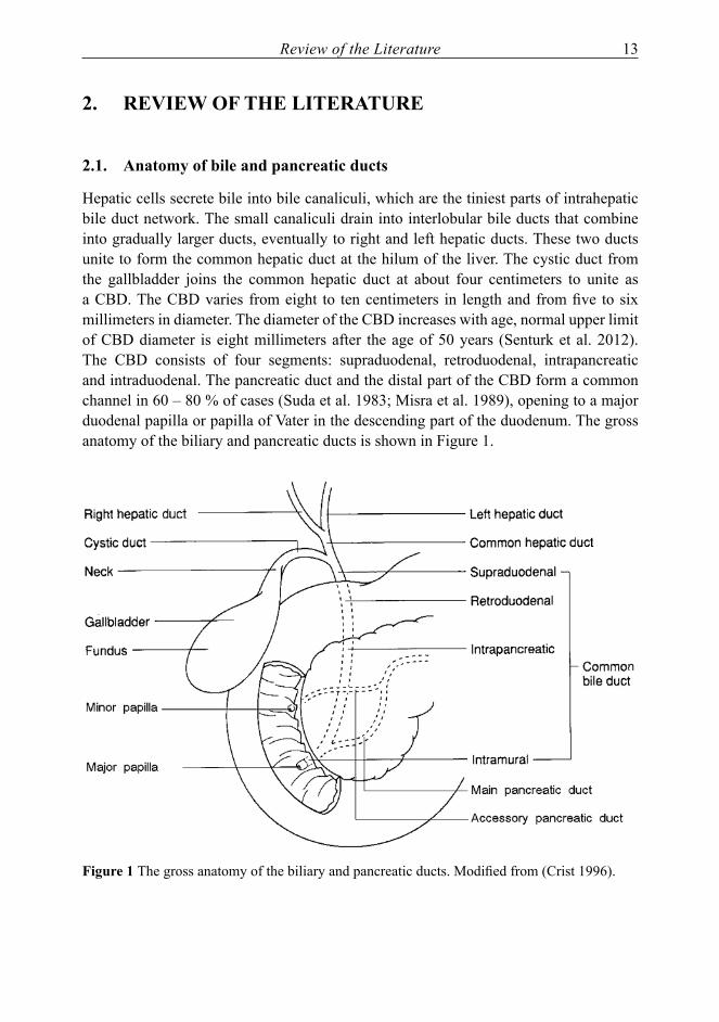
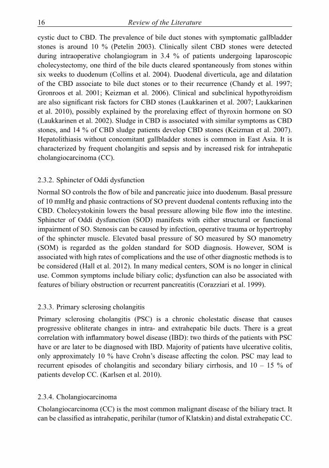
Hepatic cells secrete bile into bile canaliculi, which are the tiniest parts of intrahepatic bile duct network. The small canaliculi drain into interlobular bile ducts that combine into gradually larger ducts, eventually to right and left hepatic ducts. These two ducts unite to form the common hepatic duct at the hilum of the liver. The cystic duct from the gallbladder joins the common hepatic duct at about four centimeters to unite as a CBD. The CBD varies from eight to ten centimeters in length and from five to six millimeters in diameter. The diameter of the CBD increases with age, normal upper limit of CBD diameter is eight millimeters after the age of 50 years (Senturk et al. 2012). The CBD consists of four segments: supraduodenal, retroduodenal, intrapancreatic and intraduodenal. The pancreatic duct and the distal part of the CBD form a common channel in 60 – 80 % of cases (Suda et al. 1983; Misra et al. 1989), opening to a major duodenal papilla or papilla of Vater in the descending part of the duodenum. The gross anatomy of the biliary and pancreatic ducts is shown in Figure 1.
figure 1 The gross anatomy of the biliary and pancreatic ducts. Modified from (Crist 1996).

14 Review of the Literature
The pancreas has two major ducts: main pancreatic duct (MPD) and accessory pancreatic duct (APD, duct of Santorini). The pancreas develops from ventral and dorsal pancreatic buds. As these buds merge, the MPD and APD develop. APD is constituted from the dorsal pancreatic bud and MPD is constituted from both dorsal and ventral buds. MPD comes to close relationship to CBD, often forming a common channel called hepatopancreatic ampulla (ampulla of Vater), which opens to major papilla (MAP). APD enters the duodenum through minor papilla (MIP). The patency of APD is difficult to determine, but average reported patency is close to 50 % (Kamisawa 2004). Pancreas divisum (PD) is the most common anomaly with the pancreatic ducts. In total PD, APD and MPD do not communicate, and in partial PD there is a rudimentary connection between APD and MPD, but most drainage from APD goes through MIP. Biliary and pancreatic ducts visualized during ERCP are shown in Figure 2.
figure 2 Biliary and pancreatic ducts visualized during ERCP
MAP is a small elevation of the mucosa, where hepatopancreatic ampulla enters the duodenum. It is usually located in the descending part of duodenum (87 %), but can also be located between the descending and transverse parts of duodenum or in the transverse part of duodenum (Lindner et al. 1976). Smooth muscle surrounds the distal end of the common channel constituting the hepatopancreatic sphincter or the sphincter of Oddi (SO). SO controls the flow of bile and pancreatic juice to duodenum and the retrograde flow of duodenal material to biliary system.

Review of the Literature 15
MIP almost always exists, but it can be difficult to locate. It is usually located two centimeters proximal to MAP. In case of a non-patent APD, MIP is filled with tissue, making cannulation impossible. If ADP is patent, muscle tissue surrounds MIP as the SO surrounds the MAP.
2.2. Physiology of the pancreatobiliary system
The bile is secreted in the liver by hepatocytes; approximately 500 - 1000 ml of bile is produced every day. The bile secretion has two main functions: (1) to promote the digestion and absorption of lipids from the intestine, and (2) to eliminate substances from blood that are not excreted through the kidneys. The bile constitutes of organic and inorganic components. The main organic components are bile salts, phospholipids, cholesterol and bile pigments. Bile salts are the major component of the bile. Approximately 95 % of bile salts are returned to portal venous system from distal ileum through enterohepatic circulation to be re-excreted by hepatocytes again. Water and electrolytes are added to bile through osmotic gradient. (Strange 1984). The gallbladder stores concentrated bile between meals. Fat and protein digestion promotes the excretion of cholecystokinin from duodenum, which stimulates the gallbladder to contract, and the bile is excreted to the intestine.
Pancreas is a complex organ with both endocrine and exocrine functions, which are controlled by numerous neural and hormonal mediators. Endocrine cells of the pancreas are located throughout the whole gland in small islets. The main endocrine hormones secreted by islet cells are insuline, glucagone, somatostatin and pancreatic polypeptide. Exocrine portion of the pancreas secretes pancreatic juice through the MPD. Acinar cells, which produce digestive enzymes, comprise most of the glandular mass of the pancreas. These powerful enzymes are produced first as inactive proenzymes, and are converted to active enzymes outside the pancreas. Bicorbonate-ions and fluids are secreted by the pancreatic ductal cells; this secrete neutralizes acidity of gastric contents in the duodenum. (Chandra et al. 2009)
2.3. Pathophysiology of the pancreatobiliary system
Cholestasis is a common finding in diseases affecting the biliary tract. Increased levels of serum bilirubin cause jaundice, a distinguishable yellow color of skin and scleras. Both intra- and extraductal lesions can cause extrahepatic obstructive jaundice. Impaired bile flow can also lead to abnormal bacterial ingrowth with cholangitis and sepsis (Navaneethan et al. 2011).
2.3.1. Common bile duct stones
Gallstones are very common in Western countries. Their prevalence and location vary between different ethnical and geographical groups. CBD stones are usually secondary to gallbladder stones; stones are originated in gallbladder and are passed through
16 Review of the Literature
cystic duct to CBD. The prevalence of bile duct stones with symptomatic gallbladder stones is around 10 % (Petelin 2003). Clinically silent CBD stones were detected during intraoperative cholangiogram in 3.4 % of patients undergoing laparoscopic cholecystectomy, one third of the bile ducts cleared spontaneously from stones within six weeks to duodenum (Collins et al. 2004). Duodenal diverticula, age and dilatation of the CBD associate to bile duct stones or to their recurrence (Chandy et al. 1997; Gronroos et al. 2001; Keizman et al. 2006). Clinical and subclinical hypothyroidism are also significant risk factors for CBD stones (Laukkarinen et al. 2007; Laukkarinen et al. 2010), possibly explained by the prorelaxing effect of thyroxin hormone on SO (Laukkarinen et al. 2002). Sludge in CBD is associated with similar symptoms as CBD stones, and 14 % of CBD sludge patients develop CBD stones (Keizman et al. 2007). Hepatolithiasis without concomitant gallbladder stones is common in East Asia. It is characterized by frequent cholangitis and sepsis and by increased risk for intrahepatic cholangiocarcinoma (CC).
2.3.2. Sphincter of Oddi dysfunction
Normal SO controls the flow of bile and pancreatic juice into duodenum. Basal pressure of 10 mmHg and phasic contractions of SO prevent duodenal contents refluxing into the CBD. Cholecystokinin lowers the basal pressure allowing bile flow into the intestine. Sphincter of Oddi dysfunction (SOD) manifests with either structural or functional impairment of SO. Stenosis can be caused by infection, operative trauma or hypertrophy of the sphincter muscle. Elevated basal pressure of SO measured by SO manometry (SOM) is regarded as the golden standard for SOD diagnosis. However, SOM is associated with high rates of complications and the use of other diagnostic methods is to be considered (Hall et al. 2012). In many medical centers, SOM is no longer in clinical use. Common symptoms include biliary colic; dysfunction can also be associated with features of biliary obstruction or recurrent pancreatitis (Corazziari et al. 1999).
2.3.3. Primary sclerosing cholangitis
Primary sclerosing cholangitis (PSC) is a chronic cholestatic disease that causes progressive obliterate changes in intra- and extrahepatic bile ducts. There is a great correlation with inflammatory bowel disease (IBD): two thirds of the patients with PSC have or are later to be diagnosed with IBD. Majority of patients have ulcerative colitis, only approximately 10 % have Crohn’s disease affecting the colon. PSC may lead to recurrent episodes of cholangitis and secondary biliary cirrhosis, and 10 – 15 % of patients develop CC. (Karlsen et al. 2010).
2.3.4. Cholangiocarcinoma
Cholangiocarcinoma (CC) is the most common malignant disease of the biliary tract. It can be classified as intrahepatic, perihilar (tumor of Klatskin) and distal extrahepatic CC.

Review of the Literature 17
The diagnosis is often difficult as the carcinoma grows silently with majority of patients having symptoms only at the advanced stage of the disease. PSC, hepatolithiasis, bile duct cysts, parasitic infections and toxins are established risk factors for the development of CC, but most carcinomas appear with no detected predisposing state. Approximately 10 % of CCs are attributed to PCS. The prognosis of CC is very poor. (Tyson et al. 2011).
2.3.5. Acute pancreatitis
Acute pancreatitis is an acute inflammatory process of the pancreas. The majority of cases (approximately 80 %) are mild and resolve with supportive therapy. The remaining 20 % associate with significant morbidity and mortality and need multidisciplinary care consisting of surgeons, anesthesiologists and radiologists. The most common etiological factors causing acute pancreatitis are alcohol and gallstones. The passage of biliary stone through CBD is thought to initiate acute biliary pancreatitis. Several studies have shown increased risk of acute pancreatitis with small gallstones (≤ five mm) (Diehl et al. 1997; Venneman et al. 2005) and biliary sludge (Lee et al. 1992). Unless there is evidence of CBD obstruction in preoperative imaging or laboratory tests, the incidence of CBD stones after acute biliary pancreatitis (5 %) among patients undergoing cholecystectomy correlates to the incidental finding of CBD stones (5 %) in patients with symptomatic cholecystolithiasis (Shayan et al. 2007).
2.3.6. Chronic pancreatitis
Chronic pancreatitis is a progressive inflammatory disorder that may lead to fibrosis of pancreatic secretory parenchyma. The destruction of exocrine and endocrine functions leads to malnutrition and diabetes in an advanced state. Alcohol is regarded as the leading cause of chronic pancreatitis; recurrent attacks of acute pancreatitis can precede the chronic state (Schneider et al. 2005). Other toxins, as tobacco smoke, are also recognized as risk factors for the disease. Chronic pancreatitis usually presents with acute or recurrent acute pancreatitis, constant pain, local complications and exocrine or endocrine insufficiency. Complications of chronic pancreatitis include biliary and pancreatic duct obstruction, pancreatic fistulas and pseudocysts. Chronic pancreatitis is also a risk factor for pancreatic cancer, but among patients with chronic pancreatitis only 5 % or less develop pancreatic cancer over a 20-year period (Raimondi et al. 2010).
2.3.7. Neoplasms of the pancreas
The most frequent neoplastic tumor of the pancreas is ductal adenocarcinoma (90 %). The prognosis of pancreatic adenocarcinoma is very dismal; in Finland the re-evaluated overall five- year survival was as low as 0.2 % during 1990-1996 (Carpelan-Holmstrom et al. 2005). R0 resection and early tumor stage associate with 5 year survival of over 10% after pancreatic resection in pancreatic adenocarcinoma (Ferrone et al. 2008). The symptoms can be quite non-spesific, e.g. abdominal discomfort and nausea. The majority

18 Review of the Literature
of patients seek medical attention for jaundice as their first symptom. The presence of pain in newly diagnosed patients correlates both with tumor unresectability and with worse survival even if the tumor is resectable (Kelsen et al. 1997). Radical surgery is the only possibly curative treatment, but unfortunately 80 – 85 % of patients present with unresectable disease.
Cystic neoplasms account for approximately 5 % of primary pancreatic tumors. Non-neoplastic tumors (pseudocysts, true congenital cysts, cystic fibrosis and polycystic disease) should be distinguished from real cystic neoplastic tumors of the pancreas. Cystic neoplasms of the pancreas have malignant potential and are to be considered for radical treatment. They can be divided to four entities according to a WHO classification: serous cystic neoplasms, mucinous cystic neoplasms, intraductal papillary mucinous neoplasms and solid pseudopapillary neoplasms (Bosman et al. 2010).
2.3.8. Other neoplastic diseases
There are several other causes of extra- and intrahepatic biliary obstruction. Choledochal cysts are quite rare among Western adults. Most of these congenital cysts are extrahepatic, but both extra- and intrahepatic cysts and entirely intrahepatic cysts are more common with adults than with children and can be associated with biliary malignancy (Nicholl et al. 2004). Most common primary presenting symptoms include pain and cholangitis, but icterus and pancreatitis have also been reported (Visser et al. 2004).
Obstructive jaundice caused by hepatocellular carcinoma is not common. Biliary tract can be obstructed by tumor thrombi, hemobilia, tumor compression or diffuse tumor infiltration (Qin et al. 2003). Other metastatic diseases can cause biliary obstruction mainly by liver metastases compressing the hilum or, less frequently, by lymph nodes or peritoneal masses interfering with the bile duct (Van Laethem et al. 2003). Liver metastases usually originate from the gastrointestinal tract, breasts, kidneys or lungs.
2.4. Diagnostic methods of the pancreatobiliary diseases
The main purpose of diagnostics in extrahepatic biliary obstruction is to differentiate between a malignant and benign nature of the disease. The level and the etiology of the possible strictures combined with the information of surrounding tissues define the proper treatment protocol. Imaging of the pancreaticobiliary system has developed enormously during the past decades. Availability of the imaging methods defines the study patterns in each individual medical center.
2.4.1. Laboratory tests
There is no ideal laboratory test to differentiate between benign and malignant nature of biliary obstruction. Elevations of biochemical parameters as γ-glutamyltransferase (γ-GT), alkaline phosphatase (AP), alanine aminotransferase (ALT), bilirubin (bil) and

Review of the Literature 19
aspartate aminotransferase (AST) can be seen in biliary obstruction. These biochemical tests, especially γ-GT, can be used in predicting absence of bile duct stones with patients undergoing laparoscopic cholecystectomy (Yang et al. 2008). Bil level has been used to distinguish malignant and benign extrahepatic cholestasis: cut off value of 145 μmol/l with sensitivity of 66 % and specificity of 91 % (Karvonen et al. 2006) and 100 μmol/l with sensitivity of 72 % and specificity of 87 % (Garcea et al. 2011) have been proposed to provide optimal sensitivity and specificity in separating patients with malignant bile duct stricture from those with bile duct stones. Bilirubin level can also be elevated in non- obstructive icterus, which can be caused by, for example, liver diseases or hemolysis. Carbohydrate antigen 19-9 (CA 19-9) is a carbohydrate tumor-associated antigen which is expressed by several epithelial cancer cells and also by normal pancreatic and biliary ductal cells (Koprowski et al. 1979). The level of CA 19-9 can be increased both with malignant biliary stricture and benign cholestasis. Cut off level of 70 – 90 U/ml after successful biliary drainage has been suggested to differentiate benign obstruction from malignant pathology (Marrelli et al. 2009; Morris-Stiff et al. 2009).
2.4.2. Imaging methods
Even though ERCP is considered a golden standard in diagnosing suspected biliary obstruction, its invasiveness and complication rate restrict its use mainly to therapeutic procedures (Sahni et al. 2008). Ultrasound (US) is commonly used as an initial noninvasive imaging method in evaluating suspected biliary obstruction. It is easily accessed, relatively cheap with no radiation involved, but on the drawback it is very interpreter dependent and bowel gas or obesity interfere with the image quality. US gives reliable information concerning the dilatation of the bile ducts, but has a sensitivity of 71 – 88 % in defining the level of biliary obstruction and a sensitivity of 48 – 57 % in finding the etiology for obstruction (Blackbourne et al. 1994). The sensitivity of 75 % in detecting bile duct stones has been reported (Laing et al. 1984; Dong et al. 1987).
Computed tomography (CT) is mostly used in this connection to evaluate structures surrounding the biliary tract and possible lesions causing biliary obstruction. The sensitivity and specificity of contrast enhanced CT in detecting bile duct stones is 77 % and 73 %, respectively (Tseng et al. 2008). CT cholangiography is performed after injection of biliary contrast medium and with three-dimensional image reconstruction it gives a good visualization of the biliary tree. It offers an option for biliary imaging for patients with contraindications for MRCP. CT cholangiography may be useful for patients with normal bilirubin levels: hyperbilirubinemia can affect the visualization of the bile ducts as bilirubin excretion is impaired (Stockberger et al. 1994) .
MRCP is considered a standard biliary imaging technique using heavily T2-weighted sequences to provide magnetic resonance images from biliary tree as a noninvasive alternative for diagnostic ERCP. In a large meta-analysis MRCP provides overall sensitivity of 95 % and specificity of 97 % in detecting the level and the presence of biliary obstruction, but is less sensitive for biliary stones (92 %) and for differentiating

20 Review of the Literature
malignant conditions from benign conditions (88 %) (Romagnuolo et al. 2003). CBD stones smaller than five millimeters have given false negative results in MRCP (Kondo et al. 2005).
Percutaneus transhepatic cholangiography (PTC) is an invasive imaging method performed both in US and fluoroscopy guidance. Due to its invasiveness, PTC is considered mainly in association with therapeutic procedures, e.g. stenting of hilar strictures or rendezvous procedures (Covey et al. 2008).
Endoscopic ultrasound (EUS) was introduced in the 1980s. The ultrasound transducer is located at the tip of the echoendoscope. The close proximity to gastrointestinal wall allows a good view to gastrointestinal tumors as well as to the pancreaticobiliary system. EUS is comparable to MRCP in diagnosis of extrahepatic biliary obstruction (Materne et al. 2000). EUS and MRCP are also comparable in detection of bile duct stones (Verma et al. 2006). The good visualization of adjacent organs and tissues allows therapeutic procedures to be performed in EUS guidance.
2.5. Technical aspects of ERCP procedures
2.5.1. Duodenoscope
Duodenoscope is a flexible side viewing endoscope. The insertion tube varies in length (1235 - 1250 mm) and in diameter (7.5 - 12.1 mm). The working channel (2.0 - 4.8 mm) is usually wider than in gastroscopes (2.0 - 3.8 mm). On the tip of the duodenoscope there is an elevator that can lift the instruments that come through the working channel to facilitate the cannulation and other procedures. The endoscopist can control the elevator from the control section of the endoscope.
2.5.2. Patient and position
ERCP is usually performed under conscious sedation, but general anesthesia is used if necessary. Benzodiazepines, propofol and opiates can be used as sedative and analgesic agents during ERCP. A state of deep sedation has been suggested to ensure the stability of the patient during the procedures (Chainaki et al. 2011). A patient controlled sedation with propofol and remifentanil is a valuable option for sedation during ERCP procedures (Mazanikov et al. 2011). Intestinal motility can be suppressed with hyoscine butylbromide or glucagon.
Patients usually lie in prone position during ERCP, the supine position is reserved mostly for intubated patients. Supine position is associated with more demanding ERCPs and with more likely adverse cardiorespiratory events (Terruzzi et al. 2005). Provided that supine position is used regularly in daily basis, supine and prone position can be considered equal in terms of difficulty and cannulation success (Tringali et al. 2008). Supine position can also be useful in advancing the duodenoscope in case of Billroth II gastrectomy.

Review of the Literature 21
2.5.3. Fluoroscopy
Both cannulation and therapeutic procedures are performed under fluoroscopic visualization. Water-soluble iodine-containing contrast media is injected under fluoroscopy through cannula into the CBD to enable the visualization of the whole biliary tree, and into the pancreatic duct, if needed. Adverse reactions to contrast media administered during ERCP are exceedingly low (Draganov et al. 2008). Data on radiation exposure of the patients is scarce. In a data of twenty patients (Larkin et al. 2001) the average fluoroscopy time for diagnostic ERCP was 2.3 minutes and that for therapeutic ERCP was 10.5 minutes, the difference between times being significant (p<0.05). Combining the radiation from the fluoroscopy and from the x-ray films, the calculated average effective dose for diagnostic ERCP examination was 3.1 mSv and for therapeutic examinations 12 mSv. In the study of 54 therapeutic ERCPs (Buls et al. 2002), an average effective dose for patients was 7.3 mSv. The effective dose for abdominal CT is approximately 12 mSv. Radiation dose depends on patient size, procedure type and equipment. Also fluoroscopy time is significantly shorter during ERCP when performed by very experienced endoscopist (Jorgensen et al. 2010).
Exposure to radiation is problematic during pregnancy. ERCP can still be performed during pregnancy in case of symptomatic CBDS if radiation exposure is limited to minimum and appropriate shields to cover fetus are used (Williams et al. 2008). ERCP performed with assistance of EUS and ultrasound contrast without the need for radiation is under evaluation (Gotzberger et al. 2012).
2.5.4. Electrosurgical current
Electrosurgical current is used for endoscopic sphincterotomy (ES). In ES, high-frequency alternating current passes through the papillary tissue inducing thermal coagulation and/or cutting. The frequency, power and waveform of the electrosurgical current can be altered. The type of current has not proved to affect the risk of PEP, but primary mild bleeding is associated more with pure-cut current than with mixed current (Macintosh et al. 2004; Verma et al. 2007) and more with mixed current than with microprocessor controlled intermittent pulses (Perini et al. 2005).
2.6. Indications for ERCP procedures
Even though diagnostic ERCP is associated with less major complications than therapeutic ERCP (Loperfido et al. 1998), the complications for solely diagnostic purposes are not acceptable in the MRCP era. MRCP has mostly replaced purely diagnostic ERCP; diagnostic ERCP is restricted to patients who are in need of concurrent therapeutic ERCP (Albert et al. 2002). The use of EUS also reduces the need for diagnostic ERCP (Lee et al. 2008). Imaging techniques (US, CT, MRCP, EUS) provide diagnostic information that is needed to evaluate the need for therapeutic ERCP (NIH Consens 2002).

22 Review of the Literature
2.6.1. Biliary tract diseases
ERCP is practical in managing bile duct obstruction. Therapeutic procedures as stone removal and stenting are focused on restoring the bile flow. ERCP remains the procedure of choice in case of cholestasis and obvious need for therapeutic procedures (Hekimoglu et al. 2008) .
2.6.1.1. Bile duct stones
Approximately 10 % of patients with symptomatic gallbladder stones have CBD stones (Petelin 2003). Complications of bile duct stones include biliary colic, biliary obstruction with elevated liver function tests, cholangitis and acute biliary pancreatitis.
Jaundice, elevated liver chemistry and CBD dilatation are suggestive for CBD stones (NIH Concens 2002). If the suspicion of bile duct stones is high, it may be beneficial to proceed directly to ERCP procedure (Sharma et al. 2003). Biliary colic as a solitary symptom should necessitate other diagnostic imaging modalities rather than ERCP (Thornton et al. 1992).
Acute cholangitis that does not respond to immediate conservative treatment with fluid and antibiotics necessitates emergency ERCP procedure. In severe acute biliary pancreatitis, urgent endoscopy and ES benefited the patients with coexisting biliary sepsis (Fan et al. 1993). Patients with clinically suspected biliary obstruction without cholangitis may not benefit from early ERCP procedure (Oria et al. 2007). The recommendations for the performance of ERCP in acute biliary pancreatitis are controversial except for the concomitant cholangitis and biliary sepsis and for high suspicion of a persistent CBD stone (AGA institute 2007; Tse et al. 2012). There is no evidence that early routine ERCP significantly affects the mortality or the incidence of local or systemic complications in unselected group of patients with acute biliary pancreatitis (Tse et al. 2012).
2.6.1.2. Benign biliary strictures
Benign strictures in biliary tree arise from a wide variety of different etiologies: post-operative conditions (post-cholecystectomy, biliary anastomosis), chronic pancreatitis, PSC and other additional causes. The clinical presentation varies from acute obstructive jaundice to fluctuating abdominal pain and elevation of liver function tests. ERCP is indicated for both evaluation and treatment of benign biliary strictures.
Distal bile duct obstruction is a common complication of chronic pancreatitis. Traditionally, surgery has been the procedure of choice for persistent symptomatic biliary obstruction. However, these patients often have underlying liver disease or malnutrition and thus are not optimal candidates for operative treatment. The short-term results of endoscopic stenting with single PSs are excellent, but long-term success seems to be disappointing, with only approximately 30 % of patients without relapse after a medium of five years of follow-up (Eickhoff et al. 2001). Endoscopic dilation and biliary drainage

Review of the Literature 23
with multiple, simultaneous PSs seems to provide a successful dilation of the stricture with good long-term results (Catalano et al. 2004). Chronic calcifying pancreatitis seems to correlate with increased rate of relapse (Pozsar et al. 2004). Promising short- and long-term results have been achieved with partially covered SEMS with a median of five months stenting (Behm et al. 2009).
PSC is characterized by strictures and saccular dilatations of intra- and extrahepatic bile ducts. Many patients develop dominant stenoses (DS) in the bile duct that necessitate brush cytology and endoscopic therapy. Repeated endoscopic dilatations of DS are effective in preservation of the bile flow (Gotthardt et al. 2010). Short term stenting (approximately one week) for symptomatic DS seems to be effective and safe (Ponsioen et al. 1999), but longer stenting has been associated with increased rate of complications compared to balloon dilatation (Kaya et al. 2001).
Endoscopic stenting can be regarded as a primary treatment for postoperative bile duct strictures after open or laparoscopic cholecystectomy and for anastomotic bile duct strictures after liver transplantation. Endoscopic treatment including repeated ERCP procedures and multiple PSs combined with optional balloon dilatation give high overall success and favorable long-term results in treating CBD strictures after laparoscopic cholecystectomy (de Reuver et al. 2007; Tuvignon et al. 2011). Similarly, good results have been achieved with balloon dilatation and multiple PSs (Pasha et al. 2007) and with temporary placement of fully covered SEMS (Sauer et al. 2012) in anastomotic strictures after liver transplantation, while the randomized studies on the topic are still missing.
2.6.1.3. Malignant biliary strictures
ERCP procedure is useful both in the assessment and in the treatment of suspected malignant biliary obstruction. Brush cytology is easily accessible during ERCP, but the major disadvantage is its low negative predictive value. Combination of stricture dilatation, endoscopic needle aspiration and brush cytology can improve the diagnostic impact in malignant biliary strictures (Farrell et al. 2001). A cholangioscopy with cholangiosopically guided intraductal biopsies during duodenoscopy may help in differentiating between malignant and benign ductal lesions (Ramchandani et al. 2011). Approximately 2/3 of CCs in PSC arise in perihilar region, the area which is usually accessible through ERCP for brush cytology (Ahrendt et al. 1999). Brush cytology is usually a method with low sensitivity of detecting malignant lesions in PSC (Ponsioen et al. 1999), but one study suggested a 100 % sensitivity with lower specificity for brush cytology in detecting CC in PSC when including the brush samples with low-grade and high-grade dysplasias and carcinomas (Boberg et al. 2006).
Endoscopic biliary stenting is a commonly accepted palliation in relieving obstructive jaundice caused by malignant distal biliary strictures. In case of liver metastasis and expected short survival, PSs are a good option for palliative treatment of obstructive

24 Review of the Literature
malignant jaundice (Katsinelos et al. 2006; Gronroos et al. 2010). SEMS seems to be the intervention of choice in regard to maintaining stent patency; the cost-effectiveness being limited to patients surviving more than four months (Moss et al. 2007). Stents can be used in temporary manner bridging to surgery or as a long-term palliation in non-operable malignant disease. In complicated hilar lesions, percutaneus approach should be considered instead of endoscopic approach (Dumonceau et al. 2012).
2.6.1.4. Sphincter of Oddi dysfunction
SOD is characterized by typical biliary-type pain. According to Milwaukee classification, there are three types of SO motor dysfunctions: (I) biliary-type pain, abnormal liver function tests, dilated CBD and delayed drainage of contrast medium at ERCP; (II) biliary-type pain and only one or two other criteria seen with type I; (III) biliary-type pain, no objective abnormalities (Hogan et al. 1988). Delayed drainage during ERCP is no longer measured. Elevated basal pressure of SO measured by SOM has been the golden standard for SOD diagnosis. Manometry carries highly elevated risk for complications and therefore alternative investigation methods as biliary scintigraphy and secretin stimulated MRCP have been introduced to replace the use of SOM (Hall et al. 2012). Type I is likely to benefit from ES. Response rates of even 90 % to ES have been reported, thus SOM is not necessary (Heetun et al. 2011). The strategies of the investigation and the treatment of type II SOD are controversial, but with careful patient selection and counseling, ES may be performed even without SOM. In type III SOD, improvement of symptoms after ES is poor and invasive procedures for diagnosis and treatment cannot be recommended. (Hall et al. 2012) Conservative treatment with calcium channel antagonists can be attempted (Sand et al. 2005).
2.6.1.5. Iatrogenic bile leakage
Bile leak is a potentially serious complication after open or laparoscopic cholecystectomy. Leakage site can be located in cystic stump, in a peripheral right hepatic duct (duct of Luschka) or in other sites. The bile leak site can often be identified during ERCP. The treatment is based on the reduction of the pressure on SO, to allow the bile flow freely transpapillary instead of extravasation through the leak. Biliary stenting is commonly agreed treatment of choice for biliary leakage (Sandha et al. 2004; Kaffes et al. 2005; Karvonen et al. 2007), but ES alone has been used in low-grade leaks identified only after opacification of intrahepatic ducts (Sandha et al. 2004).
2.6.2. Pancreatic diseasesEndoscopic evaluation and treatment of pancreatic diseases are mostly focused on the treatment of acute and chronic pancreatitis and its complications. Endoscopic therapy offers a less invasive approach to pancreatic disorders compared to traditional
Review of the Literature 25
surgery. The data on endoscopic procedures concerning pancreatic duct is limited, and prospective, randomized trials are still missing.
MRCP and EUS allow good visualization to the pancreatobiliary tract. Despite of thorough evaluation and excellent imaging methods, a small part of acute pancreatitis attacks are labeled as idiopathic. Recurrent episodes of acute idiopathic pancreatitis may necessitate further evaluation with ERCP. Even though the use of manometry is controversial, according to the literature, manometry and ES for both biliary and pancreatic sphincters can be performed in case of suspected SOD (Kaw et al. 2002). SOM can reveal SOD as the cause of recurrent idiopathic pancreatitis in 30–40 % of cases (Coyle et al. 2002; Fischer et al. 2010). Bile samples may reveal microcrystals. In case of PD, ES of MIP may be helpful (Borak et al. 2009).
Pancreatic duct can be accessed in ERCP for the treatment of complications involving acute or chronic pancreatitis (pain, pseudocysts, fistulas). Pancreatic sphincterotomy is often performed in order to facilitate other concomitant procedures with pancreatic duct (Ross et al. 2010). Pancreatic duct stricture can be benign or malignant, and brush cytology may help in the clinical evaluation.
Painful chronic pancreatitis with strictures and/or stones in the pancreatic duct can be treated with endoscopic ductal decompression therapy including stenting and stone removal with successful long-term pain reduction (Rosch et al. 2002). Pancreatic duct may rupture at the main duct or its side branches during acute or chronic pancreatitis. Duct disruption can lead to the formation of a pseudocyst, pancreatic ascites or pancreaticopleural or -cutaneous fistula. Pancreatic stenting and/or pancreatic sphincterotomy are effective in treating pancreatic fistulas (Halttunen et al. 2005; Cicek et al. 2006). Symptomatic pseudocysts (pain, obstruction, infection) can be treated with a variety of different modalities including surgery, interventional radiology, and particularly using one or multiple stents with or without EUS assistance. Pseudocysts that communicate with the main pancreatic duct are optimal candidates for transpapillary stenting (Binmoeller et al. 1995); pancreatic stenting can also be considered if transmural drainage is not feasible or is contraindicated (Samuelson et al. 2012). Transmural necrosectomy is an option for open surgery in pancreatic necrosis (Coelho et al. 2008).
2.6.3. Ampullary adenomas
Papillary tumors have been treated earlier with pancreaticoduodenectomy. Endoscopic snare papillectomy is a less aggressive treatment modality. This procedure is safe and effective for benign papillary adenomas, and may be applicable with high-grade intraepithelial neoplasias and cancer confined to mucosa (Yamao et al. 2010). Prophylactic pancreatic stent placement seems to protect from postampullectomy pancreatitis (Harewood et al. 2005).

26 Review of the Literature
2.7. Cannulation methods
Since the introduction of endoscopic cannulation of the ampulla of Vater in 1968 (McCune et al. 1968), the cannulation techniques and equipment have been developed rapidly. The main targets are on increasing the cannulation rate and decreasing the incidence of PEP. Expert ERCP endoscopists can be expected to have technical success rates of 95–100 %. The review of the literature supports the use of GW cannulation over the standard cannulation method and the GW technique should be considered as the primary cannulation method (Cheung et al. 2009). If the conventional cannulation methods fail in terms of deep biliary cannulation, alternative cannulation methods, as DGW and pre-cut techniques, should be considered.
2.7.1. Standard cannulation
In standard cannulation, cannula is directed towards the papilla and the tip of the cannula is inserted to papilla a few millimeters with gently rotating to the left at 11 o’clock position. Contrast medium is then injected to visualize the biliary tract. If fluoroscopy confirms the right position, the cannula is then forwarded a little more.
2.7.2. GW cannulation
GW can be used with a cannula or sphincterotome to gain deep biliary cannulation and to maintain the position during concomitant procedures, e.g. stone extraction or stenting. GWs vary in material, length, design and diameter. GW assisted cannulation has been shown to associate with higher cannulation success of the CBD compared to conventional contrast cannulation (Bailey et al. 2008; Katsinelos et al. 2008). In a large meta-analysis GW cannulation reduces the risk of PEP compared with the use of contrast assisted cannulation (Cheung et al. 2009). GW assisted cannulation is the primary biliary cannulation method in most Nordic centers (Lohr et al. 2012). Traditional GWs are 420 - 480 cm long and thus the assistant is in control of the GW. Short GWs with length of 185 - 270 cm have been developed. Short GW ERCP systems include the ability to lock the GW in position allowing physician control over the GW. Physician controlled system has a potential of reducing the time needed for procedures during endoscopy (Reddy et al. 2009).
2.7.3. Cannulas and sphincterotomes
Standard cannulas are usually five French to seven French catheters with straight or tapered tip that can fit a 0.035-inch GW. There are also available triple lumen cannulas that can fit a preloaded GW while contrast medium is inserted through the other lumen. The limitation of a standard cannula is the inability to vary the angulation of the catheter when approaching the papilla. Steerable catheter and sphincterotome can offer a solution to this problem. Sphincterotome allows variable upward angulation, which can be useful in accessing the CBD. The standard / wire-guided sphincterotome is argued to be superior compared to standard catheter in initial cannulation success, also the mean number of cannulation
Review of the Literature 27
attempts and cannulation time are reduced while cannulating with sphincterotome (Cortas et al. 1999). Steerable-tip catheter can be angulated at its tip manually by the GW that runs through the catheter. Both steerable catheter and sphincterotome allow faster access to the bile duct with significantly better success rate for the initial cholangiogram compared to standard catheter (Laasch et al. 2003). The use of steerable catheter or sphincterotome with or without the GW has been recommended over standard cannula as the initial biliary cannulation method because of a better cannulation success (Freeman et al. 2005).
2.7.4. DGW cannulation
DGW or pancreatic GW placement in biliary cannulation was first introduced in 1998 (Dumonceau et al. 1998). The literature contains some case reports (Dumonceau et al. 1998; Gotoh et al. 2001), some case series (Gyokeres et al. 2003; Draganov et al. 2005; Ito et al. 2008), and three prospective randomized trials (Maeda et al. 2003; Herreros de Tejada et al. 2009; Angsuwatcharakon et al. 2012) on the role of the DGW technique in difficult biliary cannulation. In this alternative cannulation method a GW is left in the pancreatic duct to physically occupy it and straighten both biliary and pancreatic ducts. Then a new cannula preloaded with another GW is introduced into the papilla alongside the pancreatic GW in attempt to achieve deep biliary cannulation. Even though this method might be associated with increased risk of PEP (Herreros de Tejada et al. 2009), it offers an option in difficult biliary cannulation (Maeda et al. 2003). A modified DGW assisted cannulation can also be applied in difficult pancreatic duct cannulation, or DGW can be placed into the cystic duct instead of the pancreatic duct in biliary cannulation (Gronroos et al. 2011).
2.7.5. Pre-cut techniques
The term pre-cut technique includes several modifications in gaining access to the bile duct or less commonly to the pancreatic duct. Most widely performed techniques include needle-knife papillotomy and needle-knife fistulotomy. In needle-knife papillotomy, the needle-knife is positioned in the papillary orifice and the incision is enlarged towards 11 o’clock position. A needle-knife fistulotomy includes a puncture into the papilla above the orifice and then the opening is extended toward the papilla or upward to the cephalad direction. Bile access can also be achieved by papillary roof incision (Binmoeller et al. 1996). In this technique, a short-nosed papillotome, so called Erlangen-type papillotome, is positioned at papillary orifice to incise repeatedly the papillary roof at 11’clock position until the biliary orifice is exposed. In pancreatic sphincterotomy, a standard traction papillotome using a GW is positioned in the pancreatic orifice and the opening is extended toward the biliary orifice (Goff 1995; Halttunen et al. 2009).
Pre-cut techniques are usually considered demanding and are often used as a last resort after more conventional methods have failed in terms of deep biliary cannulation. A pre-cut is a risk factor for overall complications after ERCP, but with careful patient selection and highly skilled endoscopists this risk can be minimized (Williams et al.

28 Review of the Literature
2007). Evaluation of complications and cannulation success after pre-cut procedure is difficult since this method usually follows a number of failed cannulation attempts and cannulation is primarily considered demanding (Freeman et al. 2005).
2.7.6. Rendezvous techniques
Although cannulation success rate can be close to 100 % in expert centers, cannulation failure is still a clinical problem. Percutaneus transhepatic route assisted rendezvous technique is a very useful option in case of ERCP failure (Gronroos 2007; Liu et al. 2007). EUS guided rendezvous procedure is also a novel method in case of failed biliary cannulation (Iwashita et al. 2012). In this method, a dilated intra- or extrahepatic bile duct is punctured through the stomach or the small intestine in EUS guidance and then a GW is advanced through papilla into the duodenum. Echoendoscope is then exchanged to duodenoscope and the bile duct is cannulated with the help of the previously set GW. Laparoendoscopic rendezvous procedure may be applicable and safe in experienced hands compared to two-stage procedure with preoperative ERCP and laparoscopic cholecystectomy in treating gallbladder and bile duct stones (Tzovaras et al. 2012).
2.7.7. Special circumstances
Billroth II gastrectomy and total gastrectomy are traditional operations that alter the upper gastrointestinal anatomy. A new increasing group of patients have a roux-en-Y gastric bypass performed for severe obesity. All these patients present a problem in reaching the papilla and cannulating the desired duct. There is no well-established technique for reaching the papilla, but many different methods have been suggested, e.g. laparoscopic transgastric ERCP after gastric bypass (Bertin et al. 2011) and double balloon enteroscope (Shimatani et al. 2009). In case of altered surgical anatomy, each patient must be considered individually based on the evaluation of the number of estimated procedures, the condition of the patient and the surgical procedure performed previously and finally on the experience and technical skills presented at the institution performing ERCP.
The incidence of periampullary diverticula increases by age. Periampullary diverticula can be classified as type I (papilla inside the diverticulum), type II (papilla on the margin of the diverticulum) and type III (papilla near the diverticulum) (Boix et al. 2011). Intradiverticular papilla may cause problems in cannulation. There are case reports describing different approaches to difficult cannulation, e.g. a second cannula lifting the papilla (Garcia-Cano 2008) and endoclip-assisted biliary cannulation (Huang et al. 2010).
2.8. Procedures
Successful cannulation of the desired duct is a prerequisite for therapeutic ERCP. Appropriate procedures are performed depending on patient characteristics and the nature of the disease. In some cases, multiple ERCP sessions are needed.

Review of the Literature 29
2.8.1. Sphincterotomy
Since the introduction of endoscopic biliary sphincterotomy in 1974 (Kawai et al. 1974), it has become a standard procedure to treat bile duct stones and to facilitate biliary stenting, if necessary. ES carries a risk for bleeding, perforation and pancreatitis (Cotton et al. 1991; Freeman et al. 1996), the overall complication rate being close to 10 % (Freeman et al. 1996).
Once the selective cannulation of CBD has been reached, a biliary sphincterotomy can be commenced. Sphincterotome (papillotome) is brought to a contact with papilla with only the distal tip of the sphincterotome and with one-third of cutting wire inside the bile duct. Cutting wire is kept under traction and biliary sphincter is cut step-by-step manner in 11 o’clock direction. A sphincterotome is usually stabilized by a GW in the bile duct to hold correct position. The length of sphincterotomy depends on the indication of the procedure and on the intraduodenal length of the CBD. Periampullary diverticulum and post-operative conditions (e.g. Billroth II) can cause difficulties during sphincterotomy.
Balloon dilatation is an option, if ES is not achieved or is considered very demanding or risky. Traditional balloon dilatation of the biliary sphincter is performed with 6-8 mm balloon dilator. The balloon is inflated to maximum pressure usually for one minute and the pressure is kept until the waist has disappeared in fluoroscopy. This kind of balloon dilatation instead of sphincterotomy for stone removal should be avoided in routine practice because of increased short-term morbidity and mortality rates (Disario et al. 2004).
2.8.2. Stone removal
Almost 90 % of the CBD stones can be removed with a balloon catheter or Dormia basket after successful ES (Cotton 1980). Small stones (less that 1.5 cm) can usually be removed by balloon catheter or Dormia basket. The choice between these two methods depends largely on personal experience. The balloon catheter is not optimal in removing stones over one centimeter. On the other hand, Dormia basket may get impacted into the papilla (Binmoeller et al. 2001). In balloon removal, the balloon is filled above the stone in fluoroscopy control and traction is applied. The adequacy of the sphincterotomy can be assessed by pulling the inflated balloon through the papilla. Dormia basket offers better traction than balloon catheter. The net of the basket is opened and stone is captured inside the net. If basket with stone gets impacted into the papilla and removal of the device becomes impossible, an “emergency” -type lithotripter is used.
Difficult stone removal is often associated with large stones (over 1.5 cm) and with tapering of the distal bile duct. ES combined with large balloon dilatation (LBD) is useful in removing difficult stones (Ersoz et al. 2003). In LBD, an ES is performed prior dilatation. After that, a large balloon catheter is passed over a GW across the papilla. Balloon is then gradually dilated using diluted contrast media until the waist has disappeared under fluoroscopy control. The diameter of the balloon (from 12-15 mm to

30 Review of the Literature
15-20 mm) is selected according to the diameter of the stones and the CBD. It seems that LBD with ES is as effective as mechanical lithotripsy with fewer complications (Stefanidis et al. 2011) with no increased rate of pancreatitis (Rebelo et al. 2012).
If CBD stones cannot be removed with ES and balloon catheter / Dormia basket or with LBD, a mechanical lithotripter can be used to capture and fragment large stones. Fragmented stones can then be removed by the lithotripter itself or with balloon catheter or Dormia basket. If proper bile duct clearance has not been achieved, a temporary biliary stent can be applied to ensure biliary drainage. The remnant stones can be attempted to be removed at a repeat procedure. Surprisingly, biliary stent placement for two months has been associated with large or multiple CBD stones becoming smaller or disappearing (Horiuchi et al. 2010).
2.8.3. Brush cytology
CC and pancreatic carcinoma can both manifest with biliary stricture without any visible tumor in imaging studies. Cytologic samples from biliary and pancreatic tracts may help clinician to achieve an accurate diagnosis. Brush cytology is associated with high specificity of nearly 100%, but with low sensitivity of 15-35% for detecting malignant lesions in the pancreatobiliary tract (Kipp et al. 2004; Smoczynski et al. 2012). The stenosis is brushed approximately five times and then the brush is removed and shaken in sample liquid and cut to the same specimen.
DNA analysis for ploidity by flow cytometry combined to traditional brush cytology may help to identify malignancy in biliary strictures (Lindberg et al. 2006). Fluorescence in situ hybridization (FISH) detects specific chromosome alterations in cells, and its availability for detecting malignancy in pancreaticobiliary strictures is investigated at present. FISH was significantly more sensitive in detecting malignant biliary lesions than brush cytology alone (Kipp et al. 2004) and has improved the diagnostic accuracy of brush cytology in biliary strictures (Gonda et al. 2012).
2.8.4. Stenting and stricture dilatation
Treatment of benign strictures depends on the location and etiology of the lesion. Usually endoscopic therapy alone may be inadequate, if the hepatic bifurcation is involved. Benign etiologies vary from postoperative states (previous cholecystectomy, biliary reconstruction, liver transplantation) to pancreas related states (acute or chronic pancreatitis) and PSC. Repeated dilatations alone can be sufficient when treating DS with PSC, but data is insufficient to define optimal duration of possible stent therapy and the frequency of dilatations (Aljiffry et al. 2011). In the treatment of other benign strictures, temporary dilatation with multiple PSs is feasible in over 90% of cases, and their use is often recommended (Dumonceau et al. 2012). Uncovered SEMS should not be used in benign strictures because they cannot be removed. Fully covered SEMSs offer a promising alternative for the treatment of

Review of the Literature 31
benign strictures (Park do et al. 2008), but prospective randomized studies and data on long-term efficacy is still missing. At present, a prospective, randomized study on this topic is going on in Nordic countries within Allied Network for the Development and Research in Endoscopy (ANDRE).
In palliation of malignant biliary obstruction, endoscopic drainage is associated with lower morbidity, mortality and hospitalization time than operative biliary bypass (Smith et al. 1994). Placement of a PS is cost-effective compared to the use of SEMS in relieving malignant biliary obstruction, if expected survival is short due to for example liver metastases (Katsinelos et al. 2006). SEMSs are associated with longer patency compared to PSs (Moss et al. 2007). In case of possibly resectable pancreatic cancer, a routine preoperative stenting for moderate jaundice should be avoided as stenting associates with higher incidence of positive bile cultures and infectious complications (Iacono et al. 2012).
A sphincterotomy is performed prior to stent insertion, if needed. If the stricture is tight, a dilatation with a dilating balloon or with dilating catheters is performed before inserting the stent. A stent is then pushed through the working channel of the duodenoscope into the desired duct over a GW under fluoroscopy control. Depending on the manufacturer and the type of the stent, a pusher tube may or may not be used to glide the stent. The stent is placed one to two centimeters above the superior margin of the stricture, usually with its distal end protruding into the duodenum.
2.8.5. Manometry
SOM is the golden standard for SOD diagnosis. Unfortunately its use carries a high risk of pancreatitis. Other non-invasive diagnostic methods (biliary scintigraphy, secretin enhanced MRCP) have been introduced to reduce the morbidity induced by manometry. (Hall et al. 2012). Many centers have abandoned SOM in the evaluation of suspected SOD. SOM is performed by passing a manometry catheter through the duodenoscope during ERCP. A baseline pressure should be measured before cannulation. Catheter is then introduced into the desired duct with or without a GW and drawn backwards until the sphincter is reached and basal sphincter pressure can be measured.
2.8.6. Peroral cholangioscopy
Peroral cholangioscopy has been developed to enable direct visualization and targeted tissue sampling of the bile duct lesions in case of indeterminate diagnosis. First cholangioscopes have been “mother-baby” scopes in which a thinner endoscope is passed through the accessory channel of a duodenoscope (Parsi 2011). This system has required two endoscopists to perform the procedure. A single-operator controlled cholangioscopes have been developed. They are 8-10 Fr detachable flexible endoscope systems that consist of 4-channel lumen catheter that enables irrigation and tissue sampling in direct visual guidance (Ramchandani et al. 2011; Cennamo et al. 2012). In addition, an ultra-

32 Review of the Literature
slim upper endoscopes have been used for direct peroral cholangioscopy to evaluate and remove retained bile duct stones (Lee et al. 2012). In this method, after bile ducts have been visualized under fluoroscopy control with conventional duodenoscope, a GW is passed into the intrahepatic bile ducts, the duodenoscope is removed and an ultra-slim cholangioscope is advanced over the wire through the papilla to the bile duct. Most peroral cholangioscopies are performed for biliary stones and for indeterminate biliary duct lesions (Parsi 2011).
2.9. Biliary stents
Endoscopic biliary stents are used to treat a wide variety of different conditions. Both benign and malignant diseases can cause biliary obstruction and on the other hand, for example, postoperative conditions may involve bile leakage from bile ducts. The development of covered metal endoprostheses has expanded the use of metal stents, since uncovered metal stents were not suitable for endoscopic removal and were used only as a palliative treatment for malignant conditions. A novel biodegradable biliary stent may diminish the need for repeat procedures in benign conditions in the future as the removal of the stent is unnecessary (Laukkarinen et al. 2007)
2.9.1. Plastic stents
First plastic biliary endoprostheses were developed to treat malignant biliary obstruction (Soehendra et al. 1980). They were cut off from angiographic pigtail catheters. Since then, PSs with different materials, sizes and shapes have been developed. Stents usually have proximal and distal side flaps to prevent migration. Most stents are slightly curved to adjust to the anatomy of the CBD and duodenum.
The use of PS is limited by the tendency for early occlusions compared to SEMS (Weber et al. 2009). Several efforts have been made to prolong the stent patency. Diameter of ten French seems to optimize the patency and easy placement of the stent (Speer et al. 1988; Kadakia et al. 1992). PSs are available from 1 cm to >15 cm in length. The shortest possible stent is preferred to minimize premature occlusion. PS with or without side-holes has equal patency times (Sung et al. 1994). The role of different medications has been studied in order to prevent early stent occlusions, but drug administration seems not to be useful to prolong the stent patency (Dumonceau et al. 2012). PS can be made of polyethylene, polyurethane or polytetrafluoroethylene (Teflon). Teflon-made stents should be avoided if identical polyethylene stents are available in regard to short term (three months) patency (Dumonceau et al. 2012). Microscopic studies on occluded PSs suggest that duodenobiliary reflux may be a major factor contributing to stent clogging (Weickert et al. 2001; van Berkel et al. 2005). A plastic ARS has been developed in order to prevent early stent clogging (Dua et al. 2007). An occluded plastic stent protruding from the MAP is shown in Figure 3.

Review of the Literature 33
figure 3 An occluded plastic biliary stent protruding from the major papilla.
2.9.2. Metal stents
The working channel of duodenoscope restricts the maximum caliber of a stent usually to 11.5 French. SEMSs have the advantage of expanding to a much greater diameter enabling longer patency compared to PSs. Even though SEMSs are more expensive that PSs, their use results in decrease of endoscopic procedures as their patency is longer than that of PSs (Davids et al. 1992). Disadvantages include occlusion caused by tumor ingrowth and, if uncovered, inability to be removed. Small data suggests that treatment of occluded biliary SEMS with new SEMS provides longer patency and decreases the number of subsequent ERCPs compared with PS (Rogart et al. 2008).
Biliary SEMSs are made of metal alloys such as nitinol, which are constructed in the form of mesh or braided metal wires. They differ in shortening ratio, covering, flexibility, radial force, size of the open cells and the design of the ends. The covering, if present, consists of various materials, for example silicone, polyurethane and expanded polytetrafuoroethylene. The covering may reach the total length of the stent (fully covered) or it can leave both ends uncovered (partially covered) to hinder stent migration. The covering was initially designed to prevent tumor ingrowth, but it also prevents the metal stent from adhering to adjacent tissues making it removable. This feature can be utilized in treating benign conditions. A removal mechanism may be included in the distal end
34 Review of the Literature
of the stent. Partially covered SEMS are associated with more serious adverse events (e.g. stent migration) compared to uncovered SEMSs, with no significant difference in recurrent biliary obstruction (Telford et al. 2010). Initial studies with drug-eluting SEMS (Suk et al. 2007) and antireflux SEMS (Hu et al. 2011) have been published.
2.9.3. Stent migration
A PS may migrate either proximally (into the duct) or distally (into the bowel). Incidence rates of 10 % have been reported (Johanson et al. 1992). The risk of stent migration is higher with benign strictures than with malignant strictures: short stent and distal strictures are risk factors for proximal migration and long stent and proximal strictures are risk factors for distal migration (Arhan et al. 2009). Fully covered SEMS and partially covered SEMS can also present with stent migration. Migration rate with fully covered SEMS in treating benign biliary strictures varies between 4 - 38 % (Tarantino et al. 2012) and with partially covered SEMS in treating both benign and malignant biliary lesions the migration rate is approximately 8 % (Ho et al. 2010), most of these migrations being distal. Stent migration may result in loss of drainage capability, infection or biliary or duodenal perforation.
2.9.4. Stent occlusion
Stent clogging causes the need for repeat procedures for stent exchange, increases the number for stent-associated hospital admissions and exposes the patient at increased risk of cholangitis and biliary sepsis. PSs tend to occlude after four months while SEMSs are associated with a much longer median patency (Davids et al. 1992). The patency rates of 78% after 12 months of stent placement have been reported with covered SEMS in treating malignant distal biliary obstruction (Kahaleh et al. 2005). The higher occlusion rate of PSs is related to the caliber of the stents: the diameter of a duodenoscope restricts the caliber of a PS to a maximum of 11.5 French as the lumen diameter of a SEMS can be ten millimeters.
Late stent obstruction of a PS is usually caused by biliary sludge consisting of bacteria, biliary components and dietary fibers (Groen et al. 1987). Duodenobiliary reflux is considered to be the major factor contributing to stent clogging (Weickert et al. 2001; van Berkel et al. 2005). The migration of a PS can also cause stent dysfunction. The causes for stent occlusion with SEMS can be categorized into four types: tumor ingrowth, tumor overgrowth, stent clogging with biliary sludge and stent migration. Tumor ingrowth has been reported to be the most common cause to initial SEMS occlusion (Shah et al. 2012). In case of occluded SEMS, a second SEMS insertion can provide a longer patency time compared to a PS or PTBD (Rogart et al. 2008; Ridtitid et al. 2010).
2.10. Complications
ERCP is a demanding procedure with a relatively high complication rate; a total complication rate after ERCP is 10–15 % with PEP being the most common complication

Review of the Literature 35
(Christensen et al. 2004; Enochsson et al. 2010). Precut papillotomy, multiple cannulation attempts and suspected SOD (Williams et al. 2007) and biliary sphincterotomy (Cotton et al. 2009) have been found to be risk factors for overall complications after ERCP. ERCP related mortality varies from 0.008 % to 1 % (Christensen et al. 2004; Salminen et al. 2008). Poor health status, obesity, suspected or known CBD stones, pancreatic manometry procedures and complex procedures associate with severe or fatal complications (Cotton et al. 2009). ERCP seems to be relatively safe during pregnancy, but it may be associated with higher rate of PEP (Tang et al. 2009). The risk of aspiration during the 2nd and the 3rd trimesters of pregnancy is increased.
2.10.1. Post-ERCP pancreatitis
PEP is the most common complication after ERCP. Its incidence varies from two to seven percent (Vandervoort et al. 2002; Williams et al. 2007), but multiple risk factors may increase the risk for PEP as high as up to 40 % (Freeman et al. 2001). Risk factors for PEP include patient related factors as prior PEP, suspected SOD, female gender and procedure related factors such as multiple attempts to cannulate papilla and main pancreatic duct cannulation and opacification (Freeman et al. 2001; Cheng et al. 2006; Testoni et al. 2010).
PEP is defined as a new pancreatic-type pain associated with at least three-fold elevation in serum amylase levels at 24 hours after ERCP necessitating hospitalization. The severity of PEP can be classified as mild (hospitalization 2–3 days), moderate (4–10 days of hospitalization) and severe (>10 days of hospitalization or hemorrhagic pancreatitis, pancreatic necrosis, pseudocyst or need for percutaneous drainage or surgical intervention). (Cotton et al. 1991). PEP is usually mild (45 %) or moderate (44 %), but in approximately 10 % of cases it is severe or even fatal (Andriulli et al. 2007).
The prevention of PEP has yielded a great number of studies from cannulation and procedural techniques, operator experience and prophylactic pancreatic stents to various pharmacological agents. From all studies on pharmacological agents, rectally administered indomethacin and diclofenac as non-steroidal anti-inflammatory drugs have been proven to be effective in preventing PEP (Elmunzer et al. 2008). Routine rectal admistration of 100 mg of diclofenac or indomethacin immediately before or after ERCP has been recommended (Dumonceau et al. 2010). GW cannulation is recommended for deep biliary cannulation (Dumonceau et al. 2010); GW cannulation reduces the rate of PEP compared to standard cannulation with catheter (Cennamo et al. 2009; Cheung et al. 2009). Prophylactic pancreatic stent placement reduces the risk of PEP with high risk patients (Mazaki et al. 2010). A five French pancreatic PS seems to be associated with easier placement than a three French PS (Zolotarevsky et al. 2011).
2.10.2. Bleeding
Bleeding is a frequent complication after ES. The incidence of post-ES bleeding varies from 1 - 2 % (Kuran et al. 2006; Cotton et al. 2009) to a much higher percentage. The

36 Review of the Literature
reported bleeding incidence varies because different definitions for post-ES bleeding have been used. Bleeding can be classified as immediate, being apparent at the time of the ES, or delayed, presenting hours or days after ES. Clinically significant bleeding can be graded to mild (clinical bleeding with haemoglobin (Hb) drop <30 g/l with no blood transfusion needed), moderate (transfusion needed ≤ four units) and severe (transfusion of ≥ five units or intervention) (Cotton et al. 1991). Risk factors for post ES bleeding include coagulopathy, active cholangitis, anticoagulant therapy and any observed bleeding during the procedure (Freeman et al. 1996).
The rate of endoscopically observed bleeding has decreased after the use of microprocessor-controlled electrosurgery in which electrosurgical current is delivered in intermittent pulses that cut and coagulate in rapid cycles (Perini et al. 2005). However, no difference in clinically evident bleeding has been seen. There is no accepted strategy for the treatment of post-ES bleeding. Immediate bleedings may cease spontaneously, but epinephrine injections (Tsou et al. 2009), heat probe (Kuran et al. 2006) and angiographic embolization (So et al. 2012) can be used for persistent or life-threatening bleeding.
2.10.3. Cholangitis
Cholangitis is defined as body temperature rise over 38◦C with an elevation in blood liver parameters without any evidence of true cholecystitis. Cholecystitis can also manifest after ERCP procedure. The incidence of cholangitis after ERCP is 1 - 5 % (Freeman et al. 1996; Christensen et al. 2004). Significant risk factors for cholangitis are combined percutaneous-endoscopic procedures, failed biliary access or drainage and stenting of malignant strictures (Freeman et al. 1996). Treatment includes proper antibiotic treatment with adequate biliary drainage and supportive therapy.
2.10.4. Perforation
ERCP related perforations are quite rare. Incidence of duodenal perforations is under 1 % (Mao et al. 2008; Avgerinos et al. 2009), but perforation related mortality can be even 20 % (Avgerinos et al. 2009). Perforation can also occur in the esophagus, stomach or in the biliary or pancreatic tract. Esophageal and gastric perforations are duodenoscope related and biliary and pancreatic tract perforations are caused by procedure related instrumentation (Enns et al. 2002). The treatment of perforation depends on the location and on the severity of the lesion (Polydorou et al. 2011).

Aims of the Study 37
3. AIMS Of THE STUDY
This study was carried out to investigate current and controversial issues in biliary cannulation and stenting. The specific aims of the present study were:
1) To evaluate the feasibility and safety of DGW assisted cannulation technique in difficult biliary cannulation.
2) To study the association of female gender with difficult biliary cannulation which may predispose females to an increased risk for PEP.
3) To compare the performance of an angled tipped guidewire (AGW) and a straight tipped guidewire (SGW) in facilitating biliary cannulation.
4) To analyze the patency of a plastic biliary ARS compared with a conventional biliary PS.

38 Patients and Methods
4. PATIENTS AND METHODS
4.1. Patients and data collection
In all of the studies I-V, patients admitted for ERCP with intended deep biliary cannulation were evaluated for study inclusion. All patients were treated at the Department of Surgery in Turku University Hospital between April 2008 and September 2010. Turku University Hospital is a tertiary referral center providing all ERCP procedures in the hospital district area. Exclusion criteria for all the studies included were previous procedures on the papilla (i.e. sphincterotomy and stenting) and inability to reach papilla due to gastrointestinal obstruction or surgically altered route to the papilla. In all of the studies all the procedural data including patient demographics, pre- and postprocedural diagnosis, indications, sedative medications, cannulation method(s), success of cannulation, the cannulation time (from the first touch to papilla to successful deep biliary cannulation measured by an assisting radiographer), the duration of the entire procedure, findings, other procedures and immediate complications were recorded in a Microsoft® Excel file created for quality and study purposes in our endoscopy unit. For studies I and II, the medical records were also reviewed to identify all attempted DGW cannulations, as usually only the cannulation method leading to successful deep biliary cannulation was recorded automatically. Similarly, all medical records were reviewed for late complications in all of the studies. All the ERCPs were performed or supervised (one surgeon receiving ERCP training) by four experienced endoscopists, except in studies IV and V the trainee was allowed to perform ERCPs independently.
4.1.1. Studies I and IІ
In 2009, there were 452 patients admitted for ERCP. Out of these, 284 patients met the inclusion criteria (Study I) and 151 (53 %) were female and 133 (47 %) male. The mean age of the patients was 65 years (range 13–95 years). The exclusion criteria were previous papillary procedures (n = 125), gastrointestinal obstruction or previous surgery (n = 27) and intended pancreatic cannulation (n = 16). The DGW method was applied in 50 patients (31 females, 19 males), and this group was further analyzed to evaluate the use of DGW technique in difficult biliary cannulation. The majority of ERCPs were therapeutic, only five (10 %) diagnostic ERCPs were performed. In one biliary cannulation was not achieved.
Out of these 452 patients, 168 (37 %) ERCPs were performed by a single experienced endoscopist (J.G.). After applying the exclusion criteria, altogether 105 patients with both intended biliary cannulation and unhindered access to a native papilla constituted this sub-group population used to analyze the feasibility and safety of the novel three-step protocol for biliary cannulation (Study II). The mean age of the patients was 64 years (range 23–91 years) and 48 (46 %) were female and 57 (54 %) were male. Ninety-

Patients and Methods 39
three ERCPs were therapeutic, eleven were diagnostic and one deep biliary cannulation was unsuccessful (1 %). The exclusion criteria were previous procedures on the papilla (n = 50), gastrointestinal obstruction or previous surgery (n = 7) and intended pancreatic duct cannulation (n = 6).
4.1.2. Study III
Between April 2008 and September 2009, 663 consecutive patients were admitted for ERCP. They were evaluated for inclusion in a comparative single center study (Study III). All ERCPs performed or attempted by two trainees were excluded (n = 50) to eliminate variaty on cannulation time caused by inexperience. Altogether 364 patients met the inclusion criteria and were divided into study groups based on gender (185 female patients and 179 male patients). The median age of the patients was 67 years (range 15–95). Exclusion criteria were prior sphincterotomy and/or biliary or pancreatic stent (n = 179), an intended pancreatic procedure (n = 31) and gastrointestinal obstruction or surgically altered route to papilla (n = 26). In addition, four patients refused to have endoscopy, one patient presented with anomalous papilla and eight ERCPs were repeat procedures during the study period.
The number of attempted ERCPs performed by each of the four surgeons was 145, 109, 62 and 48. Main findings were biliary stones (n = 120), malignant stricture (n = 95) and normal biliary tract (n = 42)
4.1.3. Study IV
A prospective, randomized controlled study comparing the performance of an AGW and a SGW in biliary cannulation was conducted between October 2009 and September 2010. An informed consent was obtained from all patients. All consecutive patients aged 18–89 years with intended biliary cannulation were assessed for eligibility to study inclusion. The estimated sample size was 300 patients referring to the number of primary biliary cannulations performed annually at our institution. During the study period, 375 patients were admitted to our hospital for ERCP with biliary cannulation intention. Exclusion criteria were previous procedures on papilla, surgically altered route to papilla, gastrointestinal obstruction prior papilla, pregnancy and inability to give informed consent. Excluding the patients with previous procedures involving the papilla, 239 patients were assessed for eligibility to the study. Fourteen patients were excluded from the randomization: inability to give informed consent (n =6), age (n = 4), known gastroduodenal obstruction (n = 2), imbalanced warfarin treatment (n = 1), Billroth II (n = 1) and one patient refused to participate in the study. Sixty-nine patients were not evaluated for the study enrollment because of the recruiting problems in the ward. Finally, altogether 155 patients were randomized to the study; 72 patients in the AGW arm and 83 in the SGW arm. Of those 72 patients in the AGW arm, two were excluded after randomization because a proper cannulation position could not be reached

40 Patients and Methods
due to the location of the papilla in a large duodenal diverticulum in one patient, and due to a massive hiatal hernia in the other patient. The study consort flow chart is shown in Figure 4. The number of attempted ERCPs performed by experienced endoscopists was 65, 29, 21 and 17 and the number attempted by trainee was 21.
4.1.4. Study V
All consecutive patients admitted to hospital with suspected unresectable malignant distal biliary stricture between October 2009 and September 2010 were to be evaluated for enrollment in a prospective, randomized controlled study comparing the patency of ARS and conventional PS in palliation of malignant biliary obstruction. Inclusion criteria were jaundice or elevated liver enzymes secondary to inoperable distal malignant CBD stricture. Exclusion criteria were previous procedures on papilla, active cholangitis, altered route to papilla due to previous surgery, pregnancy and age under 18 or over 89 years. Altogether 15 patients were randomized: seven to the ARS arm and eight to the conventional PS arm from October 2009 to May 2010. The study was prematurely terminated on the basis of the results of the interim analysis with no further enrollment permitted after May 2010. One patient was excluded after randomization from the ARS arm because the stricture proved to be of benign nature. One patient was excluded similarly from the PS arm, after undergoing a successful pancreaticoduodenectomy.
figure 4. The flow diagram from study IVReproduced with the permission of the copyright holders.(Vihervaara et al., in press)

Patients and Methods 41
4.2. Methods
4.2.1. ERCP procedures
Following an overnight fast, the patients were positioned on the left side for the ERCP. All patients were given prophylactic antibiotic (cefuroxime 1.5 g) intravenous (i.v.), pethidine (50 mg) and atropine (0.6 mg) subcutaneously one hour prior to the procedure. ERCP was performed under conscious sedation with midatzolam (i.v.) and fentanyl (i.v.). Heart rate, blood pressure and oxygen saturation were monitored during the procedure. After the procedure, asymptomatic patients were released from the hospital during the same day. In case of suspected complication or patient’s poor general condition, the patients were kept in the hospital at least overnight for observation and treatment.
Biliary cannulation was first attempted by GW and cannula (Study I-IV) or with cannula alone (Study I and III). If the conventional cannulation method with GW failed and the GW was introduced twice to the pancreatic duct, DGW method was applied. In this technique, the first GW is left into the pancreatic duct to physically occupy it and to straighten both biliary and pancreatic ducts. Then a cannula preloaded with a second GW is passed beside the other GW and is attempted to be introduced to the bile ducts. If the DGW method proved unsuccessful in terms of deep biliary cannulation, or if the GW did not enter either bile or pancreatic duct, a precut assisted cannulation was attempted. In our hospital, we consistently use needle-knife fistulotomy (Gullichsen et al. 2005). In this method, the incision is directed at the center and along the longitudinal axis of the papilla. The incision is not extended to the papillary orifice, but is directed towards the neck of the papilla if needed.
The complications associated to ERCP procedures were defined according to standard criteria (Cotton et al. 1991). PEP was defined as the presence of a new or a worsened abdominal pain associated with a threefold or greater increase in plasma amylase activity above the upper limit of reference interval at 24 hours necessitating hospitalization. The severity of PEP was classified as mild (2–3 days of hospitalization), moderate (4–10 days of hospitalization) or severe (>10 days of hospitalization or hemorrhagic pancreatitis, pancreatic necrosis, pseudocyst or need for percutaneous drainage or surgical intervention). (Cotton et al. 1991). Bleeding was classified as immediate or delayed and the need for endoscopic intervention or blood transfusions was recorded. Cholangitis was defined by a typical clinical picture including hyperthermia above 38oC for over 24 hours and jaundice or elevated liver enzymes. Early mortality (<30 days) was recorded.
4.2.2. Study design
Three of the studies were based on a retrospective data review comprising a prospective data collection (Study I-III). Two were prospective, randomized controlled studies (Study IV and V).
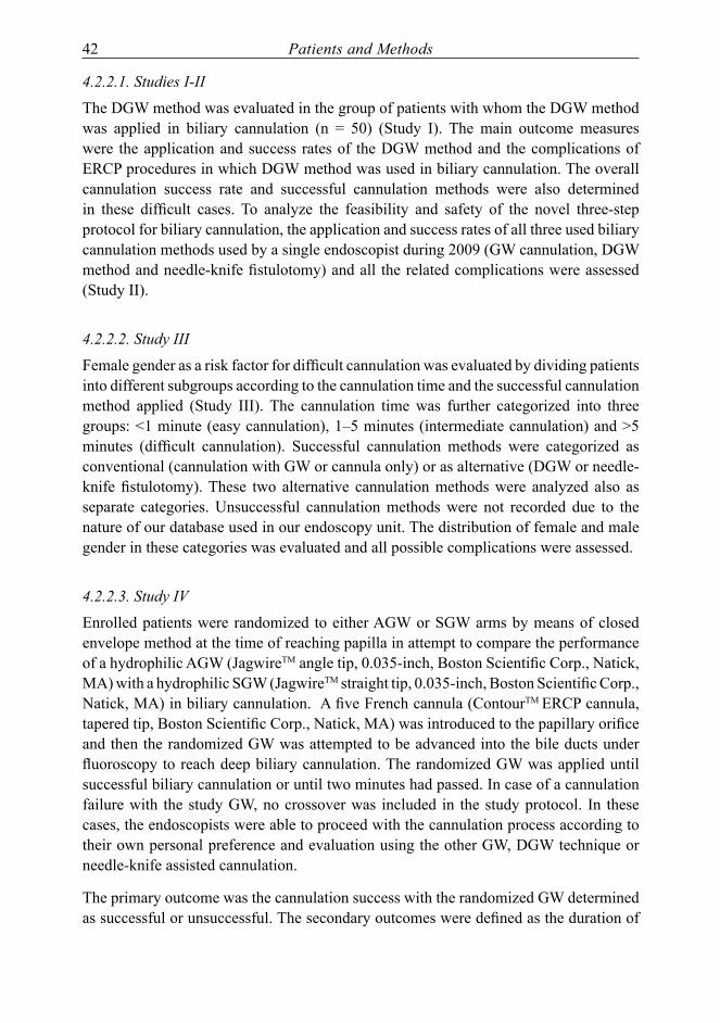
42 Patients and Methods
4.2.2.1. Studies I-II
The DGW method was evaluated in the group of patients with whom the DGW method was applied in biliary cannulation (n = 50) (Study I). The main outcome measures were the application and success rates of the DGW method and the complications of ERCP procedures in which DGW method was used in biliary cannulation. The overall cannulation success rate and successful cannulation methods were also determined in these difficult cases. To analyze the feasibility and safety of the novel three-step protocol for biliary cannulation, the application and success rates of all three used biliary cannulation methods used by a single endoscopist during 2009 (GW cannulation, DGW method and needle-knife fistulotomy) and all the related complications were assessed (Study II).
4.2.2.2. Study III
Female gender as a risk factor for difficult cannulation was evaluated by dividing patients into different subgroups according to the cannulation time and the successful cannulation method applied (Study III). The cannulation time was further categorized into three groups: <1 minute (easy cannulation), 1–5 minutes (intermediate cannulation) and >5 minutes (difficult cannulation). Successful cannulation methods were categorized as conventional (cannulation with GW or cannula only) or as alternative (DGW or needle-knife fistulotomy). These two alternative cannulation methods were analyzed also as separate categories. Unsuccessful cannulation methods were not recorded due to the nature of our database used in our endoscopy unit. The distribution of female and male gender in these categories was evaluated and all possible complications were assessed.
4.2.2.3. Study IV
Enrolled patients were randomized to either AGW or SGW arms by means of closed envelope method at the time of reaching papilla in attempt to compare the performance of a hydrophilic AGW (JagwireTM angle tip, 0.035-inch, Boston Scientific Corp., Natick, MA) with a hydrophilic SGW (JagwireTM straight tip, 0.035-inch, Boston Scientific Corp., Natick, MA) in biliary cannulation. A five French cannula (ContourTM ERCP cannula, tapered tip, Boston Scientific Corp., Natick, MA) was introduced to the papillary orifice and then the randomized GW was attempted to be advanced into the bile ducts under fluoroscopy to reach deep biliary cannulation. The randomized GW was applied until successful biliary cannulation or until two minutes had passed. In case of a cannulation failure with the study GW, no crossover was included in the study protocol. In these cases, the endoscopists were able to proceed with the cannulation process according to their own personal preference and evaluation using the other GW, DGW technique or needle-knife assisted cannulation.
The primary outcome was the cannulation success with the randomized GW determined as successful or unsuccessful. The secondary outcomes were defined as the duration of

Patients and Methods 43
cannulation and the whole ERCP procedure and the immediate and late complications. The endoscopists were categorized as experienced (n = 4) or trainees (n = 1).
4.2.2.4. Study V
To compare the patency of an ARS with conventional PS, enrolled patients (n = 15) were randomized by closed envelope method just prior stent insertion to receive either a ten French ARS (Fusion® Marathon™ Anti-Reflux Biliary Stent, Cook Endoscopy, Winston-Salem, NC) or a conventional ten French PS [QuickPlace V™, DoubleLayer, Olympus Medical Systems Corp., Tokyo, Japan (n = 7) or Flexima™, Boston Scientific Corp., Natick, MA (n = 1)]. An ES and dilatation of the stricture were performed, if needed, prior the stent application. The stent was introduced through the stricture along with the GW. The stent was positioned 1–2 cm proximal to the stricture and its distal end protruding from duodenum with possible anti-reflux mechanism open. The ARS consists of a plastic Tannenbaum stent equipped with an antireflux part in its duodenal end designed to prevent duodenal reflux. This part is made of expanded polytetrafluoroethylene. Tannenbaum stent consists of stainless steel mesh between an inner Teflon coating and outer polyamid layer. DoubleLayer stent is similarly constructed of three layers: the inner layer is made of chemically smoothed Teflon and the outer layer is made of polyamide elastomer and between the outer and inner layers is correspondingly stainless steel mesh. Flexima is made of polyurethane based polymer.
The primary endpoint of the study was stent patency. The stent patency was recorded in days from the stent insertion to the replacement of an occluded stent. Stent occlusion was defined as jaundice that improved after stent replacement. In case of patient dying jaundiced, the stent was regarded as occluded. The follow-up time was considered the patency time if the stent was still functioning after a follow-up period of six months. In case of patient dying with a functioning stent, the time from stent placement to death was regarded as the patency time. The secondary endpoint was cholangitis. Patients were followed up by phone interviews at one, three and six months after stent placement or until stent replacement or death. At the time of each follow-up, blood haemoglobin (B-Hb), fasting blood white blood cell count (fB-WBC), plasma C-reactive protein (P-CRP), plasma alkaline phosphatase (P-AP), plasma bilirubin (P-bil) and serum CA19-9 (S-CA19-9) were measured.
4.2.3. Statistics
Continuous variables were characterized using medians and range of values and categorical variables using frequencies and percents (Study I-V). Differences between genders in continuous variables were tested using Mann-Whitney U-test and the associations between categorical variables were statistically tested using Pearson’s chi-squared test (Study III). Differences between ARS and PS arms in continuous variables were tested using Kruskal-Wallis test and the associations between categorical variables were tested

44 Patients and Methods
using Pearson’s chi-squared test or Fisher’s exact test (Study IV). Differences between stent groups in normally distributed continuous variables were tested using independent sample t-test and Mann-Whitney U-test when appropriate (Study V). Kaplan-Meier curves between stents were compared using log rank test (Study V). P-values less than 0.05 were considered statistically significant. Statistical analyses were performed using SAS system for Windows, Version 9.2 (SAS Institute Inc., Cary, NC) (Study III-V).
4.2.4. Ethics
The review of the medical records and data of the patients was approved by the committee on clinical research of our hospital (Study I-V). The studies IV and V were approved by Ethics Committee of Turku University Hospital and registered in Clinical Trials.gov. The registration numbers were NCT01002404 and NCT00990366, respectively.

Results 45
5. RESULTS
5.1. Biliary cannulation (Studies I-IV)
5.1.1. Cannulation methods
The DGW technique was applied in 18 % (Study I) and 19 % (Study II) of all attempted biliary cannulations with unhindered access to a native papilla. Success rate of the DGW technique was 66 % (Study I) and 65 % (Study II). Successful DGW method accounted for 12 % (13/105) in study II, 10 % (36/364) in study III, and 17 % (26/153) in study IV of all attempted biliary cannulations.
Cannulation success rate with conventional techniques was for cannula with GW 80 % (84/105) in study II and 64 % (98/153) in study IV and for cannula with or without GW 74 % (270/364) in study III. All cannulations were primarily attempted with conventional methods (Study II-IV).
Needle-knife technique was used in 7/105 (7 %) with 100 % success rate in study II. In terms of a failed DGW cannulation, needle-knife fistulotomy was used in 13/17 cases (76 %) and was successful with 12/13 patients (92 %) in study I. The failure was associated with needle-knife associated bleeding, that ceased without a need for endoscopic interventions or blood transfusions, and in repeat ERCP procedure later, the cannulation was successful using conventional technique. Successful needle-knife technique was used in 47 out of 364 patients (13 %) in study III and in 19 out of 153 patients (12 %) in study IV, respectively.
The overall cannulation success for deep biliary cannulation was 99 % (Study II), 97 % (Study III), and 93 % (Study IV).
5.1.2. Cannulation and procedure related time
Median biliary cannulation time with successful DGW method was eight minutes (range 1–31 minutes) and the median duration of entire procedure was 23 minutes (range 11–60) in study I. The median biliary cannulation time with a single endoscopist was one minute (range 0–27) and median duration of entire procedure was 13 minutes (range 3–45) in study II. In study III, the median time for deep biliary cannulation was two minutes (range 0–40) and median procedure duration was 14 minutes (range 3–67 minutes). For study IV, the cannulation process was affected by the study protocol.
5.1.3. Complications
The complications after an attempted DGW technique (n = 50) included one moderate PEP (2%) and one bleeding after needle-knife fistulotomy (Study I).

46 Results
The rate for PEP was 3/105 (3 %) and the rate for cholangitis was 2/105 (2 %) with the total complication rate of 5 % in study II. All the PEPs were of moderate severity: one patient with sclerosing cholangitis and one with suspected bile duct stones were cannulated with cannula and GW within one and nine minutes respectively, and one with bile duct stones was cannulated with DGW technique within eight minutes. Both cases of cholangitis resolved with antibiotic treatment within four days. All of the ERCPs were therapeutic.
ERCP procedure related complications for 364 patients in study III included nine (2 %) PEPs (five mild, two moderate and two severe), five (1 %) bleedings (two patients needing both two units of blood transfusions) and three (1 %) cholangitis (all resolved within four days with antibiotic treatment). The total complication rate was 5%. With the nine cases of PEP, the successful cannulation methods included two cannulations with standard cannula only (both mild), four GW cannulations (one moderate, three mild), two DGW cannulations (both severe) and one precut (moderate).
There were eight (5 %) cases of PEP (four mild and four moderate), five (3 %) immediate bleedings, two (1 %) perforations and three (2 %) cases of unspecified pain in study IV patients with the total complication rate of 10%. The successful cannulation methods associating with PEPs were GW (one mild) and DGW (two mild, two moderate). Three PEPs (two mild, one moderate) were associated with unsuccessful cannulation attempts. One perforation of the CBD associated with the use of AGW and resolved with conservative therapy only and the other perforation resulted from stenting a tight hilar stricture necessitating laparotomy and drainage. No blood transfusions were needed to treat immediate bleedings.
There was no procedure related mortality in any of the studies.
5.2. Associationoffemalegenderanddifficultcannulation(StudyIII)
Deep biliary cannulation was achieved in 353/364 patients with the overall success rate of 97 % in the study population. Cannulation was unsuccessful in eleven patients (five female patients and six male patients) (p = 0.718). The cannulation times of 352/353 patients were analyzed to compare the difference between genders (the data on cannulation time for one patients was not recorded). The median time needed for deep biliary cannulation was two minutes (range 0–40 min) in female and one minute (range 0–22 min) in male patients (p= 0.061). The distribution of the female and male patients into different subgroups according to the time needed for deep biliary cannulation is shown in Table 1. Although the cannulation times seemed to be longer in female patients, no statistically significant difference (p = 0.147) was found between the genders in these subgroups.

Results 47
Table 1. The distribution of the female and male patients [n (%)] into subgroups according to the time needed to achieve deep biliary cannulation defining the cannulation difficulty.
<1 mineasy
1-5 minintermediate
>5 mindifficult Total
female 54 (30%) 73 (41%) 52 (29%) 179male 65 (38%) 72 (42%) 36 (21%) 173
Reproduced with the permission of the copyright holders.(Vihervaara et al. 2011)
The division of the female and male patients into three subgroups according to the particular successful cannulation technique (conventional techniques, needle-knife technique and DGW technique) is shown in Table 2. No significant difference was found between the genders comparing these subgroups. Also the use of the needle-knife technique and DGW technique were also combined in attempt to analyze the difference between female and male patients using alternative techniques compared with conventional techniques (cannula with or without GW) in biliary cannulation; there was a trend towards statistical significance in this comparison (p = 0.054).
Table 2. The division of the female and male patients [n (%)] into three subgroups according to the cannulation technique that proved successful (conventional techniques, needle-knife technique and double-guidewire technique).
conventional techniques
needle-knife technique
double-guidewire technique
total
female 130 (72%) 27 (15%) 23 (13%) 180male 140 (81%) 20 (12%) 13 (8%) 173
Reproduced with the permission of the copyright holders.(Vihervaara et al. 2011)
The median duration of the entire ERCP procedures were 14 minutes (range 3–49 minutes) in female patients and 15 minutes (range 4–67 minutes) in male patients (p = 0.522). ERCP-related complications in female and male gender are shown in Table 3. The incidence of PEP was 2.2 % among females and 2.8 % among males.
Table 3. Complications related to ERCP procedures in female (n=185) and male (n=179) patients.
female 4 post-ERCP pancreatitis 1 mild, 2 moderate, 1 severe3 bleedings 1 needed 2 units of blood transfusions, 2 were observed
in the ward1 cholangitis/infection needle-knife was introduced accidentally into common
bile duct, 4 days at hospital for antibiotic treatment male 5 post-ERCP pancreatitis 4 mild, 1 severe
2 bleedings 1 needed 2 units of blood transfusions, 1 was observed in the ward
2 cholangitis both 3 days at hospital for antibiotic treatmentReproduced with the permission of the copyright holders.(Vihervaara et al., in press)

48 Results
5.3. Biliary cannulation with AGW or SGW (Study IV)
Primary cannulations with the randomized GW technique within two-minute cannulation time were successful in 42/70 (60 %) in the AGW group and in 54/83 (65 %) in the SWG group (p = 0.615). The median time to deep biliary cannulation was 20 seconds with the AGW and 63 seconds with the SGW (p = 0.014). Trainee endoscopist attempted 21/153 (14 %) of the biliary cannulations with a cannulation success rate of 63 % in the AGW arm and 46 % in the SGW arm (p = 0.659). Correspondingly the deep biliary cannulation success rate was 37/62 (60 %) in the AGW arm and 48/70 (69 %) in the SGW arm with the experienced endoscopists (p = 0.362).
Two GW cannulations were successful after crossover to the other GW. DGW technique was applied successfully in 26/153 patients (17 %) and needle-knife fistulotomy resulted in successful biliary cannulation with 19/153 patients (12 %). The overall deep biliary cannulation rate was 93 %. Overall deep biliary cannulation rate in AGW arm was 67/70 (96 %) and in SGW arm 76/83 (92 %) (p = 0.356). There was no statistical difference between the AGW and the SGW arms in the overall cannulation success rate of the trainee endoscopist (p = 0.796) or in that of the experienced endoscopists (p = 0.235).
The occurrence of complications was similar in both study arms. The incidence of complications is shown in Table 4. One PEP occurred after a successful SGW cannulation, but no PEPs were detected after successful AGW cannulation. The incidence of PEP was 8/153 (5 %) in the whole study population.
Table 4 The number (percentage) of complications in the study groups
AGW SGWBleeding* 3 (4.3 %) 2 (2.4 %)Perforation 2 (2.9 %) 0 (0.0 %)Pancreatitis 3 (4.3 %) 5 (6.0 %)Unspecified pain 2 (2.9 %) 1 (1.2 %)
AGW angled tipped guidewire; SGW straight tipped guidewire: * all immediateReproduced with the permission of the copyright holders.(Vihervaara et al. 2011)
5.4. ARS or conventional PS in malignant distal strictures (Study V)
Between October 2009 and May 2010, fifteen jaundiced patients with non-resectable malignant biliary stricture were randomized to receive either an ARS or a PS. Stent insertion was successful in all but one patient, who underwent several unsuccessful insertion attempts of an ARS and finally a conventional PS was inserted. This patient was later excluded from the study due to the benign nature of the stricture (chronic pancreatitis). One patient was also excluded from the PS arm after successful pancreaticoduodenectomy.

Results 49
An interim analysis was performed in May 2010 after the enrollment of these 15 patients based on a clinical suspicion of early occlusions in the ARS arm. After excluding those two aforementioned patients, 13 patients were included in the analysis, seven patients in the PS arm and six patients in the ARS arm. Stent patency was significantly shorter in the ARS arm (p = 0.0003). The median stent patency in the ARS arm was 34 (8–49) days compared to the median patency of 167 (38–214) days in the PS arm. One patient with an ARS had persistent jaundice after stent placement and was considered a primary failure. An early re-intervention (irrigation) was performed eight days after stent placement and this time period was used in the analysis as the patency time. Based on these interim analysis results, the use of ARS was evaluated unethical and the study was prematurely closed with no further enrollment permitted.
The pre-stent laboratory values between the study arms did not differ statistically. CRP and AP values were significantly higher (p = 0.007 and p = 0.028, respectively) in the ARS arm compared to the PS arm at one month follow-up. Detailed laboratory values before stent insertion and at one month follow-up are shown in Table 5. No further analysis on laboratory values was made, because all ARSs were exchanged to a new stent after one month.
Table 5 Laboratory values [median, range (number analyzed)] before stent insertion (-0) and at one month follow-up (-1) and p-values between antireflux and conventional plastic stents
Parameter ARS PS p-value normal rangeB-Hb-0 113, 82-138 (6)F 129, 101-152 (7) 0.14 117-167 g/LB-Hb-1 121, 88-131 (5) 140, 94-147 (7) 0.17
fB-WBC-0 6.1, 4.4-8.8 (6) 6.0, 3.3-7.5 (7) 0.44 3.4 – 8.2 E9/LfB-WBC-1 6.6, 5.3-8.9(5) 7.4, 3.1-12.6 (7) 0.90
P-CRP-0 5, 1-14 (6) 10, 2-32 (7) 0.06 < 10 mg/LP-CRP-1 27, 25-69 (4) 3, 1-8 (7) 0.007
P-AP-0 389, 256-1079 (6) 507, 240-957 (7) 0.86 35-105 U/LP-AP-1 530, 222-943 (4) 109, 75-173 (7) 0.03
P-Bil-0 162, 21-243 (6) 109, 52-297 (7) 0.77 < 21 µmol/LP-Bil-1 30, 13-260 (5) 14, 10-34 (7) 0.06
S-CA19-9-0 92, 5.7-48430 (5) 265, 26-1727 (6) 0.86 < 27 kU/LS-CA19-9-1 137, 87-10000 (3) 123, 13-8585 (6) 0.44
B-Hb, blood haemoglobin, fB-WBC, fasting blood white blood cell count; P-CRP, plasma C-reactive protein; P-AP, plasma alkaline phosphatase; P-Bil, plasma bilirubin; S-CA19-9, serum carbohydrate antigen 19-9; ARS, antireflux stent; PS, plastic stent
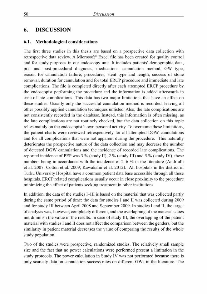
50 Discussion
6. DISCUSSION
6.1. Methodological considerations
The first three studies in this thesis are based on a prospective data collection with retrospective data review. A Microsoft® Excel file has been created for quality control and for study purposes in our endoscopy unit. It includes patients’ demographic data, pre- and post-procedural diagnosis, medications, cannulation method, GW type, reason for cannulation failure, procedures, stent type and length, success of stone removal, duration for cannulation and for total ERCP procedure and immediate and late complications. The file is completed directly after each attempted ERCP procedure by the endoscopist performing the procedure and the information is added afterwards in case of late complications. This data has two major limitations that have an effect on these studies. Usually only the successful cannulation method is recorded, leaving all other possibly applied cannulation techniques unlisted. Also, the late complications are not consistently recorded in the database. Instead, this information is often missing, as the late complications are not routinely checked, but the data collection on this topic relies mainly on the endoscopist’s own personal activity. To overcome these limitations, the patient charts were reviewed retrospectively for all attempted DGW cannulations and for all complications that were not apparent during the procedure. This naturally deteriorates the prospective nature of the data collection and may decrease the number of detected DGW cannulations and the incidence of recorded late complications. The reported incidence of PEP was 3 % (study II), 2 % (study III) and 5 % (study IV), these numbers being in accordance with the incidence of 2–6 % in the literature (Andriulli et al. 2007; Cotton et al. 2009; Kawakami et al. 2012). All hospitals in the district of Turku University Hospital have a common patient data base accessible through all these hospitals. ERCP related complications usually occur in close proximity to the procedure minimizing the effect of patients seeking treatment in other institutions.
In addition, the data of the studies I–III is based on the material that was collected partly during the same period of time: the data for studies I and II was collected during 2009 and for study III between April 2008 and September 2009. In studies I and II, the target of analysis was, however, completely different, and the overlapping of the materials does not diminish the value of the results. In case of study III, the overlapping of the patient material with studies I and II does not affect the comparison between the genders, but the similarity in patient material decreases the value of comparing the results of the whole study population.
Two of the studies were prospective, randomized studies. The relatively small sample size and the fact that no power calculations were performed present a limitation in the study protocols. The power calculation in Study IV was not performed because there is only scarcely data on cannulation success rates on different GWs in the literature. The
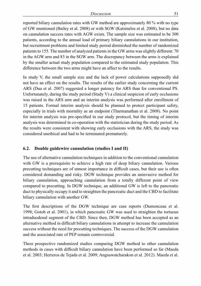
Discussion 51
reported biliary cannulation rates with GW method are approximately 80 % with no type of GW mentioned (Bailey et al. 2008) or with SGW (Katsinelos et al. 2008), but no data on cannulation success rates with AGW exists. The sample size was estimated to be 300 patients, according to the annual load of primary biliary cannulations in our institution, but recruitment problems and limited study period diminished the number of randomized patients to 155. The number of analyzed patients in the GW arms was slightly different: 70 in the AGW arm and 83 in the SGW arm. The discrepancy between the arms is explained by the smaller actual study population compared to the estimated study population. This difference between the two arms might have an affect to the results.
In study V, the small sample size and the lack of power calculations supposedly did not have an effect on the results. The results of the earlier study concerning the current ARS (Dua et al. 2007) suggested a longer patency for ARS than for conventional PS. Unfortunately, during the study period (Study V) a clinical suspicion of early occlusions was raised in the ARS arm and an interim analysis was performed after enrollment of 15 patients. Formal interim analysis should be planned to protect participant safety, especially in trials with mortality as an endpoint (Tharmanathan et al. 2008). No point for interim analysis was pre-specified in our study protocol, but the timing of interim analysis was determined in co-operation with the statistician during the study period. As the results were consistent with showing early occlusions with the ARS, the study was considered unethical and had to be terminated prematurely.
6.2. Double guidewire cannulation (studies I and II)
The use of alternative cannulation techniques in addition to the conventional cannulation with GW is a prerequisite to achieve a high rate of deep biliary cannulation. Various precutting techniques are of utmost importance in difficult cases, but their use is often considered demanding and risky. DGW technique provides an uninvasive method for biliary cannulation, approaching cannulation from a totally different point of view compared to precutting. In DGW technique, an additional GW is left to the pancreatic duct to physically occupy it and to straighten the pancreatic duct and the CBD to facilitate biliary cannulation with another GW.
The first descriptions of the DGW technique are case reports (Dumonceau et al. 1998; Gotoh et al. 2001), in which pancreatic GW was used to straighten the tortuous intraduodenal segment of the CBD. Since then, DGW method has been accepted as an alternative method in difficult biliary cannulations in attempt to increase the cannulation success without the need for precutting techniques. The success of the DGW cannulation and the associated rate of PEP remain controversial.
Three prospective randomized studies comparing DGW method to other cannulation methods in cases with difficult biliary cannulation have been performed so far (Maeda et al. 2003; Herreros de Tejada et al. 2009; Angsuwatcharakon et al. 2012). Maeda et al.

52 Discussion
compared the DGW method to conventional cannulation with cannula only, Herreros de Tejada et al. compared the DGW method to the continuation of the GW assisted cannulation and, finally, Angsuwatcharakon et al. randomized DGW method with precut fistulotomy. The success rate of the DGW technique (27 patients) was significantly higher than that of the conventional technique (93 % vs. 58 %) in the single-center pilot trial of Maeda et al.; no PEP occurred in either of groups. Herreros de Tehada et al. concluded in their multicenter trial that the DGW technique (97 patients) was not superior to the standard GW technique (success rates 47 % and 56 %, respectively) and might be associated with higher risk for PEP (17 % and 8 %, respectively). The cannulation of rate of the DGW technique (23 patients) and the precut technique were comparable (74 % vs. 81 %, respectively) in the single-center trial of Angsuwatcharakon et al. with a tendency for more pancreatitis in the DGW group (17 % vs. 10 %). Case series presenting the DGW technique in difficult biliary cannulation include success rates of 73 % with 113 patients (Ito et al. 2008) and 83 % with 12 patients (Draganov et al. 2005). The rates of PEP were 12 % and 0 %, respectively. Also an additional case series of 24 patients with no success rate reported has been written (Gyokeres et al. 2003). The rate of PEP was 8 %.
The series of 50 patients in study I is one of the largest materials on DGW cannulation in the literature. The success rate for the attempted DGW cannulations was 66 % with the rate of PEP only 2 %. Even though the success rate of the DGW method was relatively low compared to previous studies, the overall cannulation rate was as high as 98 % reflecting the expert level. The low success rate of the DGW technique (47 %) and the relatively high risk for PEP (17 %) in the multicenter trial of Herreros de Tejada et al. (Herreros de Tejada et al. 2009) can be explained by too many low-volume centers participating (n = 6). Also, the median cannulation time was 16 minutes in the former multicenter study, clearly longer compared to eight minutes in study I. In the study of Angsuwatcharakon et al. (Angsuwatcharakon et al. 2012) the rate of PEP was surprisingly high (17 %) with the median cannulation time of only three minutes. However, in their study, ten minutes of standard cannulation was attempted before randomization thus increasing the median cannulation time to 13 minutes. In addition, the number of possible pancreatic duct involvements was not recorded in the preceding ten minutes. In studies I and II, the DGW method was applied after two unintended insertions of GW into pancreatic duct.
The results of study I favor the use of the DGW technique as an alternative cannulation technique. Even though the success rate of DGW technique was only 66%, the overall cannulation rate was 98%. DGW technique offers an alternative to precut, but if the DGW method seems unsatisfactory, additional cannulation methods must be used. The cannulations in study I were performed only by very experienced endoscopists. This naturally can have an affect to the results in favor of the DGW technique. On the other hand, the cannulation rate of the desired duct(s) can be as low as 84% associated with low-volume centers with variable experience (Kapral et al. 2008). Accordingly, the success rate of the DGW method was relatively low (47%) in the multicenter trial of

Discussion 53
Herreros de Tejada et al. including probably too many low-volume centers (Herreros de Tejada et al. 2009). The results of our study reflect the expert level seen in total cannulation rate and in the low rate of pancreatitis. It remains unclear whether the DGW method should be used irrespective of local level of experience according to the results of our study. In experienced hands, the DGW method seems safe and feasible as an alternative cannulation technique.
6.2.1. Three-step protocol
Traditionally, a two-step protocol has been used for deep biliary cannulation, i.e. standard cannula or sphincterotome with or without GW and an alternative technique with at least one of the modifications of pre-cut papillotomy (Siegel 1980). The inclusion of the DGW technique provides a new approach to the previous protocol. Study II attempts to clarify the feasibility and the safety of the novel three-step protocol. In this biliary cannulation protocol, conventional cannulation with cannula and GW is attempted primarily. If the conventional method fails and the GW passes into the pancreatic duct more than once, the DGW method is applied. If the DGW assisted cannulation fails or neither biliary nor pancreatic duct can be cannulated, the needle-knife assisted cannulation is attempted. A similar protocol has been described in a single hospital using a sphincterotome preloaded with a GW in a primary cannulation setting (Xinopoulos et al. 2011). In their study, GW assisted cannulation was restricted to five attempts within 15 minutes. In case of a failed standard cannulation and repeated pancreatic duct insertions with GW, a DGW method was performed up to three times. The needle-knife technique was used as the last resort in case of DGW method failing or if no bile or pancreatic duct insertions were achieved. In study II, there were no time or attempt limits restricting the cannulation process.
The results of the study II and of the previous study (Xinopoulos et al. 2011) support each other. In the study of Xinopoulos et al. the conventional method with sphincterotome and GW resulted in a high cannulation success rate of 92 % (2153/2332). The DGW technique was successful in 44 % (49 out of 112 patients), and precut was successful with 73 % (46/63) of patients after a failed DGW cannulation, and with 81 % (54/67) when neither bile nor pancreatic duct insertion was successful. The success rates of the traditional GW method, the DGW method and the needle-knife fistulotomy were 80 % (84/105), 65 % (13/20) and 100 % (7/7), respectively, in study II. The total cannulation success was as high as 99 % in both of the studies. The total rate of PEP was 3 % in study II and 5 % in the study of Xinoupoulos et al. The novel sequential three-step protocol for biliary cannulation seems to be effective and safe in expert hands according to these studies.
A recent meta-analysis of randomized controlled trials (Cennamo et al. 2010) suggests that early implementation of precut technique vs. persistent biliary cannulation result in similar cannulation rates (90 % in both groups) and total complication rates (5 % in the early precut group and 6 % in persistence group), but the early precut significantly reduces the rate of PEP compared to the persistent biliary cannulation (2 % vs. 5

54 Discussion
%, respectively) in expert hands. In study II, the rate of PEP and similarly the total complication rate were 5 % (1/21) in the group of alternative cannulation techniques. Also, in the same study the median cannulation time was only one minute with the median total procedural time of 13 minutes. The median time to successful DGW cannulation was nine minutes and the median time to successful needle-knife assisted cannulation was 16 minutes. Taken together, in order to keep the PEP rate as low as possible and the cannulation rate as high as possible, it seems to be important to proceed to alternative cannulation techniques relatively quickly in case of difficulties with conventional or DGW cannulation techniques.
6.3. Cannulationdifficulty(StudyIII)
Difficult cannulation involving repeated cannulation attempts and prolonged mechanical manipulation of the papilla carries an increased risk for PEP (Freeman et al. 2001; Vandervoort et al. 2002; Testoni et al. 2010). Difficult cannulation can be regarded as a situation where regularly used cannulation methods fail after a certain time or an attempt limit (Udd et al. 2010). There is no generally accepted definition for difficult cannulation, and thus comparison between different studies is difficult. Time limits between 10 and 15 minutes are often used (Maeda et al. 2003; Kaffes et al. 2005; Xinopoulos et al. 2011), but longer time limits as 20 minutes have also been applied (Fukatsu et al. 2008). Difficult cannulation can also be defined as a number of unsuccessful cannulation attempts; the limit ranging from five (Herreros de Tejada et al. 2009) to 15 (Freeman et al. 2001) attempts. Also the number of pancreatic injections with contrast media (Kaffes et al. 2005) and number of unintended pancreatic duct insertions (Lee et al. 2009) can be used to set a limit for a difficult biliary cannulation.
The time limit for difficult biliary cannulation in study III was only five minutes; the limit which is not so commonly used in the literature. We find that the avoidance of prolonged and repeated trauma to the papilla and to the pancreatic duct is of pivotal importance for successful and complication-free biliary cannulation. The cannulation time was categorized into three groups because we wanted to distinguish not only the difficult cannulations but also the easy cannulations causing only minimal trauma to papilla. Another approach to difficult cannulation in study III was to categorize biliary cannulations into three subgroups according to the successful cannulation method used: conventional cannulation with or without GW, DGW and needle-knife technique. By using the cut-off level of five minutes for difficult biliary cannulation, 25 % of the successful cannulations were considered difficult. This percentage reflects also the use of alternative cannulation methods (24 %) in the same study. The rate of difficult biliary cannulations varies between 10 and 20 % in the literature, depending on the chosen effective primary cannulation technique and the chosen limit for difficult cannulation (Artifon et al. 2007; Lee et al. 2009; Xinopoulos et al. 2011). Even though the percentage of difficult biliary cannulations (25 %) in study III was relatively high, the rate of PEP was only 2 %. The percentage of difficult cannulations defined by alternative cannulation

Discussion 55
methods was also influenced by the tendency of proceeding to alternative cannulation techniques relatively fast, which can be seen in the short cannulation times (median cannulation time of two minutes in study III). By using the cut-off level of five minutes for difficult cannulation, 25 % of all cannulations (88/352) were considered difficult, whereas, for example, only 11 % (37/352) would have been considered difficult if the cut-off level was 10 minutes.
Female gender has been indentified as an independent predictor for PEP (Freeman et al. 2001; Williams et al. 2007), but also as a predictor of difficult biliary cannulation in one previous study (Fukatsu et al. 2008). Theoretically, the elasticity and laxity of female tissues may cause difficulties in biliary cannulation process (Gronroos et al. 2008). Although the cannulation times seemed to be longer and alternative cannulation techniques seemed to be needed more frequently for successful cannulation in female patients compared to male patients in study III, no statistically significant differences between the genders could be found in our study. The expert level of ERCP endoscopists at our institution may have affected the detection of statistically significant difference in cannulation time between the genders.
6.4. Guidewire cannulation (Study IV)
The successful cannulation rate with GW using cannula or sphincterotome varies usually between 80–90 % in expert hands (Bailey et al. 2008; Katsinelos et al. 2008; Xinopoulos et al. 2011), but can reach even the rate of almost 100 % (Karamanolis et al. 2005). There are no studies in the literature comparing cannulation success between different GWs. The biliary cannulation rate in study IV with AGW and SGW was surprisingly low, 60% and 65%, respectively. This difference in the cannulation rates between our and former studies possibly relates to differences in study protocols. Compared to ten minutes of GW assisted cannulation in the previous studies (Bailey et al. 2008; Katsinelos et al. 2008), only two minutes of cannulation with the randomized GW was assumed in study IV. The total success rate for biliary cannulation including the alternative methods used was 93 %. There are two aspects that might have an effect on the relatively low rate of cannulation success compared to the previous studies (Studies II and III): (1) the cannulation process may have been altered towards aborting the GW method faster since the study protocol included only two minutes of cannulation time with the study GW and (2) this study included independent trainee involvement on the contrary to the studies I - III. Cannulation times were short. The median cannulation time with AGW was only 20 seconds and with SGW 63 seconds. The median cannulation time including all successful methods was 100 seconds, and even for alternative cannulation methods the median time for cannulation was only eight minutes. Longer time limit for the primary cannulation method in the study protocol and persistence in cannulation process might have increased the cannulation success rate.
56 Discussion
The rate of PEP was also slightly higher in study IV (5%) compared to studies I (2%) and III (2%). In study IV, trainee involvement does not explain the slight increase in the PEP rate; only one PEP was associated with trainee cannulating. The cannulation protocol might have had an affect to the PEP rate: if the endoscopists felt uncomfortable cannulating with the randomized GW, they might have aborted the GW cannulation technique and proceeded to the alternative methods earlier than normal. The low success rate of GW cannulation supports this assumption.
The AGW (20 seconds) resulted in faster biliary cannulation than the SGW (63 seconds). The reason for this remains unclear. Speculatively AGW might offer benefit in biliary cannulation in tortuous CBD or in case of distal strictures or impacted stones, but this study was not designed to detect such possible differences.
6.5. ARS vs. conventional PS in malignant distal strictures (Study V)
The exact mechanisms for the PS propensity to clog remain unclear. Dietary fibers and plant material have been found in occluded PSs suggesting that duodenobiliary reflux contributes to stent occlusion (Weickert et al. 2001; van Berkel et al. 2005). The problem with early occlusions has been attempted to be solved by a novel antireflux mechanism in PS (Dua et al. 2007). This antireflux mechanism consists of a windsock-shaped tubular valve, made of expanded polytetrafluoroethylene, attached to the duodenal end of a PS. The first results from the inventor of the current antireflux stent were promising with the median patency of 145 days for ARS and 101 days for PS (p = 0.002) (Dua et al. 2007). Unfortunately, the results of the study V were exactly opposite with a significantly shorter stent patency for ARS compared to a conventional PS, leading to the termination of the study after an interim analysis.
The reason for the discrepancy between these two studies is unclear. In the study V, ES was performed to all patients except one, as in the study by Dua et al. (Dua et al. 2007) none of the patients underwent ES. Even though the placement of a 10 French PS does not usually necessitate ES, the sphincterotomy has neither been shown to affect to the patency of a PS (Giorgio et al. 2004). In study V, the valves of the occluded ARSs were found to be filled with sludge and often twisted 180º, obstructing the bile flow mechanically. In the former study by Dua et al., all the valves were fully extended in the patients with repeat ERCP. The biofilm formation on the inner side of the PS consisting of protein and bacteria may be the initial event in stent clogging (Leung et al. 1988; Speer et al. 1988). The polytetrafluoroethylene antireflux valve may itself increase the friction and the turbulence within the stent, provoking the attachment of bacteria and duodenal contents leading to stent clogging. Primary results of a novel antireflux metal stent with a silicone valve have been promising with a median stent patency of 14 months (Hu et al. 2011).

Conclusions 57
7. CONCLUSIONS
The following conclusions can be made from the present data:
1) The DGW assisted biliary cannulation is a feasible and safe method in expert hands as an alternative cannulation technique in difficult biliary cannulation. It can be used as a part of a three-step cannulation protocol in addition to conventional cannulation with or without GW and pre-cut techniques.
2) Although the cannulation times seemed to be longer and alternative cannulation techniques seemed to be needed more frequently in female patients, no statistically significant association was found between female gender and difficult biliary cannulation. Thus, the mechanism predisposing females to PEP remains speculative.
3) The success and complication rates of biliary cannulation were similar with AGW and SGW. The median time to deep biliary cannulation was significantly shorter with AGW than SGW. Thus, AGW may facilitate biliary cannulation.
4) The patency of the ARS was significantly shorter compared to conventional PSs, concluding that the studied antireflux mechanism does not prolong the patency of a biliary PS.

58 Acknowledgements
8. ACKNOWLEDGEMENTS
This study was carried out at the Department of Surgery, Turku University Hospital, University of Turku, Finland during the years 2009-2012.
I express my gratitude to Professor Matti Laato for giving me the opportunity to do research in his department.
My deepest and sincerest gratitude belongs to my two supervisors, Docent Juha Grönroos and Docent Paulina Salminen. Your constant scientific support and encouragement have made this thesis possible. Thank you, Juha, for your kind and supportive attitude that made me believe in my work. Your effectiveness and preciseness made me want to work harder to be worthy of your excellent guidance. And thank you, Paulina, for pushing me always when I needed it. I admire your capability of remembering all the important and practical things through the whole course of this study even though you had so many other things to remember at the same time. You have been an excellent supervisor and a kind-hearted friend, thank you for that.
I want to thank Docent Johanna Laukkarinen and Docent Markku Heikkinen for reviewing my thesis. Your constructive and professional criticism improved essentially the final content of this thesis.
I wish to thank all my wonderful co-authors. Docent Risto Gullichsen is especially thanked for his translating skills that helped to produce patient information sheets in Swedish. I want to thank Simo Laine, MD, PhD, for the help in data collection. Risto and Simo, it has been a great privilege to follow your vast experience in the field of ERCP, thank you for performing big part of the ERCPs for the study. I have been very fortunate to have been able to work with both of you. You have been great instructors in my yet so short surgical career. I am sincerely grateful to Jukka Karvonen, MD, PhD for his endless computer guidance and picture editing. Special thanks go to Saija Hurme, MSc, and Mari Koivisto, MSc, for their statistical expertise in studies III-V.
I want to thank Mika Helmiö, MD, for doing his share in performing ERCPs in studies IV and V.
I am grateful to Tuulia Vihanto, BA, for her excellent help in the linguistic revision of the thesis.
I want to thank Docent Jari Ovaska, Head of the Unit of Gastroenterological Surgery, for the opportunity to work under his supervision and for being encouraging towards both clinical and scientific work. I sincerely want to thank all my past and present colleges in the Surgical Units 216 and 233. You have made my journey to gastrointestinal surgery enjoyable. Especially I want to thank Saila Kauhanen, MD, PhD, for the friendship and valuable advice.

Acknowledgements 59
The personnel of the Endoscopy unit 227 are warmly thanked for the help with this study and for always being so happy and supportive towards my work. I express my thanks to the personnel at Surgical Unit 216, nurse Anne Laaksonen is especially acknowledged for the help in recruiting the patients for studies IV and V. The colleges and personnel of the Department of Surgery, especially Surgical Units 233 and 250 are thanked for the warm attitude towards me; it has been a pleasure to work with all of you. Secretary Anne Mäkinen is thanked for her help in secretarial matters.
I express my gratitude to all my dear friends, especially Katja, Salla, Sanna, Pauliina, Tanja, Riikka and Hanna. Good friends have been very valuable to me, and I hope that there are still lots of good memories to come. I also want to acknowledge my dear cousin, Kati, who has been there for the ups and downs during past years.
I sincerely thank my father, Tapio Vihervaara and his spouse Kirsi Nurmi for their love and support and, of course, for helping to take care of the children to provide me some time to do my research. My grandmother, Alli Vihervaara is thanked for bringing stability in my life. My warm thanks also go to my god-mother Hannele Helistö for being there during these years.
Finally, I want to thank Ville for giving me the greatest joys of my life. Lotta and Minna, my close-to-daughters, it has been an honour to be able to grow with you and to see you grow. Thank you for letting me to be part of your life. Rebekka and Eliel, my dear children, I thank you everyday for giving me the smile on my face. You are my inspiration.
This study was financially supported by the EVO Foundation of the Department of Surgery, the EVO Foundation of Turku University Hospital, the Gastrointestinal Disorders Research Foundation and the Finnish Medical Foundation.
Turku, December 2012
Hanna Vihervaara

60 References
9. REfERENCES
AGA Institute (2007). “AGA Institute medical position statement on acute pancreatitis.” Gastroenterology 132(5): 2019-2021.
Ahrendt, S. A., H. A. Pitt, A. Nakeeb, A. S. Klein, K. D. Lillemoe, A. N. Kalloo and J. L. Cameron (1999). “Diagnosis and management of cholangiocarcinoma in primary sclerosing cholangitis.” J Gastrointest Surg 3(4): 357-367; discussion 367-358.
Albert, J. G. and J. F. Riemann (2002). “ERCP and MRCP--when and why.” Best Pract Res Clin Gastroenterol 16(3): 399-419.
Aljiffry, M., P. D. Renfrew, M. J. Walsh, M. Laryea and M. Molinari (2011). “Analytical review of diagnosis and treatment strategies for dominant bile duct strictures in patients with primary sclerosing cholangitis.” HPB (Oxford) 13(2): 79-90.
Andriulli, A., S. Loperfido, G. Napolitano, G. Niro, M. R. Valvano, F. Spirito, A. Pilotto and R. Forlano (2007). “Incidence rates of post-ERCP complications: a systematic survey of prospective studies.” Am J Gastroenterol 102(8): 1781-1788.
Angsuwatcharakon, P., R. Rerknimitr, W. Ridtitid, Y. Ponauthai and P. Kullavanijaya (2012). “Success rate and cannulation time between precut sphincterotomy and double-guidewire technique in truly difficult biliary cannulation.” J Gastroenterol Hepatol 27(2): 356-361.
Arhan, M., B. Odemis, E. Parlak, I. Ertugrul and O. Basar (2009). “Migration of biliary plastic stents: experience of a tertiary center.” Surg Endosc 23(4): 769-775.
Artifon, E. L., P. Sakai, J. E. Cunha, B. Halwan, S. Ishioka and A. Kumar (2007). “Guidewire cannulation reduces risk of post-ERCP pancreatitis and facilitates bile duct cannulation.” Am J Gastroenterol 102(10): 2147-2153.
Avgerinos, D. V., O. H. Llaguna, A. Y. Lo, J. Voli and I. M. Leitman (2009). “Management of endoscopic retrograde cholangiopancreatography: related duodenal perforations.” Surg Endosc 23(4): 833-838.
Bailey, A. A., M. J. Bourke, S. J. Williams, P. R. Walsh, M. A. Murray, E. Y. Lee, V. Kwan and P. M. Lynch (2008). “A prospective randomized trial of cannulation technique in ERCP: effects on technical success and post-ERCP pancreatitis.” Endoscopy 40(4): 296-301.
Behm, B., A. Brock, B. W. Clarke, K. Ellen, P. G. Northup, J. M. Dumonceau and M. Kahaleh (2009). “Partially covered self-expandable metallic stents for benign biliary strictures due to chronic pancreatitis.” Endoscopy 41(6): 547-551.
Bertin, P. M., K. Singh and M. E. Arregui (2011). “Laparoscopic transgastric endoscopic retrograde cholangiopancreatography (ERCP) after gastric bypass: case series and a description of technique.” Surg Endosc 25(8): 2592-2596.
Binmoeller, K. F. and T. W. Schafer (2001). “Endoscopic management of bile duct stones.” J Clin Gastroenterol 32(2): 106-118.
Binmoeller, K. F., H. Seifert, H. Gerke, U. Seitz, M. Portis and N. Soehendra (1996). “Papillary roof incision using the Erlangen-type pre-cut papillotome to achieve selective bile duct cannulation.” Gastrointest Endosc 44(6): 689-695.
Binmoeller, K. F., H. Seifert, A. Walter and N. Soehendra (1995). “Transpapillary and transmural drainage of pancreatic pseudocysts.” Gastrointest Endosc 42(3): 219-224.
Blackbourne, L. H., R. C. Earnhardt, C. L. Sistrom, P. Abbitt and R. S. Jones (1994). “The sensitivity and role of ultrasound in the evaluation of biliary obstruction.” Am Surg 60(9): 683-690.
Boberg, K. M., P. Jebsen, O. P. Clausen, A. Foss, L. Aabakken and E. Schrumpf (2006). “Diagnostic benefit of biliary brush cytology in cholangiocarcinoma in primary sclerosing cholangitis.” J Hepatol 45(4): 568-574.
Boix, J. and V. Lorenzo-Zuniga (2011). “Radiation dose to patients during endoscopic retrograde cholangiopancreatography.” World J Gastrointest Endosc 3(7): 140-144.
Borak, G. D., J. Romagnuolo, M. Alsolaiman, E. W. Holt and P. B. Cotton (2009). “Long-term clinical outcomes after endoscopic minor papilla therapy in symptomatic patients with pancreas divisum.” Pancreas 38(8): 903-906.
Bosman F. T., F. Carneiro, R. H, Hruban and N. D. Theise , eds. (2010). “WHO Classification of Tumours of the Digestive System.”Lyon, IARC press
Buls, N., J. Pages, F. Mana and M. Osteaux (2002). “Patient and staff exposure during endoscopic retrograde cholangiopancreatography.” Br J Radiol 75(893): 435-443.
Carpelan-Holmstrom, M., S. Nordling, E. Pukkala, R. Sankila, J. Luttges, G. Kloppel and C. Haglund (2005). “Does anyone survive pancreatic ductal adenocarcinoma? A nationwide study re-evaluating the data of the Finnish Cancer Registry.” Gut 54(3): 385-387.
Catalano, M. F., J. D. Linder, S. George, E. Alcocer and J. E. Geenen (2004). “Treatment of symptomatic

References 61
distal common bile duct stenosis secondary to chronic pancreatitis: comparison of single vs. multiple simultaneous stents.” Gastrointest Endosc 60(6): 945-952.
Cennamo, V., L. Fuccio, R. M. Zagari, L. H. Eusebi, L. Ceroni, L. Laterza, C. Fabbri and F. Bazzoli (2009). “Can a wire-guided cannulation technique increase bile duct cannulation rate and prevent post-ERCP pancreatitis?: A meta-analysis of randomized controlled trials.” Am J Gastroenterol 104(9): 2343-2350.
Cennamo, V., L. Fuccio, R. M. Zagari, L. H. Eusebi, L. Ceroni, L. Laterza, C. Fabbri and F. Bazzoli (2010). “Can early precut implementation reduce endoscopic retrograde cholangiopancreatography-related complication risk? Meta-analysis of randomized controlled trials.” Endoscopy 42(5): 381-388.
Cennamo, V., C. Luigiano, C. Fabbri, A. Maimone, F. Bazzoli, L. Ceroni, C. Morace and E. Jovine (2012). “Cholangioscopy using a new type of cholangioscope for the diagnosis of biliary tract disease: a case series.” Endoscopy 44(9): 878-881.
Chainaki, I. G., M. M. Manolaraki and G. A. Paspatis (2011). “Deep sedation for endoscopic retrograde cholangiopacreatography.” World J Gastrointest Endosc 3(2): 34-39.
Chandra, R. and R. A. Liddle (2009). “Neural and hormonal regulation of pancreatic secretion.” Curr Opin Gastroenterol 25(5): 441-446.
Chandy, G., W. J. Hart and I. C. Roberts-Thomson (1997). “An analysis of the relationship between bile duct stones and periampullary duodenal diverticula.” J Gastroenterol Hepatol 12(1): 29-33.
Cheng, C. L., S. Sherman, J. L. Watkins, J. Barnett, M. Freeman, J. Geenen, M. Ryan, H. Parker, J. T. Frakes, E. L. Fogel, W. B. Silverman, K. S. Dua, G. Aliperti, P. Yakshe, M. Uzer, W. Jones, J. Goff, L. Lazzell-Pannell, A. Rashdan, M. Temkit and G. A. Lehman (2006). “Risk factors for post-ERCP pancreatitis: a prospective multicenter study.” Am J Gastroenterol 101(1): 139-147.
Cheung, J., K. K. Tsoi, W. L. Quan, J. Y. Lau and J. J. Sung (2009). “Guidewire versus conventional contrast cannulation of the common bile duct for the prevention of post-ERCP pancreatitis: a systematic review and meta-analysis.” Gastrointest Endosc 70(6): 1211-1219.
Christensen, M., P. Matzen, S. Schulze and J. Rosenberg (2004). “Complications of ERCP: a prospective study.” Gastrointest Endosc 60(5): 721-731.
Cicek, B., E. Parlak, D. Oguz, S. Disibeyaz, A. S. Koksal and B. Sahin (2006). “Endoscopic treatment of pancreatic fistulas.” Surg Endosc 20(11): 1706-1712.
Coelho, D., J. C. Ardengh, J. M. Eulalio, J. E. Manso, K. Monkemuller and J. F. Coelho (2008).
“Management of infected and sterile pancreatic necrosis by programmed endoscopic necrosectomy.” Dig Dis 26(4): 364-369.
Collins, C., D. Maguire, A. Ireland, E. Fitzgerald and G. C. O’Sullivan (2004). “A prospective study of common bile duct calculi in patients undergoing laparoscopic cholecystectomy: natural history of choledocholithiasis revisited.” Ann Surg 239(1): 28-33.
Corazziari, E., E. A. Shaffer, W. J. Hogan, S. Sherman and J. Toouli (1999). “Functional disorders of the biliary tract and pancreas.” Gut 45 Suppl 2: II48-54.
Cortas, G. A., S. N. Mehta, N. S. Abraham and A. N. Barkun (1999). “Selective cannulation of the common bile duct: a prospective randomized trial comparing standard catheters with sphincterotomes.” Gastrointest Endosc 50(6): 775-779.
Cotton, P. B. (1980). “Non-operative removal of bile duct stones by duodenoscopic sphincterotomy.” Br J Surg 67(1): 1-5.
Cotton, P. B., D. A. Garrow, J. Gallagher and J. Romagnuolo (2009). “Risk factors for complications after ERCP: a multivariate analysis of 11,497 procedures over 12 years.” Gastrointest Endosc 70(1): 80-88.
Cotton, P. B., G. Lehman, J. Vennes, J. E. Geenen, R. C. Russell, W. C. Meyers, C. Liguory and N. Nickl (1991). “Endoscopic sphincterotomy complications and their management: an attempt at consensus.” Gastrointest Endosc 37(3): 383-393.
Covey, A. M. and K. T. Brown (2008). “Percutaneous transhepatic biliary drainage.” Tech Vasc Interv Radiol 11(1): 14-20.
Coyle, W. J., B. C. Pineau, P. R. Tarnasky, W. L. Knapple, L. Aabakken, B. J. Hoffman, J. T. Cunningham, R. H. Hawes and P. B. Cotton (2002). “Evaluation of unexplained acute and acute recurrent pancreatitis using endoscopic retrograde cholangiopancreatography, sphincter of Oddi manometry and endoscopic ultrasound.” Endoscopy 34(8): 617-623.
Crist, D. W., Gadacz, T.R. (1996). Anatomy, embryology, congenital anomalies, and physiology of the gallbladder and extrahepatic biliary ducts, in Shackleford’s Surgery of the Alimentary Tract. 4th ed. Philadelphia, W.B. Saunders.
Davids, P. H., A. K. Groen, E. A. Rauws, G. N. Tytgat and K. Huibregtse (1992). “Randomised trial of self-expanding metal stents versus polyethylene stents for distal malignant biliary obstruction.” Lancet 340(8834-8835): 1488-1492.
de Reuver, P. R., E. A. Rauws, M. Vermeulen, M. G. Dijkgraaf, D. J. Gouma and M. J. Bruno (2007). “Endoscopic treatment of post-surgical bile duct injuries: long term outcome and predictors of success.” Gut 56(11): 1599-1605.

62 References
Diehl, A. K., D. R. Holleman, Jr., J. B. Chapman, W. H. Schwesinger and W. E. Kurtin (1997). “Gallstone size and risk of pancreatitis.” Arch Intern Med 157(15): 1674-1678.
Disario, J. A., M. L. Freeman, D. J. Bjorkman, P. Macmathuna, B. T. Petersen, P. E. Jaffe, T. G. Morales, L. J. Hixson, S. Sherman, G. A. Lehman, M. M. Jamal, F. H. Al-Kawas, M. Khandelwal, J. P. Moore, G. A. Derfus, P. A. Jamidar, F. C. Ramirez, M. E. Ryan, K. L. Woods, D. L. Carr-Locke and S. C. Alder (2004). “Endoscopic balloon dilation compared with sphincterotomy for extraction of bile duct stones.” Gastroenterology 127(5): 1291-1299.
Dong, B. and M. Chen (1987). “Improved sonographic visualization of choledocholithiasis.” J Clin Ultrasound 15(3): 185-190.
Draganov, P., D. A. Devonshire and J. T. Cunningham (2005). “A new technique to assist in difficult bile duct cannulation at the time of endoscopic retrograde cholangiopancreatography.” JSLS 9(2): 218-221.
Draganov, P. V. and C. E. Forsmark (2008). “Prospective evaluation of adverse reactions to iodine-containing contrast media after ERCP.” Gastrointest Endosc 68(6): 1098-1101.
Dua, K. S., N. D. Reddy, V. G. Rao, R. Banerjee, B. Medda and I. Lang (2007). “Impact of reducing duodenobiliary reflux on biliary stent patency: an in vitro evaluation and a prospective randomized clinical trial that used a biliary stent with an antireflux valve.” Gastrointest Endosc 65(6): 819-828.
Dumonceau, J. M., A. Andriulli, J. Deviere, A. Mariani, J. Rigaux, T. H. Baron and P. A. Testoni (2010). “European Society of Gastrointestinal Endoscopy (ESGE) Guideline: prophylaxis of post-ERCP pancreatitis.” Endoscopy 42(6): 503-515.
Dumonceau, J. M., J. Deviere and M. Cremer (1998). “A new method of achieving deep cannulation of the common bile duct during endoscopic retrograde cholangiopancreatography.” Endoscopy 30(7): S80.
Dumonceau, J. M., A. Tringali, D. Blero, J. Deviere, R. Laugiers, D. Heresbach and G. Costamagna (2012). “Biliary stenting: indications, choice of stents and results: European Society of Gastrointestinal Endoscopy (ESGE) clinical guideline.” Endoscopy 44(3): 277-298.
Eickhoff, A., R. Jakobs, A. Leonhardt, J. C. Eickhoff and J. F. Riemann (2001). “Endoscopic stenting for common bile duct stenoses in chronic pancreatitis: results and impact on long-term outcome.” Eur J Gastroenterol Hepatol 13(10): 1161-1167.
Elmunzer, B. J., A. K. Waljee, G. H. Elta, J. R. Taylor, S. M. Fehmi and P. D. Higgins (2008). “A meta-analysis of rectal NSAIDs in the prevention of post-ERCP pancreatitis.” Gut 57(9): 1262-1267.
Enns, R., M. A. Eloubeidi, K. Mergener, P. S. Jowell, M. S. Branch, T. M. Pappas and J. Baillie (2002). “ERCP-related perforations: risk factors and management.” Endoscopy 34(4): 293-298.
Enochsson, L., F. Swahn, U. Arnelo, M. Nilsson, M. Lohr and G. Persson (2010). “Nationwide, population-based data from 11,074 ERCP procedures from the Swedish Registry for Gallstone Surgery and ERCP.” Gastrointest Endosc 72(6): 1175-1184, 1184 e1171-1173.
Ersoz, G., O. Tekesin, A. O. Ozutemiz and F. Gunsar (2003). “Biliary sphincterotomy plus dilation with a large balloon for bile duct stones that are difficult to extract.” Gastrointest Endosc 57(2): 156-159.
Fan, S. T., E. C. Lai, F. P. Mok, C. M. Lo, S. S. Zheng and J. Wong (1993). “Early treatment of acute biliary pancreatitis by endoscopic papillotomy.” N Engl J Med 328(4): 228-232.
Farrell, R. J., A. K. Jain, S. L. Brandwein, H. Wang, R. Chuttani and D. K. Pleskow (2001). “The combination of stricture dilation, endoscopic needle aspiration, and biliary brushings significantly improves diagnostic yield from malignant bile duct strictures.” Gastrointest Endosc 54(5): 587-594.
Ferrone, C. R., M. F. Brennan, M. Gonen, D. G. Coit, Y. Fong, S. Chung, L. Tang, D. Klimstra and P. J. Allen (2008). “Pancreatic adenocarcinoma: the actual 5-year survivors.” J Gastrointest Surg 12(4): 701-706.
Fischer, M., A. Hassan, B. W. Sipe, E. L. Fogel, L. McHenry, S. Sherman, J. L. Watkins, S. Schmidt, L. Lazzell-Pannell and G. A. Lehman (2010). “Endoscopic retrograde cholangiopancreatography and manometry findings in 1,241 idiopathic pancreatitis patients.” Pancreatology 10(4): 444-452.
Freeman, M. L., J. A. DiSario, D. B. Nelson, M. B. Fennerty, J. G. Lee, D. J. Bjorkman, C. S. Overby, J. Aas, M. E. Ryan, G. S. Bochna, M. J. Shaw, H. W. Snady, R. V. Erickson, J. P. Moore and J. P. Roel (2001). “Risk factors for post-ERCP pancreatitis: a prospective, multicenter study.” Gastrointest Endosc 54(4): 425-434.
Freeman, M. L. and N. M. Guda (2005). “ERCP cannulation: a review of reported techniques.” Gastrointest Endosc 61(1): 112-125.
Freeman, M. L., D. B. Nelson, S. Sherman, G. B. Haber, M. E. Herman, P. J. Dorsher, J. P. Moore, M. B. Fennerty, M. E. Ryan, M. J. Shaw, J. D. Lande and A. M. Pheley (1996). “Complications of endoscopic biliary sphincterotomy.” N Engl J Med 335(13): 909-918.
Fukatsu, H., H. Kawamoto, H. Kato, K. Hirao, N. Kurihara, T. Nakanishi, O. Mizuno, Y. Okamoto, T. Ogawa, E. Ishida, H. Okada and K. Sakaguchi (2008). “Evaluation of needle-knife precut
References 63
papillotomy after unsuccessful biliary cannulation, especially with regard to postoperative anatomic factors.” Surg Endosc 22(3): 717-723.
Garcea, G., W. Ngu, C. P. Neal, A. R. Dennison and D. P. Berry (2011). “Bilirubin levels predict malignancy in patients with obstructive jaundice.” HPB (Oxford) 13(6): 426-430.
Garcia-Cano, J. (2008). “ERCP cannulation of a hidden papilla within a duodenal diverticulum.” Endoscopy 40 Suppl 2: E53.
Giorgio, P. D. and L. D. Luca (2004). “Comparison of treatment outcomes between biliary plastic stent placements with and without endoscopic sphincterotomy for inoperable malignant common bile duct obstruction.” World J Gastroenterol 10(8): 1212-1214.
Goff, J. S. (1995). “Common bile duct pre-cut sphincterotomy: transpancreatic sphincter approach.” Gastrointest Endosc 41(5): 502-505.
Gonda, T. A., M. P. Glick, A. Sethi, J. M. Poneros, W. Palmas, S. Iqbal, S. Gonzalez, S. V. Nandula, J. C. Emond, R. S. Brown, V. V. Murty and P. D. Stevens (2012). “Polysomy and p16 deletion by fluorescence in situ hybridization in the diagnosis of indeterminate biliary strictures.” Gastrointest Endosc 75(1): 74-79.
Gong, B., L. Hao, L. Bie, B. Sun and M. Wang (2010). “Does precut technique improve selective bile duct cannulation or increase post-ERCP pancreatitis rate? A meta-analysis of randomized controlled trials.” Surg Endosc 24(11): 2670-2680.
Gotoh, Y., K. Tamada, T. Tomiyama, S. Wada, A. Ohashi, Y. Satoh, T. Higashizawa, T. Miyata, K. Ido and K. Sugano (2001). “A new method for deep cannulation of the bile duct by straightening the pancreatic duct.” Gastrointest Endosc 53(7): 820-822.
Gotthardt, D. N., G. Rudolph, P. Kloters-Plachky, H. Kulaksiz and A. Stiehl (2010). “Endoscopic dilation of dominant stenoses in primary sclerosing cholangitis: outcome after long-term treatment.” Gastrointest Endosc 71(3): 527-534.
Gotzberger, M., M. Pichler and V. Gulberg (2012). “Contrast-enhanced US-guided ERCP for treatment of common bile duct stones in pregnancy.” Gastrointest Endosc.
Groen, A. K., T. Out, K. Huibregtse, B. Delzenne, F. J. Hoek and G. N. Tytgat (1987). “Characterization of the content of occluded biliary endoprostheses.” Endoscopy 19(2): 57-59.
Gronroos, J. M. (2007). “Unsuccessful endoscopic stenting in iatrogenic bile duct injury: remember rendezvous procedure.” Surg Laparosc Endosc Percutan Tech 17(3): 186-189.
Gronroos, J. M., R. Gullichsen, J. Karvonen and P. Salminen (2011). “Double-guidewire-assisted
cannulation in ERCP: novel applications.” Surg Laparosc Endosc Percutan Tech 21(3): e107-109.
Gronroos, J. M., R. Gullichsen, S. Laine and P. Salminen (2010). “Endoscopic palliation of malignant obstructive jaundice in extremely elderly patients: plastic stent is enough.” Minim Invasive Ther Allied Technol 19(2): 122-124.
Gronroos, J. M., M. M. Haapamaki and R. Gullichsen (2001). “Effect of the diameter of the common bile duct on the incidence of bile duct stones in patients with recurrent attacks of right epigastric pain after cholecystectomy.” Eur J Surg 167(10): 767-769.
Gronroos, J. M., S. Laine, P. Salminen, J. Karvonen, M. Lavonius and R. Gullichsen (2008). “Female gender may give rise to difficulties in endoscopic and laparoscopic biliary surgery.” Surg Endosc 22(12): 2761-2762.
Gullichsen, R., M. Lavonius, S. Laine and J. Gronroos (2005). “Needle-knife assisted ERCP.” Surg Endosc 19(9): 1243-1245.
Gyokeres, T., J. Duhl, M. Varsanyi, R. Schwab, M. Burai and A. Pap (2003). “Double guide wire placement for endoscopic pancreaticobiliary procedures.” Endoscopy 35(1): 95-96.
Hall, T. C., A. R. Dennison and G. Garcea (2012). “The diagnosis and management of Sphincter of Oddi dysfunction: a systematic review.” Langenbecks Arch Surg 397(6): 889-898.
Halttunen, J., I. Keranen, M. Udd and L. Kylanpaa (2009). “Pancreatic sphincterotomy versus needle knife precut in difficult biliary cannulation.” Surg Endosc 23(4): 745-749.
Halttunen, J., L. Weckman, E. Kemppainen and M. L. Kylanpaa (2005). “The endoscopic management of pancreatic fistulas.” Surg Endosc 19(4): 559-562.
Harewood, G. C., N. L. Pochron and C. J. Gostout (2005). “Prospective, randomized, controlled trial of prophylactic pancreatic stent placement for endoscopic snare excision of the duodenal ampulla.” Gastrointest Endosc 62(3): 367-370.
Heetun, Z. S., F. Zeb, G. Cullen, G. Courtney and A. R. Aftab (2011). “Biliary sphincter of Oddi dysfunction: response rates after ERCP and sphincterotomy in a 5-year ERCP series and proposal for new practical guidelines.” Eur J Gastroenterol Hepatol 23(4): 327-333.
Hekimoglu, K., Y. Ustundag, A. Dusak, Z. Erdem, B. Karademir, S. Aydemir and S. Gundogdu (2008). “MRCP vs. ERCP in the evaluation of biliary pathologies: review of current literature.” J Dig Dis 9(3): 162-169.
Herreros de Tejada, A., J. L. Calleja, G. Diaz, V. Pertejo, J. Espinel, G. Cacho, J. Jimenez, I. Millan, F. Garcia and L. Abreu (2009). “Double-guidewire technique

64 References
for difficult bile duct cannulation: a multicenter randomized, controlled trial.” Gastrointest Endosc 70(4): 700-709.
Ho, H., A. Mahajan, S. Gosain, A. Jain, A. Brock, M. E. Rehan, K. Ellen, V. M. Shami and M. Kahaleh (2010). “Management of complications associated with partially covered biliary metal stents.” Dig Dis Sci 55(2): 516-522.
Hogan, W. J. and J. E. Geenen (1988). “Biliary dyskinesia.” Endoscopy 20 Suppl 1: 179-183.
Horiuchi, A., Y. Nakayama, M. Kajiyama, N. Kato, T. Kamijima, D. Y. Graham and N. Tanaka (2010). “Biliary stenting in the management of large or multiple common bile duct stones.” Gastrointest Endosc 71(7): 1200-1203 e1202.
Hu, B., T. T. Wang, Z. M. Shi, S. Z. Wang, R. Lu, Y. M. Pan, H. Huang and S. P. Wang (2011). “A novel antireflux metal stent for the palliation of biliary malignancies: a pilot feasibility study (with video).” Gastrointest Endosc 73(1): 143-148.
Huang, C. H., Y. K. Tsou, C. H. Lin and J. H. Tang (2010). “Endoscopic retrograde cholangiopancreatography (ERCP) for intradiverticular papilla: endoclip-assisted biliary cannulation.” Endoscopy 42 Suppl 2: E223-224.
Iacono, C., A. Ruzzenente, T. Campagnaro, L. Bortolasi, A. Valdegamberi and A. Guglielmi (2012). “Role of Preoperative Biliary Drainage in Jaundiced Patients Who Are Candidates for Pancreatoduodenectomy or Hepatic Resection: Highlights and Drawbacks.” Ann Surg.
Ito, K., N. Fujita, Y. Noda, G. Kobayashi, T. Obana, J. Horaguchi, O. Takasawa, S. Koshita and Y. Kanno (2008). “Pancreatic guidewire placement for achieving selective biliary cannulation during endoscopic retrograde cholangio-pancreatography.” World J Gastroenterol 14(36): 5595-5600; discussion 5599.
Iwashita, T., J. G. Lee, S. Shinoura, Y. Nakai, D. H. Park, V. R. Muthusamy and K. J. Chang (2012). “Endoscopic ultrasound-guided rendezvous for biliary access after failed cannulation.” Endoscopy 44(1): 60-65.
Johanson, J. F., M. J. Schmalz and J. E. Geenen (1992). “Incidence and risk factors for biliary and pancreatic stent migration.” Gastrointest Endosc 38(3): 341-346.
Jorgensen, J. E., J. H. Rubenstein, M. M. Goodsitt and G. H. Elta (2010). “Radiation doses to ERCP patients are significantly lower with experienced endoscopists.” Gastrointest Endosc 72(1): 58-65.
Kaassis, M., J. Boyer, R. Dumas, T. Ponchon, D. Coumaros, R. Delcenserie, J. M. Canard, J. Fritsch, J. F. Rey and P. Burtin (2003). “Plastic or metal stents for malignant stricture of the common bile duct? Results of a randomized prospective study.” Gastrointest Endosc 57(2): 178-182.
Kadakia, S. C. and E. Starnes (1992). “Comparison of 10 French gauge stent with 11.5 French gauge stent in patients with biliary tract diseases.” Gastrointest Endosc 38(4): 454-459.
Kaffes, A. J., L. Hourigan, N. De Luca, K. Byth, S. J. Williams and M. J. Bourke (2005). “Impact of endoscopic intervention in 100 patients with suspected postcholecystectomy bile leak.” Gastrointest Endosc 61(2): 269-275.
Kaffes, A. J., P. V. Sriram, G. V. Rao, D. Santosh and D. N. Reddy (2005). “Early institution of pre-cutting for difficult biliary cannulation: a prospective study comparing conventional vs. a modified technique.” Gastrointest Endosc 62(5): 669-674.
Kahaleh, M., J. Tokar, M. R. Conaway, A. Brock, T. Le, R. B. Adams and P. Yeaton (2005). “Efficacy and complications of covered Wallstents in malignant distal biliary obstruction.” Gastrointest Endosc 61(4): 528-533.
Kamisawa, T. (2004). “Clinical significance of the minor duodenal papilla and accessory pancreatic duct.” J Gastroenterol 39(7): 605-615.
Kapral, C., C. Duller, F. Wewalka, E. Kerstan, W. Vogel and F. Schreiber (2008). “Case volume and outcome of endoscopic retrograde cholangiopancreatography: results of a nationwide Austrian benchmarking project.” Endoscopy 40(8): 625-630.
Karamanolis, G., A. Katsikani, N. Viazis, G. Stefanidis, S. Manolakopoulos, S. Sgouros, E. Papadopoulou and A. Mantides (2005). “A prospective cross-over study using a sphincterotome and a guidewire to increase the success rate of common bile duct cannulation.” World J Gastroenterol 11(11): 1649-1652.
Karlsen, T. H., E. Schrumpf and K. M. Boberg (2010). “Primary sclerosing cholangitis.” Best Pract Res Clin Gastroenterol 24(5): 655-666.
Karvonen, J., R. Gullichsen, S. Laine, P. Salminen and J. M. Gronroos (2007). “Bile duct injuries during laparoscopic cholecystectomy: primary and long-term results from a single institution.” Surg Endosc 21(7): 1069-1073.
Karvonen, J., V. Kairisto and J. M. Gronroos (2006). “Stone or stricture as a cause of extrahepatic cholestasis--do liver function tests predict the diagnosis?” Clin Chem Lab Med 44(12): 1453-1456.
Kasher, J. A., J. G. Corasanti, P. R. Tarnasky, L. McHenry, E. Fogel and J. Cunningham (2011). “A multicenter analysis of safety and outcome of removal of a fully covered self-expandable metal stent during ERCP.” Gastrointest Endosc 73(6): 1292-1297.
Katsinelos, P., D. Paikos, J. Kountouras, G. Chatzimavroudis, G. Paroutoglou, I. Moschos, A.

References 65
Gatopoulou, A. Beltsis, C. Zavos and B. Papaziogas (2006). “Tannenbaum and metal stents in the palliative treatment of malignant distal bile duct obstruction: a comparative study of patency and cost effectiveness.” Surg Endosc 20(10): 1587-1593.
Katsinelos, P., G. Paroutoglou, J. Kountouras, G. Chatzimavroudis, C. Zavos, I. Pilpilidis, G. Tzelas and G. Tzovaras (2008). “A comparative study of standard ERCP catheter and hydrophilic guide wire in the selective cannulation of the common bile duct.” Endoscopy 40(4): 302-307.
Kaw, M. and G. J. Brodmerkel, Jr. (2002). “ERCP, biliary crystal analysis, and sphincter of Oddi manometry in idiopathic recurrent pancreatitis.” Gastrointest Endosc 55(2): 157-162.
Kawai, K., Y. Akasaka, K. Murakami, M. Tada and Y. Koli (1974). “Endoscopic sphincterotomy of the ampulla of Vater.” Gastrointest Endosc 20(4): 148-151.
Kawakami, H., H. Maguchi, T. Mukai, T. Hayashi, T. Sasaki, H. Isayama, Y. Nakai, I. Yasuda, A. Irisawa, T. Niido, Y. Okabe, S. Ryozawa, T. Itoi, K. Hanada, M. Kikuyama, Y. Arisaka and S. Kikuchi (2012). “A multicenter, prospective, randomized study of selective bile duct cannulation performed by multiple endoscopists: the BIDMEN study.” Gastrointest Endosc 75(2): 362-372, 372 e361.
Kaya, M., B. T. Petersen, P. Angulo, T. H. Baron, J. C. Andrews, C. J. Gostout and K. D. Lindor (2001). “Balloon dilation compared to stenting of dominant strictures in primary sclerosing cholangitis.” Am J Gastroenterol 96(4): 1059-1066.
Keizman, D., M. Ish-Shalom and F. M. Konikoff (2007). “The clinical significance of bile duct sludge: is it different from bile duct stones?” Surg Endosc 21(5): 769-773.
Keizman, D., M. Ish Shalom and F. M. Konikoff (2006). “Recurrent symptomatic common bile duct stones after endoscopic stone extraction in elderly patients.” Gastrointest Endosc 64(1): 60-65.
Kelsen, D. P., R. Portenoy, H. Thaler, Y. Tao and M. Brennan (1997). “Pain as a predictor of outcome in patients with operable pancreatic carcinoma.” Surgery 122(1): 53-59.
Kipp, B. R., L. M. Stadheim, S. A. Halling, N. L. Pochron, S. Harmsen, D. M. Nagorney, T. J. Sebo, T. M. Therneau, G. J. Gores, P. C. de Groen, T. H. Baron, M. J. Levy, K. C. Halling and L. R. Roberts (2004). “A comparison of routine cytology and fluorescence in situ hybridization for the detection of malignant bile duct strictures.” Am J Gastroenterol 99(9): 1675-1681.
Kondo, S., H. Isayama, M. Akahane, N. Toda, N. Sasahira, Y. Nakai, N. Yamamoto, K. Hirano, Y. Komatsu, M. Tada, H. Yoshida, T. Kawabe, K. Ohtomo and M. Omata (2005). “Detection of
common bile duct stones: comparison between endoscopic ultrasonography, magnetic resonance cholangiography, and helical-computed-tomographic cholangiography.” Eur J Radiol 54(2): 271-275.
Koprowski, H., Z. Steplewski, K. Mitchell, M. Herlyn, D. Herlyn and P. Fuhrer (1979). “Colorectal carcinoma antigens detected by hybridoma antibodies.” Somatic Cell Genet 5(6): 957-971.
Kuran, S., E. Parlak, D. Oguz, B. Cicek, S. Disibeyaz and B. Sahin (2006). “Endoscopic sphincterotomy-induced hemorrhage: treatment with heat probe.” Gastrointest Endosc 63(3): 506-511.
Laasch, H. U., A. Tringali, L. Wilbraham, A. Marriott, R. E. England, M. Mutignani, V. Perri, G. Costamagna and D. F. Martin (2003). “Comparison of standard and steerable catheters for bile duct cannulation in ERCP.” Endoscopy 35(8): 669-674.
Laing, F. C., R. B. Jeffrey and V. W. Wing (1984). “Improved visualization of choledocholithiasis by sonography.” AJR Am J Roentgenol 143(5): 949-952.
Larkin, C. J., A. Workman, R. E. Wright and T. C. Tham (2001). “Radiation doses to patients during ERCP.” Gastrointest Endosc 53(2): 161-164.
Laukkarinen, J., G. Kiudelis, M. Lempinen, S. Raty, H. Pelli, J. Sand, E. Kemppainen, C. Haglund and I. Nordback (2007). “Increased prevalence of subclinical hypothyroidism in common bile duct stone patients.” J Clin Endocrinol Metab 92(11): 4260-4264.
Laukkarinen, J., I. Nordback, J. Mikkonen, P. Karkkainen and J. Sand (2007). “A novel biodegradable biliary stent in the endoscopic treatment of cystic-duct leakage after cholecystectomy.” Gastrointest Endosc 65(7): 1063-1068.
Laukkarinen, J., J. Sand, S. Aittomaki, I. Porsti, P. Koobi, J. Kalliovalkama, O. Silvennoinen and I. Nordback (2002). “Mechanism of the prorelaxing effect of thyroxine on the sphincter of Oddi.” Scand J Gastroenterol 37(6): 667-673.
Laukkarinen, J., J. Sand, V. Autio and I. Nordback (2010). “Bile duct stone procedures are more frequent in patients with hypothyroidism. A large, registry-based, cohort study in Finland.” Scand J Gastroenterol 45(1): 70-74.
Lee, S. P., J. F. Nicholls and H. Z. Park (1992). “Biliary sludge as a cause of acute pancreatitis.” N Engl J Med 326(9): 589-593.
Lee, T. H., H. Park do, J. Y. Park, E. O. Kim, Y. S. Lee, J. H. Park, S. H. Lee, I. K. Chung, H. S. Kim, S. H. Park and S. J. Kim (2009). “Can wire-guided cannulation prevent post-ERCP pancreatitis? A prospective randomized trial.” Gastrointest Endosc 69(3 Pt 1): 444-449.

66 References
Lee, Y. N., J. H. Moon, H. J. Choi, S. K. Min, H. I. Kim, T. H. Lee, Y. D. Cho, S. H. Park and S. J. Kim (2012). “Direct peroral cholangioscopy using an ultraslim upper endoscope for management of residual stones after mechanical lithotripsy for retained common bile duct stones.” Endoscopy 44(9): 819-824.
Lee, Y. T., F. K. Chan, W. K. Leung, H. L. Chan, J. C. Wu, M. Y. Yung, E. K. Ng, J. Y. Lau and J. J. Sung (2008). “Comparison of EUS and ERCP in the investigation with suspected biliary obstruction caused by choledocholithiasis: a randomized study.” Gastrointest Endosc 67(4): 660-668.
Leung, J. W., T. K. Ling, J. L. Kung and J. Vallance-Owen (1988). “The role of bacteria in the blockage of biliary stents.” Gastrointest Endosc 34(1): 19-22.
Lindberg, B., L. Enochsson, B. Tribukait, U. Arnelo and A. Bergquist (2006). “Diagnostic and prognostic implications of DNA ploidy and S-phase evaluation in the assessment of malignancy in biliary strictures.” Endoscopy 38(6): 561-565.
Lindner, H. H., V. A. Pena and R. A. Ruggeri (1976). “A clinical and anatomical study of anomalous terminations of the common bile duct into the duodenum.” Ann Surg 184(5): 626-632.
Liu, Y. D., Z. Q. Wang, X. D. Wang, Y. S. Yang, E. Q. Linghu, W. F. Wang, W. Li and F. C. Cai (2007). “Stent implantation through rendezvous technique of PTBD and ERCP: the treatment of obstructive jaundice.” J Dig Dis 8(4): 198-202.
Lohr, J. M., L. Aabaken, U. Arnelo, J. Gronroos, J. Halttunen, T. Hauge, E. Jonas, P. M. Kleveland, P. Nordblad Schmidt, F. Swahn, A. Saarela, E. Toth and S. Meisner (2012). “How to cannulate? A survey of the Scandinavian Association for Digestive Endoscopy (SADE) in 141 endoscopists.” Scand J Gastroenterol 47(7): 861-869.
Loperfido, S., G. Angelini, G. Benedetti, F. Chilovi, F. Costan, F. De Berardinis, M. De Bernardin, A. Ederle, P. Fina and A. Fratton (1998). “Major early complications from diagnostic and therapeutic ERCP: a prospective multicenter study.” Gastrointest Endosc 48(1): 1-10.
Macintosh, D. G., J. Love and N. S. Abraham (2004). “Endoscopic sphincterotomy by using pure-cut electrosurgical current and the risk of post-ERCP pancreatitis: a prospective randomized trial.” Gastrointest Endosc 60(4): 551-556.
Maeda, S., H. Hayashi, O. Hosokawa, K. Dohden, M. Hattori, M. Morita, E. Kidani, N. Ibe and S. Tatsumi (2003). “Prospective randomized pilot trial of selective biliary cannulation using pancreatic guide-wire placement.” Endoscopy 35(9): 721-724.
Mao, Z., Q. Zhu, W. Wu, M. Wang, J. Li, A. Lu, Y. Sun and M. Zheng (2008). “Duodenal perforations after endoscopic retrograde cholangiopancreatography:
experience and management.” J Laparoendosc Adv Surg Tech A 18(5): 691-695.
Marrelli, D., S. Caruso, C. Pedrazzani, A. Neri, E. Fernandes, M. Marini, E. Pinto and F. Roviello (2009). “CA19-9 serum levels in obstructive jaundice: clinical value in benign and malignant conditions.” Am J Surg 198(3): 333-339.
Materne, R., B. E. Van Beers, J. F. Gigot, J. Jamart, A. Geubel, J. Pringot and P. Deprez (2000). “Extrahepatic biliary obstruction: magnetic resonance imaging compared with endoscopic ultrasonography.” Endoscopy 32(1): 3-9.
Mazaki, T., H. Masuda and T. Takayama (2010). “Prophylactic pancreatic stent placement and post-ERCP pancreatitis: a systematic review and meta-analysis.” Endoscopy 42(10): 842-853.
Mazanikov, M., M. Udd, L. Kylanpaa, O. Lindstrom, P. Aho, J. Halttunen, M. Farkkila and R. Poyhia (2011). “Patient-controlled sedation with propofol and remifentanil for ERCP: a randomized, controlled study.” Gastrointest Endosc 73(2): 260-266.
McCune, W. S., P. E. Shorb and H. Moscovitz (1968). “Endoscopic cannulation of the ampulla of vater: a preliminary report.” Ann Surg 167(5): 752-756.
Misra, S. P., P. Gulati, V. K. Thorat, J. C. Vij and B. S. Anand (1989). “Pancreaticobiliary ductal union in biliary diseases. An endoscopic retrograde cholangiopancreatographic study.” Gastroenterology 96(3): 907-912.
Morris-Stiff, G., M. Teli, N. Jardine and M. C. Puntis (2009). “CA19-9 antigen levels can distinguish between benign and malignant pancreaticobiliary disease.” Hepatobiliary Pancreat Dis Int 8(6): 620-626.
Moss, A. C., E. Morris, J. Leyden and P. MacMathuna (2007). “Do the benefits of metal stents justify the costs? A systematic review and meta-analysis of trials comparing endoscopic stents for malignant biliary obstruction.” Eur J Gastroenterol Hepatol 19(12): 1119-1124.
Moss, A. C., E. Morris, J. Leyden and P. MacMathuna (2007). “Malignant distal biliary obstruction: a systematic review and meta-analysis of endoscopic and surgical bypass results.” Cancer Treat Rev 33(2): 213-221.
Navaneethan, U., V. Jayanthi and P. Mohan (2011). “Pathogenesis of cholangitis in obstructive jaundice-revisited.” Minerva Gastroenterol Dietol 57(1): 97-104.
Nicholl, M., H. A. Pitt, P. Wolf, J. Cooney, M. Kalayoglu, J. Shilyansky and L. F. Rikkers (2004). “Choledochal cysts in western adults: complexities compared to children.” J Gastrointest Surg 8(3): 245-252.
NIH Consens State Sci Statements. (2002). “NIH state-of-the-science statement on endoscopic retrograde

References 67
cholangiopancreatography (ERCP) for diagnosis and therapy.” NIH Consens State Sci Statements 19(1): 1-26.
Ong, T. Z., J. L. Khor, D. S. Selamat, K. G. Yeoh and K. Y. Ho (2005). “Complications of endoscopic retrograde cholangiography in the post-MRCP era: a tertiary center experience.” World J Gastroenterol 11(33): 5209-5212.
Oria, A., D. Cimmino, C. Ocampo, W. Silva, G. Kohan, H. Zandalazini, C. Szelagowski and L. Chiappetta (2007). “Early endoscopic intervention versus early conservative management in patients with acute gallstone pancreatitis and biliopancreatic obstruction: a randomized clinical trial.” Ann Surg 245(1): 10-17.
Park do, H., M. H. Kim, S. H. Moon, S. S. Lee, D. W. Seo and S. K. Lee (2008). “Feasibility and safety of placement of a newly designed, fully covered self-expandable metal stent for refractory benign pancreatic ductal strictures: a pilot study (with video).” Gastrointest Endosc 68(6): 1182-1189.
Parsi, M. A. (2011). “Peroral cholangioscopy in the new millennium.” World J Gastroenterol 17(1): 1-6.
Pasha, S. F., M. E. Harrison, A. Das, C. C. Nguyen, H. E. Vargas, V. Balan, T. J. Byrne, D. D. Douglas and D. C. Mulligan (2007). “Endoscopic treatment of anastomotic biliary strictures after deceased donor liver transplantation: outcomes after maximal stent therapy.” Gastrointest Endosc 66(1): 44-51.
Perini, R. F., R. Sadurski, P. B. Cotton, R. S. Patel, R. H. Hawes and J. T. Cunningham (2005). “Post-sphincterotomy bleeding after the introduction of microprocessor-controlled electrosurgery: does the new technology make the difference?” Gastrointest Endosc 61(1): 53-57.
Petelin, J. B. (2003). “Laparoscopic common bile duct exploration.” Surg Endosc 17(11): 1705-1715.
Polydorou, A., A. Vezakis, G. Fragulidis, D. Katsarelias, C. Vagianos and G. Polymeneas (2011). “A tailored approach to the management of perforations following endoscopic retrograde cholangiopancreatography and sphincterotomy.” J Gastrointest Surg 15(12): 2211-2217.
Ponsioen, C. Y., K. Lam, A. W. van Milligen de Wit, K. Huibregtse and G. N. Tytgat (1999). “Four years experience with short term stenting in primary sclerosing cholangitis.” Am J Gastroenterol 94(9): 2403-2407.
Ponsioen, C. Y., S. M. Vrouenraets, A. W. van Milligen de Wit, P. Sturm, M. Tascilar, G. J. Offerhaus, M. Prins, K. Huibregtse and G. N. Tytgat (1999). “Value of brush cytology for dominant strictures in primary sclerosing cholangitis.” Endoscopy 31(4): 305-309.
Pozsar, J., P. Sahin, F. Laszlo, G. Forro and L. Topa (2004). “Medium-term results of endoscopic
treatment of common bile duct strictures in chronic calcifying pancreatitis with increasing numbers of stents.” J Clin Gastroenterol 38(2): 118-123.
Prat, F., O. Chapat, B. Ducot, T. Ponchon, G. Pelletier, J. Fritsch, A. D. Choury and C. Buffet (1998). “A randomized trial of endoscopic drainage methods for inoperable malignant strictures of the common bile duct.” Gastrointest Endosc 47(1): 1-7.
Qin, L. X. and Z. Y. Tang (2003). “Hepatocellular carcinoma with obstructive jaundice: diagnosis, treatment and prognosis.” World J Gastroenterol 9(3): 385-391.
Raimondi, S., A. B. Lowenfels, A. M. Morselli-Labate, P. Maisonneuve and R. Pezzilli (2010). “Pancreatic cancer in chronic pancreatitis; aetiology, incidence, and early detection.” Best Pract Res Clin Gastroenterol 24(3): 349-358.
Ramchandani, M., D. N. Reddy, R. Gupta, S. Lakhtakia, M. Tandan, S. Darisetty, A. Sekaran and G. V. Rao (2011). “Role of single-operator peroral cholangioscopy in the diagnosis of indeterminate biliary lesions: a single-center, prospective study.” Gastrointest Endosc 74(3): 511-519.
Rebelo, A., P. M. Ribeiro, A. P. Correia and J. Cotter (2012). “Endoscopic papillary large balloon dilation after limited sphincterotomy for difficult biliary stones.” World J Gastrointest Endosc 4(5): 180-184.
Reddy, S. C. and P. V. Draganov (2009). “ERCP wire systems: the long and the short of it.” World J Gastroenterol 15(1): 55-60.
Ridtitid, W., R. Rerknimitr, A. Janchai, P. Kongkam, S. Treeprasertsuk and P. Kullavanijaya (2010). “Outcome of second interventions for occluded metallic stents in patients with malignant biliary obstruction.” Surg Endosc 24(9): 2216-2220.
Rogart, J. N., A. Boghos, F. Rossi, H. Al-Hashem, U. D. Siddiqui, P. Jamidar and H. Aslanian (2008). “Analysis of endoscopic management of occluded metal biliary stents at a single tertiary care center.” Gastrointest Endosc 68(4): 676-682.
Romagnuolo, J., M. Bardou, E. Rahme, L. Joseph, C. Reinhold and A. N. Barkun (2003). “Magnetic resonance cholangiopancreatography: a meta-analysis of test performance in suspected biliary disease.” Ann Intern Med 139(7): 547-557.
Rosch, T., S. Daniel, M. Scholz, K. Huibregtse, M. Smits, T. Schneider, C. Ell, G. Haber, J. F. Riemann, R. Jakobs, R. Hintze, A. Adler, H. Neuhaus, M. Zavoral, F. Zavada, V. Schusdziarra and N. Soehendra (2002). “Endoscopic treatment of chronic pancreatitis: a multicenter study of 1000 patients with long-term follow-up.” Endoscopy 34(10): 765-771.
Ross, A. S. and R. A. Kozarek (2010). “Therapeutic pancreatic endoscopy.” Dig Liver Dis 42(11): 749-756.

68 References
Sahni, V. A. and K. J. Mortele (2008). “Magnetic resonance cholangiopancreatography: current use and future applications.” Clin Gastroenterol Hepatol 6(9): 967-977.
Salminen, P., S. Laine and R. Gullichsen (2008). “Severe and fatal complications after ERCP: analysis of 2555 procedures in a single experienced center.” Surg Endosc 22(9): 1965-1970.
Samuelson, A. L. and R. J. Shah (2012). “Endoscopic management of pancreatic pseudocysts.” Gastroenterol Clin North Am 41(1): 47-62.
Sand, J., P. Arvola and I. Nordback (2005). “Calcium channel antagonists and inhibition of human sphincter of Oddi contractions.” Scand J Gastroenterol 40(12): 1394-1397.
Sandha, G. S., M. J. Bourke, G. B. Haber and P. P. Kortan (2004). “Endoscopic therapy for bile leak based on a new classification: results in 207 patients.” Gastrointest Endosc 60(4): 567-574.
Sauer, P., F. Chahoud, D. Gotthardt, W. Stremmel, K. H. Weiss, M. Buchler, P. Schemmer, J. Weitz and A. Schaible (2012). “Temporary placement of fully covered self-expandable metal stents in biliary complications after liver transplantation.” Endoscopy 44(5): 536-538.
Schneider, A. and M. V. Singer (2005). “Alcoholic pancreatitis.” Dig Dis 23(3-4): 222-231.
Senturk, S., T. C. Miroglu, A. Bilici, H. Gumus, R. C. Tekin, F. Ekici and G. Tekbas (2012). “Diameters of the common bile duct in adults and postcholecystectomy patients: a study with 64-slice CT.” Eur J Radiol 81(1): 39-42.
Shah, T., S. Desai, M. Haque, H. Dakik and D. Fisher (2012). “Management of occluded metal stents in malignant biliary obstruction: similar outcomes with second metal stents compared to plastic stents.” Dig Dis Sci 57(11): 2765-2773.
Sharma, S. K., K. A. Larson, Z. Adler and M. A. Goldfarb (2003). “Role of endoscopic retrograde cholangiopancreatography in the management of suspected choledocholithiasis.” Surg Endosc 17(6): 868-871.
Shayan, H., D. Kopac and C. B. Sample (2007). “The role of intraoperative cholangiogram in the management of patients recovering from acute biliary pancreatitis.” Surg Endosc 21(9): 1549-1552.
Shimatani, M., M. Matsushita, M. Takaoka, M. Koyabu, T. Ikeura, K. Kato, T. Fukui, K. Uchida and K. Okazaki (2009). “Effective “short” double-balloon enteroscope for diagnostic and therapeutic ERCP in patients with altered gastrointestinal anatomy: a large case series.” Endoscopy 41(10): 849-854.
Siegel, J. H. (1980). “Precut papillotomy: a method to improve success of ERCP and papillotomy.” Endoscopy 12(3): 130-133.
Smith, A. C., J. F. Dowsett, R. C. Russell, A. R. Hatfield and P. B. Cotton (1994). “Randomised trial of endoscopic stenting versus surgical bypass in malignant low bileduct obstruction.” Lancet 344(8938): 1655-1660.
Smoczynski, M., A. Jablonska, A. Matyskiel, J. Lakomy, M. Dubowik, I. Marek, W. Biernat and J. Limon (2012). “Routine brush cytology and fluorescence in situ hybridization for assessment of pancreatobiliary strictures.” Gastrointest Endosc 75(1): 65-73.
So, Y. H., Y. H. Choi, J. W. Chung, H. J. Jae, S. Y. Song and J. H. Park (2012). “Selective embolization for post-endoscopic sphincterotomy bleeding: technical aspects and clinical efficacy.” Korean J Radiol 13(1): 73-81.
Soehendra, N. and V. Reynders-Frederix (1980). “Palliative bile duct drainage - a new endoscopic method of introducing a transpapillary drain.” Endoscopy 12(1): 8-11.
Speer, A. G., P. B. Cotton and K. D. MacRae (1988). “Endoscopic management of malignant biliary obstruction: stents of 10 French gauge are preferable to stents of 8 French gauge.” Gastrointest Endosc 34(5): 412-417.
Speer, A. G., P. B. Cotton, J. Rode, A. M. Seddon, C. R. Neal, J. Holton and J. W. Costerton (1988). “Biliary stent blockage with bacterial biofilm. A light and electron microscopy study.” Ann Intern Med 108(4): 546-553.
Stefanidis, G., N. Viazis, D. Pleskow, S. Manolakopoulos, L. Theocharis, C. Christodoulou, N. Kotsikoros, J. Giannousis, S. Sgouros, M. Rodias, A. Katsikani and R. Chuttani (2011). “Large balloon dilation vs. mechanical lithotripsy for the management of large bile duct stones: a prospective randomized study.” Am J Gastroenterol 106(2): 278-285.
Stockberger, S. M., J. L. Wass, S. Sherman, G. A. Lehman and K. K. Kopecky (1994). “Intravenous cholangiography with helical CT: comparison with endoscopic retrograde cholangiography.” Radiology 192(3): 675-680.
Strange, R. C. (1984). “Hepatic bile flow.” Physiol Rev 64(4): 1055-1102.
Suda, K., T. Miyano, I. Konuma and M. Matsumoto (1983). “An abnormal pancreatico-choledocho-ductal junction in cases of biliary tract carcinoma.” Cancer 52(11): 2086-2088.
Suk, K. T., J. W. Kim, H. S. Kim, S. K. Baik, S. J. Oh, S. J. Lee, H. G. Kim, D. H. Lee, Y. H. Won and D. K. Lee (2007). “Human application of a metallic stent covered with a paclitaxel-incorporated membrane for malignant biliary obstruction: multicenter pilot study.” Gastrointest Endosc 66(4): 798-803.

References 69
Sung, J. J., S. C. Chung, C. P. Tsui, A. L. Co and A. K. Li (1994). “Omitting side-holes in biliary stents does not improve drainage of the obstructed biliary system: a prospective randomized trial.” Gastrointest Endosc 40(3): 321-325.
Tang, S. J., M. J. Mayo, E. Rodriguez-Frias, L. Armstrong, L. Tang, J. Sreenarasimhaiah, L. F. Lara and D. C. Rockey (2009). “Safety and utility of ERCP during pregnancy.” Gastrointest Endosc 69(3 Pt 1): 453-461.
Tarantino, I., B. Mangiavillano, R. Di Mitri, L. Barresi, F. Mocciaro, A. Granata, E. Masci, G. Curcio, M. Di Pisa, A. Marino and M. Traina (2012). “Fully covered self-expandable metallic stents in benign biliary strictures: a multicenter study on efficacy and safety.” Endoscopy 44(10): 923-927.
Telford, J. J., D. L. Carr-Locke, T. H. Baron, J. M. Poneros, B. C. Bounds, P. B. Kelsey, R. H. Schapiro, C. S. Huang, D. R. Lichtenstein, B. C. Jacobson, J. R. Saltzman, C. C. Thompson, D. G. Forcione, C. J. Gostout and W. R. Brugge (2010). “A randomized trial comparing uncovered and partially covered self-expandable metal stents in the palliation of distal malignant biliary obstruction.” Gastrointest Endosc 72(5): 907-914.
Terruzzi, V., F. Radaelli, G. Meucci and G. Minoli (2005). “Is the supine position as safe and effective as the prone position for endoscopic retrograde cholangiopancreatography? A prospective randomized study.” Endoscopy 37(12): 1211-1214.
Testoni, P. A., A. Mariani, A. Giussani, C. Vailati, E. Masci, G. Macarri, L. Ghezzo, L. Familiari, N. Giardullo, M. Mutignani, G. Lombardi, G. Talamini, A. Spadaccini, R. Briglia and L. Piazzi (2010). “Risk factors for post-ERCP pancreatitis in high- and low-volume centers and among expert and non-expert operators: a prospective multicenter study.” Am J Gastroenterol 105(8): 1753-1761.
Tharmanathan, P., M. Calvert, J. Hampton and N. Freemantle (2008). “The use of interim data and Data Monitoring Committee recommendations in randomized controlled trial reports: frequency, implications and potential sources of bias.” BMC Med Res Methodol 8: 12.
Thornton, J. R., A. J. Lobo, D. J. Lintott and A. T. Axon (1992). “Value of ultrasound and liver function tests in determining the need for endoscopic retrograde cholangiopancreatography in unexplained abdominal pain.” Gut 33(11): 1559-1561.
Tringali, A., M. Mutignani, A. Milano, V. Perri and G. Costamagna (2008). “No difference between supine and prone position for ERCP in conscious sedated patients: a prospective randomized study.” Endoscopy 40(2): 93-97.
Tringali, A., M. Mutignani, V. Perri, G. Zuccala, L. Cipolletta, M. A. Bianco, G. Rotondano,
M. Philipper, B. Schumacher, H. Neuhaus, A. Schmit, J. Deviere and G. Costamagna (2003). “A prospective, randomized multicenter trial comparing DoubleLayer and polyethylene stents for malignant distal common bile duct strictures.” Endoscopy 35(12): 992-997.
Tse, F. and Y. Yuan (2012). “Early routine endoscopic retrograde cholangiopancreatography strategy versus early conservative management strategy in acute gallstone pancreatitis.” Cochrane Database Syst Rev 5: CD009779.
Tseng, C. W., C. C. Chen, T. S. Chen, F. Y. Chang, H. C. Lin and S. D. Lee (2008). “Can computed tomography with coronal reconstruction improve the diagnosis of choledocholithiasis?” J Gastroenterol Hepatol 23(10): 1586-1589.
Tsou, Y. K., C. H. Lin, N. J. Liu, J. H. Tang, K. F. Sung, C. L. Cheng and C. S. Lee (2009). “Treating delayed endoscopic sphincterotomy-induced bleeding: epinephrine injection with or without thermotherapy.” World J Gastroenterol 15(38): 4823-4828.
Tuvignon, N., C. Liguory, T. Ponchon, B. Meduri, J. Fritsch, J. Sahel, J. Boyer, J. L. Legoux, J. Escourrou, C. Boustiere, J. P. Arpurt, M. Barthet, P. Tuvignon, G. Bommelaer, B. Ducot and F. Prat (2011). “Long-term follow-up after biliary stent placement for postcholecystectomy bile duct strictures: a multicenter study.” Endoscopy 43(3): 208-216.
Tyson, G. L. and H. B. El-Serag (2011). “Risk factors for cholangiocarcinoma.” Hepatology 54(1): 173-184.
Tzovaras, G., I. Baloyiannis, E. Zachari, D. Symeonidis, D. Zacharoulis, A. Kapsoritakis, G. Paroutoglou and S. Potamianos (2012). “Laparoendoscopic rendezvous versus preoperative ERCP and laparoscopic cholecystectomy for the management of cholecysto-choledocholithiasis: interim analysis of a controlled randomized trial.” Ann Surg 255(3): 435-439.
Udd, M., L. Kylanpaa and J. Halttunen (2010). “Management of difficult bile duct cannulation in ERCP.” World J Gastrointest Endosc 2(3): 97-103.
van Berkel, A. M., J. van Marle, A. K. Groen and M. J. Bruno (2005). “Mechanisms of biliary stent clogging: confocal laser scanning and scanning electron microscopy.” Endoscopy 37(8): 729-734.
Van Laethem, J. L., S. De Broux, P. Eisendrath, M. Cremer, O. Le Moine and J. Deviere (2003). “Clinical impact of biliary drainage and jaundice resolution in patients with obstructive metastases at the hilum.” Am J Gastroenterol 98(6): 1271-1277.
Vandervoort, J., R. M. Soetikno, T. C. Tham, R. C. Wong, A. P. Ferrari, Jr., H. Montes, A. D. Roston, A. Slivka, D. R. Lichtenstein, F. W. Ruymann, J.

70 References
Van Dam, M. Hughes and D. L. Carr-Locke (2002). “Risk factors for complications after performance of ERCP.” Gastrointest Endosc 56(5): 652-656.
Weber, A., T. Mittermeyer, S. Wagenpfeil, R. M. Schmid and C. Prinz (2009). “Self-expanding metal stents versus polyethylene stents for palliative treatment in patients with advanced pancreatic cancer.” Pancreas 38(1): e7-e12.
Weickert, U., T. Venzke, J. Konig, J. Janssen, K. Remberger and L. Greiner (2001). “Why do bilioduodenal plastic stents become occluded? A clinical and pathological investigation on 100 consecutive patients.” Endoscopy 33(9): 786-790.
Venneman, N. G., W. Renooij, J. F. Rehfeld, G. P. VanBerge-Henegouwen, P. M. Go, I. A. Broeders and K. J. van Erpecum (2005). “Small gallstones, preserved gallbladder motility, and fast crystallization are associated with pancreatitis.” Hepatology 41(4): 738-746.
Verma, D., A. Kapadia and D. G. Adler (2007). “Pure versus mixed electrosurgical current for endoscopic biliary sphincterotomy: a meta-analysis of adverse outcomes.” Gastrointest Endosc 66(2): 283-290.
Verma, D., A. Kapadia, G. M. Eisen and D. G. Adler (2006). “EUS vs MRCP for detection of choledocholithiasis.” Gastrointest Endosc 64(2): 248-254.
Vihervaara, H, J. M. Gronroos, M. Koivisto, R. Gullichsen and P. Salminen. “Angled-or straight-tipped hydrophilic guidewire in biliary cannulation: a prospective randomized controlled trial.” Surg Endosc, in press, DOI : 10.1007/s00464-012-2596-6.
Vihervaara, H., P. Salminen, S. Hurme, R. Gullichsen, S. Laine and J. M. Gronroos (2011). “Female gender and post-ERCP pancreatitis: is the association caused by difficult cannulation?” Scand J Gastroenterol 46(12): 1498-1502.
Williams, E. J., J. Green, I. Beckingham, R. Parks, D. Martin and M. Lombard (2008). “Guidelines on the management of common bile duct stones (CBDS).” Gut 57(7): 1004-1021.
Williams, E. J., S. Taylor, P. Fairclough, A. Hamlyn, R. F. Logan, D. Martin, S. A. Riley, P. Veitch, M. L. Wilkinson, P. R. Williamson and M. Lombard (2007). “Risk factors for complication following ERCP; results of a large-scale, prospective multicenter study.” Endoscopy 39(9): 793-801.
Visser, B. C., I. Suh, L. W. Way and S. M. Kang (2004). “Congenital choledochal cysts in adults.” Arch Surg 139(8): 855-860; discussion 860-852.
Xinopoulos, D., S. P. Bassioukas, D. Kypreos, D. Korkolis, A. Scorilas, K. Mavridis, D. Dimitroulopoulos and E. Paraskevas (2011). “Pancreatic duct guidewire placement for biliary cannulation in a single-session therapeutic ERCP.” World J Gastroenterol 17(15): 1989-1995.
Yamao, T., H. Isomoto, S. Kohno, Y. Mizuta, M. Yamakawa, K. Nakao and J. Irie (2010). “Endoscopic snare papillectomy with biliary and pancreatic stent placement for tumors of the major duodenal papilla.” Surg Endosc 24(1): 119-124.
Yang, M. H., T. H. Chen, S. E. Wang, Y. F. Tsai, C. H. Su, C. W. Wu, W. Y. Lui and Y. M. Shyr (2008). “Biochemical predictors for absence of common bile duct stones in patients undergoing laparoscopic cholecystectomy.” Surg Endosc 22(7): 1620-1624.
Zolotarevsky, E., S. M. Fehmi, M. A. Anderson, P. S. Schoenfeld, B. J. Elmunzer, R. S. Kwon, C. R. Piraka, E. J. Wamsteker, J. M. Scheiman, S. J. Korsnes, D. P. Normolle, H. M. Kim and G. H. Elta (2011). “Prophylactic 5-Fr pancreatic duct stents are superior to 3-Fr stents: a randomized controlled trial.” Endoscopy 43(4): 325-330.