Endoplasmic reticulum in developing seeds of Vicia faba
7
Planta 146, 63-69 (1979) Planta by Springer-Verlag 1979 Endoplasmic Reticulum in Developing Seeds of Vicia faba A High Voltage Electron Microscope Study Nick Harris University of Durham, Department of Botany, Durham DH1 3LE, U.K. Abstract. The changes in endoplasmic reticulum (ER) morphology during seed development have been fol- lowed using a thick section electron microscope tech- nique. The tissues were stained with a zinc iodine- osmium tetroxide complex which preferentially accu- mulated in the lumen between double membranes. Sections up to 2 lain in thickness were examined in a high voltage electron microscope (HVEM) with tilt facility to produce stereo pairs. The micrographs from HVEM showed an increase in the extent of interconnecting tubular and cisternal ER during the protein deposition phase of seed maturation with sub- sequent degeneration of the cisternae to a reticular form during the final seed maturation phase. No evi- dence of cisternal ER vesicles was found, instead our work suggests that such structures are artefacts of thin sectioning with the so-called vesicles representing the interconnection of cisternal and tubular ER. The results are discussed with reference to the transport of storage protein from its site of synthesis, the rough cisternal ER, to that of accumulation, the vacuolar protein bodies. Key words: Endoplasmic reticulum- High voltage electron microscopy - Storage protein - Vicia. Introduction The reserves of starch, oil and protein found in the seeds of many plant species are synthesised and depo- sited during the development and maturation of the seed. Seed storage proteins are of considerable impor- tance as a source of dietry protein and as a conse- quence, seed development and the events of protein synthesis and storage have received considerable re- search attention (see reviews by Dure, 1975; Boulter, 1978). Legume seeds are particularly rich in total Abbreviations: ER = endoplasmic reticulum ; HVEM = high voltage electron microscopy protein although the biological value is limited by deficiency of certain essential amino acids. The seed food reserves are largely accumulated in the seed cotyledons and a general pattern of cellular development has been described for a range of le- gumes including Pisum sativum (Bain and Mercer, 1966), Phaseolus vulgaris (Opik, 1968), Vicia faba (Briarty et al., 1969), and Vigna unguiculata (Harris and Boulter, 1976). A phase of cell division is followed by a phase of cell expansion when the cells become highly vacuolate. A period of intense synthetic activity then occurs during which reserves are synthesised and deposited. The reserve proteins are synthesised on the rough endoplasmic reticulum (ER) and subse- quently accumulated in single membrane bound sacs referred to as protein bodies. The seed then enters a maturing phase of dehydration. The origin of many of the protein bodies lies in the numerous small cytoplasmic vesicles which are formed during the protein storage phase (Harris and Boulter, 1976). It is assumed that these vesicles co- alesce to form the larger protein bodies ; some protein bodies may be formed by subdivision of the initital large vacuoles but this is considered to represent only a small fraction of the final protein body population. In studies of legume seed development it has been frequently assumed that the transport of proteins shown to be synthesised on the rough ER (Bailey et al., 1970), to the protein bodies, shown to be the site of storage by Graham and Gunning (1970), is mediated by vesicles formed at the edges of the cister- nal ER. An alternative pathway, via the dictyosome, has been suggested (Dieckert and Dieckert, 1972; Harris and Boulter, 1976). In an attempt to obtain some critical information on the nature of the transport of storage protein we have re-examined the structure of the endoplasmic reticulum using a thick section technique allied to high voltage electron microscopy. The advantage of a thick section study is in the comparative ease of obtaining, by use of stereopairs, a three dimensional 0032-0935/79/0146/0063/$01.40
-
Upload
nick-harris -
Category
Documents
-
view
214 -
download
1
Transcript of Endoplasmic reticulum in developing seeds of Vicia faba
Planta 146, 63-69 (1979) Planta �9 by Springer-Verlag 1979
Endoplasmic Reticulum in Developing Seeds of Vicia faba
A High Voltage Electron Microscope Study
Nick Harris University of Durham, Department of Botany, Durham DH1 3LE, U.K.
Abstract. The changes in endoplasmic reticulum (ER) morphology during seed development have been fol- lowed using a thick section electron microscope tech- nique. The tissues were stained with a zinc iodine- osmium tetroxide complex which preferentially accu- mulated in the lumen between double membranes. Sections up to 2 lain in thickness were examined in a high voltage electron microscope (HVEM) with tilt facility to produce stereo pairs. The micrographs from HVEM showed an increase in the extent of interconnecting tubular and cisternal ER during the protein deposition phase of seed maturation with sub- sequent degeneration of the cisternae to a reticular form during the final seed maturation phase. No evi- dence of cisternal ER vesicles was found, instead our work suggests that such structures are artefacts of thin sectioning with the so-called vesicles representing the interconnection of cisternal and tubular ER. The results are discussed with reference to the transport of storage protein from its site of synthesis, the rough cisternal ER, to that of accumulation, the vacuolar protein bodies.
Key words: Endoplasmic ret iculum- High voltage electron microscopy - Storage protein - Vicia.
Introduction
The reserves of starch, oil and protein found in the seeds of many plant species are synthesised and depo- sited during the development and maturation of the seed. Seed storage proteins are of considerable impor- tance as a source of dietry protein and as a conse- quence, seed development and the events of protein synthesis and storage have received considerable re- search attention (see reviews by Dure, 1975; Boulter, 1978). Legume seeds are particularly rich in total
Abbreviations: ER = endoplasmic reticulum ; HVEM = high voltage electron microscopy
protein although the biological value is limited by deficiency of certain essential amino acids.
The seed food reserves are largely accumulated in the seed cotyledons and a general pattern of cellular development has been described for a range of le- gumes including Pisum sativum (Bain and Mercer, 1966), Phaseolus vulgaris (Opik, 1968), Vicia faba (Briarty et al., 1969), and Vigna unguiculata (Harris and Boulter, 1976). A phase of cell division is followed by a phase of cell expansion when the cells become highly vacuolate. A period of intense synthetic activity then occurs during which reserves are synthesised and deposited. The reserve proteins are synthesised on the rough endoplasmic reticulum (ER) and subse- quently accumulated in single membrane bound sacs referred to as protein bodies. The seed then enters a maturing phase of dehydration.
The origin of many of the protein bodies lies in the numerous small cytoplasmic vesicles which are formed during the protein storage phase (Harris and Boulter, 1976). It is assumed that these vesicles co- alesce to form the larger protein bodies ; some protein bodies may be formed by subdivision of the initital large vacuoles but this is considered to represent only a small fraction of the final protein body population. In studies of legume seed development it has been frequently assumed that the transport of proteins shown to be synthesised on the rough ER (Bailey et al., 1970), to the protein bodies, shown to be the site of storage by Graham and Gunning (1970), is mediated by vesicles formed at the edges of the cister- nal ER. An alternative pathway, via the dictyosome, has been suggested (Dieckert and Dieckert, 1972; Harris and Boulter, 1976).
In an attempt to obtain some critical information on the nature of the transport of storage protein we have re-examined the structure of the endoplasmic reticulum using a thick section technique allied to high voltage electron microscopy. The advantage of a thick section study is in the comparative ease of obtaining, by use of stereopairs, a three dimensional
0032-0935/79/0146/0063/$01.40

64 N. Harris: ER in DeveIoping Seeds
image of the organelles under investigation. The prob- lems associated with high voltage microscopy and the possible advantages of selective staining have been reviewed by Glauert (1974). A zinc iodine-osmium tetroxide complex has been used for fixation and staining. This complex preferentially stains organelles with double membranes and gives good contrast in thick sections for endoplasmic reticulum, dictyosome cisternae, mitochondria, nuclear envelope and plastid membranes (Marty, 1973; Harris, 1978). Because of the difficulty in spatially resolving the immense amount of ultrastructural detail section thickness in this study has been limited to 2 ~tm.
Materials and Methods
Viciafaba (line NS76, P.B.I. Cambridge) was grown in the field. The tissues were taken from cotyledon leaves of seeds at different stages of maturity. Fixation was as described previously (Harris, 1978). Primary fixation in 2.5% glutaraldehyde and 1.5% formalde- hyde in sodium cacodylate (pH 7.0, 0.05 M) was followed by a buffer wash and post fixation in a freshly made mixture of equal volumes of 2% aqueous osmium tetroxide and zinc iodine. The zinc iodine was prepared freshly by mixing 3 g zinc powder in 20 ml water and adding 1 g resublimed iodine. Sections were cut on glass knives and mounted on uncoated, acid washed copper grids. Thin sections were examined in an AE1 EM 6B at 80 kV after staining with uranyl acetate and alkaline lead citrate and thick sections in the AE1 EM7 with 60 ~ tilt stage at the Imperial College of Science and Technology. Thick sections were not post- stained.
Results
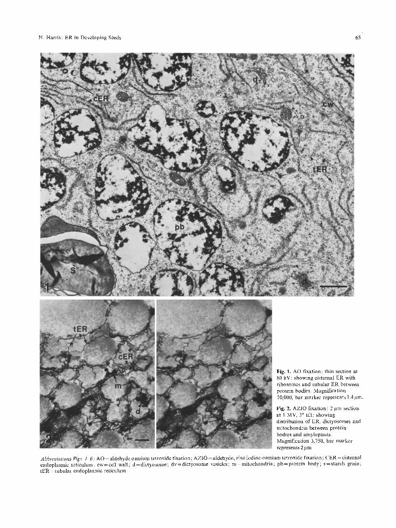
The appearance, in thin sections, of part of a cotyle- don cell f rom a developing seed at the storage protein deposition phase after conventional aldehyde fixation and osmium tetroxide post fixation is shown in Fig. 1. Numerous rough ER cisternae are seen in the cyto- plasm between the vacuoles/protein bodies which show accumulation of some storage protein. The floc- ular appearance of the protein body contents is char- acteristic of this stage of protein body development. Some tubular ER is also to be seen, particularly on the right hand side of the micrograph. Two dictyo- somes, both showing associated vesicles some of which have electron dense contents, are also labelled.
Examination of earlier stages of cellular develop- ment showed a similar pattern to that described for a range of legumes including V. faba (Briarty et al., 1969) i.e. cotyledon cells becoming highly vacuolate during the phase of cell expansion with the cytoplasm limited to a thin lining adjacent to the cell wall and a few transcellular sheets or strands (results not presented).
The appearance of a thick (2 ~tm) section of part of a cotyledon cell after fixation with aldehyde and post fixation with zinc iodine-osmium tetroxide corn-
plex is shown in Fig. 2. By examing the pair of micro- graphs through a stereo viewer the three dimensional relationship of the organelles can be seen. The single membrane surrounding the protein bodies is not stained by the procedure used, but the cisternae of the ER can be seen throughout the cytoplasm between the amorphous slightly electron dense regions which represent segments of protein bodies. Mitochondria, many of which are " d u m b e l l " shaped, are distributed throughout the cytoplasm. The numerous dictyo- somes may be identified by the aggregations of vesi- cles seen between the protein bodies; the fixation used only stains some of the cisternae. Some dictyosome vesicles may have expanded during fixation; this is the only difference in organelle sizes and distribution shown by the two fixation methods.
Two dictyosomes are seen close to the centre of Fig. 3 which is a micrograph of an approximately 300 nm section. Some of the dictyosome cisternae have accumulated stain although substantially less than that which marks the cisternae and tubules of the ER. Two types of vesicle are associated with the dictyosomes, the larger having fine granular contents and the smaller having contents with a uniform elec- tron density. Similar small vesicles are seen in the cytoplasm apparently away from dictyosomes al- though when examined further these are usually close to dictyosomes of adjacent sections. Within the protein bodies small vesicles similar in size and stain- ing to the small dictyosome vesicles are occasionally seen (e.g. Fig. 3). With zinc iodine osmium-tetroxide staining the intra protein body vesicles have a greater electron density than the storage protein and thus are clearly distinguishable. With only osmium tetrox- ide for post fixation the material within the protein bodies has a similar high electron density and conse- quently such a distinction cannot be made.
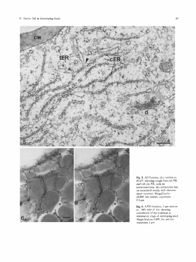
The cisternal ER and tubular ER appear to be interconnected in Fig. 3 although it is possible that this could have resulted from organelle overlap in the 300 nm section. Examination of the stereo pair in Fig. 4 however shows that many tubules are con- nected to the edges of the ER cisternae and it is apparent in this and other unpublished stereo pairs that cisternae are interconnected by the network of tubules. In contrast no evidence was found of vesicles at the periphery of the ER cisternae. The diameter of the tubules seen in Fig. 3 and 4 is greater than the lumen of the cisternae. Examination of the "con- vent ional" thin section seen in Fig. 5 shows that a corresponding difference in dimensions is seen be- tween the rough cisternal ER and tubular ER. Unlike the cisternal ER which has numerous attached ribo- somes the tubular ER is generally free of associated ribosomes. Swelling at the edges of some ER cis- ternae are seen in this micrograph, which are inter-
N. Harris: ER in Developing Seeds 65
Fig. 1. AO fixation; thin section at 80 kV: showing cisternal ER with ribosomes and tubular ER between protein bodies. Magnification 10,000, bar marker represents 1.4 lain.
Fig. 2. AZIO fixation; 2 gm section at 1 MV, 3 ~ tilt: showing distribution of ER, dictyosomes and mitochondria between protein bodies and amyloplasts. Magnification 3,750, bar marker represents 2 ~m
Abbreviations Figs. l 6: AO = aldehyde osmium tetroxide fixation; AZIO = aldehyde, zinc iodine-osmium tetroxide fixation; CER = cisternal endoplasmic reticulmn ; cw = cell wall; d = dictyosome; dv = dictyosome vesicles ; m = mitochondria; pb = protein body ; s = starch grain; tER =tubular endoplasmic reticulum

66 N. Harris: ER in Developing Seeds
Fig. 3. AZIO fixation; 0.3 gm section at 80 kV: showing morphology of cisternaI and tubular ER and also dictyosomes with associated besicles. Magnification 14,000, bar marker represents 1 g m
Fig. 4. AZIO fixation; 1 gm section at 1 MV with 6 ~ tilt: showing interconnections of tubular ER with periphery o f ER cisternae. Magnification 12,500, bar marker represents 1 gm
N. Harris: ER in Developing Seeds 67
Fig. 5. AO fixation; thin section at 80 kV: showing rough cisternal ER and tubular ER, with an interconnection; the dictyosome has an associated vesicle with electron dense contents. Magnification 28,000, bar marker represents 0,5 gm
Fig. 6. AZIO fixation; 1 Itm section at 1 MV with 6 ~ tilt: showing reticulation of the cisternae at maturat ion stage of developing seed. Magnification 8,400, bar marker represents 1 !am

68 N. Harris: ER in Developing Seeds
preted as interconnections with tubular ER. The ste- reo pair in Fig. 4 shows that the tubules arise at a variety of angles from the undulating cisternae and consequently the chance of cutting an intersection to show cisternal ER and a length of tubular ER in (thin) section is small. One such section is seen in Fig. 5 (arrowed) where the tubule is cut partly in section and partly along a membrane surface close to the cisterna.
Evidence from serial thin sectioning (not presented here) has been obtained demonstrating that the cister- nal swellings are not vesicles. The ER tubules are only slightly greater in diameter (approx. 100 nm) that the sections are thin (approx. 80 nm) so in three serial sections the following are frequently seen: a) cisterna without peripheral swelling b) cisterna with peripheral swelling and c) cisterna without peripheral swelling but with adjacent "vesicle" which is really a section of tubule.
An extensive interconnecting network of cisternal and tubular ER was found throughout the stage of storage protein deposition. It has been widely reported that this stage of seed development is fol- lowed by a period of seed maturation when the dry weight increases only slightly but the fresh weight reduces as seed dehydration occurs. Thick section micrographs of zinc iodine osmium-tetroxide fixed maturing cotyledons show that the cisternae develop into a reticular form during the late stage of seed development (Fig. 6).
Discussion
Comparison of the three dimensional images from stereo pairs of thick section micrographs with the conventional thin section micrographs shows that there is considerable potential for misinterpretation of the morphology of the endoplasmic reticulum if only the latter are used. In thin section micrographs parallel, ribosome studded membranes of cisternae are easily recognised. The unevenly distributed "vesicles" of different diameters which represent cross sections of the tubular ER are less obvious and a consequent underestimate of the relative proportion of tubular ER is possible. Another misinterpretation made in previous work in this field concerns the widely reported "vesicles" described at the periphery of the rough cisternal ER. The stereo pair images from thick sections show that a system of tubular ER arises from the periphery of the cisternae but no evidence of pe- ripheral vesicles was found. Comparison of thick and thin sections shows that the peripheral "vesicles" rep- resent the interconnections of the cisternal and tubu- lar ER.
In both conventional thin section work and thick section studies we have found the widely reported
increase in cisternal ER immediately prior to and during the period of storage protein deposition. This increase in rough ER has been quantified by Briarty (1973) who showed that in Phaseolus vulgaris a rapid increase in the surface area of the rough ER has been preceeded by a doubling of the S/V ratio of the ER but a comparison of the relative fractions of rough cisternal ER and smooth tubular ER was not reported. Preliminary results from a quantitative study in this laboratory show that at the beginning of storage protein deposition approximately 40% of the total volume of endoplasmic reticulum is tubular ER although this fraction represents less than 30% of the total ER surface area (Harris, unpublished).
From a static series of electron micrographs we can only speculate as to the dynamic nature of the changes observed. The term transition element has been used to refer to those parts of the endomembrane system intermediate between rough ER and tonoplast and plasmalemma. The most general form of transi- tion element is smooth ER which is directly contin- uous with rough ER. Evidence from animal studies suggests that smooth ER may be derived from rough ER either by loss of ribosomes or by outgrowths of ribosome free regions from rough ER (see review, MorrO, 1975). In V.faba the cisternal ER is studded with ribosomes and the smooth ER is present as a tubular form. If smooth ER acts as a transition form in membrane biogenesis as suggested by Morr6 (1975) it would seem that in the cotyledon cells of developing legume seeds the transition may be from smooth tubu- lar ER to rough cisternal ER at the time of increase in ER surface to volume ratio quantified by Briarty (1973). The reverse transition appears to occur during the drying out or maturation phase of the seed. This latter stage of seed development is less well described in the literature, probably because of difficulties with specimen preparation. There is general agreement that during the final maturation phase biosynthetic activ- ity, as measured by rate of dry weight gain, is reducing and there is a reduction in the extent of the rough ER (see review, Millerd, 1975). Thick section micro- graphs suggest much of the cisternal ER is forming into an interconnecting reticular system at this time and that this form of ER remains in the seed until germination. Because of the high cytoplasmic density in mature dry seeds it is impossible to visualize confi- dently the tubular ER by either thick or thin section techniques, however tubular ER is observable imme- diately seeds are imbibed (Harris and Chrispeels, in preparation).
The lack of any evidence of vesicles at the periph- ery of the rough cisternal ER reintroduces the prob- lem of transport of storage protein from the site of synthesis to the site of storage, the vacuolar protein bodies. Proteins synthesised by membrane bound
N. Harris: ER in Developing Seeds 69
po ly r i bosomes are secreted into the lumen o f the E R ( R e d m a n et al., 1966). I t is poss ible that the synthe- sised p ro te in m a y be re leased f rom the E R and diffuse freely th rough the cy top l a sm e.g. Chert and Jones (1974) c la im tha t bar ley a -amylase is made on the rough E R and then re leased into the cy toso l before being secreted f rom the cell, a l t hough the work has been cr i t ic ized (Chrispeels , 1976). E x a m i n a t i o n o f nu- merous thin sect ions o f legume co ty ledons has fai led to show any evidence of direct connec t ion o f E R lumen with p ro te in bod ies ; this cont ras t s with the f indings f rom cereals where direct connec t ion o f E R to p ro te in bodies has been shown for e.g. rice (Har r i s and Jul iano, 1977) and maize (Lark ins and H u r k m a n , 1978). O u r results suggest tha t an a l te rna t ive p a t h w a y to a soluble m o d e o f t r a n s p o r t m a y be avai l- able for reserve p ro te ins synthesised in deve lop ing V. faba . This p a t h w a y would involve t r anspo r t o f the synthesised p ro te ins f rom the rough cis ternal E R via the t ubu la r E R to d i c tyosome vesicles. N u m e r o u s dic- tyosome vesicles con ta in e lec t ron dense mater ia l which is s imi lar in appea rance to the p ro te in body contents . The Golg i a p p a r a t u s has been shown to be involved in synthesis and secret ion o f po lysaccha- rides and g lycopro te ins (for review see Chrispeels , 1976) a l though two recent repor t s have shown local- iza t ion of glycosyl t ransferase in ER. N a g a h a s h i and Beevers (1978) have shown tha t in pea is local ized in the E R f rac t ion and Me l lo r and Lo rd (1978) have found g lycosy la t ion local ized in ge rmina t ing cas to r bean e n d o s p e r m E R fract ion. I t has been suggested tha t the a p p a r e n t differences in organel le d i s t r ibu t ion in different deve lop ing legumes may be a ref lect ion o f the type o f s to rage p ro te ins being synthesised (Harr i s and Boulter , 1976) and s imilar ly there is no reason to imagine tha t d i f ferent classes o f s torage p ro te in wou ld be t r a n s p o r t e d in ident ica l m a n n e r even though the f inal des t ina t ion , a p ro te in body with a mixture o f s torage p ro te ins ( G r a h a m and Gunning ,
1970), is the same. A n invo lvement in the t r a n s p o r t of synthesised
p ro te in would not exclude the t ubu la r E R f rom a poss ib le role in in t race l lu la r t r a n s p o r t of smal l mole- cules. Othe r p l an t cells r epo r t ed to have an extensive ne twork o f s m o o t h E R tubules all special ize in the t r anspo r t a n d / o r b iosynthes is o f small molecules (for review see G u n n i n g and Steer, 1975). The tubu la r E R present in co ty l edon cells dur ing the pe r iod o f s tarch and reserve p ro te in depos i t ion m a y also be involved in the m o v e m e n t o f small molecules (e.g. su- crose and amides) within the cells.
The high voltage microscopy was carried out with the Science Research Council supported EM7 microscope at the Imperial Col- lege of Science and Technology, London. Travelling expenses were paid by the University of Durham Research Fund Committee.
I should like to thank Professor D. Boulter for his continuing interest and support with these studies.
References
BaiIey, C.J., Cobb, A., Boulter, A. : A cotyledon slice system for the elctron autoradiographic study of the synthesis and intracel- lular transport of seed storage protein of Vicia faba. Planta 95, 103-118 (1970)
Bain, J.M., Mercer, F.V. : Subcellular organisation of the develop- ing cotyledons of Pisum sativum. Aust. J. Biol. Sci. 19, 49-67 (1966)
Briarty, L.G. : Stereology in seed development studies: Some pre- liminary work. Caryologica 25, suppl. 289-301 (1973)
Briarty, L.G., Coult, D.A., Boulter, D. : Protein bodies of develop- ing seeds of Viciafaba. J. Exp. Bot. 20, 358-372 (1969)
Boulter, D.: Protein synthesis and deposition in Seeds. In: 6th Long Ashton Symposium, p. 359-368. New York, London: Academic Press 1978
Chert, R., Jones, R.L.: Studies on the release of barley aleurone cell proteins: autoradiography. Planta 119, 207-220 (1974)
Chrispeels, M.J. : Biosynthesis, intracellular transport and secretion of extracellular macromolecules. Ann. Rev. Plant Physiol. 27, 19-36 (1976)
Dieckart, J.W., Dieckert, M.C.: In Symposium: seed proteins, Inglett, E.D., ed. The Avi Publishing Company, Conn. 1972
Dure, L.S. : Seed formation. Ann Rev. Plant Physiol. 26, 259-278 (1975)
Glauert, A.M.: The high voltage electron microscope in biology. J. Cell Biol. 63, 717-748 (1974)
Graham, T.A., Gunning, B.E.S. : Localization of legumin and vicil- lin in bean cotyledons cells using fluorescent antibodies. Nature (London) 228, 81-82 (1970)
Gunning, B.E.S.o Steer, M.W.: Ultrastructure and the biology of plant cells. London: Arnold 1975
Harris, N.: Nuclear pore distribution and relation to adjacent cytoplasmic organelles in cotyledon cells of developing Vicia faba. Planta 141, 121-128 (1978)
Harris, N., Boulter, D.: Protein body formation in cotyledons of developing cowpea (Vigna unguiculata) seeds Ann. Bot. 40, 739 744 (1976)
Harris, N., Juliano, B.O.: Ultrastructure of endosperm protein bodies in developing rice grains differing in protein content. Ann. Bot. 41, 1-5 (1977)
Larkins, B.A., Hurkman, W.J.: Synthesis and deposition of zein in protein bodies of maize endosperm. Plant Physiol. 62, 256-263 (1978)
Marty, M.F. : Sites reactifs a l'iodure de zinc-tetroxyde d'osmium dans les cellules de ia racine d'Euphorbia characias. C.R. Acad. Sci. 277, 1317-1320 (1973)
Mellor, R.B., Lord, J.M. : Incorporation of D-(I~C) galactose into organelles glycoprotein in castor bean endosperm. Planta 141, 329 332 (1978)
Millerd, A. : Biochemistry of legume seed proteins. Ann. Rev. Plant Physiol. 26, 53-72 (1975)
Morr6, D.J. : Membrane biogenesis. Ann. Rev. Plant Physiol. 26, 441-481 (1975)
Nagahashi, J., Beevers, L.: Subcellular localization of glycosyl transferases involved in glycoprotein biosynthesis in the cotyle- dons of Pisum sativum. Plant Physiol. 61,451-459 (1978)
Opik, H. : Development of the cotyledon cell structure in ripening Phaseolus vulgaris seeds. J. Exp. Bot. 19, 64 76 (1968)
Redman, C.M., Siekevitz, P., Palade, G.E. : Synthesis and transfer of amylase in pigeon pancreatic microsomes. J. Biol. Chem. 241, 1150 1158 (1966)
Received 10 December 1978 ; accepted 23 January 1979