Benign Endometrial Hyperplasia Sequence and Endometrial Intraepithelial Neoplasia
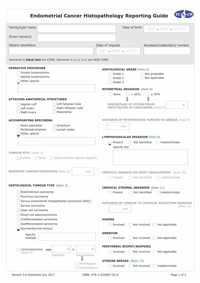
Endometrial Cancer Histopathology Reporting Guide
Version 3.0 Published July 2017 ISBN: 978-1-925687-02-6 Page 1 of 2
Family/Last name Date of birth
Given name(s)
Patient identifiers Date of request Accession/Laboratory number
OPERATIVE PROCEDURE
ACCOMPANYING SPECIMENS
TUMOUR SITE (Note 1)
Elements in black text are CORE. Elements in grey text are NON-CORE.
Simple hysterectomy Radical hysterectomy Other, specify
Vaginal cuffLeft ovary Right ovary
Fundus Body Isthmus/lower uterine segment
ATTACHED ANATOMICAL STRUCTURES
Left fallopian tubeRight fallopian tubeParametria
None submittedPeritoneal biopsiesOther, specify
Omentum Lymph nodes
MAXIMUM TUMOUR DIMENSION (Note 2)
mm
HISTOLOGICAL TUMOUR TYPE (Note 3)
Endometrioid carcinoma
Mucinous carcinoma
Serous endometrial intraepithelial carcinoma (SEIC)
Serous carcinoma
Clear cell carcinoma
Mixed cell adenocarcinoma
Undifferentiated carcinoma
Dedifferentiated carcinoma
Neuroendocrine tumour
Specify subtype
% %&
Epithelial Sarcomatous
Homologous Heterologous
Grade 1 Grade 2 Grade 3
HISTOLOGICAL GRADE (Note 5)
MYOMETRIAL INVASION (Note 6)
None < 50% ≥ 50%
PERCENTAGE OF MYOMETRIUM INFILTRATED BY CARCINOMA (Note 7)
%
LYMPHOVASCULAR INVASION (Note 9)
CERVICAL STROMAL INVASION (Note 11)
VAGINA
Specify site
DISTANCE OF TUMOUR TO CERVICAL RESECTION MARGINS (Note 12)
mm
UTERINE SEROSA (Note 13)
PERITONEAL BIOPSY/BIOPSIES
OMENTUM
Carcinosarcoma (Note 4)
CERVICAL SURFACE OR CRYPT INVOLVEMENT (Note 10)
DISTANCE OF MYOINVASIVE TUMOUR TO SEROSA (Note 8)
mm
Present Not identified Indeterminate
Involved Not involved Not applicable
Present Not identified Indeterminate
Involved Not involved Not applicable
Involved Not involved Not applicable
Involved Not involved Indeterminate
Present Not identified Indeterminate
Not gradeableNot applicable
DD – MM – YYYY
DD – MM – YYYY
Version 3.0 Published July 2017 ISBN: 978-1-925687-02-6 Page 2 of 2
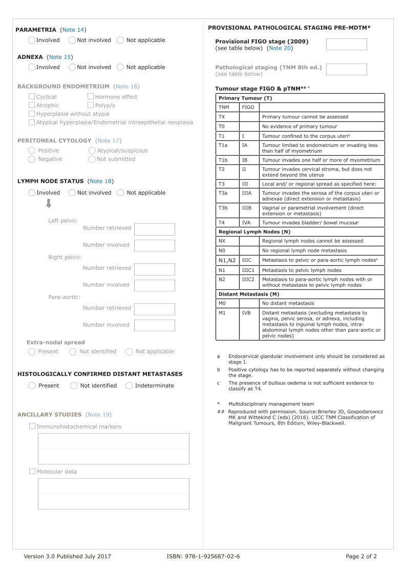
Primary Tumour (T)
TNM FIGO
TX Primary tumour cannot be assessed
T0 No evidence of primary tumour
T1 I Tumour confined to the corpus uteria
T1a IA Tumour limited to endometrium or invading less than half of myometrium
T1b IB Tumour invades one half or more of myometrium
T2 II Tumour invades cervical stroma, but does not extend beyond the uterus
T3 III Local and/ or regional spread as specified here:
T3a IIIA Tumour invades the serosa of the corpus uteri or adnexae (direct extension or metastasis)
T3b IIIB Vaginal or parametrial involvement (direct extension or metastasis)
T4 IVA Tumour invades bladder/ bowel mucosac
Regional Lymph Nodes (N)
NX Regional lymph nodes cannot be assessed
N0 No regional lymph node metastasis
N1,N2 IIIC Metastasis to pelvic or para-aortic lymph nodesb
N1 IIIC1 Metastasis to pelvic lymph nodes
N2 IIIC2 Metastasis to para-aortic lymph nodes with or without metastasis to pelvic lymph nodes
Distant Metastasis (M)
M0 No distant metastasis
M1 IVB Distant metastasis (excluding metastasis to vagina, pelvic serosa, or adnexa, including metastasis to inguinal lymph nodes, intra-abdominal lymph nodes other than para-aortic or pelvic nodes)
Tumour stage FIGO & pTNM## ^
ANCILLARY STUDIES (Note 19)
BACKGROUND ENDOMETRIUM (Note 16)
Cyclical Hormone effectAtrophic Polyp/sHyperplasia without atypia Atypical hyperplasia/Endometrial intraepithelial neoplasia
PERITONEAL CYTOLOGY (Note 17)
Positive Atypical/suspicous Negative Not submitted
LYMPH NODE STATUS (Note 18)
Number retrieved Number involved
Left pelvic:
Number retrieved Number involved
Right pelvic:
Number retrieved Number involved
Para-aortic:
Extra-nodal spread
Provisional FIGO stage (2009) (see table below) (Note 20)
Pathological staging (TNM 8th ed.)(see table below)
HISTOLOGICALLY CONFIRMED DISTANT METASTASES
Immunohistochemical markers
Molecular data
PROVISIONAL PATHOLOGICAL STAGING PRE-MDTM*
ADNEXA (Note 15)
PARAMETRIA (Note 14)
Involved Not involved Not applicable
Involved Not involved Not applicable
Involved Not involved Not applicable
Present Not identified Not applicable
Present Not identified Indeterminate
a Endocervical glandular involvement only should be considered as stage I.
b Positive cytology has to be reported separately without changing the stage.
c The presence of bullous oedema is not sufficient evidence to classify as T4.
* Multidisciplinary management team## Reproduced with permission. Source:Brierley JD, Gospodarowicz
MK and Wittekind C (eds) (2016). UICC TNM Classification of Malignant Tumours, 8th Edition, Wiley-Blackwell.
Scope This dataset has been developed for resection specimens of endometrial cancers. It is not applicable for small endometrial biopsy specimens.
Note 1 - Tumour site (Non-core) Reason/Evidentiary Support: There may be an association between lower uterine segment/isthmic tumours and Lynch syndrome.
1,2
Back
Note 2 - Maximum tumour dimension (Non-core) Reason/Evidentiary Support: There is a significant correlation between primary tumour diameter >20 mm and peritoneal failure. This does not yet reach III-2 evidence level.
3
Back
Note 3 - Histological tumour type (Core) Reason/Evidentiary Support: Endometrial carcinomas should be typed according to the 2014 World Health Organisation (WHO) Classification.
4
Accurate typing is necessary in both biopsies and resection specimens. Diagnosis of aggressive tumours such as serous carcinoma, clear cell carcinoma, carcinosarcoma, undifferentiated carcinoma and grade 3 endometrioid adenocarcinoma will usually result in full surgical staging including pelvic and para-aortic lymphadenectomy and omentectomy. Mucinous adenocarcinoma refers to a subtype of endometrial adenocarcinoma in which more than 50% of the tumour cells contain intracytoplasmic mucin. Many endometrioid adenocarcinomas contain focal mucinous areas and endometrioid and mucinous adenocarcinomas form part of a spectrum. Although carcinosarcomas (malignant mixed Müllerian tumours) are still classified as mixed epithelial and mesenchymal tumours in the 2014 WHO Classification,
4 their behaviour is similar to other high grade endometrial carcinomas and they are treated in the
same way as aggressive endometrial carcinomas. Carcinosarcomas are believed to be epithelial neoplasms that have undergone sarcomatous metaplasia, the epithelial elements being the ‘driving force’. The 2014 WHO classification of endometrial carcinomas (see below) now includes serous endometrial intraepithelial carcinoma (serous EIC).
4 Even in the absence of demonstrable stromal invasion, malignant cells can
shed from serous EIC and metastasise widely to extra-uterine sites. Neuroendocrine tumours are also a new addition to the 2014 WHO Classification.
4 They are rare primary uterine neoplasms and the diagnosis should be
confirmed immunohistochemically, although some small cell neuroendocrine carcinomas may not express neuroendocrine markers (see note on ANCILLARY STUDIES). Neuroendocrine neoplasms of the endometrium are divided into low-grade neuroendocrine tumour (carcinoid tumour) which is extremely rare and high-grade neuroendocrine carcinoma (small cell and large cell neuroendocrine carcinoma) which is more common but also rare. Large cell neuroendocrine carcinoma should demonstrate a neuroendocrine growth pattern in at least part of the tumour, and show expression of one or more neuroendocrine markers (chromogranin, synaptophysin, CD56, PGP9.5) in >10% of the tumour. Undifferentiated carcinoma
5,6 is defined by WHO as a ‘malignant epithelial
neoplasm with no differentiation’,4 and may show immunohistochemical evidence of epithelial differentiation in
only occasional tumour cells (see notes on ancillary studies). Dedifferentiated carcinoma7 is defined as an
undifferentiated carcinoma that contains a second component of either FIGO grade 1 or 2 endometrioid adenocarcinoma; in such cases, it is believed that the undifferentiated component develops as a result of dedifferentiation in the low-grade endometrioid component.
Mixed carcinomas must contain two or more different histological types of endometrial carcinoma recognisable on H&E-stained sections. At least one of the subtypes must be a type II tumour and the second component, according to the 2014 WHO Classification,
4 must comprise at least 5% of the neoplasm. The most common mixture
is endometrioid and serous carcinoma. Immunohistochemistry may assist in confirming the presence of a second, morphologically distinct subtype. All subtypes should be specified in the histopathology report, even if <5% of the neoplasm is composed of type II tumour, because the behaviour of these tumours is determined by the highest grade component.
4
In cases where there is no residual tumour in the hysterectomy specimen or where there is a significant discrepancy between the reported tumour type in the biopsy and that in the hysterectomy, it may be necessary to review the prior biopsy. If high-risk/aggressive variants of carcinoma e.g. serous carcinoma, carcinosarcoma etc., are confirmed in the endometrial biopsy but are not identified in the final hysterectomy specimen, the carcinoma should be categorised according to the worst histology. Adequate sampling of the tumour is required (minimum of 4 blocks) to allow meaningful assessment of this data item.
WHO histological classification (2014)4
Endometrial carcinoma - Epithelial tumours
ICD-O code
Endometrioid carcinoma 8380/3
Squamous differentiation 8570/3
Villoglandular 8263/3
Secretory 8382/3
Mucinous carcinoma 8480/3
Serous endometrial intraepithelial carcinoma (SEIC) 8441/2*
Serous carcinoma 8441/3
Clear cell carcinoma 8310/3
Neuroendocrine tumours
Low-grade neuroendocrine tumour
Carcinoid tumour 8240/3
High-grade neuroendocrine carcinoma
Small cell neuroendocrine carcinoma 8041/3
Large cell neuroendocrine carcinoma 8013/3
Mixed cell adenocarcinoma 8323/3
Undifferentiated carcinoma 8020/3
Dedifferentiated carcinoma
Mixed epithelial and mesenchymal tumours
Carcinosarcoma 8980/3
* This new code was approved by the International Agency for Research on Cancer (IARC) /WHO committee for
ICD-O in 2013.
© World Health Organisation. Reproduced with permission.
Back
Note 4 - Carcinosarcoma (Non-core) Reason/Evidentiary Support: A recent study has shown that the presence of heterologous elements in stage I carcinosarcomas is an important adverse prognostic feature; this does not yet reach III-2 evidence level.
8
Back
Note 5 - Histological grade (Core) Reason/Evidentiary Support: The FIGO grading system for endometrioid adenocarcinomas of the uterine corpus is based on the following architectural features:
9
Grade 1: 5% or less non-squamous solid growth pattern
Grade 2: 6% to 50% non-squamous solid growth pattern
Grade 3: >50% non-squamous solid growth pattern
Notable nuclear atypia, which exceeds that which is routinely expected for the architectural grade, increases the tumour grade by 1. Notable nuclear atypia should be present in >50% of the tumour.
10
In addition, the following guidelines should be used in grading:
(1) Non-gland forming squamous elements should be disregarded for grading purposes. (2) Endometrioid and mucinous carcinomas should be graded using the FIGO grading system. (3) Serous, clear cell and undifferentiated carcinomas and carcinosarcomas are not graded but are
regarded as high grade neoplasms.11
When the dataset is being completed, these should be designated as “not applicable” for histologic grade.
(4) In mixed carcinomas, the highest grade should be assigned.
In general, if there is a discrepancy between the grade of an endometrioid adenocarcinoma in the pre-operative biopsy and the final resection specimen, the final histological tumour grade should be based on findings in the hysterectomy specimen, which usually contains a larger volume of tumour for assessment. This is particularly important if the hysterectomy specimen contains abundant low-grade tumour and the biopsy showed grade 3 endometrioid adenocarcinoma. In this specific situation, application of the guidelines for FIGO grading may result in the tumour being downgraded, although this will not always be the case; for example, where the biopsy contained abundant grade 3 endometrioid adenocarcinoma and the hysterectomy a limited amount of low-grade tumour, the final diagnosis might still be grade 3 endometrioid adenocarcinoma. Back
Note 6 - Myometrial invasion (Core) Reason/Evidentiary Support: Depth of invasion should be measured from the endomyometrial junction (not the surface of exophytic tumours) to the deepest focus of tumour invasion. Measurement of the depth of invasion may be rendered difficult by irregularity of the endomyometrial junction, polypoid tumour growth, intramural leiomyomas, adenomyosis and uncommonly by smooth muscle metaplasia within polypoid neoplasms.
12 Deep myometrial invasion has
repeatedly been shown to be an important poor prognostic indicator in endometrial carcinoma. This is an independent predictor of haematogenous dissemination by endometrial carcinoma and it is therefore an important determinant of adjuvant therapy.
13
Back
Note 7 - Percentage of myometrium infiltrated by carcinoma (Non-core) Reason/Evidentiary Support: Tumour-free distance (to the uterine serosa) and percentage of myometrium infiltrated are independent prognostic factors for lymph node metastasis in endometrial carcinoma but studies do not reach level III-2 evidence.
14
The percentage of myometrium infiltrated by carcinoma is defined as the percentage of myometrium involved as determined by the depth of myometrial invasion from the endomyometrial junction to the deepest focus of invasive carcinoma in comparison to the overall myometrial thickness.
Back
Note 8 - Distance of myoinvasive tumour to serosa (Non-core) Reason/Evidentiary Support: Tumour-free distance and percentage of myometrium infiltrated are independent prognostic factors for lymph node metastasis in endometrial carcinoma; studies do not reach level III-2 evidence.
14
Back
Note 9 - Lymphovascular invasion (Core) Reason/Evidentiary Support: Lymphovascular invasion is a predictor of tumour recurrence and lymph node metastasis.
15 However,
lymphovascular space invasion does not alter the tumour stage. For example, if an endometrial adenocarcinoma is confined to the inner half of the myometrium but shows lymphovascular invasion in the outer half of the myometrium, this should still be staged as FIGO 1A. Similarly lymphovascular invasion alone in cervical, parametrial or para-ovarian vessels does not upstage the tumour. There is an increased incidence of vascular pseudoinvasion in laparoscopic hysterectomy specimens associated with the use of an intrauterine balloon manipulator.
15,16
Back
Note 10 - Cervical surface or crypt involvement (Non-core) Reason/Evidentiary Support: Not necessary for staging but some oncologists administer vault brachytherapy if this is identified. Level III-2 evidence currently not available.
Back
Note 11 - Cervical stromal invasion (Core) Reason/Evidentiary Support: Cervical stromal infiltration by endometrial carcinoma is associated with a risk of recurrence and is a predictor of pelvic lymph node metastases.
17,18
Back
Note 12 - Distance of tumour to cervical resection margins (Non-core) Reason/Evidentiary Support: Close margins may indicate a need for vault brachytherapy. Vascular invasion at cervical resection margin should be documented but does not upstage the tumour.
Back
Note 13 - Uterine serosa (Core) Reason/Evidentiary Support: Carcinoma should penetrate through the serosa in order to be classified as serosal involvement. Involvement of the serosa (FIGO stage IIIA) carries a higher risk of locoregional recurrence than does adnexal involvement (also FIGO stage IIIA).
19
Back
Note 14 - Parametria (Core) Reason/Evidentiary Support: Most hysterectomies for endometrial cancer will be simple hysterectomies and will not have parametrial resections. Endometrial carcinomas with parametrial invasion are staged as FIGO IIIB. Although not an independent prognostic indicator, parametrial involvement by direct extension is a poor prognostic factor and also correlates with other poor prognostic factors. The presence of lymphovascular invasion in parametrial tissues should be documented but does not constitute parametrial involvement.
20,21
Back
Note 15 - Adnexa (Core) Reason/Evidentiary Support: FIGO staging is based on tumour involvement of either the fallopian tube or ovary (stage IIIA). Especially with low-grade endometrioid adenocarcinomas, involvement of the uterine corpus and adnexa may indicate synchronous, independent neoplasms rather than metastasis from the endometrium to the adnexa; a variety of pathological parameters is useful in the distinction between synchronous independent and metastatic neoplasms. As for other sites in the gynaecological tract in which lymphovascular invasion by endometrial adenocarcinoma may be identified e.g., myometrium and parametrial tissue, the identification of lymphovascular space invasion alone in adnexal structures does not alter the tumour stage i.e. endometrial carcinoma should not be upstaged if there is vascular involvement in the adnexa in the absence of tumour outside of vascular channels.
Back
Note 16 - Background endometrium (Non-core) Reason/Evidentiary Support: The appearance of the background endometrium and the presence of abnormalities such as hyperplasia or polyps, should be documented.
Back

Note 17 - Peritoneal cytology (Non-core) Reason/Evidentiary Support: This data item is not necessary for staging but there is lack of consensus in the literature regarding the prognostic significance of positive peritoneal washings in the absence of other evidence of extrauterine spread. A recommendation is made by FIGO and the Union for International Cancer Control (UICC) to record positive peritoneal washings without altering the tumour stage.
22,23
Back
Note 18 - Lymph node status (Core and Non-core) Reason/Evidentiary Support: Pelvic and para-aortic node status should be recorded separately since this affects tumour stage. Pelvic node involvement without para-aortic involvement is stage IIIC1 while para-aortic node involvement is stage IIIC2.
24,25
Note that micrometastases (greater than 0.2 mm but not greater than 2.0 mm in diameter) are regarded as lymph node involvement and N1mi or N2mi while metastases greater than 2.0 mm in maximum dimension are classified as N1a or N2a. Isolated Tumour Cells (ITCs), in common with TNM8 staging practices at other tumour sites, are regarded as node negative (N0(i+)). The number of nodes involved and the site of involvement is prognostically important and may direct adjuvant treatment.
Back
Note 19 - Ancillary studies (Non-core) Reason/Evidentiary Support: Immunohistochemistry may be useful in certain diagnostic scenarios. For example, a panel of markers (ER, PR, vimentin, CEA, p16) may be useful in the distinction between a primary endometrial and cervical adenocarcinoma.
26-27 Other markers (ER, PR, p53, p16, PTEN, IMP3) may be useful in the distinction between an
endometrioid and a serous adenocarcinoma.28-29
p53 and p16 may help to highlight serous EIC and distinguish this from surface atypias which can mimic it. Immunohistochemistry for mismatch repair proteins (MLH1, MSH2, MSH6, PMS2) may be useful in helping to establish whether endometrial carcinomas are associated with underlying mismatch repair gene abnormalities and Lynch syndrome (hereditary non-polyposis colorectal cancer) . 30-31
Undifferentiated endometrial carcinomas are often only focally, but characteristically intensely, positive with broad spectrum cytokeratins, CK18 and epithelial membrane antigen (EMA). This may be useful in the distinction from an undifferentiated sarcoma or other neoplasms and may also help to establish a diagnosis of dedifferentiated carcinoma when a component of low-grade endometrioid adenocarcinoma is present.
5-7 Some
undifferentiated carcinomas exhibit focal expression of neuroendocrine markers.32
High-grade neuroendocrine carcinomas are usually positive with the neuroendocrine markers chromogranin, synaptophysin, CD56 and PGP9.5. Some small cell neuroendocrine markers are negative with these markers but usually at least one is positive. Large cell neuroendocrine carcinomas should be positive with at least one of these markers in >10% of tumour cells. Different morphological subtypes of endometrial adenocarcinoma are associated with distinct molecular abnormalities. However, at present molecular analysis has little role in diagnosis or as an independent prognostic or predictive factor. However, this may change in the future and it is likely that targeted therapies will be developed against carcinomas exhibiting specific molecular abnormalities.
Back

Note 20 - Provisional Pathological FIGO Stage Pre-MDTM* (Core and Non-core) Reason/Evidentiary Support: Staging is provisional since final stage should be determined at multidisciplinary team/tumour board meeting when all relevant clinical and radiological information is available.
11,33 Since serous EIC is regarded as a type of
endometrial carcinoma, it is staged as FIGO stage IA (T1a). The reference document TNM Supplement: A commentary on uniform use, 4th Edition (C Wittekind editor) may be of assistance when staging.
34
* Multidisciplinary management team
Back
References
1 Westin SN, Lacour RA, Urbauer DL, Luthra R, Bodurka DC, Lu KH and Broaddus RR (2008). Carcinoma of the lower uterine segment: a newly described association with Lynch syndrome. J Clin Oncol 26(36):5965-5971.
2 Masuda K, Banno K and Yanokura M et al (2011). Carcinoma of the Lower Uterine Segment
(LUS): Clinico-pathological characteristics and association with Lynch Syndrome. Curr Genomics 12:25-29.
3 Mariani A, Webb MJ, Keeney GL, Aletti G and Podratz KC (2003). Endometrial cancer: predictors
of peritoneal failure. Gyneco Oncol 89:236-242.
4 Kurman RJ, Carcangiu ML, Herrington CS and Young RH (2014). WHO classification of tumours of
the female reproductive organs. IARC press, Lyon.
5 Silva EG, Deavers MT and Malpica A (2007). Undifferentiated carcinoma of the endometrium: a
review. Pathology 39(1):134-138.
6 Tafe LJ, Garg K, Chew I, TornosA. C and Soslow R (2010). Endometrial and ovarian carcinomas
with undifferentiated components: clinically aggressive and frequently underrecognized neoplasms. Mod Pathol 23(6):781-789.
7 Silva EG, Deavers MT, Bodurka DC and Malpica A (2006). Association of low-grade endometrioid
carcinoma of the uterus and ovary with undifferentiated carcinoma: a new type of dedifferentiated carcinoma? Int J. Gynecol Pathology 25(1):52-58.
8 Ferguson SE, Tornos C, Hummer A, Barakat R and Soslow R (2007). Prognostic features of
surgical stage I uterine carcinosarcoma. Am J Surg Pathol 31(11):1653-1661.
9 Creasman W, Odicino F and Maisonneuve P et al (2001). Carcinoma of the corpus uteri: FIGO
Annual Report. J Epidemiol Biostat 6:45-86.
10 Zaino RJ, Kurman RJ, Diana KL and Morrow CP (1991). The utility of the revised International
Federation of Gynecology and Obstetrics histologic grading of endometrial adenocarcinoma using a defined nuclear grading system. A Gynecologic Oncology Group study. Cancer 75(1):81-86.
11 FIGO Committee on Gynecological Cancer (2009). Revised FIGO staging for carcinoma of the
vulva, cervix and endometrium. Int. J. Gynecol. Obstet. 105:103-104.
12 Ali A, Black D and Soslow R (2007). Difficulties in Assessing the Depth of Myometrial Invasion in
Endometrial Carcinoma Int J. Gynecol Pathology 26:155-123.
13 Mariani A, Webb MJ, Keeney GL, Calori G and Podratz KC (2001). Hematogenous dissemination
in corpus cancer. Gynecol Oncol 80:233−238.

14 Kondalsamy-Chennakesavan S, van Vugt S, Sanday K, Nicklin J, Land R, Perrin L, Crandon A and
Obermair A (2010). Evaluation of tumor-free distance and depth of myometrial invasion as prognostic factors for lymph node metastases in endometrial cancer. Int J Gynecol Cancer 20:1217-1221.
15 Watari H, Todo Y, Takeda M, Ebina Y, Yamamoto R and Sakuragi N (2005). Lymph_vascular space
invasion and number of positive para_aortic node groups predict survival in node_positive patients with endometrial cancer. Gynecol Oncol 96:651−657.
16 Logani S, Herdman AV, Little JV and Moller KA (2008). Vascular "pseudo invasion" in
laparoscopic hysterectomy specimens: a diagnostic pitfall. Am J Surg Pathol 32:560-565.
17 Mariani A, Webb MJ, Keeney GL and Podratz KC (2001). Routes of lymphatic spread: a study of
112 consecutive patients with endometrial cancer. Gynecol Oncol 81:100−104.
18 Fanning J, Alvarez PM, Tsukada Y and Piver MS (1991). Prognostic significance of the extent of
cervical involvement by endometrial cancer. Gynecol Oncol 40:46−47.
19 Jobsen JJ, Naudin Ten Cate L, Lybeert ML, Scholten A, van der Steen-Banasik EM, van der Palen
J, Stenfert Kroese MC, Slot A, Schutter EM and Siesling S (2011). Outcome of Endometrial Cancer Stage IIIA with Adnexa or Serosal Involvement Only. Obstet Gynecol Int.:doi: 10.1155/2011/962518.
20 Sato R, Jobo T and Kuramoto H (2003). Parametrial spread is a prognostic factor in endometrial
carcinoma. Eur J Gynaecol Oncol 24:241−245.
21 Yura Y, Tauchi K, Koshiyama M, Konishi I, Yura S and Mori T et al (1996). Parametrial
involvement in endometrial carcinomas: its incidence and correlation with other histological parameters. Gynecol Oncol 63:114−119.
22 Grimshaw RN, Tupper WC, Fraser RC, Tompkins MG and Jeffrey JF (1990). Prognostic value of
peritoneal cytology in endometrial carcinoma. Gynecol Oncol 36:97−100.
23 Fadare O, Mariappan MR, Hileeto D, Wang S, McAlpine JN and Rimm DL (2005). Upstaging
based solely on positive peritoneal washing does not affect outcome in endometrial cancer. Mod Pathol 18:673−680.
24 Morrow CP, Bundy BN, Kurman RJ, Creasman WT, Heller P and Homesley HD et al (1991).
Relationship between surgical_pathological risk factors and outcome in clinical stage I and II carcinoma of the endometrium: a Gynecologic Oncology Group study. Gynecol Oncol 40:55−65.
25 Hoekstra AV, Kim RJ, Small W Jr, Rademaker AW, Helenowsky IB and Singh DK et al (2009). FIGO
stage IIIC endometrial carcinoma: Prognostic factors and outcomes. Gynecol Oncol 114:273−278.
26 Castrillon DH, Lee KR and Nucci MR (2002). Distinction between endometrial and endocervical
adenocarcinoma: an immunohistochemical study. Int J Gynecol Pathol 21:4-10.
27 McCluggage WG, Sumathi VP, McBride HA and Patterson A (2002). A panel of
immunohistochemical stains, including carcinoembryonic antigen, vimentin, and estrogen receptor, aids the distinction between primary endometrial and endocervical adenocarcinomas. Int J Gynecol Pathol 21:11-15.
28 McCluggage WG (2007). Immunohistochemistry as a diagnostic aid in cervical pathology.
Pathology 39(1):97-111.
29 Yemelyanova A, Hongxiu J, Shih I, Wang T, Wu L and Ronnett B (2009). Utility of p16 expression
for distinction of uterine serous carcinomas from endometrial endometrioid and endocervical adenocarcinomas. Am J Surg Pathol 33:1504-1514.
30 Walsh MD, Cummings MC, Buchanan DD, Dambacher WM, Arnold S, McKeone AS and Byrnes R
et al (2008). Molecular, pathologic, and clinical features of early-onset endometrial cancer: identifying presumptive Lynch syndrome patients. Clin. Cancer Res. 14(6):1692-1700.
31 Garg K and Soslow RA (2009). Lynch syndrome (hereditary non-polyposis colorectal cancer) and
endometrial carcinoma. J Clin Pathol 62:679-684.
32 Taraif SH, Deavers MT, Malpica A and Silva EG (2009). The significance of neuroendocrine
expression in undifferentiated carcinoma of the endometrium. Int J. Gynecol Pathology 28(2):142-147.
33 Amin MB, Edge SB and Greene FL et al (eds) (2017). AJCC Cancer Staging Manual. 8th ed.,
Springer, New York.
34 Wittekind C (ed) (2012). TNM Supplement : A Commentary on Uniform Use, The Union for
International Cancer Control (UICC), Wiley-Blackwell.