ENDODONTICS IN FOCUS - ccendo.netccendo.net/files/2015/03/endo3.pdf · ENDODONTICS IN FOCUS ......
9
PPD February 2011 137 ENDODONTICS IN FOCUS ENDODONTIC DIAGNOSIS: THE THIRD DIMENSION Contacts North Mohammed Naeem 07815 781 625 South Petra Helgesen 07977 120 031 Pirooz Zia (left) Reza Farshey (right) CONT S uccessful endodontic treatment starts with an accurate diagnosis, which is defined as the process of determining by examination, the nature and circumstances of a diseased condition. is process involves compiling subjective data – information gathered from a patient’s perspective; and objective data – information gathered independent of the patient’s perspective (Table 1). e clinician then needs to fit the pieces of the puzzle together to arrive at an accurate diagnosis and determine the appropriate treatment and likely prognosis. One of the most important objective tools in formulating an endodontic diagnosis is the dental radiograph. Since its introduction to dentistry in 1896, dental imaging has improved exponentially, both in quality and radiation hygiene. e advent of digital radiography, almost two decades ago, enabled clinicians to expose quality images, which can be magnified and enhanced instantly, and was a remarkable leap forward. However, there are two vital limitations inherent to traditional dental radiography: • Superimposition of important structures. Masking or superimposition of anatomical landmarks occurs because a 3D object is presented in a 2D picture. is can create great difficulty in accurately interpreting a radiographic finding. A common example is when we are unable to see the palatal root of a maxillary molar clearly because the zygomatic arch is blocking the apical third of the root, despite good radiographic technique (Figures 1-2). • Active disease may not be readily apparent. According to a classic study by Bender, Table 1: The most common subjective and objective data used to make an appropriate diagnosis Subjective data: patient derived Patient’s chief complaint (eg: ‘My tooth hurts when I drink coffee’) History of present complaint Patient response to stimuli testing (cold, percussion, palpation, bite) based on reproducing the patient’s chief complaint Objective data: clinician derived Relevant dental and medical history (eg recent pulp exposure, trauma, etc) Examination findings (eg quality of the restoration, periodontal pocketing, presence of swelling, sinus tract) Radiographic findings Reza Farshey and Pirooz Zia discuss how accurate diagnosis is the key to well-executed endodontic treatment Dr Pirooz Zia completed his Masters degree in Endodontics at Boston University in 1995. He is a Diplomate of the American Board of Endodontics, and lectures nationally and internationally on current endodontic techniques. He and Dr Farshey maintain a practice in Chevy Chase, Maryland. Dr Reza Farshey worked as a general dental practitioner until 2005, before pursuing his interest in endodontics. He completed his endodontic training at Boston University in 2007, and has been practising in the Washington metropolitan area since. He also lectures on various endodontic topics to general dentists locally and regionally.
Transcript of ENDODONTICS IN FOCUS - ccendo.netccendo.net/files/2015/03/endo3.pdf · ENDODONTICS IN FOCUS ......

PPD February 2011 137
ENDODONTICSIN FOCUS
eNDODONTIc DIaGNOsIs: The ThIRD DIMeNsION
Contacts
North Mohammed Naeem 07815 781 625South Petra Helgesen 07977 120 031
pirooz Zia (left) Reza Farshey (right)
CONT
Successful endodontic treatment starts with an accurate diagnosis, which is defined as the process of determining by
examination, the nature and circumstances of a diseased condition. This process involves compiling subjective data – information gathered from a patient’s perspective; and objective data – information gathered independent of the patient’s perspective (Table 1).
The clinician then needs to fit the pieces of the puzzle together to arrive at an accurate diagnosis and determine the appropriate treatment and likely prognosis.
One of the most important objective tools in formulating an endodontic diagnosis is the dental radiograph. Since its introduction to dentistry in 1896, dental imaging has improved exponentially, both in quality and radiation hygiene. The advent of digital radiography, almost two decades ago, enabled clinicians to expose quality images, which can be magnified and enhanced instantly, and was a remarkable leap forward.
However, there are two vital limitations inherent to traditional dental radiography:• Superimposition of important structures. Masking or superimposition of anatomical landmarks occurs because a 3D object is presented in a 2D picture. This can create great difficulty in accurately interpreting a radiographic finding. A common example is when we are unable to see the palatal root of a maxillary molar clearly because the zygomatic arch is blocking the apical third of the root, despite good radiographic technique (Figures 1-2).• Active disease may not be readily apparent. According to a classic study by Bender,
Table 1: The most common subjective and objective data used to make an appropriate diagnosis
Subjective data: patient derived
patient’s chief complaint (eg: ‘My tooth hurts when I drink coffee’)
history of present complaint
patient response to stimuli testing (cold, percussion, palpation, bite) based on reproducing the patient’s chief complaint
Objective data: clinician derived
Relevant dental and medical history (eg recent pulp exposure, trauma, etc)
examination findings (eg quality of the restoration, periodontal pocketing, presence of swelling, sinus tract)
Radiographic findings
Reza Farshey and Pirooz Zia discuss how accurate diagnosis is the key to well-executed endodontic treatment
Dr Pirooz Zia completed his Masters degree in Endodontics at Boston University in 1995. He is a Diplomate of the American Board of Endodontics, and lectures nationally and internationally on current endodontic techniques. He and Dr Farshey maintain a practice in Chevy Chase, Maryland.
Dr Reza Farshey worked as a general dental practitioner until 2005, before pursuing his interest in endodontics. He completed his endodontic training at Boston University in 2007, and has been practising in the Washington metropolitan area since. He also lectures on various endodontic topics to general dentists locally and regionally.
138 February 2011 PPD
CONT
Figure 3: a lesion is allowed to appear only when it encroaches the endosteal cortex
Figure 4: veraviewepocs 3D outputs a panoramic image. From this, a region of interest is selected and a 3D scan is obtained: axial plane (red box), sagittal plane (blue box) and coronal plane (green box) (J.Morita corporation)
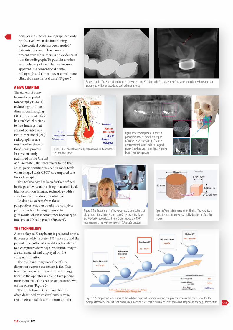
Figures 1 and 2: The p root of tooth #14 is not visible in the pa radiograph. a coronal slice of the same tooth clearly shows the root anatomy as well as an associated peri-radicular lucency
Figure 5: The footprint of the veraviewepocs is identical to that of a panoramic machine. a small cone X-ray beam irradiates the FpD for 9.4 seconds, while the c-arm makes one 180˚ rotation around the region of interest (J.Morita corporation)
Figure 6: voxel: Minimum unit for 3D data. The voxel is an isotropic cube that provides a highly detailed, artifact-free image
Figure 7: a comparative table outlining the radiation figures of common imaging equipments (measured in micro-sieverts). The average effective dose of radiation from a cBcT machine is less than a full mouth series and within range of an analog panoramic film
bone loss in a dental radiograph can only be observed when the inner lining of the cortical plate has been eroded.1 Extensive disease of bone may be present even when there is no evidence of it in the radiograph. To put it in another way, only very chronic lesions become apparent in a conventional dental radiograph and almost never corroborate clinical disease in ‘real time’ (Figure 3).
A NEW CHAPTERThe advent of cone-beamed computed tomography (CBCT) technology or three-dimensional imaging (3D) in the dental field has enabled clinicians to ‘see’ findings that are not possible in a two-dimensional (2D) radiograph, or at a much earlier stage of the disease process. In a recent study published in the Journal of Endodontics, the researchers found that apical periodontitis was seen in more teeth when imaged with CBCT, as compared to a PA radiograph.2
This technology has been further refined in the past few years resulting in a small field, high-resolution imaging technology with a very low effective dose of radiation.
Looking at an area from three perspectives, one can obtain the ‘complete picture’ without having to resort to guesswork, which is sometimes necessary to interpret a 2D radiograph (Figure 4).
THE TECHNOLOGYA cone shaped X-ray beam is projected onto a flat sensor, which rotates 180o once around the patient. The collected raw data is transferred to a computer where high-resolution images are constructed and displayed on the computer monitor.
The resultant images are free of any distortion because the sensor is flat. This is an invaluable feature of this technology because the operator is able to take precise measurements of an area or structure shown on the screen (Figure 5).
The resolution of CBCT machines is often described by its voxel size. A voxel (volumetric pixel) is a minimum unit for
PPD February 2011 141
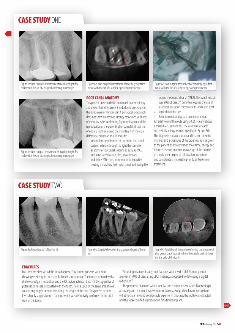
Figure 8a: Non-surgical retreatment of maxillary right first molar with the aid of a surgical operating microscope
Figure 8b: Non-surgical retreatment of maxillary right first molar with the aid of a surgical operating microscope
Figure 8c: Non-surgical retreatment of maxillary right first molar with the aid of a surgical operating microscope
Figure 8d: Non-surgical retreatment of maxillary right first molar with the aid of a surgical operating microscope
ROOT CANAL ANATOMY This patient presented with continued heat sensitivity and discomfort after a recent endodontic procedure in the right maxillary first molar. a periapical radiograph does not show an obvious lucency associated with any of the roots. after confirming (by examination and the reproduction of the patient’s chief complaint) that the offending tooth is indeed the maxillary first molar, a differential diagnosis should include: • Incomplete debridement of the entire root canal system. schilder brought to light the complex anatomy of root canal systems as early as 1967, including lateral canals, fins, anastamoses, and deltas.4 The most common omission when treating a maxillary first molar is not addressing the
second mesiobuccal canal (MB2). This canal exists in over 90% of cases,5,6 but often requires the use of a surgical operating microscope to locate and treat• vertical root fracture • Recontamination due to a poor coronal seal.an axial view of the roots using a cBcT clearly shows a missed MB2 (Figure 8b). The case was retreated successfully using a microscope (Figure 8c and 8d). The diagnosis is made quickly and in a non-invasive manner, and a clear idea of the prognosis can be given to the patient prior to investing more time, energy and finances. having an exact knowledge of the number of canals, their degree of calcification, curvature and complexity is invaluable prior to embarking on treatment.
Figure 9a: pa radiograph of tooth #18 Figure 9b: sagittal slice depicting a greater degree of bone loss
Figure 9c: axial slice of the tooth confirming the presence of a horizontal crack extending from the distal marginal ridge into the pulp of the tooth
FRACTURESFractures are often very difficult to diagnose. This patient presents with mild chewing sensitivity in the mandibular left second molar. The tooth is restored with a shallow amalgam restoration and the pa radiograph is, at best, mildly suggestive of potential bone loss, associated with the tooth. here, a cBcT of the same area shows an amazing degree of bone loss along the length of the root. This pattern of bone loss is highly suggestive of a fracture, which was definitively confirmed in the axial view of the tooth.
according to a recent study, root fractures with a width of 0.2mm or greater are seen in 70% of cases using cBcT imaging, as opposed to 43% using a digital radiograph.7
The prognosis of a tooth with a root fracture is often unfavourable. Diagnosing it accurately and in a non-invasive manner (versus a surgical exploratory procedure) will save chair time and considerable expense. In this case, the tooth was extracted and the socket grafted in preparation for a future implant.
CONT
CASE STUDY ONe
CASE STUDY TWO

142 February 2011 PPD
CASE STUDY ThRee
Figure 10b: sagittal slice of right central incisor highlighting a fracture in buccal plate (red arrow)
Figure 10a: pa radiograph of the maxillary anterior region. The right central incisor was luxated and the left central incisor was avulsed and subsequently replanted, following a traumatic injury to the anterior maxillary region
Figure 10c: sagittal slice of left central incisor showing an obliterated buccal plate. Note also the gap seen between the tooth and the socket
TRAUMA In this case, the pa radiograph taken of a 15-year-old who has sustained trauma to her maxillary anterior teeth shows a significant widening of the periodontal space around the left central incisor. sagittal slices of a cBcT of the anterior maxilla depict a much more complex situation. The buccal plate adjacent to the upper right central incisor is fractured (Figure 10b), which will prolong the splinting time required for treatment. Figure 10c shows an obliterated buccal plate adjacent to the left central incisor, thus rendering its prognosis as hopeless.
3D data, analogous to a pixel for 2D image. Generally speaking, a smaller voxel size yields a higher resolution because more voxels (data) can fill a given area (Figure 6).
RADIATIONA general rule is the smaller the field of view (FOV) the lower the absorbed radiation. In fact, absorbed radiation is a function of the FOV cubed. So, the small field CBCT machines provide for less radiation absorbed by tissues and organs as compared to the large field CBCT machines. According to a recent published study, the Veraviewepocs recorded an effective dose of 29.78 μSv for the panoramic and 40x40mm 3D protocol3 (Figure 7). So, does this new technology actually make a meaningful difference in the diagnostic process? The four clinical cases in this article seen at our endodontic practice will illustrate the value of the additional information gained by viewing an area of interest from different perspectives.
PPD February 2011 143
ENDODONTICSINFOCUS
ppd
Dr pirooz Zia will be presenting a seminar on 12 march 2011 at seminars@38, London, entitled: a practical view of modern endodontic diagnosis. www.seminarsthirtyeight.com
CASE STUDY FOuR
Figure 11b: example of an external cervical inflammatory root resorption
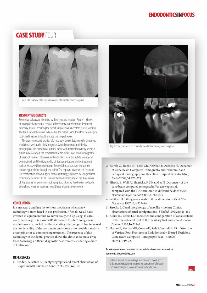
Figure 11a: example of an external cervical inflammatory root resorption
RESORPTIvE DEFECTS Resorptive defects are identified by their type and location. Figure 11 shows an example of an external cervical inflammatory root resorption. Treatment generally involves repairing the defect surgically with Geristore, a resin ionomer. The cBcT shows the defect to be within the pulpal space; therefore, non-surgical root canal treatment should precede the surgical repair.
The type, extent and location of a resorptive defect determine the treatment modality as well as the likely prognosis. careful examination of the pa radiograph of this mandibular left first molar with thermal sensitivity reveals a subtle radiolucency in the coronal third of the mesial root, which is suggestive of a resorptive defect. however, without a cBcT scan, this subtle lucency can go unnoticed, and therefore lead to clinical complications during treatment, such as excessive bleeding through the mesiobuccal canal, or extrusion of sodium hypochlorite through the defect. The requisite treatment on this tooth is a combination of non-surgical root canal therapy, followed by a surgical root repair using Geristore. a cBcT scan of the tooth clearly shows the dimensions of this external inflammatory root resorption, allowing the clinician to decide beforehand whether treatment would have a favourable outcome.
CONCLUSIONIt is necessary and healthy to show skepticism when a new technology is introduced in our profession. After all, we all have invested in equipment that we never really end up using. Is CBCT really necessary, or is it overkill? We believe this technology is as revolutionary in our field as the operating microscope. It has increased the predictability of the treatments and allows us to provide a realistic prognosis prior to commencing treatment. The presence of this technology in the dental practice allows the clinician to move away from predicting a difficult diagnostic case towards rendering a more definitive one.
REFERENCES1. Bender IB, Seltzer S. Roentgenographic and direct observation of experimental lesions on bone: JADA 1961;62:152
2. Estrela C, Bueno M, Leles CR, Azevedo B, Azevedo JR. Accuracy of Cone Beam Computed Tomography and Panoramic and Periapical Radiography for Detection of Apical Periodontitis: J Endod 2008;34:273-2793. Hirsch, E, Wolf, U, Heinicke, F, Silva, M A G Dosimetry of the cone beam computed tomography Veraviewepocs 3D compared with the 3D Accuitomo in different fields of view: Dentomaxillofac Radiol 2008;37: 268-2734. Schilder H. Filling root canals in three dimensions. Dent Clin North Am 1967;Nov:723–44.5. Stropko J. Canal morphology of maxillary molars: Clinical observations of canal configurations. J Endod 1999;25:446-4506. Kulild JD, Peters DD. Incidence and configuration of canal systems in the mesiobuccal root of the maxillary first and second molars. J Endod 1990;16:311–7.7. Hassan B, Metska ME, Ozok AR, Stelt P, Wesselink PR. Detection of Vertical Root Fractures in Endodontically Treated Teeth by a Cone Beam Computed Tomography Scan. J Endod 2009;35:719-722
To ask a question or comment on this article please send an email to: [email protected]

8 Endodontic practice Volume 5 Number 4
Practice profile
What can you tell us about your background?I was born in Iran, to parents who were both physicians. In 1979, when I was 10 years old, my family and I moved to England after the revolution hit. I spent the next 14 years living and studying in London. While initially the move was a big change, over time, I grew to love all of the positive aspects of the UK. I received my dental degree at University of London (Guy’s Hospital) in 1991. From there, I went to Boston University Goldman School of Dental Medicine Boston (BUGSDM) to become an endodontist, where I was trained by Herb Schilder. Looking back, I feel that I’m really blessed to have lived in three different continents and to have been exposed to three different cultures. As a result, I will encourage my children to get out of their usual environment once in awhile. I want them to learn how to adapt and overcome the feeling of being an outsider. It’s possible to embrace the best elements of each culture. Now, I know a bit about carpets, making a nice cup of tea, and baseball! After finishing the program at Boston, I moved to the Washington DC area, and I’ve lived here for the past 17 years. It is a very cosmopolitan city and a great place to live and raise a family.
Is your practice limited to endodontics?Yes. My practice focuses on endodontics only. It does, however, make a great deal of sense for endodontists to get involved in implantology. By nature, endodontists are exacting people. They have a great understanding of the anatomy as well as the skill to perform delicate surgery. Endodontists who wish to expand their services and place implants must invest in serious training first. I hope to see meaningful educational opportunities in the future — perhaps a part-time residency program for practicing endodontists — to get the necessary training. For me, in this area’s competitive environment when so many skilled specialists can place implants, without prolonged training, I wouldn’t be comfortable providing that service.
Why did you decide to focus on endodontics?I decided to focus on endodontics because I was attracted to the “micro” and precise nature of the specialty. A millimeter is a long way in our field and is the difference between success and failure.
Also, in endodontics, we can create and complete outstanding results on a daily basis. The results and the gratification are pretty immediate. In other specialties like prosthodontics or orthodontics, it can take months or even years to get an outcome. Dr. Stephen Buchanan once described himself as an “impatient perfectionist.” I can relate to that! Most importantly, we have the opportunity to meet people in significant pain and distress and provide them with relief and effective care. That is very gratifying. Through our patients, we can make a meaningful difference and a friend for life! How long have you been practicing, and what systems do you use?I have been a dentist for 21 years and an endodontist for 17 years, and use many different systems:1. For cleaning and shaping—Protaper® (Dentsply Tulsa Dental Specialties). 2. For obturation: Vertical compaction of warm gutta percha. I use the System B for downpacking and the Calamus® for backpacking of the canals.3. Improved visibility: In endodontics, microscopy should be the standard of care. We have microscopes in every operatory — we don’t even have overhead lights!4. Improved imaging: We use DEXIS® digital radiography with DEXIS Platinum sensors. Digital radiography is a “no-brainer.” This imaging method gives me instant, clear images, lower radiation, and the opportunity to sit side-by-side with a patient and communicate. The new revolution in endodontics is the ability to see in three dimensions. I have a Veraviewepocs® 3De (J. Morita) CBCT. It is a fantastic unit with terrific image quality and relatively low radiation. It has increased the predictability of our treatments and allows us to provide a realistic prognosis prior to commencing treatment. I can see it becoming very prevalent in
Through the keyho eDr. Pirooz ZiaA practice filled with canals, culture,
compassion, and camaraderie
Volume 5 Number 4 Endodontic practice 9
Practice profile
dentistry.5. ASI carts: It’s important to have a clean, uncluttered, organized, calm ambiance in your office and the operatories. The amalgamation of all the various tools into one unit is very helpful. Technology has made endodontics easier and has leveled the playing field. But, good technology does not make the distinction between who is good and who is great. You also need exceptional patience, intentionality, and awareness to do good work. Dr. Schilder used to create outstanding shapes with just hand-held files and reamers. He filled many portals of exit and lateral canals without fancy gadgets. Even now, I make a concerted effort to slow down and be methodical.
How has your experience and affiliations affected your practice philosophy?I feel that my specific program at BU was very focused on producing good clinicians versus good academicians. I liked that, and it was one of the reasons I chose the program. But, the process of board certification (ABE, which I completed in 2002) completed the circle when I became familiar with the research. I definitely recommend that all young endodontists go through the process. It’s a challenge, but well worth it! My partner, Reza Farshey, also a BU-trained endodontist, is a sharp guy. We took the time to get to know each other as people, and made sure that our philosophies were aligned. That is the most important aspect of making partnerships work in dentistry — whether you have the same core values and outlook in life. We push each other to get better every day. Teaching is the best way to learn. I taught the endodontic residents at University of Maryland for 6 years. I was responsible for teaching vertical compaction of warm gutta percha. There was still a lot of resistance to that technique at the time as most schools taught lateral condensation. I loved it when a student would fill his/her first lateral canal, and suddenly a light bulb would go off! One of the best teachers I ever had was the program director at BU, Carlo Castellucci. I still hear his voice in my head when I’m negotiating a calcified canal. We have some terrific practitioners in our area with a wealth of knowledge. Lecturing locally keeps me up-to-date especially in other specialties, and this is essential. I don’t want to be a one-
tooth dentist; I want to know a lot about other disciplines and get involved in the treatment planning process. My last, but definitely not least philosophy: stay an eternal student — you are never done learning!
Who has inspired you?1. Dr. Herb Schilder: He had a very clear sense of the realities of the root canal system and what it takes to address them. He then held himself (the clinician) as “cause in the matter.” He proposed that lesions of endodontic origin had a 100% potential for healing (if the tooth was structurally and periodontally sound) and that our ability to deal with the root canal system was the main factor in the success or failure of the case. I loved his clarity, his vision, his passion, and his willingness to take responsibility.2. Drs. John West and Cliff Ruddle have taken Dr. Schilder’s ideas and have worked hard to make them relevant to today’s practice through creating innovative and efficient systems. They have done this without losing the essence of exceptional endodontics as initially described by Schilder.3. Marc Cooper (www.masteryofpractice.com), a periodontist by training, is my practice coach. He is all about looking in the mirror and being mindful. I really recommend finding someone who can help you develop the non-clinical skills that we all need to run a successful practice. I feel strongly that even endodontic training programs should include non-clinical skills training in their curriculum. Besides being familiar with the research, it makes sense to educate endodontists in how to lead and manage, especially in this rough economy. 4. My father was a surgeon with exceptional skills, but more importantly, he taught me that people don’t care how much you know until they know how much you care. We need to understand that caring is an indispensable part of health care.
What is the most satisfying aspect of your practice?I get to work as part of a team with my referring colleagues, some wonderfully committed professionals, who have over the years become friends. We work on treatment planning and delivering exceptional results. I feel I’m an extension of their practices and really enjoy that. Endodontists are like sculptors, working on the internal anatomy of the tooth. In almost all cases, I can look at
(Top left) This is my amazing team. From the right: my partner Dr. Farshey, my office manager Jennifer, Rhea, Claudia, Maritza, Cindy and Kirsten. They are an exceptional group of people and make me look good every day
(Top right) Our J. Morita CBCT unit
(Bottom left) The office is designed to exude a clean, calm feel
(Bottom right) I have used Global microscopes since 1995
10 Endodontic practice Volume 5 Number 4
Practice profile
a preoperative image and form a “future memory” of what the postoperative image will look like. Then, with the help of my extraordinary team, I create it! To see a mental image become reality is very fulfilling. I think it’s up to our generation of endodontists to undo the historical negative image of root canals. To exceed a patient’s expectation as far as the totality of their experience is a wonderful interaction.
Professionally, what are you most proud of?I’m certainly proud of having come to a very “saturated” area in terms of the ratio of endodontists to restorative dentists and creating a practice that is successful, consistent, and stable. We ask every single one of our patients for feedback, and it has been overwhelmingly positive. So, I’m proud of having worked hard to earn the respect and trust of my patients and colleagues. It’s something we have to strive for every day and never take it for granted. I never get comfortable! The essence of life is relationships. Mostly, I’m proud of the phenomenal relationships that have been formed around me, whether it’s with colleagues, my partner, or my wonderful staff. We have each other’s back. I’m surrounded by givers and proud to be “in the trenches” with them. These things mean a lot more to me than any awards or recognition I may have received.
What do you think is unique about your practice?We are uncompromising about our commitment to excellence. We define it as the natural result of consistently adhering to the core fundamentals without cutting corners. We have embraced the technology that allows us to deliver great care, such as equipping all rooms with operating microscopes and being among the first practices in the area to acquire the CBCT. The quality of the endodontic care is like the quality of the food at a restaurant. The food needs to be great, but it’s only part of the experience. We are focused on providing exceptional service and are tuned into all aspects of the patient’s senses — what they feel, hear, see, and smell. We have a flat screen TV in
the waiting room with calming messages and beautiful scenery. Patients arriving from the hubbub and traffic are treated to calm, subliminal messages. The quality of our communication is our “secret sauce.” How many times in your life, when it comes to interactions, do people sit down and listen to you, explain something to you, do what they say they would do in a timely manner? They should be respectful of your time and comfort and follow up long after you
have paid the bill. Besides calling right after the procedure, we call our patients a month after the root canal and ask if they got the final restoration done, and if they are comfortable. My imaging methods also help me achieve this goal. I am a very visual person and use PowerPoint presentations and preoperative imaging to educate my patients. This connects me to my patients and makes me accountable.
What has been your biggest challenge?It has taken me a long time to understand that it’s about energy management versus time management. We have to wear many hats concurrently, as leaders, managers, clinicians, and “rainmakers.” I have also had to learn to prioritize effectively, to deliver my best at the office and still have enough energy left in the “tank” for the people who matter most — my wife and kids!
What would you have become if you had not become a dentist?A professional tennis player! I really loved tennis as a kid and definitely dreamt of raising the Wimbledon trophy. I was “gently encouraged” by my folks to pursue “something a bit more sensible,” so here we are. I get to raise several
mental trophies a day with each successful result. Great tennis champions are perceptive, proactive, and reactive all at the same time; the same qualities that you need to be a good endodontist — I just use files instead of a racket! I am really grateful for my profession and the opportunities that it has afforded me.
What is the future of endodontics and dentistry?Endodontics is more exciting than ever. We have a profound understanding of the elements that are needed for success, and we have fabulous technology and innovative techniques to satisfy them. Endodontics has never been more predictable, efficient, and comfortable for the patient. I know that we are struggling with a deep downturn in the economy that has affected all of us significantly. But, I know that “this too shall pass,” and through it we can learn to become better, provide more value, and be of more service to others. Although it seems like a cliché, out of every adversity comes abundance. There will always remain an appreciation for quality, patient-centered dental care. I see the pendulum swinging back on the issue of teeth being “sacrificed” for implants. I think it is crucial that endodontists make the effort to educate our colleagues about the predictability of a properly performed endodontic procedure. We must know all that we can about the alternatives so that we can provide our patients with intelligent and cost-effective treatment options. We can’t be passive observers. We must be advocates of what serves our patients best. I have joined forces with other endodontists in the
(Right) Playing tennis with the boys on the grass courts of London
(Left) My wife Ladan and my sons Cameron and Cyrus
community to educate our colleagues with a uniform message. I am happy that implants are an option for certain dental conditions, but the public also deserves information on quality endodontics. Endodontists should make a case for our specialty as a whole, not just for their specific practice.
What are your top tips for maintaining a successful practice?1. Have a clear, authentic, patient-centered vision that your team can commit to rather than comply with. This is the main job of a leader, and you must lead!2. Work hard on learning to listen and being more “relational” vs. “transactional.” People are generally not listened to very well. Listening can be powerful, and you run the risk of actually learning something!3. Take the time to develop and implement sensible managerial systems. Write the “managerial software” that will make the practice run — that way you can spend more time doing what you are best at — endodontics.4. Be committed to “Kaizen” (Japanese for continuous improvement). Every day ask yourself: “What did I do to improve the practice?” and “What did I do to grow the practice?”5. Don’t listen to coaches that tell you to deliver doughnuts to your referrers. You’ll only kill them sooner of a heart attack! And, that’s not good for business. If you are truly committed to the success of your referring colleagues, then you will see the reciprocation.
What advice would you give to budding endodontists?1. Create the time to sit down and form a clear vision of what you want your practice to be like and who you want to be. “Being” is a lot more important than “doing.” So many of us have a to-do list. Only a few people take the time to make a to-be list. This will define your core values. Then stick to them! Don’t let anything/anyone, not the insurance companies, your referring doctors, or your patients, talk you into compromising your values.2. Beyond that, it’s about working hard to consistently deliver value to your patients and referral colleagues. The trust placed in you by them is worthy of your absolute best effort, every day! 3. Don’t be afraid of failure. A productive failure is more valuable than an unproductive success.4. Don’t focus on generating income. Focus on providing value by doing great work.5. Surround yourself with good mentors and teachers. Then, be coachable!6. Look for ways of giving back to your profession and your community. 7. Always be grateful and humble.
What are your hobbies, and what do you do in your spare time?I follow tennis pretty closely and play as often as I can. It used to be a lot more often before we had kids! (Time is a thief!) Luckily, both my sons are getting into playing tennis now, so they tag along. My 7-year-old loves it. I’m banking on him winning Wimbledon in 15 years (who needs a retirement plan!). I also love skiing. The mountain air clears my head. I don’t do the double-diamond runs anymore. A few blue runs and then a nice cup of hot chocolate! I’m trying to stay fit and take care of my back. I absolutely detest going to the gym, and have a trainer who truly enjoys inflicting pain and suffering. (I can’t believe that we endodontists are the ones with the bad reputation). Beyond that, I love spending time with my family and
Pirooz Zia, BDS, MScD, maintains full-time private practice in Chevy Chase, Maryland, and he presents seminars nationally and internationally on current endodontic techniques. He was an associate clinical professor at the University of Maryland School of Dentistry, and has lectured at Boston University, the University of Michigan and the University of London, England. Dr. Zia is a Diplomate of the American Board of Endodontics and an active member of the American Association of Endodontists, the American Dental Association, and the DC Dental Society. He is the 2001 recipient of the District of Columbia Dental Society’s David Mast Memorial Award for excellence in continuing education. He was voted one of Washington’s “Top Dentists” in a survey of his peers conducted by Washingtonian magazine and was voted among “America’s Top Dentists” in a similar national survey. He is a Fellow of the International College of Dentistry, which is dedicated to advancing the science and art of dentistry worldwide.
Top Ten List:
1. An amazing team of people who understands and is committed to a common vision.
2. The ASI units (Advance Integrated Systems). I hate clutter, and these units are extremely well made.
3. Great educational software to help me communicate effectively with my patients.
4. DEXIS® digital radiography and sensors. The instant images which are produced with less radiation are a “no brainer” for diagnosis and clarity of communication.
5. Veraviewepocs® 3De CBCT (J. Morita). It is fantastic. The ability to remove superimpositions and get multiple perspectives on a tooth has revolutionized our field. We have a much clearer idea of pitfalls and prognoses before we start a case. No pilot should take off without a flight plan!
6. ProTaper® rotary files.
7. The electronic apex locator.
8. Chlor-XTRA™ (Vista Dental Products), 6% sodium hypochlorite with surface modifiers. This is far more effective than regular bleach in dissolving tissue because of the lower surface tension that allows it to really “bathe all the canal irregularities.”
9. System B for vertical compaction of warm gutta percha to seal all portals of exit and Calamus® to “backpack.”
10. A sense of humor! Take what you do seriously without taking yourself too seriously.
friends. There is a difference between success and significance. Nido Qubein, president of High Point University, once told me that success is about fans, fortune, and fame. Significance is about family, friends and faith. At the end of the day, these are the things that will matter most. EP
12 Endodontic practice Volume 5 Number 4
Practice profile