Endocrinology for the Surgeon Dr. Jeremy Gilbert Sunnybrook Health Sciences Centre.
68
Endocrinology for the Surgeon Dr. Jeremy Gilbert Sunnybrook Health Sciences Centre
-
Upload
zechariah-excell -
Category
Documents
-
view
255 -
download
5
Transcript of Endocrinology for the Surgeon Dr. Jeremy Gilbert Sunnybrook Health Sciences Centre.

Endocrinology for the Surgeon
Dr. Jeremy GilbertSunnybrook Health Sciences Centre

ObjectivesBy the end of this presentation, participants will be
able to:• Review diagnosis and treatment of thyroid
emergencies• Discuss diagnosis and management of adrenal
insufficiency– Focus on steroid tapering
• Review workup for adrenal incidentaloma– Focus on pheochromocytoma
• Discuss an approach to hypercalcemia diagnosis and management
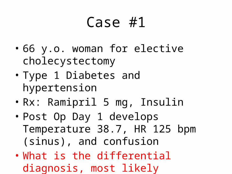
Case #1
• 66 y.o. woman for elective cholecystectomy• Type 1 Diabetes and hypertension• Rx: Ramipril 5 mg, Insulin • Post Op Day 1 develops Temperature 38.7, HR
125 bpm (sinus), and confusion• What is the differential diagnosis, most likely
diagnosis and management
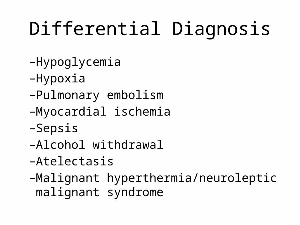
Differential Diagnosis
–Hypoglycemia–Hypoxia–Pulmonary embolism–Myocardial ischemia–Sepsis–Alcohol withdrawal–Atelectasis–Malignant hyperthermia/neuroleptic malignant
syndrome

Thyroid Storm- Definition
• Rare, life threatening condition manifesting as exaggeration of thyrotoxicosis
• Usually patients have underlying hyperthyroidism with an acute precipitant (surgery, trauma, infection)
• Maybe related to poor compliance and low SES
• CLINICAL DIAGNOSIS (NOT LABORATORY)

Uptodate 2012
SensitiveNot Specific
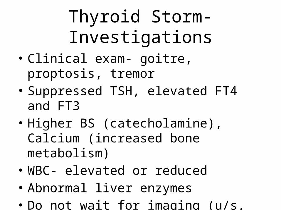
Thyroid Storm- Investigations
• Clinical exam- goitre, proptosis, tremor• Suppressed TSH, elevated FT4 and FT3• Higher BS (catecholamine), Calcium (increased
bone metabolism)• WBC- elevated or reduced• Abnormal liver enzymes• Do not wait for imaging (u/s, RAIU and scan)
to make diagnosis!

Thyroid Storm- Management
• Monitored Setting (10-30% mortality rate)• Same as for hyperthyroidism, except meds
given in higher doses, more often• Treat comorbidities (eg. infection)• Supportive care- tylenol, cooling blanket,
fluids or diuretics
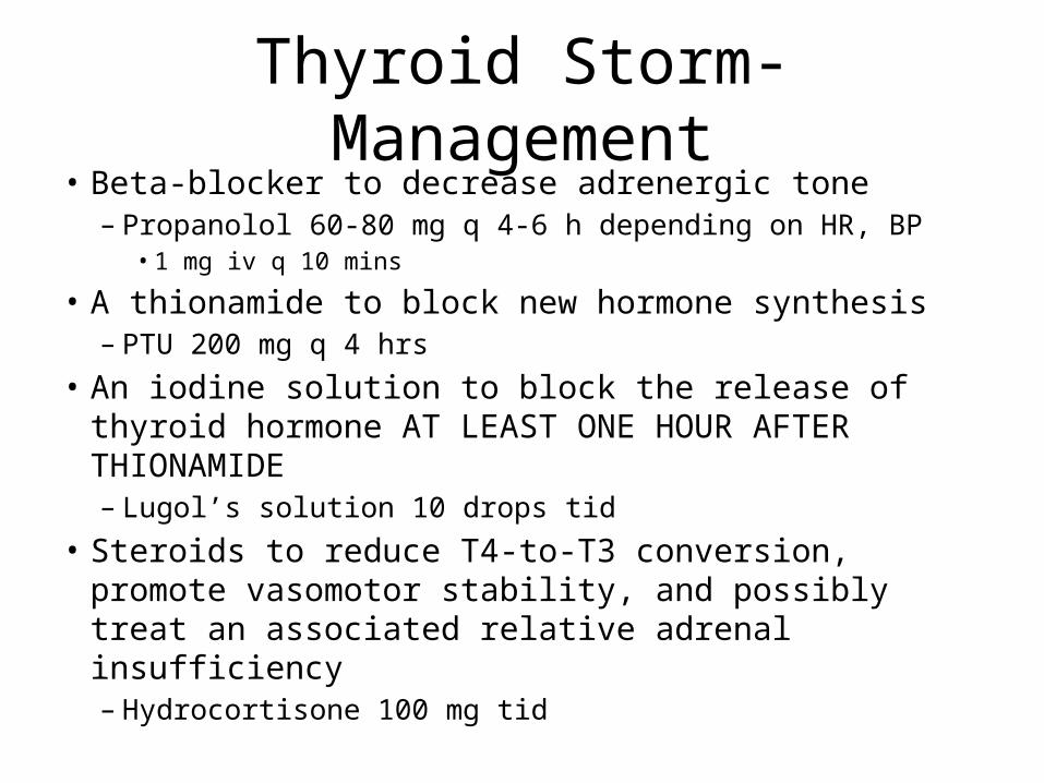
Thyroid Storm- Management• Beta-blocker to decrease adrenergic tone
– Propanolol 60-80 mg q 4-6 h depending on HR, BP• 1 mg iv q 10 mins
• A thionamide to block new hormone synthesis– PTU 200 mg q 4 hrs
• An iodine solution to block the release of thyroid hormone AT LEAST ONE HOUR AFTER THIONAMIDE– Lugol’s solution 10 drops tid
• Steroids to reduce T4-to-T3 conversion, promote vasomotor stability, and possibly treat an associated relative adrenal insufficiency– Hydrocortisone 100 mg tid

Thyroid Storm
• Suspect it• Review History• Complete Physical Exam• Appropriate blood work• Management
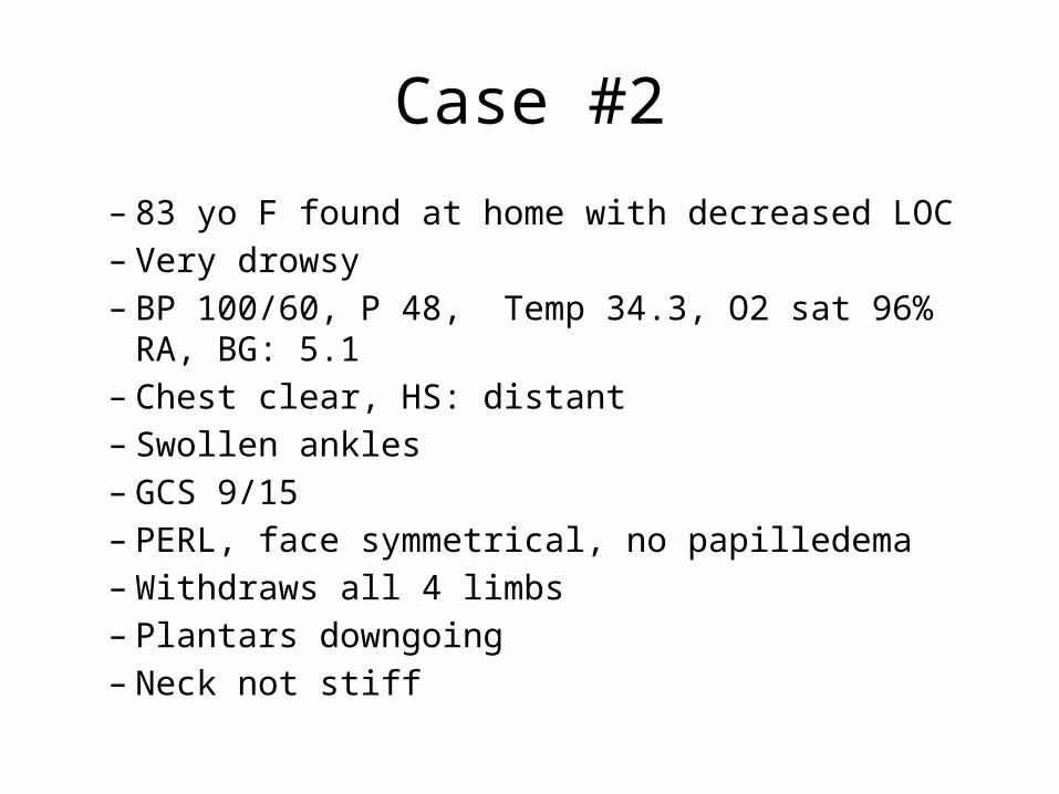
Case #2
– 83 yo F found at home with decreased LOC – Very drowsy– BP 100/60, P 48, Temp 34.3, O2 sat 96% RA, BG: 5.1– Chest clear, HS: distant– Swollen ankles– GCS 9/15– PERL, face symmetrical, no papilledema– Withdraws all 4 limbs– Plantars downgoing– Neck not stiff
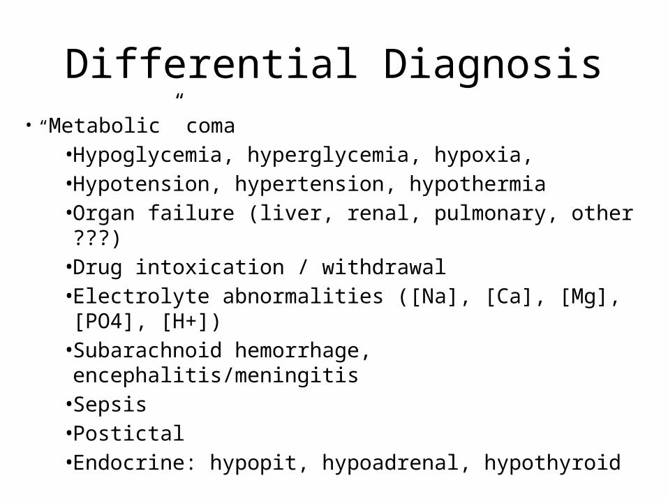
Differential Diagnosis• “Metabolic” coma
• Hypoglycemia, hyperglycemia, hypoxia, • Hypotension, hypertension, hypothermia• Organ failure (liver, renal, pulmonary, other ???)• Drug intoxication / withdrawal• Electrolyte abnormalities ([Na], [Ca], [Mg], [PO4],
[H+])• Subarachnoid hemorrhage, encephalitis/meningitis• Sepsis• Postictal• Endocrine: hypopit, hypoadrenal, hypothyroid
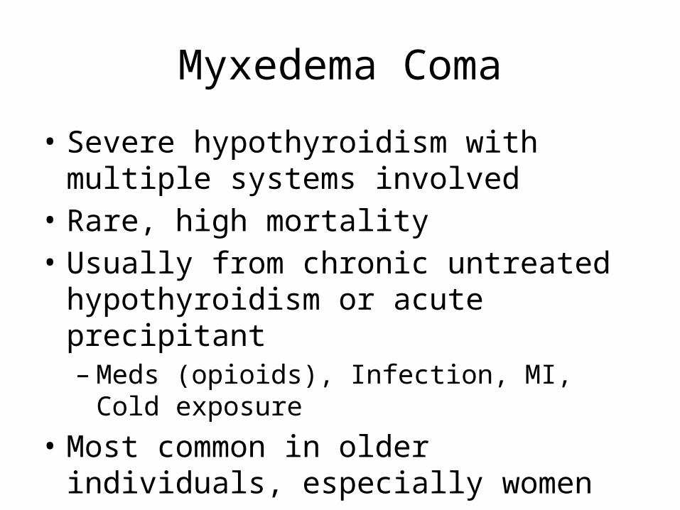
Myxedema Coma
• Severe hypothyroidism with multiple systems involved
• Rare, high mortality• Usually from chronic untreated
hypothyroidism or acute precipitant – Meds (opioids), Infection, MI, Cold exposure
• Most common in older individuals, especially women

Myxedema Coma- Presentation
• Decreased LOC (often not coma)• Hypothermia• Hyponatremia• Hypotension• Hypoglycemia• Hypoventilation• Bradycardia• Puffiness- myxedema- mucin deposits
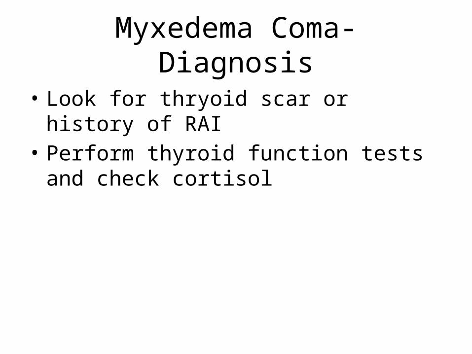
Myxedema Coma- Diagnosis
• Look for thryoid scar or history of RAI• Perform thyroid function tests and check
cortisol


Adrenal Disorders
Prepared by: Drs Jeannette Goguen, Robert Silver and Jeremy Gilbert
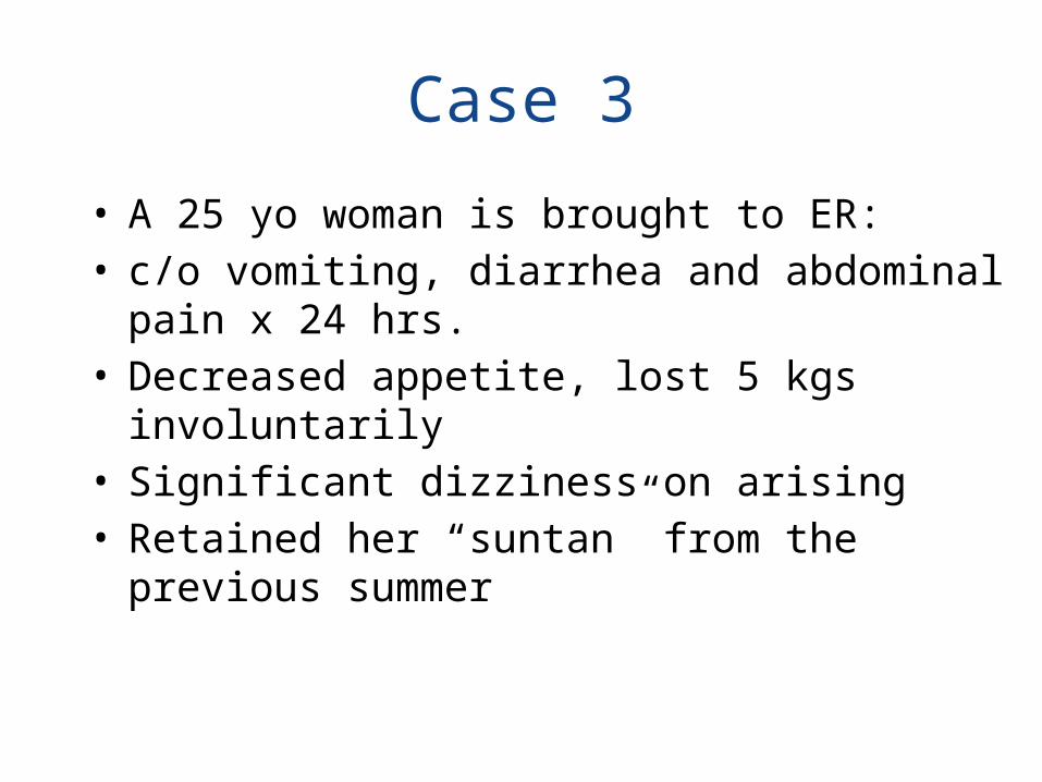
Case 3
• A 25 yo woman is brought to ER:• c/o vomiting, diarrhea and abdominal pain x 24 hrs.• Decreased appetite, lost 5 kgs involuntarily• Significant dizziness on arising • Retained her “suntan” from the previous summer

Case 3 continued . . .• O/E: she looks chronically unwell• HR120/minute; BP is 90/60 supine and 60/30 upright• Her JVP is not visible• Diffuse abdominal tenderness with no peritoneal signs • Large, dark freckles over her cheeks and darkened
palmer creases• Large patches of vitiligo
• Preliminary labs: Na=125, K= 5.2, glucose = 2.5.
1. What is the likeliest diagnosis? What is in your differential diagnosis?
1. What is the likeliest diagnosis? What is in your differential diagnosis?
• The likeliest diagnosis: • acute on chronic adrenal insufficiency presumably precipitated by an
acute viral gastroenteritis.
• The differential of weight loss and malaise is very broad, and includes: • Malignancy• Endocrine: Diabetes mellitus, thyrotoxicosis• Organ failure (liver, kidney)• Inflammatory disorders• Infections (eg, TB)

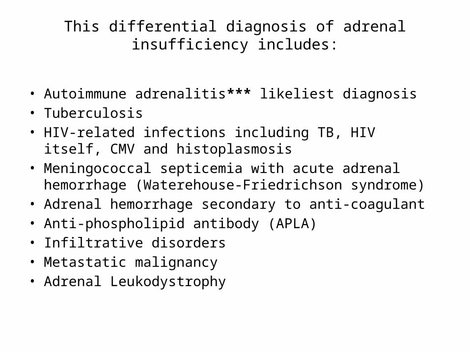
This differential diagnosis of adrenal insufficiency includes:
This differential diagnosis of adrenal insufficiency includes:
• Autoimmune adrenalitis*** likeliest diagnosis• Tuberculosis• HIV-related infections including TB, HIV itself, CMV and
histoplasmosis• Meningococcal septicemia with acute adrenal hemorrhage
(Waterehouse-Friedrichson syndrome)• Adrenal hemorrhage secondary to anti-coagulant• Anti-phospholipid antibody (APLA)• Infiltrative disorders• Metastatic malignancy• Adrenal Leukodystrophy
2. What additional laboratory testing would you order?

• Other baseline labs: – Creatinine 124– Urea 10 – Hgb 98, increased lymphocytes and eosinophils
• Baseline cortisol = 88 nmol/L (next day)• Baseline ACTH = 100 (normal < 20) (1 month later)• Formal Cortrosyn stim test: 1 hr cortisol= 120 (next day)
– Cortisol level over 500-550 nmol/L at either baseline or 60 minutes post-injection of Cortrosyn and
– A rise in Cortisol of 250 nmol/L above baseline. (baseline cortisol > 500 nmol/L rules out adrenal insufficiency)
• TFT’s with thyroid antibodies: TSH 10, +++anti-thyroid Ab
2. What additional laboratory testing would you order?

3. How would you differentiate primary from secondary (pituitary failure) adrenal insufficiency (AI)?

3. How would you differentiate primary from secondary (pituitary failure) adrenal insufficiency (AI)?
Primary AI Secondary AIHyperpigmentation? Yes NoOther autoimmune disorders
Often Rarely
Evidence of pituitary insufficiency/mass effect
No Maybe
Hyponatremia? Yes YesHyperkalemia? Yes NoACTH level High Low

4. How would you acutely manage this patient?

4. How would you acutely manage this patient?
• IV fluids with normal saline and glucose running wide open until BP has stabilized and hypoglycemia has resolved
• Administration of 4-8 mg of IV Dexamethasone once baseline ACTH and cortisol drawn, do Cortrosyn Stimulation test
• DDAVP 1-2 ug iv or sc and sodium load when significantly low baseline plasma Na < 120
• Once hemodynamically stable and able to eat, stress dose steroid coverage can be aborted and oral administration of Hydrocortisone can begin

5. What advice for long-term management of AI would
you give after discharge?

5. What advice for long-term management of AI would
you give after discharge?
• Hydrocortisone- 25 mg daily in split doses, try to reduce to lowest tolerated dose, typically 10 mg QAM, 5 mg QPM (dose is weight-dependent)
• Florinef- 0.1 mg daily• Meds must be taken every day.• For a mild febrile illness, double the dosage of
Hydrocortisone for 3 days then see doctor if still unwell.• If persistently nauseated or vomiting, go immediately to a
local emergency room for intravenous glucocorticoid steroid
• Get a Medic Alert bracelet• Purchase injectable Dexamethasone for remote travelling

Steroids
• Glucocorticoid dosing equivalences
– cortisone acetate 25 mg– hydrocortisone (Solucortef) 20 mg– prednisone 5 mg – methyl-prednisolone (Solumedrol) 4 mg– dexamethasone (Decadron) 0.75 mg

Steroid tapering
• Steroids suppress the H-P-A axis based on duration, potency, dose– Likely if on prednisone 20 mg or its equivalent for more than 3
weeks or who looks Cushingoid– Unlikely if on steroids for < 3 weeks or on alternate day regimens– Uncertain if prednisone 10-20 mg for < 3 weeks or
• If uncertain and going for surgery, it may be worth checking their HPA axis via ACTH stim test
• Individuals vary in how tapering affects them (age, ethnicity- slower in Blacks, elderly)– Consider stability of disease and general health status
• Too expensive and not practical to be following cortisols
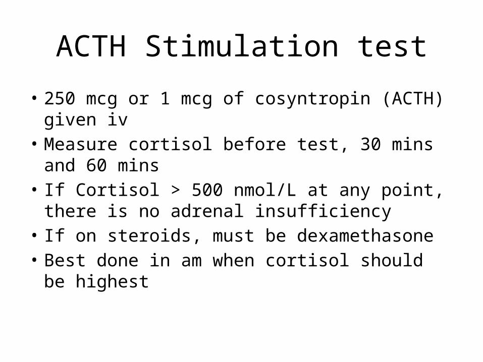
ACTH Stimulation test
• 250 mcg or 1 mcg of cosyntropin (ACTH) given iv
• Measure cortisol before test, 30 mins and 60 mins
• If Cortisol > 500 nmol/L at any point, there is no adrenal insufficiency
• If on steroids, must be dexamethasone• Best done in am when cortisol should be
highest

Tapering- paucity of evidence
• 5 to 10 mg/day every one to two weeks from an initial dose above 40 mg of prednisone or equivalent per day.
• 5 mg/day every one to two weeks at prednisone doses between 40 and 20 mg/day.
• 2.5 mg/day every two to three weeks at prednisone doses between 20 and 10 mg/day.
• 1 mg/day every two to four weeks at prednisone doses between 10 and 5 mg/day.
• 0.5 mg/day every two to four weeks at prednisone doses from 5 mg/day down. This can be achieved by alternating daily doses, eg, 5 mg on day 1 and 4 mg on day 2.

Tapering-alternate days
• If prednisone between 20-30 mg, can try alternate days at 10 mg by reducing by 5 mg every 1-2 weeks
• Decrease alternate day dose by 2.5 mg every 1-2 weeks until the alternate day dose is 0 mg
• Reduce the other dose as you would on a daily regimen

Case 4:
• Mrs S is a 55 yo woman with kidney stones who has been found to have a right 4 cm adrenal mass on routine CT
• She has a past 1-year history of hypertension, BP today is 165/108 on:– Amlodipine 10 mg daily– Ramipril 10 mg daily

1. What are your priorities regarding the adrenal mass?

1. What are your priorities regarding the adrenal mass?
• Is this a benign or malignant tumor?• Is it hormone-secreting? – Catecholamines– Cortisol– Aldosterone– Estrogen or androgen
2. What do benign adenomas look like on imaging?

2. What do benign adenomas look like on imaging?
• Small (typically < 4 cm)• Regular shape, no hemorrhage or calcification• Lack of growth over time• Lipid-rich• Advanced answer: on CT, low Hounsfeld units
(< 10); rapid wash out of contrast (>50% in 10 minutes)
3. What lifestyle factors can contribute to poorly controlled hypertension?
3. What lifestyle factors can contribute to poorly controlled hypertension?
• Dietary: salt, alcohol, licorice• Lack of exercise• Obesity with sleep apnea• Over the counter meds: pseudoephedrine,
NSAIDs• Cocaine, amphetamines

Case Continued. . .
• Mrs S has a family history of “dangerous tumors” in the adrenal gland: both her father, paternal uncle and cousin had these removed. It had been recommended to her that she get her urine tested for adrenaline and that she consider genetic testing, but she has felt well overall and has been too busy with her law practice to get the testing done.

What are you concerned about?

What are you concerned about?
• Pheochromocytoma

What genetic syndromes are associated with this disorder?

What genetic syndromes are associated with this disorder?
• MEN 2A and 2B• Von Hippel Lindau• Advanced answer:• Neurofibromatosis type 1• Familial paraganglioma syndromes (mutations
in SDH genes)

What should you ask her on history?

What should you ask her on history?
• Triad: Spells with the 3 P’s: – Headache (“pain”), palpitations, perspiration
• Symptoms associated with a neck mass (Medullary thyroid cancer)

What would you look for on physical exam?

What would you look for on physical exam?
• BP in both arms, postural hypotension• End-organ complications from hypertensionAdvanced answer:• Thyroid mass (MEN 2 A and B)• Mucosal neuromas on lips, Marfanoid habitus
(MEN 2B)• Retinal, cerebellar findings
(hemangioblastomas with vHL)• Neuromas, café au lait markings (NF-1)
What lab tests would you do to confirm Dx?

What lab tests would you do to confirm Dx?• 24 hour urine catecholamines, metanephrines and creatinine (if
negative, repeat with symptoms if gets spells, but asymptomatic with first collection)– Results: volume 2.5 L, creatinine 8 mmol/day– Metanephrine normal– Normetanephrine 29.3 (normal 0-3.3)– Epinephrine < 10 (normal)– Norepinephrine 5173 (normal 0-500)Can do plasma metanephrine levels if available
Advanced answer:– MIBG nuclear scan can be used to localize tumors of no masses
on adrenal imaging, or if suspect multiple tumors (if familial condition)
– Genetic counseling/genetic testing/screen for other associated tumors if suspect genetic disorder

What is definitive therapy and how would you prep her?
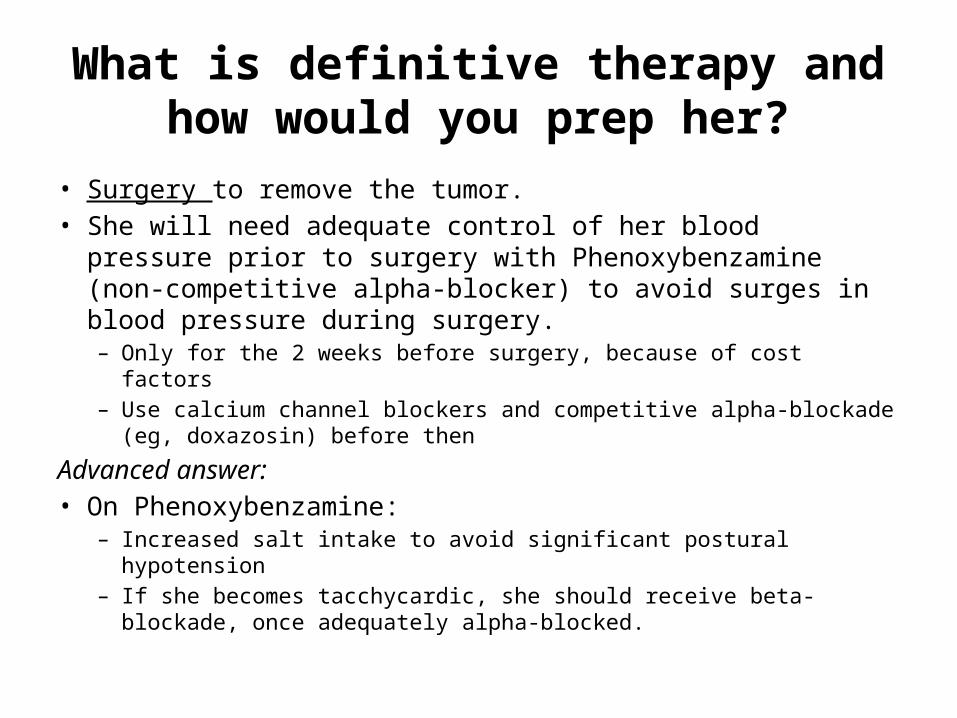
What is definitive therapy and how would you prep her?
• Surgery to remove the tumor.• She will need adequate control of her blood pressure prior to
surgery with Phenoxybenzamine (non-competitive alpha-blocker) to avoid surges in blood pressure during surgery. – Only for the 2 weeks before surgery, because of cost factors– Use calcium channel blockers and competitive alpha-blockade (eg,
doxazosin) before then
Advanced answer:• On Phenoxybenzamine:
– Increased salt intake to avoid significant postural hypotension– If she becomes tacchycardic, she should receive beta-blockade, once
adequately alpha-blocked.

Case 5
• 75 yo M brought by EMS to ED as wife noted patient had decreased LOC
• O/E dry MM, BP 100/50, HR 110• Chest clear, Normal CVS, abdo Normal• Ca+ 3.3, creatinine 125, albumin 29
• Management?

Symptoms• Bones: fractures, osteoporosis, osteitis fibrosa,
arthritis• Stones: renal stones, polyuria, polydypsia (DI),
nephrocalcinosis, renal insufficiency• Groans: constipation, nausea, pancreatitis, peptic
ulcer disease, abdo pain• Moans: lethargy, depression, fatigue, stupor, coma• Neuromuscular: proximal muscle weakness,
myopathy• Cardio: bradycardia, shortened QT, hyperT

What is your differential diagnosis?
• PTH Mediated• 1˚HPT: PTH adenoma/hyperplasia/carcinoma• 3˚ HPT• Familial Hyopcalciuric Hypercalcemia (FHH)• Lithium
• Non-PTH Mediated

Hypercalcemia Ddx• Non-PTH Mediated
• Malignancy» PTHrP (SCC)» Osteolysis (myeloma, breast Ca)» 1-alpha hydroxylase of Vitamin D (lymphoma)
• Granulomatous Disease• Drugs
» Vitamin D, A» Calcium antacids (milk alkali)» Thiazides
• Endocrinopathies: Adrenal insufficiency, Pheo, Thyrotoxicosis, Paget’s (immobility)
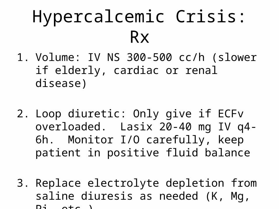
Hypercalcemic Crisis: Rx
1. Volume: IV NS 300-500 cc/h (slower if elderly, cardiac or renal disease)
2. Loop diuretic: Only give if ECFv overloaded. Lasix 20-40 mg IV q4-6h. Monitor I/O carefully, keep patient in positive fluid balance
3. Replace electrolyte depletion from saline diuresis as needed (K, Mg, Pi, etc.)

Hypercalcemic Crisis: Rx
• Calcitonin• 1 IU SC test dose: skin rxn by 15 min• 4 IU/kg SC/IM q12h• If no response by 24-48h increase to max dose 8
IU/kg q6h• Rapid effect (begins 4-6h) but transient (2-3d) due to
tachyphylaxsis• Effective in 60-70% of cases, lowers Ca by 0.3-0.5
mmol/L

Hypercalcemic Crisis: Rx
• Bisphosphonates• Pamidronate
– Ca < 3.0 mM: 30 mg in 500cc NS IV over 4h– Ca > 3.0 mM: 60-90 mg in 500cc NS IV over 24h
• Effect peaks @ 2-4d, lasts 1-6 wk (can retreat q1-6wk)• Can also use zoledronic acid
• Steroids• Useful in Vitamin D intoxication, granuloma, lymphoproliferative
disorders• Prednisone 40-80 mg/d• Takes 5-10d to see treatment effect
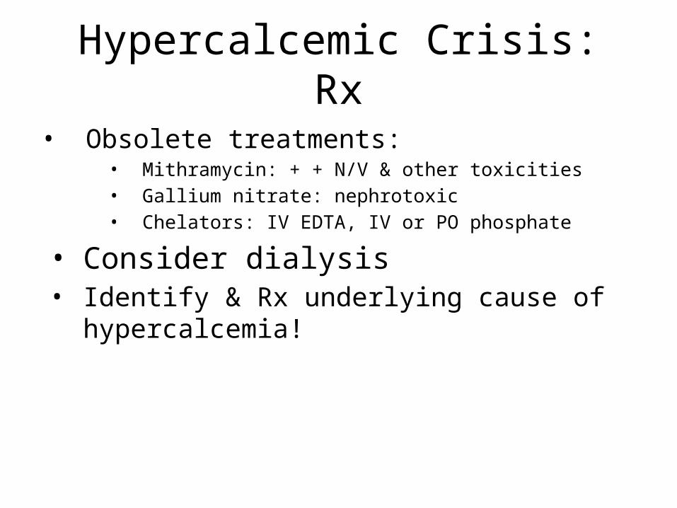
Hypercalcemic Crisis: Rx
• Obsolete treatments:• Mithramycin: + + N/V & other toxicities• Gallium nitrate: nephrotoxic• Chelators: IV EDTA, IV or PO phosphate
• Consider dialysis• Identify & Rx underlying cause of hypercalcemia!
TREATMENT- Summary
• FLUIDS FLUIDS FLUIDS• FLUIDS FLUIDS FLUIDS• FLUIDS FLUIDS FLUIDS• FLUIDS FLUIDS FLUIDS• FLUIDS FLUIDS FLUIDS• FLUIDS FLUIDS FLUIDS
• Lasix• Other
Summary of Objectives
• Review diagnosis and treatment of thyroid emergencies
• Discuss diagnosis and management of adrenal insufficiency– Focus on steroid tapering
• Review workup for adrenal incidentaloma– Focus on pheochromocytoma
• Discuss an approach to hypercalcemia diagnosis and management

Thank you!!!!
QUESTIONS??????