Endocrine Abstracts vol 54 are ‘master regulator complexes’ that coordinate the activation of...
31
Online version available at www.endocrine-abstracts.org Endocrine Abstracts published by bioscientifica February 2018 Volume 54 ISSN 1479-6848 (online) Nuclear Receptors: New Roles for Nuclear Receptors in Development, Health and Disease Conference 2018 27 February – 2 March 2018, Cancun, Mexico
Transcript of Endocrine Abstracts vol 54 are ‘master regulator complexes’ that coordinate the activation of...
Online version available at www.endocrine-abstracts.org
Endocrine Abstracts
published by
bioscientifica
February 2018 Volume 54 ISSN 1479-6848 (online)
Nuclear Receptors: New Roles for Nuclear Receptors in Development, Health and Disease Conference 201827 February – 2 March 2018, Cancun, Mexico

Endocrine AbstractsVolume 54
February 2018
Nuclear Receptors: New Rolesfor Nuclear Receptors inDevelopment, Health and DiseaseConference 2018
27 February 2018 – 2 March 2018
Cancun, Mexico
Programme Organising Committee
Charlotte Bevan, London (POC Chair)
Karen Knudsen, Pennsylvania
Simak Ali, London
Wayne Tilley, Adelaide
Only abstracts with agreed publishing consents are published from this conference

Sponsors and other supporters
Gold
Bronze
Media Partners
Endorsing Partners
Endocrine Abstracts (2018) Vol 54
Nuclear Receptors: New Roles for Nuclear Receptors in Development, Health and Disease Conference 2018
Nuclear Receptors: New Roles for Nuclear Receptors in Development, Health and Disease Conference 2018
Endocrine Abstracts (2018) Vol 54
CONTENTS
Nuclear Receptors: New Roles for Nuclear Receptors in Development, Health and Disease Conference 2018
Plenary Lectures . . . . . . . . . . . . . . . . . . . . . . . . . . . . . . . . . . . . . . . . . . . . . . . . . . . . . PL1–PL3
Invited Speaker . . . . . . . . . . . . . . . . . . . . . . . . . . . . . . . . . . . . . . . . . . . . . . . . . . . . . IS1–IS14
Oral Communications . . . . . . . . . . . . . . . . . . . . . . . . . . . . . . . . . . . . . . . . . . . . . . . . . OC1–OC8
Poster Presentations . . . . . . . . . . . . . . . . . . . . . . . . . . . . . . . . . . . . . . . . . . . . . . . . . . . . P1–P7
INDEX OF AUTHORS

Nuclear Receptors: New Roles for Nuclear Receptors in Development, Health and Disease Conference 2018
Plenary Lectures
Endocrine Abstracts (2018) Vol 54
Nuclear Receptors: New Roles for Nuclear Receptors in Development, Health and Disease Conference 2018
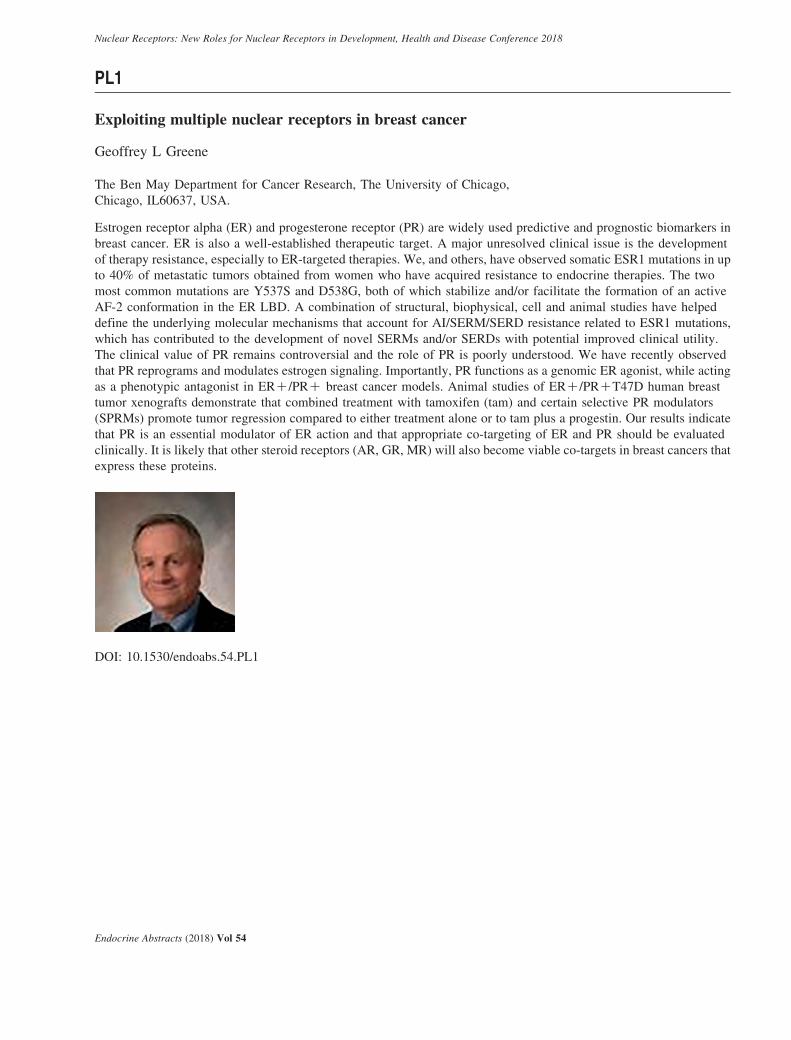
PL1
Exploiting multiple nuclear receptors in breast cancer
Geoffrey L Greene
The Ben May Department for Cancer Research, The University of Chicago,
Chicago, IL60637, USA.
Estrogen receptor alpha (ER) and progesterone receptor (PR) are widely used predictive and prognostic biomarkers in
breast cancer. ER is also a well-established therapeutic target. A major unresolved clinical issue is the development
of therapy resistance, especially to ER-targeted therapies. We, and others, have observed somatic ESR1 mutations in up
to 40% of metastatic tumors obtained from women who have acquired resistance to endocrine therapies. The two
most common mutations are Y537S and D538G, both of which stabilize and/or facilitate the formation of an active
AF-2 conformation in the ER LBD. A combination of structural, biophysical, cell and animal studies have helped
define the underlying molecular mechanisms that account for AI/SERM/SERD resistance related to ESR1 mutations,
which has contributed to the development of novel SERMs and/or SERDs with potential improved clinical utility.
The clinical value of PR remains controversial and the role of PR is poorly understood. We have recently observed
that PR reprograms and modulates estrogen signaling. Importantly, PR functions as a genomic ER agonist, while acting
as a phenotypic antagonist in ERC/PRC breast cancer models. Animal studies of ERC/PRCT47D human breast
tumor xenografts demonstrate that combined treatment with tamoxifen (tam) and certain selective PR modulators
(SPRMs) promote tumor regression compared to either treatment alone or to tam plus a progestin. Our results indicate
that PR is an essential modulator of ER action and that appropriate co-targeting of ER and PR should be evaluated
clinically. It is likely that other steroid receptors (AR, GR, MR) will also become viable co-targets in breast cancers that
express these proteins.
DOI: 10.1530/endoabs.54.PL1
Endocrine Abstracts (2018) Vol 54
Nuclear Receptors: New Roles for Nuclear Receptors in Development, Health and Disease Conference 2018
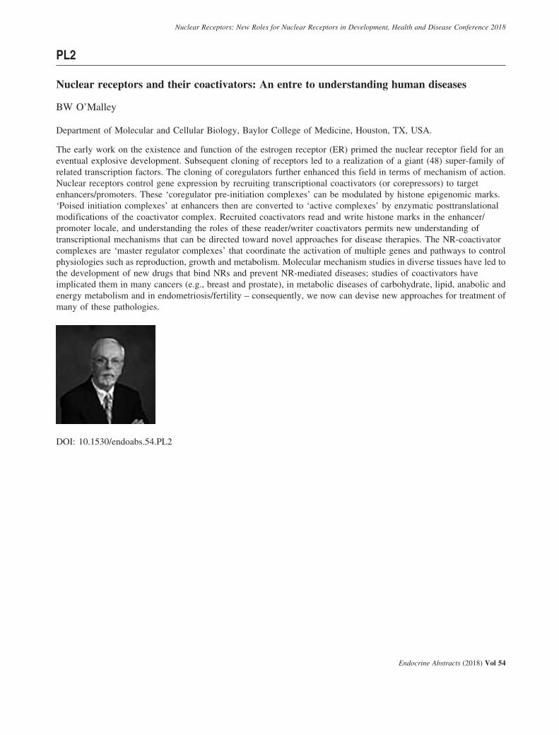
PL2
Nuclear receptors and their coactivators: An entre to understanding human diseases
BW O’Malley
Department of Molecular and Cellular Biology, Baylor College of Medicine, Houston, TX, USA.
The early work on the existence and function of the estrogen receptor (ER) primed the nuclear receptor field for an
eventual explosive development. Subsequent cloning of receptors led to a realization of a giant (48) super-family of
related transcription factors. The cloning of coregulators further enhanced this field in terms of mechanism of action.
Nuclear receptors control gene expression by recruiting transcriptional coactivators (or corepressors) to target
enhancers/promoters. These ‘coregulator pre-initiation complexes’ can be modulated by histone epigenomic marks.
‘Poised initiation complexes’ at enhancers then are converted to ‘active complexes’ by enzymatic posttranslational
modifications of the coactivator complex. Recruited coactivators read and write histone marks in the enhancer/
promoter locale, and understanding the roles of these reader/writer coactivators permits new understanding of
transcriptional mechanisms that can be directed toward novel approaches for disease therapies. The NR-coactivator
complexes are ‘master regulator complexes’ that coordinate the activation of multiple genes and pathways to control
physiologies such as reproduction, growth and metabolism. Molecular mechanism studies in diverse tissues have led to
the development of new drugs that bind NRs and prevent NR-mediated diseases; studies of coactivators have
implicated them in many cancers (e.g., breast and prostate), in metabolic diseases of carbohydrate, lipid, anabolic and
energy metabolism and in endometriosis/fertility – consequently, we now can devise new approaches for treatment of
many of these pathologies.
DOI: 10.1530/endoabs.54.PL2
Endocrine Abstracts (2018) Vol 54
Nuclear Receptors: New Roles for Nuclear Receptors in Development, Health and Disease Conference 2018
PL3
Targeting DNA repair-AR crosstalk dysfunction in advanced prostate cancer
Karen E Knudsen1,2,3,4,5
1Departments of Cancer Biology, Thomas Jefferson University, Philadelphia, Pennsylvania, USA; 2Departments of
Urology, Thomas Jefferson University, Philadelphia, Pennsylvania, USA; 3Departments of Medical Oncology, Thomas
Jefferson University, Philadelphia, Pennsylvania, USA; 4Departments of Radiation Oncology, Thomas Jefferson
University, Philadelphia, Pennsylvania, USA; 5Departments of Kimmel Cancer Center, Thomas Jefferson University,
Philadelphia, Pennsylvania, USA.
Prostatic adenocarcinoma (PCa) is the 2nd leading cause of cancer death in US men. Organ-confined PCa can be
effectively managed, but there is no durable treatment for advanced disease. Advanced PCa is treated through androgen
deprivation therapy, often coupled with direct AR antagonists, as PCa is exquisitely dependent on androgen receptor
(AR) activity for survival. Furthermore, recent studies identified AR as a major effector of DNA repair, manifest
through the ability of the receptor to regulate DNAPK expression and activity. While AR directed therapeutics
effectively suppress the pro-proliferative, pro-survival, and pro-DNA repair functions of AR and result in tumor
remission, relapse is common. Recurrent disease arises largely due to resurgent AR activity with 2–3 years, and there is
no cure for this castration-resistant phase (CRPC, castration-resistant PCa). Thus, there is a significant need to develop
new means for targeting recurrent AR activity or develop adjuvant therapies in advanced PCa.
Emerging data from our laboratory and others strongly support the concept that AR regulates DNA repair pathway,
and that alterations in DNA damage repair (DDR) pathways are more common than previously thought in sporadic
PCa, thus uncovering new, potentially more effective means of therapeutic intervention. New studies to be discussed
will address underlying mechanisms of action with regard to the pathway, and identify clinically actionable
ramifications of AR-DNA repair dysfunction. Major concepts to be considered include new discoveries regarding
PARP1 function and activity in lethal disease, and that a newly identified gene signature of PARP1-regulated networks
is associated with poor outcome. Further, mechanistic investigation revealed new insight into the means by which
PARP1 inhibitors likely act as single agents in advanced prostate cancer, manifest through both DNA repair and
transcriptional regulatory functions. Findings to be discussed strongly support a model wherein selected DDR pathways
can be developed as therapeutic targets in concert with AR-targeting strategies to tailor treatment for prostate cancer
and improve outcome for advanced disease.
DOI: 10.1530/endoabs.54.PL3
Endocrine Abstracts (2018) Vol 54

Nuclear Receptors: New Roles for Nuclear Receptors in Development, Health and Disease Conference 2018
Invited Speaker
Endocrine Abstracts (2018) Vol 54

Nuclear Receptors: New Roles for Nuclear Receptors in Development, Health and Disease Conference 2018
IS1
MicroRNA regulation of androgen signalling
Claire E Fletcher1, Ailsa Sita-Lumsden1, Alwyn Dart2, Akifumi Shibakawa1, Eric Sulpice3,Stephanie Combe3, Damien A Leach1, Johann de Bono4, SE Lupold5, SE McGuire6, Xavier Gidrol3 &Charlotte L Bevan1
1Imperial College London, London, UK; 2Cardiff University, Cardiff, UK; 3CEA, Grenoble, France; 4Institute of
Cancer Research, Sutton, UK; 5Johns Hopkins University School of Medicine, Baltimore, MD, USA; 6Baylor College
of Medicine, Houston, TX, USA.
Androgens initially drive prostate tumour growth. Although in advanced disease there is no longer dependence on
circulating androgens, the androgen receptor (AR) remains a key driver of this lethal stage thus new ways to inhibit its
activity are required. MicroRNAs play vital roles in prostate cancer (PCa) development, progression and metastasis.
Previous studies have examined microRNAs dysregulated in PCa, and also identified androgen-regulated microRNAs.
We approached microRNAs in PCa from the other angle. Having previously identified microRNA-27a as an androgen-
regulated microRNA that in turn affects AR signalling, inhibition of which inhibits PCa cell proliferation, we
hypothesised that microRNAs modulating AR activity in lethal castration-resistant PCa represent novel therapeutic targets.
Such microRNAs were systematically identified using high-throughput screens to examine effects of w1000 microRNA
inhibitors on AR activity in hormone-responsive and -resistant PCa cell lines. Results were cross-referenced with a database
of microRNAs impacting PCa cell growth. Eighty significantly altered AR activity, eight in both cell lines. Upon validation,
inhibition of selected identified microRNAs significantly reduced AR activity up to 90%, accompanied by reduced AR
mRNA/protein and AR target gene expression. At the cellular level it also increased apoptosis (up to 800%), and reduced cell
growth, migration and invasion. Opposing effects were observed upon microRNA overexpression. Inhibition of
AR-modulatory microRNAs showed additive effects with AR silencing or anti-androgen treatment, suggesting potential
combinatorial applications for PCa treatment.
Pathway analysis of AGO-PAR-CLIP-identified mRNA targets of these microRNAs identified roles in DNA replication and
repair, cell cycle, signal transduction and immune function. In addition, inhibition of AR-modulatory microRNAs induced
epithelial markers, while reducing levels of mesenchymal markers. Other targets include purported tumour suppressors:
silencing of these phenocopies effects of the microRNAs, confirming their physiological relevance. Interestingly, the
microRNAs appear to upregulate certain oncogenes as well as AR itself, suggesting novel regulation of the AR 3’UTR and
contrary to the accepted dogma that microRNAs reduce expression of their targets via translational repression and/or
transcript degradation.
In summary, we have identified microRNAs that significantly modulate AR activity in models of hormone-responsive and
castration-resistant PCa. Inhibitors of these dramatically reduce AR activity and growth, migration and invasion of PCa cells,
thus represent potential novel PCa therapeutics. Given androgen regulation of microRNA expression, our previous findings
that androgen signalling can regulate microRNA biogenesis, and the ‘hormomiR’ hypothesis that microRNAs themselves
can mediate long-range effects, this supports two-way crosstalk between these highly influential systems.
DOI: 10.1530/endoabs.54.IS1
Endocrine Abstracts (2018) Vol 54

Nuclear Receptors: New Roles for Nuclear Receptors in Development, Health and Disease Conference 2018
IS2
Androgen and estrogen receptors in breast tissues: opponents or teammates?
Theresa E Hickey
Dame Roma Mitchell Cancer Research Laboratories, University of Adelaide, Adelaide, Australia.
The balance of androgen and estrogen hormone activity determines the degree of breast development in males and
females. A predominance of androgen action impedes whereas a predominance of estrogen action promotes breast
development. This sex hormone antagonism is mechanistically mediated by androgen and estrogen receptors (AR, ER).
The alpha form of ER (ERa) is required for normal breast development and is the driving oncogene in the majority of
breast cancers. The AR is not required for female breast development but is required for suppression of breast growth in
males and modulates post-pubertal breast growth in females. The role of AR in breast cancer is complex, giving rise to
ongoing controversies about how best to leverage it as a therapeutic target. In the context of primary ERa positive
breast cancer, evidence from previous studies and unpublished data from our laboratory support the concept that AR
signalling opposes oncogenic ERa signalling in breast cancer cells and is a viable therapeutic strategy. In contrast,
evidence from other studies indicate that AR signalling facilitates ERa signalling so that AR antagonism would also be
a viable therapeutic strategy. Similar contradictory findings are present in the arena of endocrine-resistant breast
cancers. Currently, both therapeutic strategies (AR agonism and antagonism) are being tested in clinical trials of ERa-
positive breast cancer. In this talk, I will present data describing the interaction between ERa and AR on chromatin and
its functional consequences in terms of gene regulation and cell cycle control in different models of ERa positive breast
cancer, with view to explaining some of the controversies about AR signalling in this form of disease.
DOI: 10.1530/endoabs.54.IS2
Endocrine Abstracts (2018) Vol 54

Nuclear Receptors: New Roles for Nuclear Receptors in Development, Health and Disease Conference 2018
IS3
Nuclear receptors, transcriptional enhancers, and gene regulation
Hector L Franco, Shino Murakami, Tim Y Hou, Yasmin M Vasquez, Ziying Liu, Anusha Nagari,Venkat S Malladi, Tulip Nandu & W Lee Kraus
Cecil H and Ida Green Center for Reproductive Biology Sciences and Department of Obstetrics and Gynecology,
University of Texas Southwestern Medical Center, Dallas, Texas 75390, USA.
Transcriptional enhancers, which function as nucleation sites for the assembly of transcription-regulating complexes
across the genome, drive cell type-specific patterns of gene expression that underlie the distinct biological properties of
different cell types. Although many features of active enhancers (e.g., H3K4me1, H3K27ac, enrichment of p300/CBP
and Mediator, and enhancer RNA production) have been defined by genomic assays, the roles of these features in ERaenhancer function are not well understood. The Kraus lab has had a long-standing interest in enhancer biology, in
particular the molecular mechanisms and kinetics of enhancer assembly in signal-regulated systems. In particular, we
are interested in cell type-specific enhancers that drive biological outcomes in reproductive tissues and in hormone-
dependent cancers. We have focused on enhancers formed by estrogen receptor alpha (ERa), a ligand-regulated,
sequence-specific DNA-binding transcription factor that nucleates de novo enhancer formation in cells in response to
estrogen signaling, as well as other transcription factors (TFs), such as Sox2, FOSL1, and PLAG1. We have used a
variety of molecular, biochemical, genomic, genetic, and computational approaches to determine (1) where enhancers
are formed by specific TFs across the genome, (2) the kinetics of enhancer formation and disassembly, (3) the influence
of genetic variation on enhancer formation and function, and (4) the role of the specific enhancer features noted above,
especially enhancer transcription, in enhancer function. In addition, we have developed new computational tools to
study enhancer function, such as the Total Functional Score of Enhancer Elements (TFSEE), a robust and unbiased
computational pipeline that simultaneously identifies putative subtype-specific enhancers and their cognate TFs by
integrating multiple types of genomic information. Collectively, our analyses are providing new insights into enhancer
complex assembly and function in a variety of biological systems.
This work is supported by grants from the U.S. National Institutes of Health (DK058110; HD087150) and the Cancer
Prevention and Research Institute of Texas (RP160319, RP110471-P1) to W.L.K.
DOI: 10.1530/endoabs.54.IS3
Endocrine Abstracts (2018) Vol 54

Nuclear Receptors: New Roles for Nuclear Receptors in Development, Health and Disease Conference 2018
IS4
Estrogen receptor cistromics in breast tumors: from biomarkers to novel drug targets
Koen Dorus Flach1, Manikandan Periyasamy2, Ajit Jadhav3, Theresa E Hickey4, Mark Opdam5, Hetal Patel2,Sander Canisius6, David M Wilson III7, Dorjbal Dorjsuren3, Marja Nieuwland8, Roel Kluin8, AlexeyV Zakharov3, Jelle Wesseling5, Lodewyk Frederik Ary Wessels6, Sabine Charlotte Linn5,Wayne D Tilley4, Anton Simeonov3, Simak Ali2 & Wilbert Zwart1
1Department of Oncogenomics, The Netherlands Cancer Institute, 1066 CX Amsterdam, The Netherlands;2Department of Surgery and Cancer, Imperial College London, Hammersmith Hospital Campus, Du Cane Road,
London W12 0NN, UK; 3National Center for Advancing Translational Sciences, National Institutes of Health, 9800
Medical Center Drive, MSC 3370, Maryland 20892, USA; 4Dame Roma Mitchell Cancer Research Laboratories,
Adelaide Medical School, Faculty of Health Sciences, DX Number 650 801, University of Adelaide, Adelaide,
South Australia 5005, Australia; 5Department of Molecular Pathology, The Netherlands Cancer Institute, 1066 CX
Amsterdam, The Netherlands; 6Department of Molecular Carcinogenesis, The Netherlands Cancer Institute, 1066 CX
Amsterdam, The Netherlands; 7Laboratory of Molecular Gerontology, Intramural Research Program, National Institute
on Aging, National Institutes of Health, Bayview Blvd 251, Baltimore, Maryland 21224, USA; 8Genomics Core
Facility, The Netherlands Cancer Institute, 1066 CX Amsterdam, The Netherlands.
Estrogen receptor a (ERa) is a key transcriptional regulator in the majority of breast cancers. ERa-positive patients are
frequently treated with tamoxifen, but resistance is common. Through ChIP-seq analyses, we presviously identified
direct target genes of ERa acting in complex with SRC1, SRC2 or SRC3 (Zwart et al., 2011 EMBO J). Only the 111
genes there were under direct control of ERa in conjunction with SRC3 (but not the other two p160s) predicted patient
outcome. Here, the 111-gene outcome prediction-classifier was further refined, revealing FEN1 as strongest
determining factor in ERa-positive prognostication. We demonstrate FEN1 levels are predictive of outcome in
tamoxifen-treated patients, and show FEN1 is required and sufficient for tamoxifen-resistance in ERa-positive cell
lines. We show FEN1 dictates the transcriptional-activity of ERa by facilitating the formation and repair of hormone-
induced DNA damage, ultimately resulting in DNA methylation changes. FEN1 blockade induced proteasome-
mediated degradation of activated ERa, resulting in loss of ERa-driven gene expression and eradicated tumor cell
proliferation. Finally, a high-throughput 460.000 compound screen identified a novel FEN1 inhibitor, which effectively
blocks ERa-function and inhibits proliferation of tamoxifen-resistant cell lines as well as ex-vivo cultured
ERa-positive breast tumors, providing therapeutic proof-of-principle for FEN1 blockade in tamoxifen-resistant
breast cancer.
DOI: 10.1530/endoabs.54.IS4
Endocrine Abstracts (2018) Vol 54
Nuclear Receptors: New Roles for Nuclear Receptors in Development, Health and Disease Conference 2018
IS5
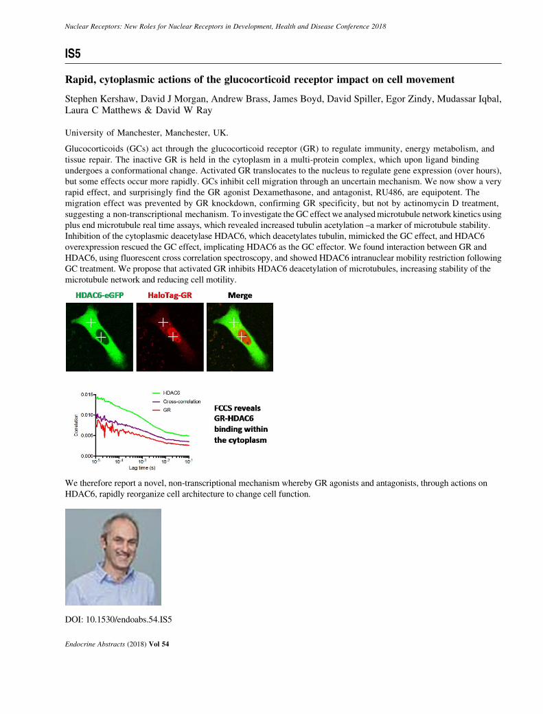
Rapid, cytoplasmic actions of the glucocorticoid receptor impact on cell movement
Stephen Kershaw, David J Morgan, Andrew Brass, James Boyd, David Spiller, Egor Zindy, Mudassar Iqbal,Laura C Matthews & David W Ray
University of Manchester, Manchester, UK.
Glucocorticoids (GCs) act through the glucocorticoid receptor (GR) to regulate immunity, energy metabolism, and
tissue repair. The inactive GR is held in the cytoplasm in a multi-protein complex, which upon ligand binding
undergoes a conformational change. Activated GR translocates to the nucleus to regulate gene expression (over hours),
but some effects occur more rapidly. GCs inhibit cell migration through an uncertain mechanism. We now show a very
rapid effect, and surprisingly find the GR agonist Dexamethasone, and antagonist, RU486, are equipotent. The
migration effect was prevented by GR knockdown, confirming GR specificity, but not by actinomycin D treatment,
suggesting a non-transcriptional mechanism. To investigate the GC effect we analysed microtubule network kinetics using
plus end microtubule real time assays, which revealed increased tubulin acetylation –a marker of microtubule stability.
Inhibition of the cytoplasmic deacetylase HDAC6, which deacetylates tubulin, mimicked the GC effect, and HDAC6
overexpression rescued the GC effect, implicating HDAC6 as the GC effector. We found interaction between GR and
HDAC6, using fluorescent cross correlation spectroscopy, and showed HDAC6 intranuclear mobility restriction following
GC treatment. We propose that activated GR inhibits HDAC6 deacetylation of microtubules, increasing stability of the
microtubule network and reducing cell motility.
We therefore report a novel, non-transcriptional mechanism whereby GR agonists and antagonists, through actions on
HDAC6, rapidly reorganize cell architecture to change cell function.
DOI: 10.1530/endoabs.54.IS5
Endocrine Abstracts (2018) Vol 54

Nuclear Receptors: New Roles for Nuclear Receptors in Development, Health and Disease Conference 2018
IS6
Androgens and endometrial function: replication, repair and regeneration
Douglas A Gibson, Ioannis Simitsidellis, Frances Collins, Arantza Esnal-Zufiaurre & Philippa TK Saunders
MRC Centre for Inflammation Research, The Queen’s Medical Research Institute, The University of Edinburgh,
Edinburgh, EH16 4TJ, UK.
The human endometrium is a complex multicellular tissue the prime function of which is to provide a receptive
environment during a fertile cycle. The tissue responds to steroid hormones exhibiting dynamic cyclical regeneration,
angiogenesis, differentiation (decidualisation) and inflammation. In the absence of an embryo the inner surface is shed
and repaired without scarring (menstruation). The endometrium exhitbits spatial and temporal expression of androgen
receptors (AR) in stromal fibroblasts and endothelial cells: upregulation of AR in epithelial cells occurs in reponse to
progesterone withdrawal (cycle) or administration of PR antagonists.
We used primary cells (human endometrial stromal cells:) and mouse models to investigate the impact of androgens
(T, DHT), AR antagonists (Flutamide) and selective AR modulators (SARMs) on key endometrial cell functions.
We discovered that local ‘intracrine’ biosynthesis of T/DHT by human stromal cells regulates AR and expression of
receptivity factors. Notably DHT inhibited cell migration but increased resistance to apoptosis with SARMs exhibiting
a range of activities on these functions and on expression of AR-regulated genes.
Administration of DHT to steroid-depleted (ovariectomised) female mice promoted a significant increase in uterine
size, induced epithelial cell proliferation, expansion of the glandular epithelium and altered uterine AR
immunoexpression. Administration of DHT in a mouse model of endometrial repair (‘menstruation’) altered regulation
of restoration of endometrial tissue homeostasis following endometrial shedding at the time of menstruation. We have
used these mouse models to test the impact of SARMs on endometrial proliferation and repair and found evidence for
selective effects of different SARMs on these functional processes.
In summary, our studies demonstrate a key role for AR in regulation of endometrial function in health and disease.
We believe SARMs may offer a novel way to target the AR for therapeutic benefit.
DOI: 10.1530/endoabs.54.IS6
Endocrine Abstracts (2018) Vol 54

Nuclear Receptors: New Roles for Nuclear Receptors in Development, Health and Disease Conference 2018
IS7
Clinically-relevant contexts for AR variants in prostate cancer
Scott M Dehm
Masonic Cancer Center, University of Minnesota, Minneapolis, Minnesota USA.
The androgen receptor (AR) functions as a master transcriptional regulator of prostate tissue homeostasis. This master
transcriptional regulator function is maintained in prostate cancer. Therefore, prostate cancer is an androgen-dependent
disease and suppression of AR transcriptional activity with androgen deprivation therapy (ADT) is an effective
systemic therapy. However, development of therapy resistance and transition to castration resistant prostate cancer
(CRPC) represents a major clinical challenge. One mechanism by which CRPC may circumvent ADT is by expression
of constitutively active AR variants (AR-Vs) that lack the ligand binding domain. The best-studied AR-V is AR-V7,
but a controversy in the field is that AR-V7 is broadly expressed and readily detectable in AR-expressing tissues,
including normal prostate. To address this controversy, we have been elucidating mechanisms governing the expression
of AR-Vs in prostate cancer, with the goal of identifying clinically-relevant contexts in which they may be functioning
as drivers of resistance. These studies have revealed heterogeneous, clonally diverse AR gene rearrangements in
clinical CRPC. Investigation of several AR gene rearrangement events has demonstrated they converge functionally by
driving stable, outlier expression of diverse tumor-specific AR-V species that are functionally equivalent to AR-V7 but
undetectable by AR-V7-specific assays currently under clinical development. A second mechanism of AR-V
expression is alternative polyadenylation, whereby AR-V7 is co-ordinately expressed in CRPC tissues with AR-V9,
AR-V1, and other annotated AR-Vs. This indicates that detection of AR-V7 in CRPC tissue or circulating tumor cells is
likely a harbinger of a broader repertoire of AR-V expression. Studies are underway to translate this knowledge to
biomarker applications for prostate cancer patients, and also for development of new therapies to combat AR-Vs.
DOI: 10.1530/endoabs.54.IS7
Endocrine Abstracts (2018) Vol 54

Nuclear Receptors: New Roles for Nuclear Receptors in Development, Health and Disease Conference 2018
IS8
Direct and indirect effects of androgens on the musculoskeletal system
Frank Claessens1, Laurent Michael1, Vanessa Dubois1, Rougin Khalil2, Ferran Jardi2 &Dirk Vanderschueren2
1Molecular Endocrinology Laboratory, Department of Cellular and Molecular Medicine, KU Leuven, Herestraat 49 PO
box 901, 3000 Leuven, Belgium; 2Clinical and Experimental Endocrinology, Department of Clinical and Experimental
Medicine, KU Leuven, Herestraat 49 PO box 902, 3000 Leuven, Belgium.
Global knockout models of the androgen receptor (ARKO) illustrates the many roles androgens and their receptor have
in the development of male reproductive organs and the gender differences in many features like the musculoskeletal
system. However, neither the global ARKO nor orchidectomy models discriminate between direct and indirect effects
of androgens. To determine direct and indirect effects of androgens on muscle, we developed a muscle-specific ARKO
(called satARKO for satellite cell-specific ARKO). In this model, we found a partial loss of androgen-responsiveness of
the levator ani as well as of other muscles. However, there is still an important response to orchidectomy in the muscle
of satARKO, which is corrected by administration of testosterone, dihydrotestosterone or the selective androgen
receptor modulator Enobosarm. Surprisingly, myostatin is one of the most responsive genes in mouse muscle and we
identified the androgen-regulated enhancer involved. This is counterintuitive as myostatin is a well known negative
regulator of muscle mass. We propose that myostatin upregulation serves to mitigate the proliferative response to
androgens. Similarly, the ARKO in bone cells did not replicate the ARKO phenotype. We are now looking at kidney
and brain as suspects for the indirect effects of androgens on the musculoskeletal system.
The observation that in human serum, sex steroids bind with high affinity to sex hormone binding globulin (SHBG) led
to the contested free hormone hypothesis. Unfortunately, rodent models do not have the higher serum levels of sex
hormone binding globulin seen in humans. We studied the free hormone hypothesis in a mouse model which
overexpresses SHBG in its circulation.
DOI: 10.1530/endoabs.54.IS8
Endocrine Abstracts (2018) Vol 54

Nuclear Receptors: New Roles for Nuclear Receptors in Development, Health and Disease Conference 2018
IS9
Progesterone receptor regulation of breast cancer cell translation
Jessica Finlay-Schultz1, Austin E Gillen2, Heather M Brechbuhl3, Shawna B Matthews1, Britta M Jacobsen1,David L Bentley2,4, Peter Kabos3 & Carol A Sartorius1
1Department of Pathology, University of Colorado Anschutz Medical Campus, Aurora, Colorado, USA; 2RNA
Biosciences Initiative, University of Colorado Anschutz Medical Campus, Aurora, Colorado, USA; 3Department of
Medicine, Division of Medical Oncology, University of Colorado Anschutz Medical Campus, Aurora, Colorado, USA;4Department of Biochemistry and Molecular Genetics, University of Colorado Anschutz Medical Campus, Aurora,
Colorado, USA.
Progesterone receptors (PR) are long recognized to suppress estrogen receptor (ER) mediated transcription in breast
cancers. However, a mechanistic basis for this repression has been lacking. Recent reports indicate this occurs, in part,
through global repositioning of ER on chromatin in the presence of selective PR modulators (SPRMS), both agonists
and antagonists [1, 2]. The goal of our studies was to further understand the mechanisms by which PR impacts
estrogen-dependent growth in solid tumor models chronically treated with SPRMs. We grew ERCPRC breast cancer
patient-derived xenografts (PDX) in the presence of E2 alone or E2 plus the natural hormone progesterone (P4) or a
synthetic SPRM medroxyprogesterone acetate (MPA) and demonstrated the SPRMs suppress tumor growth similar to
tamoxifen. In these tumors P4 and MPA alter up to half of ER regulated genes at the transcript level. However, the
majority of these genes (O80%) either show no change in ER chromatin binding by ChIP-seq or have no ER binding
sites near their promoter (G2 kb). We made the interesting discovery via PR ChIP-seq that PR (but not ER) is localized
at a large fraction of RNA polymerase III (Pol III) regulated tRNA genes. RIME for PR and subsequent IP found that
PR associates with the Pol III complex. Furthermore, select pre-tRNA transcripts and mature tRNA pools are decreased
in SPRM treated tumors [3]. We therefore speculate that PR may indirectly impede ER action through regulation of
translation. This could occur by reducing the overall bioavailability of tRNAs to reduce protein synthesis rates and
curtail tumor growth. Furthermore, PR could alter the tRNA pool to selectively change translational preference through
non-optimal codon usage, an increasingly recognized mechanism of cancer cell regulation. Studies are underway to test
these hypotheses.
DOI: 10.1530/endoabs.54.IS9
Endocrine Abstracts (2018) Vol 54
Nuclear Receptors: New Roles for Nuclear Receptors in Development, Health and Disease Conference 2018
IS10
Modulating glucocorticoid receptor function in breast and prostate cancer
E Tonsing-Carter1, T Long1, DC West1, R Harkless1, DN Dolcen1, D Hosfield2, GL Greene2,RZ Szmulewitz1 & SD Conzen1,2
1Department of Medicine, The University of Chicago, Chicago, Illinois 60637, USA; 2Ben May Department for Cancer
Biology, The University of Chicago, Chicago, Illinois 60637, USA.
In normal physiology, glucocorticoid receptor (GR) activation regulates cell type-dependent genes whose products
influence metabolism, inflammation, cell cycle and apoptosis/cell survival pathways. Synthetic GR agonists, or
glucocorticoids (GCs), are often used to treat hematologic malignancies because of GR’s ability to induce proapoptotic
gene expression, inhibit nuclear factor–kB, and induce cell cycle arrest. In contrast, recent examination of GR
expression and activity in human cancer models and clinical specimens has suggested that GR activity has remarkably
diverse roles in breast and prostate cancer subtypes. In estrogen receptor (ER)C breast cancer, GR appears to modulate
ER-regulated transcriptional activity through ER/GR receptor crosstalk resulting in antagonism of ER-associated
proliferation. However, in ER-negative breast cancer (including TNBC), high tumor GR expression is associated with
poor prognosis, anti-apoptotic signaling, and chemotherapy resistance. In addition, high GR activity in castrate-
resistant prostate cancer (CRPC) contributes to therapy-resistance to androgen receptor (AR) antagonism. Recently
described selective GR modulators will be described that have been used to dissect divergent GR mechanisms present
in breast cancer subtypes and prostate cancer evolution.
DOI: 10.1530/endoabs.54.IS10
Endocrine Abstracts (2018) Vol 54

Nuclear Receptors: New Roles for Nuclear Receptors in Development, Health and Disease Conference 2018
IS11
Protein factors involved in 3D genome organization & transcription regulation
Yijun Ruan
The Jackson Laboratory for Genomic Medicine and the Department of Genetics and Genome Sciences, UConn Health,
10 Discovery Drive, Farmington, Connecticut 06032
The human genome is over 2 meters in length, which has to be folded in micrometer-sized nuclear space for proper
functions. Although most of our understandings in the human genome are based on linear explanations, it has been
speculated that the three-dimensional (3D) and high-order organization of the genome must play important roles in
framing the mechanisms of nuclear process such as transcription regulation. Recent advance in 3D genome mapping
technologies and sophisticated computational programs have enabled us to reconstitute the 3D models of the genome,
and allowed us to investigate the functions of protein factors involved in 3D genome folding and transcription
regulation. In this effort, we developed ChIA-PET to comprehensively map specific chromatin interactions mediated by
protein factors with haplotype-specificity and nucleotide-resolution. Using ChIA-PET, we have studied the roles of
chromatin architecture factors like CTCF, nuclear receptors (NR) such as ER, AR and RARA, and transcription factors
(TF) including RNA Polymerase II (RNAPII) in 3D genome organization and transcription regulation. We
demonstrated that CTCF-mediated chromatin interaction anchors serve as 3D organizational foci, where constitutive
genes are positioned in concordance with the orientation of CTCF binding motifs, whereas RNAPII and other TFs
interacts within these structures by drawing cell-type-specific genes towards CTCF-foci for coordinated transcription.
We further found that fusion protein PML/RARA could alter the 3D genome configuration of normal cells and become
cancerous. In addition, we have shown that haplotype-resolved chromatin interactions have allelic-specific effects on
chromatin interactions, thus revise the expression of genes residing in the topological domains, and lead to different
traits or diseases. Together, these mechanistic insights establish a topological basis of 3D genome folding and
transcription regulation that links genetic variation to phenotype diversity.
DOI: 10.1530/endoabs.54.IS11
Endocrine Abstracts (2018) Vol 54

Nuclear Receptors: New Roles for Nuclear Receptors in Development, Health and Disease Conference 2018
IS12
Nuclear receptor networks in male fertility
Emmanuelle Martinot, Lauriane Sedes, Marine Baptissart, Helene Holota, De Haze Angelique,Claude Beaudoin & David H Volle
Inserm U 1103, CNRS UMR6293–Universite Clermont Auvergne, Laboratoire Genetique, Reproduction &
Developpement, Clermont-Ferrand, France
Male fertility is controlled by complex interactions between hypothalamus, pituitary, and testis. The major functions of
the testis include production of spermatozoa and synthesis of hormones. Testosterone is produced by the testicular
Leydig cells and ensures male fertility. Testosterone is involved in the development of gonad, the attainment of
puberty, the maintenance of secondary sexual characteristics as well as in spermatogenesis process. Many studies have
highlighted the complexity of the regulations of testicular homeostasis at tissue and cellular levels. Several nuclear
receptors (NRs) have been identified as key regulators of testicular physiology through the control of steroidogenesis
and germ cell differentiation. Using both genetic and pharmacologic strategies the roles of the multiple members of
the NR superfamily such as the Liver-X-Receptors, the Small Heterodimer Partner and more recently the
Farnesol-X –Receptor have been defined. We will give an overview of recent advances highlighting the identification
of a complex networks showing the interactions of NRs in the regulation of the exocrine and endocrine functions
of the testis.
DOI: 10.1530/endoabs.54.IS12
Endocrine Abstracts (2018) Vol 54

Nuclear Receptors: New Roles for Nuclear Receptors in Development, Health and Disease Conference 2018
IS13
Enhancers mapping uncovers phenotypic heterogeneity and evolution in patients with luminalbreast cancer
Darren K Patten*,1, Giacomo Corleone*,1, Balazs Gyorffy2,3, Edina Erdos4, Alina Saiakhova5,Kate Goddard6, Andrea Vingiani7, Sami Shousha8, Lorinc Sandor Pongor2, Dimitri J Hadjiminas8,Gaia Schiavon9, Peter Barry10, Carlo Palmieri11, Raul C Coombes1, Peter Scacheri5, Giancarlo Pruneri12
& Luca Magnani1
1Department of Surgery and Cancer, The Imperial Centre for Translational and Experimental Medicine, Imperial
College London, Hammersmith Campus, London, UK; 2MTA TTK Lendulet Cancer Biomarker Research Group,
Institute of Enzymology, Hungarian Academy of Sciences, 1117, Budapest, Hungary; 3Semmelweis University 2nd
Department of Pediatrics, 1094, Budapest, Hungary; 4Department of Biochemistry and Molecular Biology, Genomic
Medicine and Bioinformatic Core Facility, University of Debrecen, Debrecen 4032, Hungary; 5Department of Genetics
and Genome Sciences, Case Comprehensive Cancer Center, Case Western Reserve University, Cleveland, Ohio 44106,
USA; 6Department of Breast and General Surgery, Charing Cross Hospital, Imperial College Healthcare NHS Trust,
London, UK; 7Department of Pathology, European Institute of Oncology, Milan, Italy; 8Centre for Pathology,
Department of Medicine, Imperial, College London, Charing Cross, London, UK; 9Translational Science, IMED
Oncology, AstraZeneca, Cambridge, UK; 10Department of Breast Surgery, The Royal Marsden NHS Foundation Trust,
Orchard House, Downs Road, Sutton, SM2 5PT, UK; 11Institute of Translational Medicine University of Liverpool,
Clatterbridge Cancer Centre, NHS Foundation Trust, and Royal Liverpool University Hospital, Liverpool, Merseyside,
UK; 12Pathology Department, Fondazione IRCCS Istituto Nazionale Tumori and University of Milan, School of
Medicine, Milan, Italy; *Equal contribution.
The degree of intrinsic and interpatient phenotypic heterogeneity and its role in tumour evolution is poorly understood.
Phenotypic divergence can be achieved via the inheritance of alternative transcriptional programs. Cell-type specific
transcription is maintained through the activation of epigenetically-defined regulatory regions including promoters and
enhancers. In this work, we annotated the epigenome of 47 primary and metastatic oestrogen-receptor (ERa)-positive
breast cancer specimens from clinical samples, and developed strategies to deduce phenotypic heterogeneity from the
regulatory landscape, identifying key regulatory elements commonly shared across patients. Highly shared regions
contain a unique set of regulatory information. In vitro work shows that TF enriched in clonal enhancers are essential
for ERa transcriptional activity and defines the critical subset of functional ERa binding sites driving tumor growth in
most luminal patients. These transcription factors also control the expression of genes that mediate resistance to
endocrine treatment. Finally, we show that H3K27ac levels at active enhancer elements can be used as a surrogate of
intra-tumor phenotypic heterogeneity, and to track expansion and contraction of phenotypic subpopulations throughout
breast cancer progression. Tracking epigenetically defined clones clones in primary and metastatic lesions, we show
that endocrine therapies drive the expansion of phenotypic clones originally underrepresented at diagnosis.
Collectively, our data show that epigenetic mechanisms significantly contribute to phenotypic heterogeneity and
evolution in systemically treated breast cancer patients.
DOI: 10.1530/endoabs.54.IS13
Endocrine Abstracts (2018) Vol 54

Nuclear Receptors: New Roles for Nuclear Receptors in Development, Health and Disease Conference 2018
IS14
The structural basis of chromatin reprogramming by steroid receptors
GL Hager1, V Paakinaho1, TA Johnson1, RV Chereji2, DJ Clark2, EE Swinstead1 & DM Presman1
1Laboratory of Receptor Biology and Gene Expression, National Cancer Institute, NIH, Bethesda, Maryland 20892,
USA; 2Division of Developmental Biology, Eunice Kennedy Shriver National Institute for Child Health and Human
Development, National Institutes of Health, Bethesda, Maryland 20892, USA.
Localized transitions in chromatin structure accompany nuclear receptor binding events in mammalian cells. These
remodeling processes are critical to determine the binding landscape for steroid receptors (SRs) in cancer cells.
Multiple reports indicate that steroid receptors (ER, GR, AR, PR) can regulate the binding patterns for each other,
particularly during cancer progression. Elucidation of the mechanisms by which these ‘chromatin opening’ processes
occur is central to our understanding of steroid receptor cross-talk at the genome level. A widely accepted model
suggests that SRs function in concert with pioneer factors to open enhancer chromatin, creating DNase ‘hypersensitive’
sites (DHS), relieving the inhibitory activity of closed chromatin. These localized sites are often assumed to represent
nucleosome free regions (NFRs). We have mapped nucleosome positions at high resolution in mouse mammary
adenocarcinoma cells, and characterized GR dependent factor recruitment and changes in nucleosome structure. We
find little correlation between the extent of hypersensitivity and nucleosome presence. GR-enhancers exhibit a
complex range of states; in some cases, the receptor attacks pre-existing nucleosomes and recruits the Brg1 remodeler,
behaving at these sites as a classic pioneering activity. There is also controversy regarding the multimeric status of
SRs in their enhancer bound state. GR is typically represented as acting as a monomer or dimer. We described a
tetrameric state for GR bound to the MMTV response element in live cells, and tetrameric states for other transcription
factors have been reported. Using a mutation that mimics the DNA bound state, we have examined the chromatin
binding landscape for a putative tetrameric form of GR. This receptor dramatically increases the binding profile for GR,
penetrating chromatin that is closed in mammary cells, but available in other cell types. These findings will be
discussed in terms of a model wherein receptors and many transcription factors act to achieve chromatin remodeling
and enhancer activation through a highly dynamic mechanism termed ‘dynamic assisted loading.’
DOI: 10.1530/endoabs.54.IS14
Endocrine Abstracts (2018) Vol 54

Nuclear Receptors: New Roles for Nuclear Receptors in Development, Health and Disease Conference 2018
Oral Communications
Endocrine Abstracts (2018) Vol 54
Nuclear Receptors: New Roles for Nuclear Receptors in Development, Health and Disease Conference 2018
OC1Heterodimerization of retinoid X receptor with xenobiotic nuclearreceptors occurs in the cytoplasmic compartment of cell in a ligandindependent mannerAmit K Dash, Ashutosh S Yende & Rakesh K TyagiSpecial Centre for Molecular Medicine, Jawaharlal Nehru University,New Delhi – 110067, India.
The ‘Nuclear Receptor (NR) Super-family’ is a group of ligand modulatedtranscription factors with 48 members presently identified in human genome. NRsregulate most of the physiological processes of the body ranging from metabolismto reproduction. Retinoid X Receptor (RXR) is one of the important members ofthis NR superfamily. It serves as a heterodimeric partner of several other membersof this superfamily including two major xenobiotic nuclear receptors i.e.Pregnane and Xenobiotic Receptor (PXR) and Constitutive Androstane Receptor(CAR). The latter two receptors are primarily involved in the regulation of body’smetabolism and clearance of endobiotics and xenobiotics (including clinicaldrugs). In the present study we have investigated the exact subcellular locationresulting due to the interaction of RXR with either PXR or CAR. In order to studythis event, we have used various GFP- and RFP-tagged receptors and theirmutants of nuclear localization signal (NLS) region. The study showed that theinitial interaction of RXR-PXR and RXR-CAR occurs in the cytoplasmiccompartment of the cell and the NLS of PXR/CAR/RXR play a key role in theimport of the heterodimeric complex from the cytoplasm to the nucleus in aligand-independent manner. Our observtions exhibit that a functional NLS, alongwith respective ligand(s), are necessary for modulation of the target gene. It isobserved that RXR serves as a major driving force in importing the heterodimericcomplex to the nuclear compartment. This conclusion is based on the fact thatmutation in the NLS region of RXR severly weakens this import process. On thecontrary, mutations in the NLS regions of PXR and CAR have little or nosignificant effect. This RXR-dependent nuclear import of the RXR-PXR andRXR-CAR heterodimeric complex also modulates the individual transcriptionalactivity of PXR and CAR. Such an enhancement in the basal transcriptionalactivity of the receptor can be utilized for evaluating the diverse receptor-druginteractions.
DOI: 10.1530/endoabs.54.OC1
OC2Stage-specific and global functions of NCOR2 in prostate cancerprogressionMark D Long1, Prashant K Singh1, Gerard Llimos1, Spencer Rosario1,Dominic Smiraglia1 & Moray J Campbell21Department of Cancer Genetics, Roswell Park Cancer Institute, Buffalo,New York, 14263 USA; 2College of Pharmacy, The Ohio State University,Columbus, Ohio 43210, USA.
The corepressor NCOR2/SMRT regulates multiple nuclear receptors (NRs) andother transcription factors. Disruption to these functions are implicated in prostatecancer (PCa) progression but the details remain enigmatic. Therefore we soughtto define the global functions of NCOR2/SMRT using isogenic PCa cell models,PCa mouse models and human PCa cohorts.We mapped the NCOR2 dependent transcriptome (RNA-seq), miRnome(miRNA-seq), methylome (EPIC methylation array) and cistrome (ChIP-seq) inandrogen sensitive (LNCaP) and therapy resistant (LNCaP-C42) PCa cells, bothtreated with androgen, and stable NCOR2/SMRT knockdown. NCOR2/SMRTknockdown in LNCaP-C42 cells resulted in a striking increase in globalhypermethylation (87,078 CpGs O 10% gain, adj.Pval ! 0.01), but a divergenttranscriptome (1,491 upregulated, 1,195 downregulated). Similar patterns wereobserved in LNCaP cells, and included regulation of PPARg expression andsensitivity to PPARg ligands. NCOR2/SMRT genomic binding overlapped withAR and pioneering factor FOXA1, and modulated androgen responses in LNCaP-C42. Ongoing data integration efforts are dissecting NCOR2/SMRT methylome-cistrome-transcriptome relationships within and across models.Using clinical cohorts we revealed that NCOR2/SMRT-regulated miRNA aresignificantly altered in men progressing to PCa from high grade PIN, and elevatedNCOR2/SMRT expression on a 700 case TMA associated with worse disease freesurvival (P! 0.04; Log-rank (Mantel-Cox) test). To define NCOR2/SMRTactions in vivo, we stably knocked down NCOR2/SMRT in the CWR22 xenograftmodel. This model simulates androgen dependent primary growth and regressionupon androgen deprivation therapy (ADT). NCOR2 loss had no effect on primarytumor growth rate or size of tumor at ADT but significantly reduced regression inresponse to ADT and tumors recurred significantly quicker (PZ0.0044).Therefore loss of NCOR2/SMRT during ADT results in more aggressive tumors.
Endocrine Abstracts (2018) Vol 54
These findings suggest NCOR2/SMRT profoundly regulates the methylome, butthe consequences are divergent. At early stages, elevated expression may suppressantiproliferative signals from PPARg but during ADT NCOR2/SMRT loss andmutation drives PCa progression.
DOI: 10.1530/endoabs.54.OC2
OC3Beyond ligand activation: Disrupting LXRa phosphorylation toreprogram diet induced transcriptomes and modulate progression ofmetabolic diseasesM Gage†, N Becares†, R Louie, K Waddington & I Pineda-TorrDivision of Medicine, University College London, London WC1E6JF UK.†Equal contributors
The importance of the Liver X receptors (LXRs) as a critical modulators ofmetabolic homeostasis and immunity in health and disease has been mainlygleaned from studies evaluating the consequences of their pharmacological orgenetic manipulation. We previously showed LXRa is phosphorylated uponcholesterol loading. It is however unknown whether post-translational modifi-cations of the receptor modulate diet-induced responses and affect metabolicdiseases with an important inflammatory component. To explore the impact ofLXRa phosphorylation in disease progression we have generated two models: i) awhole-body phosphorylation-deficient mutant of LXRa at S196A (S196A) inwhich we explored the progression of non-alcoholic fatty liver disease and ii) aphospho-deficient LXRa mutant in myeloid cells (M-LXRaS196A) on theatherosclerotic LDLR null background (M-LXRaS196ALdlr-KO) in which weexamined the development of atherosclerosis.S196A mice challenged with a High Fat-High Cholesterol diet exhibit reducedhepatic inflammation and fibrosis associated with a marked protection againstcholesterol accumulation. Impaired LXRa phosphorylation in this modeluncovers novel diet-specific/phosphorylation-sensitive genes. Furthermore,M-LXRaS196ALdlr-KO fed a High-Fat diet display a significant increase inatherosclerosis burden in the absence of altered systemic lipid levels. ReducedLXRa phosphorylation during atherogenesis reprograms the macrophagetranscriptome and significantly promotes cell proliferation pathways, which is afeature of developing atherosclerotic lesions. Interestingly, the global geneexpression changes observed in response to impaired LXRa phosphorylation arefundamentally different from those revealed by ligand activation highlighting theimportance of this post-translational modification in modulating the activity of thereceptor in the context of metabolic/inflammatory diseases.Overall, we show the relevance of manipulating Ser196-LXRa phosphorylationto promote unique transcriptomes, thereby specifically modulating pathwaysimportant for the development of metabolic diseases such as non-alcoholic fattyliver disease and atherosclerosis.
DOI: 10.1530/endoabs.54.OC3
OC4Rationale targeting cell plasticity in treatment resistant prostate cancerAmina ZoubeidiDepartment of Urologic Sciences, The Vancouver Prostate Centre,University of British Columbia, Vancouver, BC, Canada.
Resistance to newly developed androgen receptor pathway inhibitors (ARPIs),such as abiraterone and enzalutamide, rapidly emerges and patients generally diewithin 2 years. In particular, a subset of patients who relapse following ARPItherapy exhibit lineage switching whereby tumours shed their dependence on ARsignaling and emerge with neuroendocrine features. These tumours, termedtreatment induced neuroendocrine prostate cancer (t-NEPC), carry an extremelypoor prognosis and, to date, treatment remains decades old cytotoxicchemotherapy which carries a short-lived response at the cost of significanttoxicity. Thus, the need to develop targeted treatments for this devastating diseaseis of paramount importance. Dr Zoubeidi will discuss how cell plasticityincluding cancer stem cells and neuroendocrine are mechanisms of ENZresistance that could be in part governed by changes in the epigenome and whythe transcription factor BRN2 is a major regulator/driver and a promising targetfor t-NEPC.
DOI: 10.1530/endoabs.54.OC4

Nuclear Receptors: New Roles for Nuclear Receptors in Development, Health and Disease Conference 2018
OC5The pathogenic role of estrogen receptor beta drives in endometriosisSang Jun Han1, Sung Yun Jung1,2, San-Pin Wu1, Mi Jin Park1, Jun Qin1,2,John P Lydon1, Sophia Y Tsai1, Ming-Jer Tsai1, Francesco J DeMayo1 &Bert W O’Malley1
1Department of Molecular and Cellular Biology, Baylor College ofMedicine, Houston, Texas 77030, USA; 2Department of Biochemistry andMolecular Biology, Baylor College of Medicine, Alkek Center forMolecular Discovery, Verna and Marrs McLean, Houston, Texas 77030,USA.
The defining feature of endometriosis is that endometrial tissues are deposited andgrown onto sites outside of the uterine cavity. The pathogenesis of endometriosis,however, remains controversial despite extensive research. Since the endome-triosis has been known as an estrogen-dependent inflammatory disease, thealterations in estrogen-mediated cellular signaling play an essential role in thepathogenesis of endometriosis. In addition to higher estrogen receptor (ER)blevels, enhanced ERb activity was detected in endometriotic tissues compared tothe normal, and the inhibition of enhanced ERb activity by an ERb-selectiveantagonist suppressed mouse ectopic lesion growth. Notably, gain of ERbfunction stimulated the progression of endometriosis. As a mechanism to evadeendogenous immune surveillance for cell survival, ERb interacts with cellularapoptotic machinery in the cytoplasm to inhibit TNFa-induced apoptosis. ERbalso interacts with components of the cytoplasmic inflammasome to increaseinterleukin-1b and thus enhance its cellular adhesion and proliferation properties.Finally, the integration of the ERb-regulated transcriptome and the ERb-cistromerevealed that the gain of ERb gene function directly enhances gene signatures ofepithelial-mesenchymal transition and Reactive Oxygen Species in ectopiclesions in addition to cell cycle gene signature, thereby increasing the invasionand proliferation activities of endometriotic tissues for the establishment ofectopic lesions. Collectively, we reveal how endometrial tissue generated byretrograde menstruation can escape immune surveillance and develop intosustained ectopic lesions in part via gain of ERb function.
DOI: 10.1530/endoabs.54.OC5
OC6Chemical systems biology analyses reveal dissociated glucocorticoidsignaling networks in skeletal muscleNelson E Bruno1, Sathish Srinivasan1, Jerome C Nwachukwu 1,Zhuang Jin2, Jason Nowak1, Theodore M Kamenecka2 &Kendall W Nettles1
1Department of Structural and Computational Biology, The ScrippsResearch Institute, Jupiter, Florida 33458, USA; 2Department of MolecularMedicine, The Scripps Research Institute, Jupiter, Florida 33458, USA.
Glucocorticoids (GC) are catabolic in skeletal muscle to provide nutrients duringfasting or other stressors, including inhibiting insulin-mediated glucose uptake,protein synthesis, and mitochondrial function, while stimulating proteosomalbreakdown of proteins. Published work in cultured myotubes required mM GCdosing to see these effects. We found GCs to show low nM activity in myotubesby using a more physiologically relevant setting, including nutrient deprivationand insulin challenge. In order to understand the molecular basis for these
Figure 1 Identification of glucocorticoids with beneficial effects onmitochondria. A) C2C12 myoblasts were starved for 24 hr duringtreatment with the indicated compounds and analyzed with highcontent imaging after labeling using mitotracker dye. B) Mito-chondrial potential was quantitated as the intensity of mitotrackerdye/mitochondrial area. C) Assay reproducibility with 22 novelglucocorticoids C controls. D) Quantitation of A) shows thatPF802, a dissociated glucocorticoid for which a prodrug is inclinical trials (Pfizer) inhibits mitochondrial potential similarly toDex. The two SR compounds (Scripps Research) have pM affinityfor GR and improve mitochondrial function. Not shown, SR1466has better in vitro anti-inflammatory activity than PF802.
phenotypic activities we generated quantitative, statistically robust bioassays inmyoblasts and myotubes and characterized a set of GCs designed to perturb theglucocorticoid receptor with several distinct structural mechanisms. The ligandsdisplayed a full range of variance across the skeletal muscle bioassays, allowingus to identify ligand-specific gene expression patterns that were highly predictivefor their effects on insulin-mediated phosphorylation of AKT, glucose disposal,mitochondrial function, and protein balance, including protein synthesis andproteosomal degradation. In vivo validation reveals that our approach, calledligand class analysis, can tie chemical and receptor structure to specifictranscriptional signaling outcomes that define glucocorticoid action in skeletalmuscle. In doing so we identified a dissociated glucocorticoid with full anti-inflammatory activity that is slightly anabolic for protein balance andmitochondria, and a full antagonist with strong anabolic activity.
DOI: 10.1530/endoabs.54.OC6
OC7Checkpoint kinase 2 and androgen receptor cross-talk regulate theDDR and prostate cancer growthHuy Q Ta, Rosalie Sleppy, Natalia Dworak, Jeffery A Allend &Daniel GioeliUniversity of Virginia, Charlottesville, Virginia, USA.
It has long been known that the AR is regulated not only by its cognate steroidhormone, but also by interactions with a constellation of co-regulatory andsignaling molecules. Checkpoint kinase 2 (CHK2) is a serine/threonine proteinkinase whose main function is regulating the DNA damage response (DDR)triggered by double-strand DNA breaks. The androgen receptor (AR) is a majordriver of prostate cancer, even at the castration–resistant stage of the disease.Our research suggests a CHK2–CDC25C–CDK1–AR phospho–S308 signalingpathway in the regulation of AR activity and prostate cancer cell growth. We havenow uncovered novel molecular interactions between CHK2 and AR that providemechanistic insight into our observation that CHK2 regulates prostate cancergrowth. The AR directly interacts with CHK2, and that interaction increases withradiation. We found that the interaction of CHK2 and AR occurs at sites of DNAdamage. The binding of CHK2 with AR can be disrupted with CHK2 kinaseinhibitors suggesting that the kinase activity of CHK2 is required. This wasverified using kinase–impaired CHK2 variants, including the K373E variantassociated with 4.2% of prostate cancer. Furthermore, the radiation–inducedincrease in CHK2–AR requires AR phosphorylation on both serine 81 and serine308. Interestingly, CHK2-depletion in LNCaP cells increases ionizing radiationinduced AR expression, AR regulation of DDR genes, and DNA damage.Together, these data provide the rationale for targeting the CHK2–AR signalingaxis to improve the effectiveness of prostate cancer therapies. The combination ofCHK2, Aurora, or CDK1 inhibitors with androgen deprivation therapy (ADT) andradiation enhances repression of tumor cell growth. Our data substantiates a newrole for CHK2 signaling and directly links a critical member of the DDR withAR–mediated transcription and proliferation in prostate cancer. The data suggestthat the CHK2–AR interaction functions to downregulate the AR mediated DDR.These findings are clinically relevant since nearly every patient with disseminatedprostate cancer will relapse following ADT and develop incurable castration–resistant prostate cancer. These data may assist in the rational application ofexisting therapies and lead to the development of novel prostate cancertherapeutics.
DOI: 10.1530/endoabs.54.OC7
OC8Structural basis of specific DNA recognition by the estrogen-relatedreceptor ERRKareeem Mohideen-Abdul, Brice Beinsteiner, Bruno Klaholz, Dino Moras& Isabelle ML BillasIGBMC, Centre National de la Recherche Scientifique (CNRS), UMR 7104,Institut National de la Sante et de la Recherche Medicale (INSERM) U964,Universite de Strasbourg (UdS), Illkirch, 67404, France.
Like other steroid hormone receptors (SHRs), the ERR binds to IR3 responseelements (REs). However, the naturally occurring ERR binding sites are
Endocrine Abstracts (2018) Vol 54
Nuclear Receptors: New Roles for Nuclear Receptors in Development, Health and Disease Conference 2018
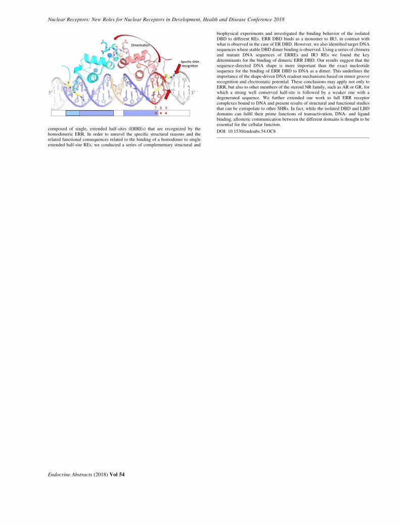
composed of single, extended half-sites (ERREs) that are recognized by thehomodimeric ERR. In order to unravel the specific structural reasons and therelated functional consequences related to the binding of a homodimer to singleextended half-site REs, we conducted a series of complementary structural and
Endocrine Abstracts (2018) Vol 54
biophysical experiments and investigated the binding behavior of the isolatedDBD to different REs. ERR DBD binds as a monomer to IR3, in contrast withwhat is observed in the case of ER DBD. However, we also identified target DNAsequences where stable DBD dimer binding is observed. Using a series of chimeraand mutant DNA sequences of ERREs and IR3 REs we found the keydeterminants for the binding of dimeric ERR DBD. Our results suggest that thesequence-directed DNA shape is more important than the exact nucleotidesequence for the binding of ERR DBD to DNA as a dimer. This underlines theimportance of the shape-driven DNA readout mechanisms based on minor grooverecognition and electrostatic potential. These conclusions may apply not only toERR, but also to other members of the steroid NR family, such as AR or GR, forwhich a strong well conserved half-site is followed by a weaker one with adegenerated sequence. We further extended our work to full ERR receptorcomplexes bound to DNA and present results of structural and functional studiesthat can be extrapolate to other SHRs. In fact, while the isolated DBD and LBDdomains can fulfil their prime functions of transactivation, DNA- and ligandbinding, allosteric communication between the different domains is thought to beessential for the cellular function.
DOI: 10.1530/endoabs.54.OC8

Nuclear Receptors: New Roles for Nuclear Receptors in Development, Health and Disease Conference 2018
Poster Presentations
Endocrine Abstracts (2018) Vol 54
Nuclear Receptors: New Roles for Nuclear Receptors in Development, Health and Disease Conference 2018
P1Single-molecule analysis of peroxisome proliferator-activated receptorg2 and a reveal subtype specific differences in chromatin bindingdynamicsRikke AM Jensen1,2, Ville Paakinaho1, Diego M Presman1, ErinE Swinstead1, R Louis Schiltz1, David A Ball1, Tatiana S Karpova1,Susanne Mandrup2 & Gordon L Hager1,*
1Laboratory of Receptor Biology and Gene Expression, NCI, NIH,Bethesda, Maryland, USA; 2Department of Biochemistry and MolecularBiology, University of Southern Denmark, Odense, Denmark.*Chief of the Laboratory of Receptor Biology and Gene Expression, NCI,NIH, Bethesda, Maryland, USA.
The peroxisome proliferator-activated receptors (PPARs) display a high degree ofconservation in the DNA- and ligand-binding domains. Despite these sequencesimilarities, the PPARs show distinct functions, even when co-expressed. Manyexperimental approaches have been employed to determine the molecularmechanisms that underlie their subtype-specific characteristics. However, theseapproaches have largely relied on cell population based studies, such as ChIP-seq.Here we have used single-molecule tracking (SMT) to investigate the unexploredintranuclear dynamics of two PPAR subtypes. Using HILO illumination,HaloTags, and the bright and stable fluorophore JF549, we have examined thebehavior of PPARg2 and PPARa in vivo at the single-molecule level. We detectslow and fast stops which we hypothesize to be functional- and non-functionalbinding events, respectively. Consistent with this model, we find that most long-lived binding events are lost upon mutation of the PPAR heterodimerization- andDNA-binding domains. The residence time and bound fraction (BF, slow stops) ofboth PPARs are unaffected by agonist or antagonist treatment. Interestinglyhowever, both the BF and the residence time are greater for PPARg2 than forPPARa, indicating that PPARg2 and PPARa display subtype-specific dynamicbinding behavior at the single-molecule level. This subtype specificity is found tobe dependent on the N-terminal domain. Furthermore, we show that the BF andresidence time of PPARg2 significantly increase in the presence of C/EBPa,which we have previously shown can facilitate PPARg binding to chromatin. Theability of C/EBPa to facilitate PPARg binding is dependent on the AF-2 domain,consistent with a model wherein the interplay between multiple TFs relies onthe recruitment of coactivators and chromatin remodelers. Overall, we havedemonstrated that SMT provides a unique ability to resolve unanswered questionsconcerning the highly dynamic properties of transcription factors, includingproperties that are linked to specific subtypes of closely related transcriptionfactors.
DOI: 10.1530/endoabs.54.P1
P2Next generation glucocorticoid receptor modulatorsKarl Edman1,2, Graham Belfield1, Matthew Dearman1, Goran Edenro1,Stefan Geschwindner2, Tove Hegelund-Myrback1, Martin Hemmerling1,Ramon Hendrickx1, Christina Keen1, Carina Karrman-Mardh1,Matti Lepisto1, Suman Mitra1, Susan Monkley1, Susanne Prothon1,Lena Ripa1, John Steele1, Outi Vaarala1, Lisa Wissler2 & Lisa Oberg1
1Respiratory, Inflammation and Autoimmunity, Innovative Medicines andEarly Development Biotech Unit, AstraZeneca, Pepparedsleden 1, Molndal43183, SE, Sweden; 2Discovery Sciences, Innovative Medicines and EarlyDevelopment Biotech Unit, AstraZeneca, Pepparedsleden 1, Molndal 43183, SE, Sweden.
Synthetic glucocorticoids bind to the glucocorticoid receptor (GR) and have beenused for nearly 70 years to treat inflammatory diseases. However, their use islimited by adverse effects such as diabetes, muscle wasting and osteoporosis.High throughput screening identified a novel non-steroidal scaffold with greatpotential for chemical optimization. Through rational design we developed theindazole ether series which combines high potency with structural motifs thatprovide vectors to key functional areas of the receptor. Exploitation of novelpockets within receptor enabled us to generate AZD7594, a GR agonist withproperties optimized for inhaled administration with high potency, long lungretention and minimal systemic exposure. AZD7594 is currently in a phase II trialfor asthma.To identify an oral agent, we modified the screening strategy to search compoundswith good bioavailablility and with different mechanistic properties. Again, westarted from the indazole ether series and used rational design to generate partialagonists with potential for differentiation. Evaluation in both human in vitrosystems and in rat in vivo models led to the discovery of AZD9567, a compound
Endocrine Abstracts (2018) Vol 54
that exhibited full inhibition of TNFa release in human whole blood after LPSstimulation but with no induction of gluconeogenic enzymes in primaryhepatocytes. AZD9567 is currently evaluated vs the effects of prednisolone inclinical studies in healthy volunteers.
DOI: 10.1530/endoabs.54.P2
P3Interactions between AR coregulators, TRIM24 and TRIM28, inCastrate Resistant Prostate Cancer (CRPC)Damien A Leach & Charlotte L BevanDivision of Cancer, Imperial Centre for Translational and ExperimentalMedicine, Imperial College London, Hammersmith Hospital Campus,London, UK.
Castrate Resistant Prostate Cancer (CRPC) is the inevitable outcome of hormonetreatment for advanced disease. Although no longer dependent on high levels ofandrogens, the androgen receptor (AR) remains active and there is evidence thatother nuclear receptors (NRs) can drive CRPC progression and/or therapyresistance. NRs share a repertoire of essential coregulators: proteins possessingthe ability to aid or repress NR action and have been proposed as a potentialmechanism for driving this inevitably lethal disease. Using publically availabledatasets we have found distinct patterns of coregulator expression between CRPCand hormone naıve disease. Importantly, we could identify a group ofcoregulators that were consistently differentially expressed across all cohorts.These could be further categorise into distinct function clusters or pathways.One such comprises TRIpartite Motif (TRIM) proteins TRIM24, TRIM28, andTRIM33. These form a unique subgroup of the larger TRIM family, in that onlythese three have Bromo-domains. Co-immunoprecipitation assay for endogenousproteins reveals that these proteins interact with each other and AR. Using ChIP-seq data we identified AR regulated genes which are also potentially TRIM24 andTRIM28 targeted. Using ChIP and RT-qPCR we were able to validate TRIM24and TRIM28 binding independently or concurrently to AR target genes VEGFA,SLC45A3, CXCR7. Silencing TRIMs can alter androgen regulation of suchgenes, and was also able to reduce proliferation and response to androgen inAR-expressing prostate cancer cells. Intriguingly in one such cell line (22RV1),silencing individual TRIMs made no difference to anti-androgen response, butsimultaneous silencing resensitized cells to the antiandrogen enzalutamide.Furthermore their expression in TCGA data sets could be used together to predictbiochemical relapse. Our data suggest that TRIM24 and TRIM28 proteinsinteract, in gene specific manners, to regulate AR activity and may provide apotential target to increase effectiveness of anti-androgen therapy.
DOI: 10.1530/endoabs.54.P3
P4Selective disruption of ERa expression in dendritic cells of lupus pronemice results in female-specific reduced survivalMelissa A Cunningham1, Jena R Wirth1, Jackie Eudaly1 &Gary S Gilkeson1,2
1Medical University of South Carolina, Charleston, South Carolina, USA;2Ralph H. Johnson Veterans Affairs Hospital, Charleston, South Carolina,USA.
Systemic lupus erythematosus (SLE) is a disease that disproportionately affectsfemales. We previously showed that a functional knockout of estrogen receptoralpha (ERaKO) resulted in significantly reduced renal disease and increasedsurvival in murine lupus. The mechanism of this effect, which requires estrogen,is not known. Interestingly, an ERaK/K (null mutant) mouse is not similarlyprotected. We and others have demonstrated a role for ERa in dendritic cell (DC)development and Toll-like receptor (TLR) responsiveness. Here we show thatselective genetic disruption of ERa in DCs of lupus prone mice results ina survival difference, but unexpectedly only in females, who die prematurelycompared with intact females. Floxed-ERa and Cre-CD11c strains werebackcrossed onto the NZM2410 lupus-prone background for 12 generations.Males and females were studied (nZ24). There was no significant difference insurvival between NZM CrePos/Floxed-ERa (DC-specific ERaKO) mice andNZM CreNeg/Floxed-ERa mice. Considered separately, however, femalesurvival was significantly different. Median age at death was 30.0 weeks(G1.8) for the CrePos and 40.4 weeks (G3.9) for the CreNeg females (P!0.04).Spleen cells were isolated and flow cytometry was performed to determinenumber and subset of DCs. Preliminary flow cytometry results revealed asignificantly reduced percent of MHCIICF480-CD11cCCD11bC DCs and

Nuclear Receptors: New Roles for Nuclear Receptors in Development, Health and Disease Conference 2018
MHCIICB220CSiglecHC pDCs in CrePos vs. CreNeg mice. There was a trendtowards increased percent of MHCIICCD11cCCD11b-CD8aC cells in CrePosmice. In summary, while selective deletion of ERa in DCs of female lupus-pronemice results in female-specific reduced survival, the etiology of this unexpectedaccelerated disease phenotype is not clear. This data joins a growing body ofevidence that ERa plays an important role in modulating immune cell function.
DOI: 10.1530/endoabs.54.P4
P5Glucocorticoid receptor inhibits ER-mediated pro-proliferative geneexpressionE Tonsing-Carter1, CR Kim1, KM Hernandez2, KR Bowie1, DC West1,S Chandarlapaty3, GL Greene4 & SD Conzen1,4
1Department of Medicine, The University of Chicago, Chicago, Illinois,USA; 2The Center for Research Informatics, The University of Chicago,Chicago, Illinois, USA; 3Department of Medicine, Memorial SloanKettering Cancer Center, New York, New York, USA; 4The Ben MayDepartment for Cancer Research, The University of Chicago, Chicago,Illinois, USA.
Early-stage ERC breast cancer (BC) with high tumor glucocorticoid receptor(GR) expression is associated with improved long term relapse-free survivalcompared to tumors with low GR expression. In addition, GR activity inhibitsER-mediated BC cell proliferation. We therefore hypothesized that GR and ERengage in nuclear receptor crosstalk to influence pro-proliferative geneexpression, thus contributing to a better outcome in ERC/GRC breast cancer.To understand the mechanisms by which ER/GR co-activation contribute to amore indolent ERC BC phenotype, we performed ChIP-sequencing and geneexpression analyses in ERC/GRC BC cell lines. We found that activation of GRwith the synthetic agonist, dexamethasone (dex), led to decreased ERC BC cellproliferation. Furthermore, ER/GR co-activation dampened cell cycle geneexpression (e.g. CDK6, CDK2, and CCND1) compared to ER-activation alone.GR and ER ChIP-sequencing revealed co-localization of ER and GR at a knowndownstream enhancer for ER-mediated CCND1 transcription suggesting directGR-mediated antagonism of ER. The highly selective GR modulators (SGRMs),CORT125134 and CORT118335 also reduced ER-driven cell proliferation andE2-mediated pro-proliferative gene expression. Moreover, MCF-7 cells engin-eered to express ER ligand-binding domain mutations (Y537S and D538G)similarly demonstrated decreased proliferation with either dex or SGRMtreatment. Taken together, these studies suggest that GR modulation deservesfurther investigation as an approach for inhibiting ER-driven BC.
DOI: 10.1530/endoabs.54.P5
P6The impact of 27-hydroxycholesterol on endometrial cancerproliferationPhilippa TK Saunders1, Frances Collins1, Fiona Cousins2,Arantza Esnal-Zufiaurre1 & Douglas A Gibson1
1MRC Centre for Inflammation Research, the Queens Medical ResearchInstitute, University of Edinburgh, 47 Little France Crescent, Edinburgh,UK; 2The Ritchie Centre, Hudson Institute of Medical Research,27-31 Wright Street, Clayton, Victoria, 3168, Australia.
Endometrial cancer (EC) is the most common gynaecological malignancy.Obesity is a major risk factor for EC and is associated with elevated cholesterol.27-Hydroxycholesterol (27HC) is a cholesterol metabolite that functions as anendogenous agonist for Liver X Receptor (LXR) and a selective estrogen receptormodulator (SERM). Exposure to estrogenic ligands increases risk of developingEC however the impact of 27HC on EC is unknown.Samples of stage 1 EC (nZ126) were collected from post-menopausal womenundergoing total abdominal hysterectomy according to a method approved by thelocal institutional ethics committee. Expression of LXRs (NR1H3, LXRa;
NR1H2, LXRb) and enzymes required for the synthesis (CYP27A1) orbreakdown (CYP7B1) of 27HC was assessed in all grades of EC. LXRa andLXRb expression was detected in EC but did not change with grade. Expressionof CYP7B1 decreased with disease severity (P!0.05) consistent with anassociation between increased bioavailability of 27HC and progression of EC.The impact of 27HC or the LXR agonist GW3965 on cell proliferation orexpression of an LXR- or ER-dependent luciferase reporter gene was assessed incell lines originating from well-, moderate- and poorly-differentiated endometrialcancers (Ishikawa, RL95, and MFE 280 respectively). Incubation with 27HCor GW3965 increased transcription via LXRE in Ishikawa, RL95 and MFE 280cells (P!0.01). 27HC selectively increased ERE reporter activity in Ishikawacells (P!0.001) and promoted proliferation of both Ishikawa and RL95 cells(P!0.001). Selective targeting of LXR with GW3965 significantly reduced cellproliferation of RL95 and MFE280 cells (P!0.0001).Altered cholesterol metabolism and exposure to 27HC may contribute toendometrial cancer risk by altering cell proliferation. These novel results suggestthat 27HC can promote proliferation of endometrial cancer epithelial cells andhighlight LXR as a potential therapeutic target in the treatment of advanceddisease.
DOI: 10.1530/endoabs.54.P6
P7MIR-96 regulates retinoic acid receptor gamma cross-talk with theandrogen receptor to drive aggressive prostate cancerMark D Long1, Spencer Rosario1, Lara E Sucheston-Campbell2,Dominic J Smiraglia1 & Moray J Campbell21Department of Cancer Genetics, Roswell Park Cancer Institute, Buffalo,New York, 14263, USA; 2College of Pharmacy, The Ohio State University,Columbus, Ohio 43210, USA.
The retinoic acid receptor gamma (RARg, encoded by NR1B3/RARG) issignificantly downregulated but not mutated in the MSKCC and PRAD prostatecancer (PCa) cohorts in the Cancer Genome Atlas (TCGA) data. We investigatedthe consequences of modifying RARg expression.Independent of ligand, stable RARg knockdown stimulated RWPE-1 and LNCaPcells proliferation, changed the cell cycle profile and significantly altered globalgene expression patterns that were enriched for AR and NF-kB signaling. RARgChIP-Seq in RWPE-1 cells identified w1300 significant binding sites, in theabsence of ligand. w800 of these sites significantly overlapped with activeenhancer histone modifications (H3K27ac, H3K4me1), AR and p65 (unit ofNF-kB) binding and were coincident with the ONECUT2 motif. CombiningRARg cistrome and transcriptome data and applying bootstrapping demonstratedRARg sustained target gene expression. Reducing RARg levels in HPr-1AR cellsdisrupted AR-dependent gene regulation; w1700 genes including MYC targets.RARg directly bound MYC, suggesting RARg directly regulates MYC, andco-regulates the AR capacity to repress MYC networks. Reduced RARg targetgene expression in the TCGA-PRAD cohort associated with higher Gleasongrade PCa.RARg reduction was driven by elevated miR-96 in PCa cells, mouse models, andTCGAPRAD, which associated with worse disease free survival. MiR-96 mimicsstimulated cell cycle progression and biotin-miR-96 pulldown identified 360miR-96 targets. The most altered miR-96 target genes in TCGA-PRAD consistedof a RARg-centric network. Finally, tumors with low RARg-network and highmiR-96 expression displayed repression of RARg target genes (e.g. SOX15) andsignificantly worse disease free survival.Together, these findings support the concept that, ligand independent RARg bindsat gene enhancer regions to govern AR signaling, including MYC networkrepression, and control proliferation. Elevated miR-96 represses the RARg-centered network and drives aggressive PCa. Conceptually, identifying miRNAthat govern NR genomic functions, independently of ligand, has the capacity tochange how these key transcription factors are studied and therapeuticallyexploited.
DOI: 10.1530/endoabs.54.P7
Endocrine Abstracts (2018) Vol 54

Nuclear Receptors: New Roles for Nuclear Receptors in Development, Health and Disease Conference 2018
Author Index
Ali, S IS4Allende, JA OC7Angelique, DH IS12
Ball, DA P1Baptissart, M IS12Barry, P IS13Beaudoin, C IS12Becares, N OC3Beinsteiner, B OC8Belfield, G P2Bentley, DL IS9Bevan, CL IS1 & P3Billas, IML OC8Bowie, KR P5Boyd, J IS5Brass, A IS5Brechbuhl, HM IS9Bruno, NE OC6
Campbell, MJ OC2 & P7Canisius, S IS4Chandarlapaty, S P5Chereji, RV IS14Claessens, F IS8Clark, DJ IS14Collins, F IS6 & P6Combe, S IS1Conzen, SD IS10 & P5Coombes, RC IS13Corleone, G IS13Cousins, F P6Cunningham, MA P4
Dart, A IS1Dash, AK OC1de Bono, J IS1Dearman, M P2Dehm, SM IS7DeMayo, FJ OC5Dolcen, DN IS10Dorjsuren, D IS4Dubois, V IS8Dworak, N OC7
Edenro, G P2Edman, K P2Erdos, E IS13Esnal-Zufiaurre, A IS6 & P6Eudaly, J P4
Finlay-Schultz, J IS9Flach, KD IS4Fletcher, CE IS1Franco, HL IS3
Gage, M OC3Geschwindner, S P2Gibson, DA IS6 & P6Gidrol, X IS1Gilkeson, GS P4Gillen, AE IS9Gioeli, D OC7Goddard, K IS13Greene, GL IS10, P5 & PL1Gyorffy, B IS13
Hadjiminas, DJ IS13Hager, GL P1 & IS14Han, SJ OC5Harkless, R IS10Hegelund-Myrback, T P2Hemmerling, M P2Hendrickx, R P2Hernandez, KM P5Hickey, TE IS2 & IS4Holota, H IS12Hosfield, D IS10Hou, TY IS3
Iqbal, M IS5
Jacobsen , BM IS9Jadhav, A IS4Jardi, F IS8Jensen, RAM P1Jin, Z OC6Johnson, T IS14Jung, SY OC5
Karrman-Mardh, C P2Kabos, P IS9Kamenecka, TM OC6Karpova, TS P1Keen, C P2Kershaw, S IS5Khalil, R IS8Kim, CR P5Klaholz, B OC8Kluin, R IS4Knudsen, KE PL3Kraus, WL IS3
Leach, DA IS1 & P3Lepisto, M P2Linn, SC IS4Liu, Z IS3Llimos, G OC2Long, MD P7 & OC2Long, T IS10
Louie, R OC3Lupold, SE IS1Lydon, JP OC5
Magnani, L IS13Malladi, VS IS3Mandrup, S P1Martinot, E IS12Matthews, SB IS9Matthews, LC IS5McGuire, SE IS1Michael, L IS8Mitra, S P2Mohideen-Abdul, K OC8Monkley, S P2Moras, D OC8Morgan, DJ IS5Murakami, S IS3
Nagari, A IS3Nandu, T IS3Nettles, KW OC6Nieuwland, M IS4Nowak, J OC6Nwachukwu, JC OC6
Oberg, L P2O’Malley, BW OC5 & PL2Opdam, M IS4
Paakinaho, V IS14 & P1Palmieri, C IS13Park, MJ OC5Patel, H IS4Patten, DK IS13Periyasamy, M IS4Pineda-Torr, I OC3Pongor, LS IS13Presman, DM IS14 & P1Prothon, S P2Pruneri, G IS13
Qin, J OC5
Ray, DW IS5Ripa, L P2Rosario, S OC2
& P7Ruan, Y IS11
Sedes, L IS12Saiakhova, A IS13Sartorius, CA IS9Saunders, PTK IS6 & P6Scacheri, P IS13
Schiavon, G IS13Schiltz, RL P1Shibakawa, A IS1Shousha, S IS13Simeonov, A IS4Simitsidellis, I IS6Singh, PK OC2Sita-Lumsden, A IS1Sleppy, R OC7Smiraglia, D OC2Smiraglia, DJ P7Spiller, D IS5Srinivasan, S OC6Steele, J P2Sucheston-Campbell, LE
P7Sulpice, E IS1Swinstead, EE IS14Swinstead, EE P1Szmulewitz, RZ IS10
Ta, HQ OC7Tilley, WD IS4Tonsing-Carter, E IS10
& P5Tsai, M-J OC5Tsai, SY OC5Tyagi, RK OC1
Vaarala, O P2Vanderschueren, D IS8Vasquez, YM IS3Vingiani, A IS13Volle, DH IS12
Waddington, K OC3Wesseling, J IS4Wessels, LFA IS4West, DC IS10 & P5Wilson, III DM IS4Wirth, JR P4Wissler, L P2Wu, S-P OC5
Yende, AS OC1
Zakharov, AV IS4Zindy, E IS5Zoubeidi, A OC4Zwart, W IS4