encephalopathy in the baboon (Papio anubis) - PubMed Central
6
British Journal of Industrial Medicine, 1974, 31, 128-133 The pathology of experimental lead encephalopathy in the baboon (Papio anubis) A. P. HOPKINS1 and A. D. DAYAN2 University Department of Clinical Neurology and Department of Neuropathology, National Hospitals for Nervous Diseases, Queen Square, London Hopkins, A. P., and Dayan, A. D. (1974). British Journal of Industrial Medicine, 31, 128-133. The pathologyof experimental leadencephalopathy in the baboon (Papio anubis). Baboons (Papio anubis) were intoxicated by intratracheal injections of lead carbonate. The main pathological findings were of widespread cerebral oedema and focal cortical necroses. The mechanism by which lead produces these changes is not known. Lead has very different effects in various species. In the rabbit for example it produces anaemia, punctate basophilia, and axonal degeneration without segmental demyelination; in the guinea-pig, segmental demyelination is the predominant lesion of the peripheral nerves; in the cat, both types of nerve lesion occur but punctate basophilia does not develop. These and other differences in other species are tabulated by Hopkins (1970). Lead encephalopathy still occurs relatively fre- quently (Whitfield, Ch'ien, and Whitehead, 1972), but the nature of the histological lesions remains confused (see discussion and review by Pentschew (1958)). We have therefore investigated this problem in a primate, the baboon (Papio anubis). Methods Two adult and one infant baboon were studied. Lead carbonate was given by repeated intratracheal injections under light anaesthesia. Details of this procedure, and details of the diet, supplements of vitamin B12, and general care of these animals are available in another publication (Hopkins, 1970). Seizures were observed either by one author (A.P.H.) or by trained animal technicians, and LPresent address: Department of Neurology, St. Bartholo- mew's Hospital, London ECIA 7BE 2Present address: Wellcome Research Laboratories, Langley Court, Beckenham, Kent, BR2 3BS daily notes were made of these and other clinical pheno- mena. Brains were removed one to three hours after death and fixed by immersion in 10% formol saline. Coronal slices were embedded in celloidin or in paraffin wax. Sections were stained by conventional techniques; frozen sections stained with Scharlach R were employed to demonstrate the breakdown of myelin. The brain of one control animal was prepared in this way, and histological examination showed no significant abnormalities. Results Observations on the changes in body weight and concentrations of haemoglobin and lead in the blood are shown in Figures 1 and 2. In spite of the high concentration of blood lead and the consider- able weight loss, there was no change in the haemo- globin concentration. Baboon 3 (Fig. 1) Female 11*7 kg. Five injections of lead carbonate, 1 g (86 mg/kg initial weight), were given on days 0, 24, 42, 59, and 122 and of 1-2 g on day 137. There was a marked increase in irritability and aggressive behaviour by this animal as early as the eleventh day. This reaction was not seen in animals which were not injected but repeatedly handled in the same way. On day 149 the first seizure was observed. 128
Transcript of encephalopathy in the baboon (Papio anubis) - PubMed Central

British Journal of Industrial Medicine, 1974, 31, 128-133
The pathology of experimental leadencephalopathy in the baboon (Papio anubis)
A. P. HOPKINS1 and A. D. DAYAN2University Department of Clinical Neurology and Department of Neuropathology,National Hospitals for Nervous Diseases, Queen Square, London
Hopkins, A. P., and Dayan, A. D. (1974). British Journal of Industrial Medicine, 31, 128-133.The pathologyof experimental leadencephalopathy in the baboon (Papio anubis). Baboons(Papioanubis) were intoxicated by intratracheal injections of lead carbonate. The main pathologicalfindings were of widespread cerebral oedema and focal cortical necroses. The mechanismby which lead produces these changes is not known.
Lead has very different effects in various species.In the rabbit for example it produces anaemia,punctate basophilia, and axonal degenerationwithout segmental demyelination; in the guinea-pig,segmental demyelination is the predominant lesionof the peripheral nerves; in the cat, both types ofnerve lesion occur but punctate basophilia does notdevelop. These and other differences in other speciesare tabulated by Hopkins (1970).Lead encephalopathy still occurs relatively fre-
quently (Whitfield, Ch'ien, and Whitehead, 1972),but the nature of the histological lesions remainsconfused (see discussion and review by Pentschew(1958)). We have therefore investigated this problemin a primate, the baboon (Papio anubis).
Methods
Two adult and one infant baboon were studied. Leadcarbonate was given by repeated intratracheal injectionsunder light anaesthesia. Details of this procedure, anddetails of the diet, supplements ofvitamin B12, and generalcare of these animals are available in another publication(Hopkins, 1970). Seizures were observed either by oneauthor (A.P.H.) or by trained animal technicians, and
LPresent address: Department of Neurology, St. Bartholo-mew's Hospital, London ECIA 7BE
2Present address: Wellcome Research Laboratories, LangleyCourt, Beckenham, Kent, BR2 3BS
daily notes were made of these and other clinical pheno-mena.
Brains were removed one to three hours after deathand fixed by immersion in 10% formol saline. Coronalslices were embedded in celloidin or in paraffin wax.Sections were stained by conventional techniques;frozen sections stained with Scharlach R were employedto demonstrate the breakdown of myelin.The brain of one control animal was prepared in this
way, and histological examination showed no significantabnormalities.
Results
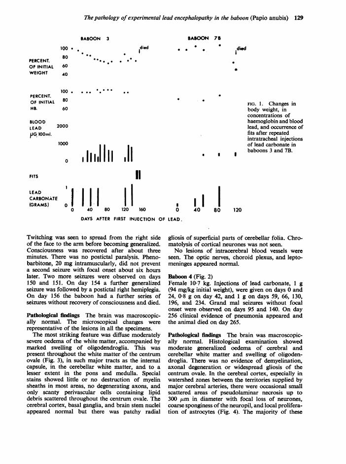
Observations on the changes in body weight andconcentrations of haemoglobin and lead in theblood are shown in Figures 1 and 2. In spite of thehigh concentration of blood lead and the consider-able weight loss, there was no change in the haemo-globin concentration.
Baboon 3 (Fig. 1)Female 11*7 kg. Five injections of lead carbonate,1 g (86 mg/kg initial weight), were given on days 0,24, 42, 59, and 122 and of 1-2 g on day 137. Therewas a marked increase in irritability and aggressivebehaviour by this animal as early as the eleventh day.This reaction was not seen in animals which werenot injected but repeatedly handled in the sameway. On day 149 the first seizure was observed.
128
Thepathology of experimental lead encephalopathy in the baboon (Papio anubis) 129
BABOON 3
died
00e0. .0
BABOON 7B0 0
Idied0 * *
a
2000
I I I
I I0 40 80
FIG. 1. Changes inbody weight, inconcentrations ofhaemoglobin and bloodlead, and occurrence offits after repeatedintratracheal injectionsof lead carbonate inbaboons 3 and 7B.
120DAYS AFTER FIRST INJECTION OF LEAD.
Twitching was seen to spread from the right sideof the face to the arm before becoming generalized.Consciousness was recovered after about threeminutes. There was no postictal paralysis. Pheno-barbitone, 20 mg intramuscularly, did not preventa second seizure with focal onset about six hourslater. Two more seizures were observed on days150 and 151. On day 154 a further generalizedseizure was followed by a postictal right hemiplegia.On day 156 the baboon had a further series ofseizures without recovery of consciousness and died.
Pathological findings The brain was macroscopic-ally normal. The microscopical changes wererepresentative of the lesions in all the specimens.The most striking feature was diffuse moderately
severe oedema of the white matter, accompanied bymarked swelling of oligodendroglia. This waspresent throughout the white matter of the centrumovale (Fig. 3), in such major tracts as the internalcapsule, in the cerebellar white matter, and to alesser extent in the pons and medulla. Specialstains showed little or no destruction of myelinsheaths in most areas, no degenerating axons, andonly scanty perivascular cells containing lipiddebris scattered throughout the centrum ovale. Thecerebral cortex, basal ganglia, and brain stem nucleiappeared normal but there was patchy radial
gliosis of superficial parts of cerebellar folia. Chro-matolysis of cortical neurones was not seen.No lesions of intracerebral blood vessels were
seen. The optic nerves, choroid plexus, and lepto-meninges appeared normal.
Baboon 4 (Fig. 2)Female 10-7 kg. Injections of lead carbonate, 1 g(94 mg/kg initial weight), were given on days 0 and24, 0-8 g on day 42, and 1 g on days 59, 66, 130,196, and 234. Grand mal seizures without focalonset were observed on days 95 and 140. On day256 clinical evidence of pneumonia appeared andthe animal died on day 265.
Pathological findings The brain was macroscopic-ally normal. Histological examination showedmoderate generalized oedema of cerebral andcerebellar white matter and swelling of oligoden-droglia. There was no evidence of demyelination,axonal degeneration or widespread gliosis of thecentrum ovale. In the cerebral cortex, especially inwatershed zones between the territories supplied bymajor cerebral arteries, there were occasional smallscattered areas of pseudolaminar necrosis up to300 /.tm in diameter with focal loss of neurones,coarse sponginess of the neuropil, and local prolifera-tion of astrocytes (Fig. 4). The majority of these
PERCENT.OF INITIALWEIGHT
100 * .
8080 0*e60
40
100 . * 0. 0 .
PERCENT.OF INITIAL 80HB. 60
BLOODLEADPG, lOOml.
FITS
LEADCARBONATE(GRAMS)
0 40
1 1
120 16080
.
1000
0 I III I

130 A. P. Hopkins and A. D. Dayan
BABOON 4
PERCENT.OF INITIALWEIGHT
100 *
80
60
40
0. *.
*0
1oo0 0 0.00PERCENT. 1 . .OF INITIAL 80 AHB. 60
2000
1000
o -1111,1 I I I I
200 240
)N OF LEAD.
III
FITS
LEADCARBONATE(GRAMS)
I I
011111 10 40 80 120 160'
DAYS AFTER FIRST INJECTIC
FIG. 2. Changes in body weight,in concentrations ofhaemoglobin and blood lead,and occurrence of fits afterrepeated intratracheal injectionsof lead carbonate in baboon 4.
I
280
FIG. 3. Baboon 3. Extensive spongy oedema of white FIG. 4. Baboon 4. Focal necrosis in cerebral cortex.
matter of centrum semiovale. (PTAH x 14). (H. and E. x 205).
00 0
0 1died0 .
BLOODLEADPJG/lOOml.
*
.

The pathology of experimental lead encephalopathy in the baboon (Papio anubis) 131
, E ' FI * , ,
%>IX ;
CZ
FIG. 5. Baboon 4. Radial gliosis in molecular layer ofcerebellar cortex. (PTAH x 255).
were in laminae 3, 4 and 5. The hippocampal andAmmon's horn regions appeared normal. In thecerebellum there were irregular scattered areas ofradial gliosis of the cortex associated with focal lossof Purkinje cells (Fig. 5). Neurons in the cerebralcortex did not show chromatolysis. No abnormalitywas seen of intracerebral blood vessels.
Baboon 7B (Fig. 1)Infant female 2-8 kg, dentition 212/212. An injectionof lead carbonate, 150 mg (54 mg/kg initial weight),was given on day 0, 400 mg on day 35, 600 mg onday 52, and 1000 mg on day 84. No seizures wereseen but weakness of hindquarters developed on day107 and the baboon died on day 117.
Pathological findings There again appeared to begeneralized oedema of the white matter and noabnormality of cerebral vessels.
Discussion
Seizures have never been observed in our controlbaboons nor in those also observed for long periodsduring a study of intoxication by acrylamide(Hopkins and Gilliatt, 1971).
Therefore the seizures observed in two out of the
three lead-intoxicated baboons reported here (8 outof 12 of all lead-intoxicated baboons observed inour laboratory (Hopkins, 1970)) clearly indicatethat lead produces an encephalopathy in thisprimate. Furthermore, there are reports of seizuresdue to lead encephalopathy in captive primates inzoos, the usual source of lead being paint from thecage (see Zook (1971) for references). Zook reporteddemyelination as a major finding in the brain andspinal cord of some of these apparently lead-intoxicated caged primates. but this might well bedue to the B12 deficiency which is known to occurin monkey colonies (Oxnard and Smith, 1966). Allour experimental animals received 1000 ,ug of B12monthly, and demyelination was not seen.The pathological findings in the present animals
may be discussed under two headings: cerebraloedema, and focal cortical necroses. Oedema wasgeneralized throughout the white matter, as shownby pallor of myelin staining and, if severe, by separa-tion of myelinated fibres by small amounts of clearfluid which sometimes formed pools around thesmaller blood vessels. It was accompanied by visibleand often quite marked swelling of oligodendro-glial cytoplasm, which produced the appearance ofperinuclear halos. The changes are common to mostforms of cerebral oedema (Blackwood et al., 1963).Similar appearances were not found in the controlanimal and so they cannot be attributed to agonalchanges. The other lesion, focal cortical necrosis,was manifested as pseudolaminar areas of spongychange and scarring in the cerebral cortex, and aszones of gliosis in the molecular layer ofthe cerebellarcortex. The nature and topographic distribution ofthese changes makes it very likely that they weredue to epileptic convulsions which may well havebeen more numerous than were observed.
Several different lesions have been considered ofimportance in lead encephalopathy (Pentschew,1958). For some authors, the primary or mostimportant pathological change lies in the nerve cell(Nissl, 1892; Ferraro and Hernandez, 1932). Thelatter authors, working with lead-intoxicated catsand monkeys, described chromatolysis, vacuoliza-tion, and liquefaction ofneurons diffusely throughoutthe cerebrum. For other workers the primary ormost important change is in the neuroglia. Vizioli(1929) described acute swelling on micro- and oligo-dendroglia in lead-intoxicated rabbits, and Roberti(1931) noted astrocytic proliferation. Other workersdescribed meningeal involvement in lead encephalo-pathy. Hassin (1921) found a proliferative menin-gitis and round cell infiltration of the meninges, andin the recent series of Whitfield et al. (1972) a thirdof 23 patients had an increased number of cells inthe cerebrospinal fluid. Yet another view is expressedin the monograph of Cantarow and Trumper (1944),who suggested that 'many if not indeed the majority

132 A. P. Hopkins and A. D. Dayan
of the clinical manifestations of lead encephalo-pathy are dependent upon, or associated withhypertension, which some authorities believe to beinvariably present in this condition'. Although itmust be admitted that the chronic nephritis causedby lead poisoning may produce hypertension andthat encephalopathy may be accompanied by papil-loedema, there are numerous reports of encephalo-pathy occurring in normotensive patients (e.g.,Akelaitis, 1941; Whitfield et al., 1972).Some reports of human and experimental lead
encephalopathy underline the importance of thevascular changes, although it has been difficult todisentangle agonal and postmortem changes in theappearances and staining properties of parenchymaland stromal cells and blood vessels. Mott (1909)described multiple haemorrhages in the brain andcord in a patient with encephalopathy which appear
to have been similar to those produced experi-mentally by Goadby and Goodbody (1909) in catsin the same year. The presence of endothelialproliferation in small blood vessels and the presence
of new capillaries and dilatation of existing ones hasbeen clearly described by a number of recent authors(see Whitfield et al. (1972) for references). Pentschew(1965) is perhaps the most vigorous proponent ofthe hypothesis that the vascular changes are a mostimportant feature. In a review of necropsy materialfrom 20 children with lead encephalopathy he des-cribed 'capillary activation' (dilatation of capillariesand swelling of endothelial cells) which was mostmarked in the molecular layer of the cerebellarcortex. Astrocytic proliferation and perivascularcuffing was also present, which Pentschew regardedas 'the earliest morphologic response to a dysfunctionof the blood-brain barrier'. Pentschew and Garro(1966) have produced lead encephalopathy in suck-ling rats. The entire cerebellum in these animalslooked reddish-brown due to large numbers ofmicroscopic haemorrhages. There was also intenseglial proliferation and serous exudates with markedcapillary proliferation. Neurons remained almostentirely normal. Injection of trypan blue revealed theextent of the breakdown of the blood-brain barrier,the white matter of the cerebral hemisphere, therostral part of the striatum, and the spinal cordstaining intensely blue. Lead-intoxicated sucklingrats have more recently been studied by Thomas,Dallenbach, and Thomas (1971). Many capillariesin the cerebellum were found to be lined by degener-ating swollen endothelial cells, and others were
occluded by aggregates of platelets and formation ofthrombi. Oedema fluid collected in the perivascularend-processes of astroglia and later coalesced to formperivascular lakes, as in the present study. Purkinjecells appeared pyknotic, and the authors consideredthis to be secondary to anoxia resulting from thedescribed changes in the capillary circulation.
There is only one previous pathological study ofthe brains of lead-intoxicated primates. Ferraro andHernandez (1932), investigating the neuropathologyof lead poisoning, administered lead carbonate totwo monkeys (species unnamed). Monkey 1 received1 03 g/day by mouth and monkey 2 1-07 g/day.Symptoms of 'vomiting, diarrhoea, anorexia, emaci-ation and general weakness, etc.' began on the 12thday in monkey 1 and on the 16th day in monkey 2.Monkey 1 lost 8% of its body weight, monkey 2lost 12%. Both animals died on the 66th day.Diffuse changes were found in the nervous systemof both animals. Chromatolysis, vacuolization, andliquefaction of the nerve cells occurred in thecerebral cortex, particularly in the layer of smallpyramidal cells; astrocytes were increased in numberand size in the white matter, and blood vessels showedendothelial swelling. Unfortunately almost all thechanges described could represent nonspecific agonaleffects of anoxia (exacerbated by convulsions),hypertension if present, and secondary reactivechanges in blood vessels in areas of cerebral oedema.The only definite changes are the occurrence of anencephalopathy associated with cerebral oedema toosevere and too widespread to be a terminal eventand of convulsions accompanied by post-epilepticbrain damage. The experiments of Pentschew andGarro (1966), in which trypan blue injected intosuckling rats with lead encephalopathy was shownto leak into the cerebellum, provide further supportfor the contention that cerebral oedema develops asan integral part of the pathological effects of leadpoisoning. Caution is required in interpreting theseexperiments, however, because the anoxia associatedwith convulsions may also cause abnormal perme-ability of blood vessels.The present experiments have not afforded any
evidence about the pathogenesis of the cerebraloedema, nor have they given any clue to possiblelinks between the cerebral lesion and the damageproduced by lead in peripheral nerves. Furthercritical studies should be of interest clinically as wellas pathologically because there are few known andcontrollable causes of oedema of the brain.
A. P. Hopkins was supported by a grant from theMedical Research Council which is gratefully acknowl-edged.
References
Akelaitis, A. J. (1941). Lead encephalopathy in children andadults: a clinico-pathological study. Journal of Nervousand Mental Diseases, 93, 313-332.
Blackwood, W., McMenemey, W. H., Meyer, A., Norman,R. M., and Russell, D. S. (1963). Greenfield's Neuro-pathology. Edward Arnold, London.
Cantarow, H. and Trumper, M. (1944). Lead Poisoning.Williams and Wilkins, Baltimore.
Ferraro, A. and Hernandez, R. (1932). Lead poisoning. (A

The pathology of experimental lead encephalopathy in the baboon (Papio anubis) 133
histopathological study of the nervous system of catsand monkeys in the acute and subacute stages.) Psychi-atric Quarterly, 6, 121-146 and 319-350.
Goadby, K. W. and Goodbody, F. W. (1909). A note onthe pathology of lead poisoning. Lancet, 2, 988-991.
Hassin, G. B. (1921). The contrast between the brain lesionsproduced by lead and other inorganic poisons and thosecaused by epidemic encephalitis. Archives of Neurologyand Psychiatry (Chicago), 6, 268-285.
Hopkins, A. P. (1970). Experimental lead poisoning in thebaboon. British Journal of Industrial Medicine, 27,130-140.and Gilliatt, R. W. (1971). Motor and sensory nerveconduction velocity in the baboon: normal values andchanges during acrylamide neuropathy. Journal ofNeurology, Neurosurgery and Psychiatry, 34, 415-426.
Mott, F. W. (1909). Examination of the nervous system ina case of chronic lead encephalitis. Archives ofNeurologyand ,'sychiatry (London), 4, 117-130.
Nissl, F. (1892). Ueber experimentell erzeugte Veranderungenan den Vorderhornzellen des Rukenmarks bei Kaninchenmit Demonstration mikroskopischer Praparate. Allge-meine Zeitschrift fdr Psychiatrie, 48, 675-682.
Oxnard, C. E. and Smith, W. T. (1966). Neurologicaldegeneration and reduced serum vitamin B12-levels incaptive monkeys. Nature, 210, 507-509.
Pentschew, A. (1958). Bleivergiftung. In Handbuch derspezielle pathologische Anatomie, edited by 0. Lubarsch,F. Henke, and R. Rossle, bd 13, 2B, pp. 1929-1970.Springer-Verlag, Berlin.
(1965). Morphology and morphogenesis of leadencephalopathy. Acta Neuropathologica, 5, 133-160.
- and Garro, F. (1966). Lead encephalo-myelopathy ofthe suckling rat and its implications on the porphyrin-opathic nervous diseases, with special reference to thepermeability disorders ofthe nervous system's capillaries.Acta Neuropathologica, 6, 266-278.
Roberti, C. E. (1931). Sul comportamento della macroglia edegli elementi nervosi nelle intossicazioni sperimentalida: istammina guanidina, acido cloridrico, acetato dipiombi e acetato talloso. Rassegna di Studi Psichiatrici,20, 7-29.
Thomas, J. A., Dallenbach, F. D., and Thomas, M. (1971).Considerations in the development of experimental leadencephalopathy. Virchows Archiv far pathologischeAnatomie und Physiologie und fdr klinische Medizin,352, 61-74.
Vizioli, F. (1929). La microglia e l'oligodendroglia nelleintossicazioni sperimentali e nelle alterazioni post-mortali. Rivista di Neurologia, 2, 365-386.
Whitfield, C. L., Ch'ien, L. T., and Whitehead, J. D. (1972).Lead encephalopathy in adults. American Journal ofMedicine, 52, 289-298.
Zook, B. C. (1971). An animal model for human disease.Comparative Pathology Bulletin, 3, 3-4.
Received for publication 9 August, 1973.Accepted for publication 26 November, 1973.