“En-Glove” Lysis of Lower Eyelid Retractors With AlloDerm ... · “En-Glove” Lysis of Lower...
5
SURGICAL TECHNIQUE “En-Glove” Lysis of Lower Eyelid Retractors With AlloDerm and Dermis-Fat Grafts in Lower Eyelid Retraction Surgery Heather S. Chang, M.D.*, Diana Lee, B.S.*, Mehryar Taban, M.D.*, Raymond S. Douglas, M.D., Ph.D.†, and Robert A. Goldberg, M.D.* *Department of Orbital and Plastic Reconstructive Surgery, Jules Stein Eye Institute, David Geffen School of Medicine at UCLA, Los Angeles, California; and †Department of Eye Plastic, Orbital and Facial Cosmetic Surgery, Kellogg Eye Center, University of Michigan, Ann Arbor, Michigan, U.S.A. Purpose: To describe a minimally invasive surgical tech- nique using AlloDerm or dermis-fat grafts for lower eyelid retraction. Methods: A retrospective review of all patients undergoing lower eyelid retraction surgery via a minimal invasive, “en-glove” technique from 2005 through 2009. Charts were reviewed for the type of graft (AlloDerm or dermis-fat) used, the etiology of lower eyelid retraction, and the follow-up time. Outcome measures included lower eyelid height (measured from the corneal light reflex to the lower eyelid margin, or MRD 2 ), reduction of lagoph- thalmos, cosmetic appearance, complications, and need for further surgery. Presurgery and postreconstruction photographs were re- viewed and graded for functional and cosmetic outcome. Results: A total of 8 patients underwent successful lower eyelid retraction surgery using this minimally invasive tech- nique. Etiologies included thyroid eye disease and cicatricial paralytic lower eyelid retraction. Mean improvement in MRD 2 was 1.5 mm for the AlloDerm group (4 patients, 7 eyelids) and 1.0 mm for the dermis-fat group (4 patients, 4 eyelids) after a minimum of 3 months’ follow-up. The cosmetic result was satisfactory in all cases. Conclusions: “En-glove” lower eyelid retraction surgical technique offers a minimally invasive approach for the release of the lower eyelid retractors and allows for volume augmen- tation using either AlloDerm or dermis-fat spacer graft. (Ophthal Plast Reconstr Surg 2011;27:137–141) L ower eyelid retraction, secondary to middle lamellar cicatrix, poses a surgical challenge. Typical etiologies include thyroid eye disease and sequelae of lower eyelid surgery (often blepharo- plasty) or trauma. Contracture and scarring of the orbital septum and/or lower eyelid retractors lead to shortening and vertical retraction of the lower eyelid. In postsurgical cases, orbicularis muscle weakness and volume collapse often play a significant role. To address this challenging problem, several surgical techniques have been developed, some of which use various graft materials as a “spacer” after elevating the lower eyelid. The spacer is designed to keep the retractors recessed and supports the tarsus, while separating the retractors from the orbital septum and preventing shifting of the overlying lamella. 1 Of note, more recently, nonsurgical elevation and “splinting” of the lower eyelid using hyaluronic acid gel have been used. 2 However, the latter is temporary and does not directly release extensive cicatrix, which occurs in some cases. We present a minimally invasive, “en-glove” surgical technique to address lower eyelid retraction, along with volume augmentation/support, using either acellular human dermis (AlloDerm; LifeCell Corporation, Branchburg, NJ, U.S.A.) or autogenous dermis-fat graft. METHODS We conducted a retrospective review of all patients undergoing lower eyelid retraction surgery via a minimally invasive “en-glove” technique by 2 surgeons (R.S.D., R.A.G.) at the Jules Stein Eye Institute in Los Angeles, CA, U.S.A., from 2005 to 2009. Charts were reviewed for patient demographics, etiology of lower eyelid retraction, type of graft (AlloDerm or dermis-fat) used, and follow-up time. Patients with less than three months’ follow-up were excluded. Out- come measures included lower eyelid height (measured from the corneal light reflex to the lower eyelid margin, or MRD 2 ), reduction of lagophthalmos, evaluation of the cosmetic appearance by the surgeon and a blind observer, complications, and need for further surgery. Preoperative and postoperative photographs were analyzed at the latest follow-up visit and graded for functional and cosmetic outcome in both groups (AlloDerm and dermis-fat). All photographs were obtained with identical lighting at primary gaze. They were analyzed using the TN5 software. 3,4 The corneal diameter and distance from the center of the pupil to the lower eyelid margin (MRD 2 ) were measured and standardized to a corneal diameter of 11.5 mm. 5 Surgical Technique. Surgery is performed under monitored local anes- thesia. The dermis-fat graft is harvested from a cosmetically hidden location (e.g., postauricular). The epithelium over the graft is hydrodis- sected with dilute anesthetic solution, and a thin layer of epithelium is dissected off the underlying dermis with a scalpel (Fig. 1). The dermis itself is then excised, including a thin cuff of subdermal fat, with typical graft size of 8 35 mm. (The AlloDerm graft size is similar.) A 1-cm horizontal incision in the skin just below or above the lateral canthal tendon is used to access the lower eyelid retractor plane. Using the Stevens scissors, blunt and sharp dissection creates a pocket to the subconjunctival space. Through the conjunctiva, the surgeon can visualize the en-glove dissection, releasing the retractors from the conjunctiva and inferior tarsus until the conjunctiva is “windowpane” thin (Fig. 1). Next, the scissor re-enters the eyelid in a more anterior plane, adjacent to the arcus marginalis of the orbital rim, which can be Accepted for publication October 7, 2009. Presented as video at Fall ASOPRS meeting, November 2008, New Orleans, LA, U.S.A. Presented as lecture at Spring ASOPRS meeting, July 2009, Laguna Niguel, California, U.S.A. The authors have no proprietary interest to disclose. Address correspondence and reprint requests to Robert A. Goldberg, M.D., Department of Orbital and Plastic Reconstructive Surgery, Jules Stein Eye Institute, David Geffen School of Medicine at UCLA, Box 957006, 100 Stein Plaza, Los Angeles, CA 90095-7006, U.S.A. E-mail: [email protected] DOI: 10.1097/IOP.0b013e3181c53d38 Ophthal Plast Reconstr Surg, Vol. 27, No. 2, 2011 137
Transcript of “En-Glove” Lysis of Lower Eyelid Retractors With AlloDerm ... · “En-Glove” Lysis of Lower...
SURGICAL TECHNIQUE
“En-Glove” Lysis of Lower Eyelid Retractors With AlloDermand Dermis-Fat Grafts in Lower Eyelid Retraction SurgeryHeather S. Chang, M.D.*, Diana Lee, B.S.*, Mehryar Taban, M.D.*, Raymond S. Douglas, M.D., Ph.D.†,
and Robert A. Goldberg, M.D.*
*Department of Orbital and Plastic Reconstructive Surgery, Jules Stein Eye Institute, David Geffen School ofMedicine at UCLA, Los Angeles, California; and †Department of Eye Plastic, Orbital and Facial Cosmetic Surgery,
Kellogg Eye Center, University of Michigan, Ann Arbor, Michigan, U.S.A.
Purpose: To describe a minimally invasive surgical tech-nique using AlloDerm or dermis-fat grafts for lower eyelidretraction.
Methods: A retrospective review of all patients undergoinglower eyelid retraction surgery via a minimal invasive, “en-glove”technique from 2005 through 2009. Charts were reviewed for thetype of graft (AlloDerm or dermis-fat) used, the etiology of lowereyelid retraction, and the follow-up time. Outcome measuresincluded lower eyelid height (measured from the corneal lightreflex to the lower eyelid margin, or MRD2), reduction of lagoph-thalmos, cosmetic appearance, complications, and need for furthersurgery. Presurgery and postreconstruction photographs were re-viewed and graded for functional and cosmetic outcome.
Results: A total of 8 patients underwent successful lowereyelid retraction surgery using this minimally invasive tech-nique. Etiologies included thyroid eye disease and cicatricialparalytic lower eyelid retraction. Mean improvement in MRD2
was 1.5 mm for the AlloDerm group (4 patients, 7 eyelids) and1.0 mm for the dermis-fat group (4 patients, 4 eyelids) after aminimum of 3 months’ follow-up. The cosmetic result wassatisfactory in all cases.
Conclusions: “En-glove” lower eyelid retraction surgicaltechnique offers a minimally invasive approach for the releaseof the lower eyelid retractors and allows for volume augmen-tation using either AlloDerm or dermis-fat spacer graft.
(Ophthal Plast Reconstr Surg 2011;27:137–141)
Lower eyelid retraction, secondary to middle lamellar cicatrix,poses a surgical challenge. Typical etiologies include thyroid
eye disease and sequelae of lower eyelid surgery (often blepharo-plasty) or trauma. Contracture and scarring of the orbital septumand/or lower eyelid retractors lead to shortening and verticalretraction of the lower eyelid. In postsurgical cases, orbicularis muscleweakness and volume collapse often play a significant role.
To address this challenging problem, several surgicaltechniques have been developed, some of which use various
graft materials as a “spacer” after elevating the lower eyelid.The spacer is designed to keep the retractors recessed andsupports the tarsus, while separating the retractors from theorbital septum and preventing shifting of the overlying lamella.1
Of note, more recently, nonsurgical elevation and “splinting” ofthe lower eyelid using hyaluronic acid gel have been used.2
However, the latter is temporary and does not directly releaseextensive cicatrix, which occurs in some cases.
We present a minimally invasive, “en-glove” surgicaltechnique to address lower eyelid retraction, along with volumeaugmentation/support, using either acellular human dermis(AlloDerm; LifeCell Corporation, Branchburg, NJ, U.S.A.) orautogenous dermis-fat graft.
METHODSWe conducted a retrospective review of all patients undergoing
lower eyelid retraction surgery via a minimally invasive “en-glove”technique by 2 surgeons (R.S.D., R.A.G.) at the Jules Stein EyeInstitute in Los Angeles, CA, U.S.A., from 2005 to 2009. Charts werereviewed for patient demographics, etiology of lower eyelid retraction,type of graft (AlloDerm or dermis-fat) used, and follow-up time.Patients with less than three months’ follow-up were excluded. Out-come measures included lower eyelid height (measured from thecorneal light reflex to the lower eyelid margin, or MRD2), reduction oflagophthalmos, evaluation of the cosmetic appearance by the surgeonand a blind observer, complications, and need for further surgery.
Preoperative and postoperative photographs were analyzed atthe latest follow-up visit and graded for functional and cosmeticoutcome in both groups (AlloDerm and dermis-fat). All photographswere obtained with identical lighting at primary gaze. They wereanalyzed using the TN5 software.3,4 The corneal diameter and distancefrom the center of the pupil to the lower eyelid margin (MRD2) weremeasured and standardized to a corneal diameter of 11.5 mm.5
Surgical Technique. Surgery is performed under monitored local anes-thesia. The dermis-fat graft is harvested from a cosmetically hiddenlocation (e.g., postauricular). The epithelium over the graft is hydrodis-sected with dilute anesthetic solution, and a thin layer of epithelium isdissected off the underlying dermis with a scalpel (Fig. 1). The dermisitself is then excised, including a thin cuff of subdermal fat, with typicalgraft size of 8 � 35 mm. (The AlloDerm graft size is similar.)
A 1-cm horizontal incision in the skin just below or above thelateral canthal tendon is used to access the lower eyelid retractor plane.Using the Stevens scissors, blunt and sharp dissection creates a pocketto the subconjunctival space. Through the conjunctiva, the surgeon canvisualize the en-glove dissection, releasing the retractors from theconjunctiva and inferior tarsus until the conjunctiva is “windowpane”thin (Fig. 1). Next, the scissor re-enters the eyelid in a more anteriorplane, adjacent to the arcus marginalis of the orbital rim, which can be
Accepted for publication October 7, 2009.Presented as video at Fall ASOPRS meeting, November 2008, New
Orleans, LA, U.S.A. Presented as lecture at Spring ASOPRS meeting, July2009, Laguna Niguel, California, U.S.A.
The authors have no proprietary interest to disclose.Address correspondence and reprint requests to Robert A. Goldberg, M.D.,
Department of Orbital and Plastic Reconstructive Surgery, Jules Stein EyeInstitute, David Geffen School of Medicine at UCLA, Box 957006, 100 SteinPlaza, Los Angeles, CA 90095-7006, U.S.A. E-mail: [email protected]
DOI: 10.1097/IOP.0b013e3181c53d38
Ophthal Plast Reconstr Surg, Vol. 27, No. 2, 2011 137
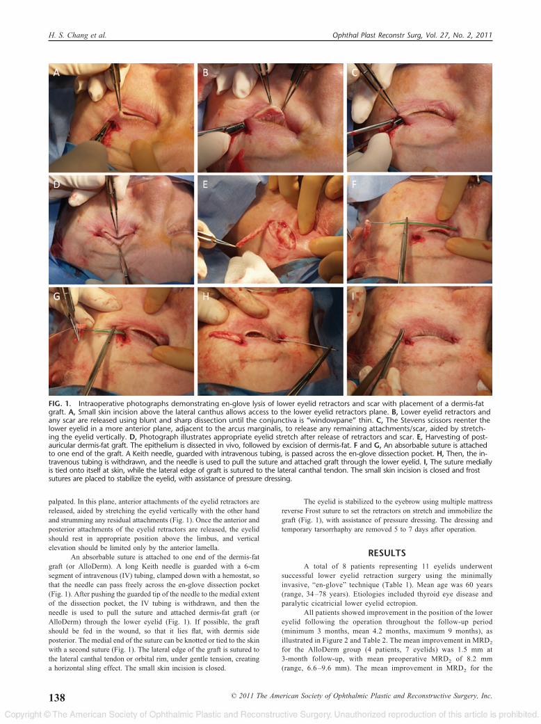
palpated. In this plane, anterior attachments of the eyelid retractors arereleased, aided by stretching the eyelid vertically with the other handand strumming any residual attachments (Fig. 1). Once the anterior andposterior attachments of the eyelid retractors are released, the eyelidshould rest in appropriate position above the limbus, and verticalelevation should be limited only by the anterior lamella.
An absorbable suture is attached to one end of the dermis-fatgraft (or AlloDerm). A long Keith needle is guarded with a 6-cmsegment of intravenous (IV) tubing, clamped down with a hemostat, sothat the needle can pass freely across the en-glove dissection pocket(Fig. 1). After pushing the guarded tip of the needle to the medial extentof the dissection pocket, the IV tubing is withdrawn, and then theneedle is used to pull the suture and attached dermis-fat graft (orAlloDerm) through the lower eyelid (Fig. 1). If possible, the graftshould be fed in the wound, so that it lies flat, with dermis sideposterior. The medial end of the suture can be knotted or tied to the skinwith a second suture (Fig. 1). The lateral edge of the graft is sutured tothe lateral canthal tendon or orbital rim, under gentle tension, creatinga horizontal sling effect. The small skin incision is closed.
The eyelid is stabilized to the eyebrow using multiple mattressreverse Frost suture to set the retractors on stretch and immobilize thegraft (Fig. 1), with assistance of pressure dressing. The dressing andtemporary tarsorrhaphy are removed 5 to 7 days after operation.
RESULTSA total of 8 patients representing 11 eyelids underwent
successful lower eyelid retraction surgery using the minimallyinvasive, “en-glove” technique (Table 1). Mean age was 60 years(range, 34 –78 years). Etiologies included thyroid eye disease andparalytic cicatricial lower eyelid ectropion.
All patients showed improvement in the position of the lowereyelid following the operation throughout the follow-up period(minimum 3 months, mean 4.2 months, maximum 9 months), asillustrated in Figure 2 and Table 2. The mean improvement in MRD2
for the AlloDerm group (4 patients, 7 eyelids) was 1.5 mm at3-month follow-up, with mean preoperative MRD2 of 8.2 mm(range, 6.6 –9.6 mm). The mean improvement in MRD2 for the
FIG. 1. Intraoperative photographs demonstrating en-glove lysis of lower eyelid retractors and scar with placement of a dermis-fatgraft. A, Small skin incision above the lateral canthus allows access to the lower eyelid retractors plane. B, Lower eyelid retractors andany scar are released using blunt and sharp dissection until the conjunctiva is “windowpane” thin. C, The Stevens scissors reenter thelower eyelid in a more anterior plane, adjacent to the arcus marginalis, to release any remaining attachments/scar, aided by stretch-ing the eyelid vertically. D, Photograph illustrates appropriate eyelid stretch after release of retractors and scar. E, Harvesting of post-auricular dermis-fat graft. The epithelium is dissected in vivo, followed by excision of dermis-fat. F and G, An absorbable suture is attachedto one end of the graft. A Keith needle, guarded with intravenous tubing, is passed across the en-glove dissection pocket. H, Then, the in-travenous tubing is withdrawn, and the needle is used to pull the suture and attached graft through the lower eyelid. I, The suture mediallyis tied onto itself at skin, while the lateral edge of graft is sutured to the lateral canthal tendon. The small skin incision is closed and frostsutures are placed to stabilize the eyelid, with assistance of pressure dressing.
H. S. Chang et al. Ophthal Plast Reconstr Surg, Vol. 27, No. 2, 2011
138 © 2011 The American Society of Ophthalmic Plastic and Reconstructive Surgery, Inc.
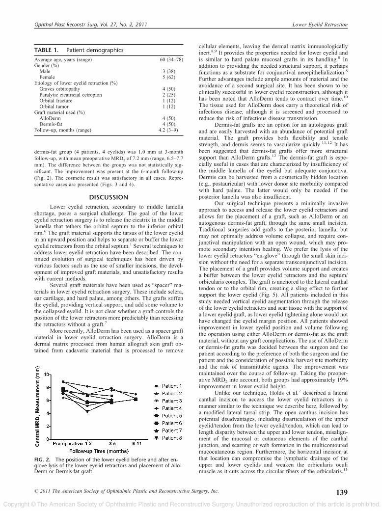
dermis-fat group (4 patients, 4 eyelids) was 1.0 mm at 3-monthfollow-up, with mean preoperative MRD2 of 7.2 mm (range, 6.5–7.7mm). The difference between the groups was not statistically sig-nificant. The improvement was present at the 6-month follow-up(Fig. 2). The cosmetic result was satisfactory in all cases. Repre-sentative cases are presented (Figs. 3 and 4).
DISCUSSIONLower eyelid retraction, secondary to middle lamella
shortage, poses a surgical challenge. The goal of the lowereyelid retraction surgery is to release the cicatrix in the middlelamella that tethers the orbital septum to the inferior orbitalrim.6 The graft material supports the tarsus of the lower eyelidin an upward position and helps to separate or buffer the lowereyelid retractors from the orbital septum.1 Several techniques toaddress lower eyelid retraction have been described. The con-tinued evolution of surgical techniques has been driven byvarious factors such as the use of smaller incisions, the devel-opment of improved graft materials, and unsatisfactory resultswith current methods.
Several graft materials have been used as “spacer” ma-terials in lower eyelid retraction surgery. These include sclera,ear cartilage, and hard palate, among others. The grafts stiffenthe eyelid, providing vertical support, and add some volume tothe collapsed eyelid. It is not clear whether a graft controls theposition of the lower retractors more predictably than recessingthe retractors without a graft.7
More recently, AlloDerm has been used as a spacer graftmaterial in lower eyelid retraction surgery. AlloDerm is adermal matrix processed from human allograft skin graft ob-tained from cadaveric material that is processed to remove
cellular elements, leaving the dermal matrix immunologicallyinert.8,9 It provides the properties needed for lower eyelid andis similar to hard palate mucosal grafts in its handling.8 Inaddition to providing the needed structural support, it perhapsfunctions as a substrate for conjunctival neoepithelialization.8
Further advantages include ample amounts of material and theavoidance of a second surgical site. It has been shown to beclinically successful in lower eyelid reconstruction, although ithas been noted that AlloDerm tends to contract over time.10
The tissue used for AlloDerm does carry a theoretical risk ofinfectious disease, although it is screened and processed toreduce the risk of infectious disease transmission.
Dermis-fat grafts are an option for an autologous graftand are easily harvested with an abundance of potential graftmaterial. The graft provides both flexibility and tensilestrength, and dermis seems to vascularize quickly.11,12 It hasbeen suggested that dermis-fat grafts offer more structuralsupport than AlloDerm grafts.12 The dermis-fat graft is espe-cially useful in cases that are characterized by insufficiency ofthe middle lamella of the eyelid but adequate conjunctiva.Dermis can be harvested from a cosmetically hidden location(e.g., postauricular) with lower donor site morbidity comparedwith hard palate. The latter would only be needed if theposterior lamella was also insufficient.
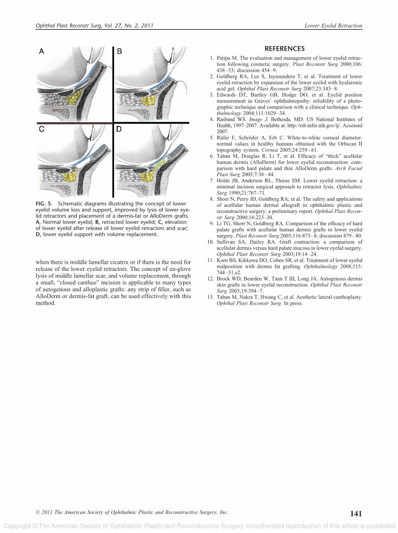
Our surgical technique presents a minimally invasiveapproach to access and release the lower eyelid retractors andallows for the placement of a graft, such as AlloDerm or anautogenous dermis-fat graft, through the same small incision.Traditional surgeries add grafts to the posterior lamella, butmay not optimally address volume collapse, and require con-junctival manipulation with an open wound, which may pro-mote secondary intention healing. We prefer the lysis of thelower eyelid retractors “en-glove” through the small skin inci-sion without the need for a separate transconjunctival incision.The placement of a graft provides volume support and createsa buffer between the lower eyelid retractors and the septum/orbicularis complex. The graft is anchored to the lateral canthaltendon or to the orbital rim, creating a sling effect to furthersupport the lower eyelid (Fig. 5). All patients included in thisstudy needed vertical eyelid augmentation through the releaseof the lower eyelid retractors and scar tissue with the support ofa lower eyelid graft, as lower eyelid tightening alone would nothave changed the eyelid margin position. All patients showedimprovement in lower eyelid position and volume followingthe operation using either AlloDerm or dermis-fat as the graftmaterial, without any graft complications. The use of AlloDermor dermis-fat grafts was decided between the surgeon and thepatient according to the preference of both the surgeon and thepatient and the consideration of possible harvest site morbidityand the risk of transmittable agents. The improvement wasmaintained over the course of follow-up. Taking the preoper-ative MRD2 into account, both groups had approximately 19%improvement in lower eyelid height.
Unlike our technique, Holds et al.7 described a lateralcanthal incision to access the lower eyelid retractors in amanner similar to the technique we describe here, followed bya modified lateral tarsal strip. The open canthus incision haspotential disadvantages, including disarticulation of the uppereyelid/tendon from the lower eyelid/tendon, which can lead tolength disparity between the upper and lower tendon, misalign-ment of the mucosal or cutaneous elements of the canthaljunction, and scarring or web formation in the multicontouredmucocutaneous region. Furthermore, the horizontal incision atthat location can compromise the lymphatic drainage of theupper and lower eyelids and weaken the orbicularis oculimuscle as it cuts across the circular fibers of the orbicularis.13
TABLE 1. Patient demographics
Average age, years (range) 60 (34–78)Gender (%)
Male 3 (38)Female 5 (62)
Etiology of lower eyelid retraction (%)Graves orbitopathy 4 (50)Paralytic cicatricial ectropion 2 (25)Orbital fracture 1 (12)Orbital tumor 1 (12)
Graft material used (%)AlloDerm 4 (50)Dermis-fat 4 (50)
Follow-up, months (range) 4.2 (3–9)
FIG. 2. The position of the lower eyelid before and after en-glove lysis of the lower eyelid retractors and placement of Allo-Derm or Dermis-fat graft.
Ophthal Plast Reconstr Surg, Vol. 27, No. 2, 2011 Lower Eyelid Retraction
© 2011 The American Society of Ophthalmic Plastic and Reconstructive Surgery, Inc. 139
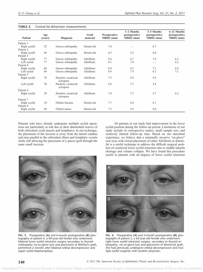
Patients who have already undergone multiple eyelid opera-tions are particularly at risk due to their diminished reserve ofboth orbicularis oculi muscle and lymphatics. In our technique,the placement of the incision is away from the lateral canthusand runs parallel to the orbicularis fibers and lymphatic vesselswhile still allowing the placement of a spacer graft through thesame small incision.
All patients in our study had improvement in the lowereyelid position during the follow-up period. Limitations of ourstudy include its retrospective nature, small sample size, andrelatively limited follow-up time. Based on our anecdotalexperience, we believe that a minimally invasive “en-glove”scar lysis with closed placement of either AlloDerm or dermis-fat is a useful technique to address the difficult surgical prob-lem of cicatricial lower eyelid retraction due to middle lamellashortage and volume collapse. We have found this procedureuseful in patients with all degrees of lower eyelid retraction
FIG. 3. Preoperative (A) and 6-month postoperative (B) pho-tographs of patient 4, a 69-year-old female who underwentbilateral lower eyelid retraction surgery secondary to thyroidorbitopathy via en-glove lysis and placement of AlloDerm graft,performed 2 months after bilateral orbital decompression andupper eyelid blepharoplasty.
FIG. 4. Preoperative (A) and 3-month postoperative (B) pho-tographs of patient 2, a 64-year-old female who underwentright lower eyelid retraction surgery, secondary to thyroid or-bitopathy, via en-glove lysis and placement of dermis-fat graft.She had previously undergone orbital decompression and mul-tiple eyelid surgeries with another physician.
TABLE 2. Central lid dimension measurements
PatientAge
(years) DiagnosisGraft
materialPreoperativeMRD2 (mm)
1–2 MonthspostoperativeMRD2 (mm)
3–5 MonthspostoperativeMRD2 (mm)
6–12 MonthspostoperativeMRD2 (mm)
Patient 1Right eyelid 52 Graves orbitopathy Dermis-fat 7.4 — 6.7
Patient 2Right eyelid 64 Graves orbitopathy Dermis-fat 6.5 5.2 6.0
Patient 3Right eyelid 77 Graves orbitopathy AlloDerm 9.6 6.7 7.9 6.3Left eyelid 77 Graves orbitopathy AlloDerm 8.1 7.0 — 6.2
Patient 4Right eyelid 69 Graves orbitopathy AlloDerm 8.9 8.5 7.1 6.9Left eyelid 69 Graves orbitopathy AlloDerm 8.9 7.9 8.1 7.1
Patient 5Right eyelid 78 Paralytic cicatricial
ectropionAlloDerm 7.5 5.9 5.9
Left eyelid 78 Paralytic cicatricialectropion
AlloDerm 6.6 5.7 5.4
Patient 6Right eyelid 39 Paralytic cicatricial
ectropionAlloDerm 7.8 5.7 5.7 6.2
Patient 7Right eyelid 34 Orbital fracture Dermis-fat 7.7 6.8 6.1
Patient 8Right eyelid 69 Orbital tumor Dermis-fat 7.2 4.3 6.0
H. S. Chang et al. Ophthal Plast Reconstr Surg, Vol. 27, No. 2, 2011
140 © 2011 The American Society of Ophthalmic Plastic and Reconstructive Surgery, Inc.
when there is middle lamellar cicatrix or if there is the need forrelease of the lower eyelid retractors. The concept of en-glovelysis of middle lamellar scar, and volume replacement, througha small, “closed canthus” incision is applicable to many typesof autogenous and alloplastic grafts: any strip of filler, such asAlloDerm or dermis-fat graft, can be used effectively with thismethod.
REFERENCES1. Patipa M. The evaluation and management of lower eyelid retrac-
tion following cosmetic surgery. Plast Reconstr Surg 2000;106:438–53; discussion 454–9.
2. Goldberg RA, Lee S, Jayasundera T, et al. Treatment of lowereyelid retraction by expansion of the lower eyelid with hyaluronicacid gel. Ophthal Plast Reconstr Surg 2007;23:343–8.
3. Edwards DT, Bartley GB, Hodge DO, et al. Eyelid positionmeasurement in Graves’ ophthalmopathy: reliability of a photo-graphic technique and comparison with a clinical technique. Oph-thalmology 2004;111:1029–34.
4. Rasband WS. Image J. Bethesda, MD: US National Institutes ofHealth, 1997–2007. Available at: http://rsb.infor.nih.gov/ij/. Accessed2007.
5. Rufer F, Schroder A, Erb C. White-to-white corneal diameter:normal values in healthy humans obtained with the Orbscan IItopography system. Cornea 2005;24:259–61.
6. Taban M, Douglas R, Li T, et al. Efficacy of “thick” acellularhuman dermis (AlloDerm) for lower eyelid reconstruction: com-parison with hard palate and thin AlloDerm grafts. Arch FacialPlast Surg 2005;7:38–44.
7. Holds JB, Anderson RL, Thiese SM. Lower eyelid retraction: aminimal incision surgical approach to retractor lysis. OphthalmicSurg 1990;21:767–71.
8. Shorr N, Perry JD, Goldberg RA, et al. The safety and applicationsof acellular human dermal allograft in ophthalmic plastic andreconstructive surgery: a preliminary report. Ophthal Plast Recon-str Surg 2000;16:223–30.
9. Li TG, Shorr N, Goldberg RA. Comparison of the efficacy of hardpalate grafts with acellular human dermis grafts in lower eyelidsurgery. Plast Reconstr Surg 2005;116:873–8; discussion 879–80.
10. Sullivan SA, Dailey RA. Graft contraction: a comparison ofacellular dermis versus hard palate mucosa in lower eyelid surgery.Ophthal Plast Reconstr Surg 2003;19:14–24.
11. Korn BS, Kikkawa DO, Cohen SR, et al. Treatment of lower eyelidmalposition with dermis fat grafting. Ophthalmology 2008;115:744–51.e2.
12. Brock WD, Bearden W, Tann T III, Long JA. Autogenous dermisskin grafts in lower eyelid reconstruction. Ophthal Plast ReconstrSurg 2003;19:394–7.
13. Taban M, Nakra T, Hwang C, et al. Aesthetic lateral canthoplasty.Ophthal Plast Reconstr Surg. In press.
FIG. 5. Schematic diagrams illustrating the concept of lowereyelid volume loss and support, improved by lysis of lower eye-lid retractors and placement of a dermis-fat or AlloDerm grafts.A, Normal lower eyelid; B, retracted lower eyelid; C, elevationof lower eyelid after release of lower eyelid retractors and scar;D, lower eyelid support with volume replacement.
Ophthal Plast Reconstr Surg, Vol. 27, No. 2, 2011 Lower Eyelid Retraction
© 2011 The American Society of Ophthalmic Plastic and Reconstructive Surgery, Inc. 141