
Empathy in Negative and Positive Interpersonal ...
16
ADVANCES IN COGNITIVE PSYCHOLOGY RESEARCH ARTICLE http://www.ac-psych.org 2017 • volume 13(1) • 105-120 105 Empathy in Negative and Positive Interpersonal Interactions. What is the Relationship Between Central (EEG, fNIRS) and Peripheral (Autonomic) Neurophysiological Responses? Michela Balconi 1,2 and Maria Elide Vanutelli 1,2 1 Research Unit in Affective and Social Neuroscience, Catholic University of the Sacred Heart, Milan, Italy 2 Department of Psychology, Catholic University of the Sacred Heart, Milan, Italy interpersonal empathy, positive/negative interac- tions, fNIRS, brain oscilla- tions; autonomic activity Emotional empathy is crucial to understand how we respond to interpersonal positive or negative situations. In the present research, we aim at identifying the neural networks and the autonomic responsiveness underlying the human ability to perceive and empathize with others’ emotions when positive (cooperative) or negative (uncooperative) interactions are observed. A multimeth- odological approach was adopted to elucidate the reciprocal interplay of autonomic (peripheral) and central (cortical) activities in empathic behavior. Electroencephalography (EEG, frequency band analysis) and hemodynamic (functional Near-Infrared Spectroscopy, fNIRS) activity were all recorded simultaneously with systemic skin conductance response (SCR) and heart rate (HR) meas- urements as potential biological markers of emotional empathy. Subjects were required to empa- thize in interpersonal interactions. As shown by fNIRS/EEG measures, negative situations elicited increased brain responses within the right prefrontal cortex (PFC), whereas positive situations elic- ited greater responses within the left PFC. Therefore, a relevant lateralization effect was induced by the specific valence (mainly for negative conditions) of the emotional interactions. Also, SCR was modulated by positive/negative conditions. Finally, EEG activity (mainly low-frequency theta and delta bands) intrinsically correlated with the cortical hemodynamic responsiveness, and they both predicted autonomic activity. The integrated central and autonomic measures better elucidated the significance of empathic behavior in interpersonal interactions. Corresponding author: Michela Balconi, Department of Psychology, Catholic University of the Sacred Heart, Largo Gemelli, 1, 20123, Milan, Italy. Phone: +39-2-72342586; fax: +30-2-72342280. E-mail: [email protected] E-mail: [email protected] ABSTRACT KEYWORDS DOI • 10.5709/acp-0211-0 INTRODUCTION e abilities to monitor and regulate emotional processes are parts of a functional model of empathic behavior (Chauhan, Mathias, & Critchley, 2008) which includes processes of emotional resonance. ese are constituted by an affective response to another person, which oſten entails knowing what another person is feeling; sharing that person’s emotional state; and, in some cases, having the intention to respond compassionately to another person’s distress (Decety & Jackson, 2006; Hooker, Verosky, Germine, Knight, & D’Esposito, 2008; Ickes, 1997; Preston & de Waal, 2002). Specifically, the emotional behavior, in ad- dition to the cognitive ability to share representations, constitute the basic components of empathy (Decety & Svetlova, 2012). However, limited previous studies explored empathy by using stim- uli consisting of real interpersonal situations. In fact, previous research This is an open access article under the CC BY-NC-ND license (http://creativecommons.org/licenses/by-nc-nd/4.0/).
Transcript of Empathy in Negative and Positive Interpersonal ...

AdvAnces in cognitive PsychologyreseArch Article
http://www.ac-psych.org2017 • volume 13(1) • 105-120105
Empathy in Negative and Positive Interpersonal Interactions. What is the Relationship Between Central (EEG, fNIRS) and Peripheral (Autonomic) Neurophysiological Responses?Michela Balconi 1,2 and Maria Elide Vanutelli 1,2
1 research Unit in Affective and social neuroscience, catholic University of the sacred heart, Milan, italy
2 department of Psychology, catholic University of the sacred heart, Milan, italy
interpersonal empathy,
positive/negative interac-
tions, fnirs, brain oscilla-
tions; autonomic activity
emotional empathy is crucial to understand how we respond to interpersonal positive or negative situations. in the present research, we aim at identifying the neural networks and the autonomic responsiveness underlying the human ability to perceive and empathize with others’ emotions when positive (cooperative) or negative (uncooperative) interactions are observed. A multimeth-odological approach was adopted to elucidate the reciprocal interplay of autonomic (peripheral) and central (cortical) activities in empathic behavior. electroencephalography (eeg, frequency band analysis) and hemodynamic (functional near-infrared spectroscopy, fnirs) activity were all recorded simultaneously with systemic skin conductance response (scr) and heart rate (hr) meas-urements as potential biological markers of emotional empathy. subjects were required to empa-thize in interpersonal interactions. As shown by fnirs/eeg measures, negative situations elicited increased brain responses within the right prefrontal cortex (PFc), whereas positive situations elic-ited greater responses within the left PFc. therefore, a relevant lateralization effect was induced by the specific valence (mainly for negative conditions) of the emotional interactions. Also, scr was modulated by positive/negative conditions. Finally, eeg activity (mainly low-frequency theta and delta bands) intrinsically correlated with the cortical hemodynamic responsiveness, and they both predicted autonomic activity. the integrated central and autonomic measures better elucidated the significance of empathic behavior in interpersonal interactions.
corresponding author: Michela Balconi, department of Psychology, catholic
University of the sacred heart, largo gemelli, 1, 20123, Milan, italy. Phone:
+39-2-72342586; fax: +30-2-72342280. e-mail: [email protected]
e-mail: [email protected]
AbstrAct
Keywords
doi • 10.5709/acp-0211-0
IntroductIon
The abilities to monitor and regulate emotional processes are parts
of a functional model of empathic behavior (Chauhan, Mathias, &
Critchley, 2008) which includes processes of emotional resonance. These
are constituted by an affective response to another person, which often
entails knowing what another person is feeling; sharing that person’s
emotional state; and, in some cases, having the intention to respond
compassionately to another person’s distress (Decety & Jackson, 2006;
Hooker, Verosky, Germine, Knight, & D’Esposito, 2008; Ickes, 1997;
Preston & de Waal, 2002). Specifically, the emotional behavior, in ad-
dition to the cognitive ability to share representations, constitute the
basic components of empathy (Decety & Svetlova, 2012).
However, limited previous studies explored empathy by using stim-
uli consisting of real interpersonal situations. In fact, previous research
this is an open access article under the cc By-nc-nd license (http://creativecommons.org/licenses/by-nc-nd/4.0/).

AdvAnces in cognitive PsychologyreseArch Article
http://www.ac-psych.org2017 • volume 13(1) • 105-120106
mainly focalized on the emotional response to generic emotional cues
and it did not include a specific empathic task (Balconi & Bortolotti,
2014; Balconi, Grippa, & Vanutelli, 2015a; Herrmann et al., 2008), or
it explored facial expressions of emotions (Balconi & Canavesio, 2013;
Herrmann et al., 2008) or empathy in specific domains (such as em-
pathic responses to pain conditions, Avenanti, Sirigu, & Aglioti, 2010;
Rêgo et al., 2015; Wang, Wang, Hu, & Li, 2014). In addition, exiguous
research monitored analytically the effect induced by different types of
empathic situations—that is, the positive versus negative valence of the
situations in which the subjects were required to empathize (Balconi &
Bortolotti, 2014; Herrmann et al., 2008; Silani & Singer, 2015). In one
case, the valence effect was explored in an empathic context, although
no specific effect was found in relation to both valence of the situation
(positive or negative) and lateralization of brain activity (left or right)
considered together. In other cases (see, e.g., Tullett, Harmon-Jones,
& Inzlicht, 2012), significant right lateralized prefrontal patterns have
been found in the case of empathy in negative circumstances. Here,
the authors suggested the mediation of feelings of sadness in the de-
velopment of the empathic mechanisms towards the suffering of other,
together with the elicitation of prefrontal asymmetry. Nonetheless,
results have often been proven to be inconsistent (see, e.g., Morelli &
Lieberman, 2013).
In the current study, we explored in an empathic context, both the
valence of the situation (positive or negative) and the lateralization of
brain activity (left or right). From a neurophysiological point of view,
it has been established that empathic responses influence both corti-
cal activity (Brüne et al., 2012; Decety & Jackson, 2006; Rameson &
Lieberman, 2009; Thirioux, Mercier, Blanke, & Berthoz, 2014) and
autonomic physiological responsiveness (Balconi & Bortolotti, 2012b;
Eisenberg et al., 1989; Prguda & Neumann, 2014). Indeed, as suggested
by empathy models, the indubitable vantage of acquiring both auto-
nomic and central activities is the possibility to better elucidate the
reciprocal interplay of these two domains (Decety & Svetlova, 2012;
Preston & de Waal, 2002). The multidimensionality of the construct of
empathy makes it less compatible with single measures. However, so
far central and peripheral measures were scarcely related to each other
in empathy research (Balconi & Bortolotti, 2012b). The current study
addresses this research gap.
Concerning cortical activity correlates of empathy, previous neu-
roimaging studies on the emotional behavior in relation to empathy
have revealed a range of areas activated in response to empathic
interactions, specifically, during general emotional processing, the
medial prefrontal cortex (MPFC, Seitz, Nickel, & Azari, 2006; Shamay-
Tsoory & Aharon-Peretz, 2007) and the dorsolateral prefrontal cortex
(DLPFC, Balconi & Bortolotti, 2012a; Balconi, Bortolotti, & Gonzaga,
2011; Brüne et al., 2012; Damasio, Everitt, & Bishop, 1996; Davidson,
2002; Ochsner & Gross, 2005; Rameson, Morelli, & Lieberman, 2012).
Moreover, electroencephalography (EEG) and lesion studies indicated
that the prefrontal cortex (PFC) plays a prominent role in mediat-
ing empathy-related behaviors. Specifically, many studies reported a
significant prefrontal involvement for the disruption of empathic be-
havior in the case of psychopathy (for a review see Pera-Guardiola et
al., 2016). For example, as has been found by Howard and McCullagh
(2007) in conditions involving both a categorization and a vigilance
task with affective stimuli, psychopaths showed significantly smaller
positive Slow Wave (pSW) amplitudes than healthy controls during the
categorization task, where they were required to discriminate between
living and nonliving stimuli, thus reflecting insensitivity to an affective
mismatch between neutral backgrounds and positive pictures. Also,
psychopaths showed a larger prefrontal negative event-related poten-
tial (ERP, N350), the amplitude of which positively correlated with the
behavioral markers of psychopathy. Similarly, Kiehl, Hare, McDonald,
and Brink (1999), by conducting a task comparing semantic and affec-
tive verbal information, reported greater centrofrontal negative-going
wave amplitudes in psychopaths than controls.
However, neither classical functional magnetic resonance imag-
ing (fMRI) nor EEG seem to have completely uncovered in depth the
physiological correlate of the emotional empathic experience, as both
of these methods have their shortcomings: a low temporal resolution
of fMRI and a low spatial resolution of activity below the cortical sur-
face plus an insensitivity to the hemodynamic response of the EEG.
Therefore, we applied optical imaging (i.e., near-infrared spectroscopy,
NIRS) as a complementary method in the study of emotions and em-
pathy. NIRS is particularly well-suited for evaluating PFC activity,
which is among the regions involved in emotional processing (i.e., the
frontopolar cortex and the DLPFC, Doi, Nishitani, & Shinohara, 2013).
Due to its high temporal resolution, a spatial resolution exceeding that
of the EEG, and its sensitivity for hemodynamic changes, NIRS seems
well suited to study the temporally evolving representation and inte-
gration among complex, extended neural networks, of the empathic
response. The temporal resolution of NIRS is high enough for meas-
uring event-related hemodynamic responses (Elwell et al., 1993), and
combined EEG/NIRS measurements allow for the complementary
examination of neural as well as hemodynamic aspects of brain activa-
tion (Balconi et al., 2015a; Biallas, Trajkovic, Haensse, Marcar, & Wolf,
2012).
Specifically, recent studies with functional NIRS (fNIRS) have
identified the PFC as a key region in the experience and regulation of
emotional responses (Brink et al., 2011; Nomura, Ogawa, & Nomura,
2010; Ogawa & Nomura, 2012). Based on this research, also a sig-
nificant lateralization effect was found, related to the positive versus
negative valence of the activating emotional context. Specifically, left
PFC areas were more activated in response to positive or approach
emotions, whereas right PFC areas were more activated in response
to negative or withdrawal emotions (Balconi et al., 2015a; Balconi,
Grippa, & Vanutelli, 2015b; Tullett et al., 2012).
Concerning EEG, frequency band analysis contributed to elucidat-
ing the role of specific cortical areas, mainly with respect to the lat-
eralization effect in emotional empathy processing, too. In fact, brain
oscillations may furnish clear brain correlates of specific empathic
contexts in terms of their valence (positive or negative) and in relation
to cortical lateralization. However, the specific role of brain oscillations
in affective and empathic behavior is partially unknown (Balconi &
Lucchiari, 2006, 2008; Başar, 1999; Vanutelli & Balconi, 2015). Only

AdvAnces in cognitive PsychologyreseArch Article
http://www.ac-psych.org2017 • volume 13(1) • 105-120107
few studies used brain oscillations to study empathy (Gutsell & Inzlicht,
2012; Moore, Gorodnitsky, & Pineda, 2012; Mu, Fan, Mao, & Han,
2008; Tullett et al., 2012). What is known from related investigations
outside empathy research proper is that, regarding the alpha frequency
band, lower-1 alpha desynchronizes in response to a warning stimulus
(Klimesch, Doppelmayr, Russegger, Pachinger, & Schwaiger, 1998).
Overall, changes in alpha power and lateralization effects related to
these changes suggested that a right frontal unbalance is associated with
negative emotions while relatively stronger left frontal activation is as-
sociated with positive emotions (Bekkedal, Rossi, & Panksepp, 2011).
An anterior asymmetry was found in alpha activity that was explained
as a correlate of changes in the affective state (Balconi, Brambilla, &
Falbo, 2009a, 2009b; Davidson, 1998; Dimberg & Petterson, 2000). In
addition, some studies showed that theta band power responds to pro-
longed visual emotional stimulation (Knyazev, 2007; Krause, Enticott,
Zangen, & Fitzgerald, 2012). Therefore, the modulation of this fre-
quency band may significantly contribute to the explanation of arousal
effects on emotional cue comprehension (Bekkedal et al., 2011). In
contrast, exiguous data concern the modulation of delta and beta band
when considering the emotional significance of a stimulus (Karakaş,
Erzengin, & Başar, 2000). In some cases, it was shown that delta could
be a marker of novelty of the emotional cues and that it can respond to
the exigency of stimulus updating in memory (Fernández et al., 1998).
Finally, as markers of spontaneous and automatic empathic be-
havior, autonomic measures are very important for understanding the
relationship between empathy and autonomic measures. It has been
observed that different degrees of empathic experience may affect
autonomic psychophysiological responses (Balconi, Falbo, & Conte,
2012; Prguda & Neumann, 2014). In those cases, participants imagined
(a) a personal experience of fear or anger from their own past, (b) an
equivalent experience from another person as if it were happening to
them, or (c) a non-emotional experience from their own past (Ruby &
Decety, 2004). Autonomic differences were found between these con-
ditions. Nevertheless, in this approach, only imagined (and not real)
empathic situations were proposed and this fact may have introduced
important variations in the subjective responses.
Systemic blood pressure (BP), heart rate (HR), and skin conduct-
ance response (SCR) were considered as potential biological markers
of emotions in empathic behavior, and recorded simultaneously with
EEG and NIRS (Tupak et al., 2014). Among the other dependent
variables, SCR provides a useful measure of limbic function (Furmark,
Fischer, Wik, Larsson, & Fredrikson, 1997; Lang, Davis, & Öhman,
2000). It is also a significant measure of arousal modulations, as has
been demonstrated previously (Balconi et al., 2009a; Balconi & Pozzoli,
2008; Bradley & Lang, 2000).
Also several NIRS studies underlined the association between PFC
activation and autonomic responses to emotional stimulation (see,
e.g., Tanida, Katsuyama, & Sakatani, 2007). Likewise, during view-
ing of trauma-related video clips, increased hemodynamic activity
(oxy-hemoglobin, O2Hb) has been found to be positively correlated
with heart rate change (Matsuo et al., 2003). Furthermore, Moghimi,
Kushki, Guerguerian, and Chau (2012) have linked the steepness of the
O2Hb peak to subjectively reported arousal levels, which is a widely
accepted indicator of autonomic system activation (Matsuo et al., 2003;
Roos, Robertson, Lochner, Vythilingum, & Stein, 2011). Moreover,
a significant correlation between ventromedial prefrontal cortex
(vmPFC) activation and SCR was found, based on stimulus content
(its threatening value, Tupak et al., 2014).
These previous studies supported the view that the prefrontal ar-
eas regulate autonomic reactions or somatic markers associated with
emotional conditions. However, such research lacked a detailed and
integrated analysis of all three levels (hemodynamic, electrophysi-
ological, and autonomic) involved in emotional processing during
empathic interactions. Only one previous study directly compared he-
modynamic, EEG, and autonomic measures, but, firstly, it focused on
generic emotional cues and, secondly, it was not on empathy (Balconi
et al., 2015a). In contrast, the present research first of all clearly focused
on empathic behavior by asking participants “to put themselves in the
shoes of another person and try to feel what this person is feeling” (as
reported in the procedural instructions). Secondly, the participants
were required to observe situations where subjects performed spe-
cific interactions and not simply a generic emotional display (such as
emotional pictures or faces). Therefore, in comparison with previous
studies, a highly empathic task was included.
In conclusion, in light of current knowledge on empathy, we
propose an integration of cortical (EEG and fNIRS) measures with
autonomic psychophysiological measures, as they have been shown
to indicate the presence of emotional tuning between subjects. In the
present study, EEG (frequency band analysis), systemic SCR and HR
were all recorded simultaneously with fNIRS measurements as poten-
tial biological markers of emotional responses to empathic situations
during a natural and interpersonal situation in which positive versus
negative contexts were represented.
A consistent prefrontal activation was expected, as indicated by
a hemodynamic modulation and brain oscillations. Both fNIRS and
brain oscillations were supposed to elicit a significant PFC response
to emotional interpersonal conditions. Specifically, as a correlate of
an empathic response, we firstly expected a higher synchronous brain
activity in low-frequency bands (delta and theta) and, in contrast, a
desynchronization of the alpha band. Moreover, based on valence and
lateralization effects of emotions (Balconi & Mazza, 2010; Russell,
2003), a significant and consistent higher prefrontal left activation
was expected for positive emotional interactions, whereas a consistent
higher prefrontal right activation was expected in response to negative
interactions. Secondly, we expected that electrodermal activity (SCR)
and HR could be significant measures of implicit reactivity to emo-
tional cues; they should consistently vary with emotional valence, with
larger responses (increased SCR and HR) elicited in either negative or
positive or both emotional conditions compared to neutral situations
(Balconi et al., 2009a; Bradley & Lang, 2000; Lang, Greenwald, Bradley,
& Hamm, 1993).
Thirdly, we expected a high coherence between the three measures
(fNIRS, EEG, and autonomic variations). Significant correlations
were hypothesized based on situational and interpersonal significance

AdvAnces in cognitive PsychologyreseArch Article
http://www.ac-psych.org2017 • volume 13(1) • 105-120108
(valence) of the empathic context. Indeed, we expected a relevant
modulation within the left and right PFC for hemodynamic activity in
concomitance with electrophysiological and autonomic response.
MaterIals and Methods
Subjects
Twenty-two subjects, 12 females and 10 males (Mage = 24.5 years; SD
= 3.53; age range from 20 to 33 years) participated in the experiment.
All subjects were right-handed (Edinburgh Handedness Inventory,
Oldfield, 1971), with normal or corrected-to-normal visual acuity.
Exclusion criteria were neurological or psychiatric pathologies of the
subjects or their close family members. Specifically, they did not show
deficits related to depression (Beck Depression Inventory II, BDI, Beck,
Steer, & Brown, 1996) and to anxiety (State-Trait Anxiety Inventory,
STAI, Spielberger, Gorsuch, Lushene, Vagg, & Jacobs, 1970): Exclusion
criterion of the BDI Inventory was 19 points or lower (M = 8.95; SD =
0.46; score range from 2 to 12 points); for the STAI 39 points or lower (M
= 28.45; SD = 1.03; score range from 27 to 45 points). No payment was
provided for participation. Participants gave informed written consent
and the research was approved by the Ethical Committee institution
where the work was carried out. The experiment was conducted in ac-
cordance with the Declaration of Helsinki, and all the procedures were
carried out with adequate understanding by the subjects. The Research
Consent Form was submitted before participation in the study.
StimuliSubjects were required to view affective images depicting real inter-
personal situations which represented two people who interacted in
a common and familiar situation (e.g., at home, in a workplace, or
on a journey). Colored images (16 cm in width and 10 cm in height)
representing positive, negative, and neutral interactions were selected.
Twenty-four pictures were used for each type of interaction. Positive
interactions represented positive and emotionally comfortable situa-
tions (such as a handshake between two people); negative interactions
represented negative and emotionally uncomfortable situations (such
as a quarrel between two people); neutral pictures represented interac-
tions without a specific emotional valence (such as two people sitting
on a couch, see Figure 1). All images were similar in their perceptual
features (i.e., their luminance, complexity, i.e., number of details in the
scene, and characters’ genders: half of the actors were male and half
were female).
In order to validate the image dataset, a pre-experimental procedure
was adopted. Each depicted scene was evaluated by four judges on
valence and arousal dimensions, using the Self-Assessment Manikin
Scale (SAM) with a five-point Likert scale (Bradley & Lang, 1994,
2007). Separately for each condition (positive, negative, and neutral),
ratings were averaged across all images presented. As shown by sta-
tistical analysis (two distinct repeated-measures analyses of variance
[ANOVAs] applied to valence and arousal), images firstly differed in
terms of valence (positive: M = 4.56, SD = 0.34; negative: M = 1.33,
SD = 0.26; neutral: M = 2.75, SD = 0.37)—positive interactions were
more positively rated than the other two categories, negative interac-
tions were more negatively rated than the other two categories, neutral
images were rated to be of intermediate valence between the other two
categories (for all significant contrast comparisons, p ≤ .01). Secondly,
with respect to arousal, the positive and negative interactions (posi-
tive: M = 4.23, SD = 0.24; negative: M = 4.72, SD = 0.25; neutral: M =
1.77, SD = 0.31) were rated as more arousing than the neutral inter-
actions (for all significant contrast comparisons, p ≤ .01). In contrast,
no significant differences were revealed between positive and negative
interactions (p = .32).
ProcedureSubjects were seated in a dimly lit room, facing an LCD computer moni-
tor that was placed at about 50 cm from the subject. The stimuli were
presented using E-Prime 2.0 software (Psychology Software Tools, Inc)
running on a laptop PC with a 15 in. screen (Acer TravelMate 250P).
Images were presented in a random order in the center of the screen for
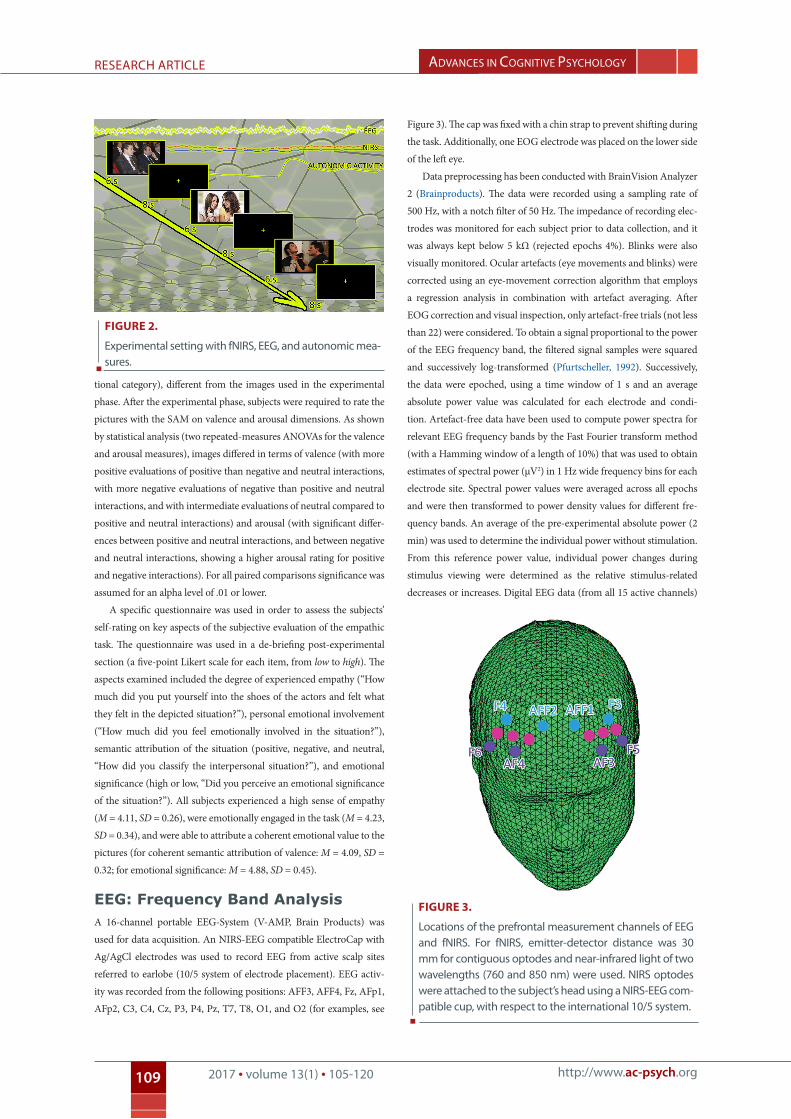
6 s, with an inter-stimulus interval of 8 s (see Figure 2).
Participants were required to view each stimulus during fNIRS/
EEG measures recording, and they were asked to attend to the inter-
personal situations during the entire time of exposition, focusing on
the emotional conditions which characterized the represented human
actors. Moreover, they were required to empathize with the two per-
sons interacting with each other (“Try to put yourself into the shoes of
the persons and to experience their feelings in this situation”). In order
to facilitate empathizing with the depicted actors, the two actors were
of about the same age as the experimental subjects.
Before scene presentations, a 2 min resting period was registered at
the beginning of the experiment. Next, a familiarization phase followed,
in which subjects saw and evaluated a set of images (one of each emo-
Figure 1.
some examples of positive, neutral, and negative interactions.
AdvAnces in cognitive PsychologyreseArch Article
http://www.ac-psych.org2017 • volume 13(1) • 105-120109
tional category), different from the images used in the experimental
phase. After the experimental phase, subjects were required to rate the
pictures with the SAM on valence and arousal dimensions. As shown
by statistical analysis (two repeated-measures ANOVAs for the valence
and arousal measures), images differed in terms of valence (with more
positive evaluations of positive than negative and neutral interactions,
with more negative evaluations of negative than positive and neutral
interactions, and with intermediate evaluations of neutral compared to
positive and neutral interactions) and arousal (with significant differ-
ences between positive and neutral interactions, and between negative
and neutral interactions, showing a higher arousal rating for positive
and negative interactions). For all paired comparisons significance was
assumed for an alpha level of .01 or lower.
A specific questionnaire was used in order to assess the subjects’
self-rating on key aspects of the subjective evaluation of the empathic
task. The questionnaire was used in a de-briefing post-experimental
section (a five-point Likert scale for each item, from low to high). The
aspects examined included the degree of experienced empathy (“How
much did you put yourself into the shoes of the actors and felt what
they felt in the depicted situation?”), personal emotional involvement
(“How much did you feel emotionally involved in the situation?”),
semantic attribution of the situation (positive, negative, and neutral,
“How did you classify the interpersonal situation?”), and emotional
significance (high or low, “Did you perceive an emotional significance
of the situation?”). All subjects experienced a high sense of empathy
(M = 4.11, SD = 0.26), were emotionally engaged in the task (M = 4.23,
SD = 0.34), and were able to attribute a coherent emotional value to the
pictures (for coherent semantic attribution of valence: M = 4.09, SD =
0.32; for emotional significance: M = 4.88, SD = 0.45).
EEG: Frequency Band Analysis A 16-channel portable EEG-System (V-AMP, Brain Products) was
used for data acquisition. An NIRS-EEG compatible ElectroCap with
Ag/AgCl electrodes was used to record EEG from active scalp sites
referred to earlobe (10/5 system of electrode placement). EEG activ-
ity was recorded from the following positions: AFF3, AFF4, Fz, AFp1,
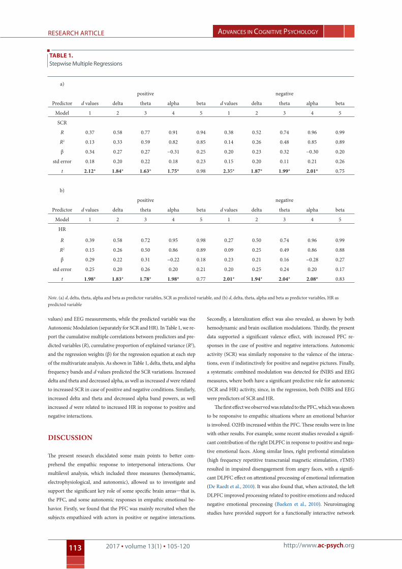
AFp2, C3, C4, Cz, P3, P4, Pz, T7, T8, O1, and O2 (for examples, see
Figure 3). The cap was fixed with a chin strap to prevent shifting during
the task. Additionally, one EOG electrode was placed on the lower side
of the left eye.
Data preprocessing has been conducted with BrainVision Analyzer
2 (Brainproducts). The data were recorded using a sampling rate of
500 Hz, with a notch filter of 50 Hz. The impedance of recording elec-
trodes was monitored for each subject prior to data collection, and it
was always kept below 5 kΩ (rejected epochs 4%). Blinks were also
visually monitored. Ocular artefacts (eye movements and blinks) were
corrected using an eye-movement correction algorithm that employs
a regression analysis in combination with artefact averaging. After
EOG correction and visual inspection, only artefact-free trials (not less
than 22) were considered. To obtain a signal proportional to the power
of the EEG frequency band, the filtered signal samples were squared
and successively log-transformed (Pfurtscheller, 1992). Successively,
the data were epoched, using a time window of 1 s and an average
absolute power value was calculated for each electrode and condi-
tion. Artefact-free data have been used to compute power spectra for
relevant EEG frequency bands by the Fast Fourier transform method
(with a Hamming window of a length of 10%) that was used to obtain
estimates of spectral power (μV2) in 1 Hz wide frequency bins for each
electrode site. Spectral power values were averaged across all epochs
and were then transformed to power density values for different fre-
quency bands. An average of the pre-experimental absolute power (2
min) was used to determine the individual power without stimulation.
From this reference power value, individual power changes during
stimulus viewing were determined as the relative stimulus-related
decreases or increases. Digital EEG data (from all 15 active channels)
Figure 2.
experimental setting with fnirs, eeg, and autonomic mea-sures.
Figure 3.
locations of the prefrontal measurement channels of eeg and fnirs. For fnirs, emitter-detector distance was 30 mm for contiguous optodes and near-infrared light of two wavelengths (760 and 850 nm) were used. nirs optodes were attached to the subject’s head using a nirs-eeg com-patible cup, with respect to the international 10/5 system.

AdvAnces in cognitive PsychologyreseArch Article
http://www.ac-psych.org2017 • volume 13(1) • 105-120110
software (Biopac Systems Inc) according to the manufacturer guide-
lines. ECG was converted to HR in number of beats per minute. The
signal was low-pass filtered at 35 Hz and highpass filtered at 0.05 Hz for
motor and ocular artefacts. For SCR, before attaching the electrodes,
the skin was cleaned with alcohol and slightly abraded. The electrodes
for SCR were attached to the distal phalanges of the first and second
finger of the left hand. SCL was recorded using two Ag/AgCl electrodes
and an isotonic gel. The signal was low-pass filtered at 10 Hz for mo-
tor, ocular, and biological artefacts. Ocular artefacts were then checked
with a visual inspection to eventually eliminate specific elements. Trials
with artefacts (2%) were excluded from the analysis. SCR elicited by
each stimulus was registered continuously with a constant voltage. It
was defined as the largest increase in conductance during emotional
image presentation, with a cut-off of at least 0.3 μS in amplitude with
respect to baseline (pre-stimulus) mean values. Baseline values were
scored during the 2 min prior to task onset.
results
The following set of analyses was performed on the data with SPSS
software for Windows (version 18): A first set of repeated-measures
ANOVAs was applied to each frequency band, a second set of analyses
was applied to hemodynamic d values, and a third set of ANOVAs was
applied to autonomic (HR, SCR) measures. Finally, stepwise multiple
regression and correlational analyses (Pearson correlations) were
applied to compare the three levels (band oscillations, d values, and
autonomic measures). Bonferroni correction was inserted for multiple
comparisons.
EEG Frequency Band AnalysisFrequency band data were entered into three-ways repeated-measures
ANOVAs, with independent variables of Lateralization (two sides: left
channels and right channels), Valence (3), and Localization (three sites:
frontal, AFF3/AFF4 and AFp1/AFp2; temporo-central, C3/C4 and T7/
T8; and parietal, P3/P4). Type I errors associated with inhomogene-
ity of variances were controlled by decreasing the degrees of freedom
using Greenhouse-Geiser epsilon. Post hoc comparisons were suc-
cessively applied to the data (contrast analyses for repeated-measures
ANOVA).
As shown by ANOVA, delta was modulated by valence, F(2, 42) =
6.16, p = .001, η2 = .27, and Lateralization × Valence, F(2, 42) = 7.23,
p = .001, η2 = .29. No other main effect or interaction was statistically
significant. Delta increased for negative and positive relative to neutral
stimuli. Moreover, it increased for negative more than for positive in-
teractions (for all comparisons, p ≤ .001). In addition, concerning the
simple effects for the two-way interaction, significant differences were
observed between positive and negative interactions, with increased
delta within the right hemisphere for negative, F(2, 42) = 5.79, p = .001,
η2 = .24, and within the left hemisphere for positive, F(2, 42) = 6.54, p =
.001, η2 = .26, interactions (see Figure 4).
For theta, the ANOVA revealed a significant main effect of valence,
F(1, 13) = 6.56, p = .001, η2 = .33, and a significant Lateralization ×
were band-pass filtered in the following frequency bands: delta (0-3),
theta (4-7), alpha (8-12), and beta (13-20). During data reduction, a
bandpass filter was applied in the 0.01-50 Hz frequency band.
fNIRSfNIRS measurements were conducted with the NIRScout System
(NIRx Medical Technologies, LLC) using a six-channel array of optodes
(four light sources/emitters and four detectors) covering the prefron-
tal area. Emitters were placed at AF3-AF4 and F5-F6 while detectors
were placed at AFF1-AFF2 and F3-F4 (see Figure 3). Emitter-detector
distance was 30 mm for contiguous optodes and a near-infrared light
of two wavelengths (760 and 850 nm) was used. NIRS optodes were
attached to the subject’s head using a NIRS-EEG compatible cup, with
respect to the international 10/5 system.
With NIRStar Acquisition Software (NIRx Medical Technologies
LLC), changes in the concentration of O2Hb and deoxygenated he-
moglobin (HHb) were recorded from a 2 min starting baseline. Signals
obtained from the six NIRS channels were measured with a sampling
rate of 6.25 Hz and analyzed and transformed according to their wave-
length and location, resulting in values for the changes in the concen-
tration of O2Hb and HHb for each channel. Haemoglobin quantity is
scaled in mM*mm, implying that all concentration changes depended
on the path length of the NIR light in the brain.
With Nirslab Software (v2014.05; NIRx Medical Technologies
LLC) the raw data of O2Hb and HHb from individual channels were
digitally band-pass filtered at 0.01–0.3 Hz. Successively, the mean con-
centration of each channel within a subject was calculated by averag-
ing data across the trials for 6 s from trial onset. Based on the mean
concentrations in the time series, we calculated the effect size in every
condition for each channel within a subject. The effect sizes (Cohen’s
d) were calculated as the differences of the means of the baseline and
trial divided by the SD of the baseline, d = (M1 − M2)/SD1. Accordingly,
M1 and M2 are the mean concentration values during the baseline and
trial, and SD1 the SD of the baseline. The mean concentration value
of the 2 s immediately before the trial was used as a baseline. Then,
the effect sizes obtained from the six channels were averaged in order
to increase the signal-to-noise ratio. Although the raw data of NIRS
were originally relative values and could not be averaged directly across
subjects or channels, normalized data, such as the effect sizes, could be
averaged regardless of the units of measurement (Matsuda & Hiraki,
2006; Schroeter, Zysset, Kruggel, & Von Cramon, 2003; Shimada &
Hiraki, 2006). In fact, the effect size is not affected by differential path-
length factor (DPF, Schroeter et al., 2003). Instead of a block design, a
continuous trial design was used in the present research.
Autonomic Measures Biopac MP 150 system (Biopac Systems Inc) was used to record the
autonomic activity. Electrocardiography (ECG) was recorded continu-
ously in lead 1 from two electrodes attached to the lower wrist, with
the positive pole on the left arm and the negative pole on the right arm.
One more reference electrode was placed over the left ankle. The ECG
signal was sampled at 1,000 Hz with the Biopac Acknowledge 3.7.1

AdvAnces in cognitive PsychologyreseArch Article
http://www.ac-psych.org2017 • volume 13(1) • 105-120111
Valence interaction, F(2, 42) = 7.76, p = .001, η2 = .29. No other effect
or interaction was statistically significant. Theta increased in response
to negative relative to positive stimuli. Concerning the two-way in-
teraction, significant differences were observed between positive and
negative interactions, with increased delta within the right hemisphere
for negative, F(2, 42) = 6.09, p = .001, η2 = .26, and within the left hemi-
sphere for positive, F(2, 42) = 6.43, p = .001, η2 = .26, interactions (see
Figure 4).
Concerning the alpha band, the valence effect was statistically
significant, F(2, 42) = 7.15, p = .001, η2 = .30. A generally decreased
alpha (increased brain activity) was observed for positive and negative
interactions. Finally, concerning beta, no significant effects were found
(see Figure 4).
fNIRSThe statistical analysis was applied to d—the dependent measure for
O2Hb and HHb-concentrations. The analysis of HHb did not reveal
any significant effects, and for this reason we report results for O2Hb
values only. The lack of any significant effect for HHb may be due to
the increase in O2Hb that is larger than the decrease in HHb (Wolf
et al., 2002). D was subjected to a repeated-measures ANOVA, with
Lateralization (2) and Valence (3) as independent variables. The data
were averaged over left (Channel 1: AF3–F3; Channel 2: AF3–AFF1;
Channel 3: F5–F3) and right (Channel 4: AF4–F4; Channel 5: AF4–
AFF2; Channel 6: F6–F4) channels.
As shown by the ANOVA, the effect of valence, F(2, 42) = 9.13, p
< .001, η2 = .41, and a Lateralization × Valence interaction, F(2, 42) =
8.13, p < .001, η2 = .32, were significant. No other effect or interaction
was statistically significant. As shown by paired comparisons, nega-
tive and positive stimuli revealed increased d values in comparison to
neutral interactions, F(1, 21) = 6.70, p < .001, η2 = .31, and F(1, 21) =
6.62, p < .001, η2 = .31, respectively. Moreover, negative interactions
showed higher d values for negative than positive interactions, F(1,
21) = 7.50, p < .001, η2 = .32. Regarding the interaction effect, positive
stimuli showed an increased brain activity within the left compared to
the right hemisphere, F(1, 21) = 8.03, p < .001, η2 = .34, whereas nega-
tive stimuli showed an increased activity within the right compared to
the left hemisphere, F(1, 21) = 8.88, p < .001, η2 = .35 (see Figure 5). In
contrast, no significant differences were found for neutral interactions
between left and right side, F(1, 21) = 1.16, p = .32, η2 = .16.
Autonomic MeasuresHR and SCR measures were analyzed with two separate repeated-
measures ANOVAs, both with Valence (3) as an independent factor.
For SCR, the valence main effect was significant, F(2, 41) = 8.88, p <
.001, η2 = .32: Negative stimuli induced an increased SCR relative to
positive and neutral conditions, F(1, 22) = 8.11, p < .001, η2 = .31, and
F(1, 22) = 6.90, p < 0.001, η2 = .28, respectively. Moreover, the positive
condition showed increased SCR values compared to neutral, F(1, 22)
= 7.13, p < .001, η2 = .30. For HR, no effect or interaction was sig-
Figure 4.
Frequency band power in response to valence and lateralization (M and SD reported; asterisks mark statistical significance, with p ≤ .05).
Figure 5.
hemodynamic states (o2hb relative concentrations) as a function of size and valence (obtained with nirslab soft-ware, data viewer section, Map tool). in response to nega-tive stimuli, the concentration of o2hb was higher for the right than the left side. Moreover, the concentration of o2hb was higher in response to negative more than posi-tive stimuli within the right side.

AdvAnces in cognitive PsychologyreseArch Article
http://www.ac-psych.org2017 • volume 13(1) • 105-120112
nificant (see Figure 6). No other effect or interaction was statistically
significant.
Correlational AnalysesPearson’s correlation analyses (across-subject correlations) were carried
out on each frequency band power and d values. Correlations were cal-
culated separately for each valence (positive/negative/neutral interac-
tions) within the left and right prefrontal area. Extensive analyses were
also applied to all the EEG and prefrontal fNIRS channels. However,
since no significant effect was found in the posterior EEG channels, for
the final analysis, we opted to compare the EEG and fNIRS data only
for the prefrontal area.
There was a significant positive correlation between d and theta (r
= .491, p < .02, Variance Inflation factor [VIF] = .460) and between
d and delta (r = .513, p < .01, VIF = .458) bands in response to nega-
tive stimuli within the right hemisphere. Moreover, significant positive
correlations between d and theta (r = .561, p < .01, VIF = .511) and
between d and delta (r = .544, p < .01, VIF = .493) bands in response to
positive stimuli were observed within the left hemisphere. Finally, the
alpha band showed a negative correlation with d (r = -.511, p < .01, VIF
= .469) within the right hemisphere in response to negative stimuli.
That is, cortical activation (alpha decreasing) was revealed within the
right hemisphere for negative interactions (see Figure 7). No other cor-
relations were statistically significant.
Regression AnalysisTwo stepwise multiple regression analyses were performed for positive
and negative interactions. Predictor variables were Hemodynamic (d
Figure 6.
Mean values for scr (up) and hr (down), with a significant effect shown for scr based on positive versus negative va-lence. (M and SD reported. Asterisks mark statistical signifi-cance, with p ≤ .05.)
Figure 7.
scatter plots of correlational analyses between hemo-dynamic and eeg measures as a function of valence and lateralization. each diamond corresponds to a single par-ticipant.
d values
d values
d values
d values
d values
d values
AdvAnces in cognitive PsychologyreseArch Article
http://www.ac-psych.org2017 • volume 13(1) • 105-120113
values) and EEG measurements, while the predicted variable was the
Autonomic Modulation (separately for SCR and HR). In Table 1, we re-
port the cumulative multiple correlations between predictors and pre-
dicted variables (R), cumulative proportion of explained variance (R2),
and the regression weights (β) for the regression equation at each step
of the multivariate analysis. As shown in Table 1, delta, theta, and alpha
frequency bands and d values predicted the SCR variations. Increased
delta and theta and decreased alpha, as well as increased d were related
to increased SCR in case of positive and negative conditions. Similarly,
increased delta and theta and decreased alpha band powers, as well
increased d were related to increased HR in response to positive and
negative interactions.
dIscussIon
The present research elucidated some main points to better com-
prehend the empathic response to interpersonal interactions. Our
multilevel analysis, which included three measures (hemodynamic,
electrophysiological, and autonomic), allowed us to investigate and
support the significant key role of some specific brain areas—that is,
the PFC, and some autonomic responses in empathic emotional be-
havior. Firstly, we found that the PFC was mainly recruited when the
subjects empathized with actors in positive or negative interactions.
Secondly, a lateralization effect was also revealed, as shown by both
hemodynamic and brain oscillation modulations. Thirdly, the present
data supported a significant valence effect, with increased PFC re-
sponses in the case of positive and negative interactions. Autonomic
activity (SCR) was similarly responsive to the valence of the interac-
tions, even if indistinctively for positive and negative pictures. Finally,
a systematic combined modulation was detected for fNIRS and EEG
measures, where both have a significant predictive role for autonomic
(SCR and HR) activity, since, in the regression, both fNIRS and EEG
were predictors of SCR and HR.
The first effect we observed was related to the PFC, which was shown
to be responsive to empathic situations where an emotional behavior
is involved. O2Hb increased within the PFC. These results were in line
with other results. For example, some recent studies revealed a signifi-
cant contribution of the right DLPFC in response to positive and nega-
tive emotional faces. Along similar lines, right prefrontal stimulation
(high frequency repetitive transcranial magnetic stimulation, rTMS)
resulted in impaired disengagement from angry faces, with a signifi-
cant DLPFC effect on attentional processing of emotional information
(De Raedt et al., 2010). It was also found that, when activated, the left
DLPFC improved processing related to positive emotions and reduced
negative emotional processing (Baeken et al., 2010). Neuroimaging
studies have provided support for a functionally interactive network
tAble 1. stepwise Multiple regressions
a)
positive negative
Predictor d values delta theta alpha beta d values delta theta alpha beta
Model 1 2 3 4 5 1 2 3 4 5
SCR
R 0.37 0.58 0.77 0.91 0.94 0.38 0.52 0.74 0.96 0.99
R2 0.13 0.33 0.59 0.82 0.85 0.14 0.26 0.48 0.85 0.89
β 0.34 0.27 0.27 −0.31 0.25 0.20 0.23 0.32 −0.30 0.20
std error 0.18 0.20 0.22 0.18 0.23 0.15 0.20 0.11 0.21 0.26
t 2.12* 1.84* 1.63* 1.75* 0.98 2.35* 1.87* 1.99* 2.01* 0.75
b)
positive negative
Predictor d values delta theta alpha beta d values delta theta alpha beta
Model 1 2 3 4 5 1 2 3 4 5
HR
R 0.39 0.58 0.72 0.95 0.98 0.27 0.50 0.74 0.96 0.99
R2 0.15 0.26 0.50 0.86 0.89 0.09 0.25 0.49 0.86 0.88
β 0.29 0.22 0.31 −0.22 0.18 0.23 0.21 0.16 −0.28 0.27
std error 0.25 0.20 0.26 0.20 0.21 0.20 0.25 0.24 0.20 0.17
t 1.98* 1.83* 1.78* 1.98* 0.77 2.01* 1.94* 2.04* 2.08* 0.83
Note. (a) d, delta, theta, alpha and beta as predictor variables, SCR as predicted variable, and (b) d, delta, theta, alpha and beta as predictor variables, HR as predicted variable

AdvAnces in cognitive PsychologyreseArch Article
http://www.ac-psych.org2017 • volume 13(1) • 105-120114
of cortico-limbic pathways that play a central role in the top-down
regulation of emotions. Indeed, a large number of studies suggested
that the PFC activates emotion regulation by inhibiting the amygdala
(Siegle, Thompson, Carter, Steinhauer, & Thase, 2007).
Results from previous fMRI studies indicated that the PFC is not
only involved in emotion induction but also in emotion regulation.
Moreover, by investigating the neural correlates of emotion regulation
processes, NIRS studies underlined the role of the PFC. For example,
the instruction to decrease the effect of negative stimuli by reinterpret-
ing the displayed situation led to an increased PFC activation and a
reduced activation of the amygdala (Banks, Eddy, Angstadt, Nathan,
& Luan Phan, 2007; Eippert et al., 2007; Kalisch et al., 2005; Lévesque
et al., 2003; Ochsner, Bunge, Gross, & Gabrieli, 2002; Ochsner et al.,
2004; Phan et al., 2005). Herrmann et al. (2002) used NIRS to compare
general emotional cue processing with processing of more specific
facial patterns. They found increased medial PFC activity during an
emotion induction paradigm which generated emotions by instructing
participants to try to feel like a person whose facial expression was dis-
played. In accordance with these results, the instruction to remember
emotional events leads to an increase of activation in the prefrontal
brain areas (Ohtani, Matsuo, Kasai, Kato, & Kato, 2005). Furthermore,
patients with post-traumatic stress disorder show increased PFC acti-
vation to disorder-related stimuli (Matsuo et al., 2003). In some cases,
the social effect of emotional face processing was considered (Nomura
et al., 2010). The study of Nomura et al. (2010) employed face stimuli
and perspective-taking, and NIRS was used to show the individual dif-
ferences in empathy that underlie the perspective taking function and
the role of the right ventrolateral PFC.
Although all these studies indicated an involvement of the PFC, for
the first time in the present research, the empathic emotional response
to interactional affective contexts was monitored. In addition, negative
versus positive situations were systematically evaluated. Indeed, we
revealed that emotional valence affected both hemodynamic activity
and brain oscillations, with a more relevant impact for the negative
interactions. In addition, this cortical activity was shown to be later-
alized within the right hemisphere in response to negative situations
and within the left hemisphere in response to positive stimuli. This
result clearly supports the view of a lateralization effect in empathic
responses to contexts of different valences when an empathic task was
administered.
Some specific brain oscillations (mainly delta and theta modula-
tion) confirmed this lateralized activation effect of stimulus valence:
Low-frequency oscillations were mainly synchronized within the right
and left side in response to negative and positive emotional interac-
tions, respectively. The increased values of delta and theta that we
found in response to positive and mainly negative interactions may
support the hypothesis that delta plays a main role in regulating the
attentional behavior in the case of salient stimuli. In line with this hy-
pothesis, in previous studies, delta band was related to the relevance of
the material being processed and to the degree of attention involved in
visual stimuli processing (Balconi & Pozzoli, 2005; Keil et al., 2001).
Therefore, in our case, brain responses to negative interactions could
suggest that subjects could have perceived them as the most relevant
emotional context, since they have a potentially threatening value.
It should be noted that in the present research we did not find a
significant and specific effect for higher frequency bands (beta). This is
in contrast with previous research (Balconi & Pozzoli, 2009). These dif-
ferent results may be due to the adoption of different methodological
approaches (e.g., task differences) and to different range limits used for
the computation of the oscillations.
A similar profile was observed for O2Hb measure, and the present
results thus confirmed the homogeneity of the emotional empathic
behavior in response to interpersonal situations by considering the
hemodynamic level of analysis. These results were also supported by
a consistent cortical lateralization effect for O2Hb, in combination
with a specific prefrontal effect. A general left/right positive/negative
distinction was observed in the subjects. That is, the subjects showed
a distinct cortical lateralized response based on the emotional valence
of the interactions: more left localized for positive situations; more
right localized for negative situations. Indeed, increased brain activity
was found to be based on stimulus valence. It has to be noted though
that, compared with some previous research on neuroimaging and
NIRS (Herrmann et al., 2008; Hoshi, 2009), we found that valence was
relevant for hemispheric lateralization during processing of emotional
cues. However, some differences were found based on valence (with
increased activation for negative situations), as previously shown by
EEG analysis. To account for the differences, we may assume that
the most salient contexts to be processed are related to negative in-
terpersonal interactions. Due to this higher degree of salience, higher
cortical activation could have been evoked by more negative interac-
tions. In general, it might be concluded that the fNIRS/EEG measures
showed a broad sensitivity to the motivational significance of social
interactions, varying as a function of the degree of negativity/positivity
attributed to the emotional situations. A general right/negative asso-
ciation was observed in the subjects, and it was mainly supported by
the right hemisphere—that is, negative, aversive interactions showed a
more consistent lateralized brain activation when compared to other
emotional situations (i.e., positive situations). Previous research under-
lined that human emotions are organized by two cortically lateralized
systems: the appetitive and defensive motivation systems, presum-
ably evolved from primitive approach and withdrawal tendencies
(Balconi & Bortolotti, 2014; Davidson, 1995; Davidson, Ekman, Saron,
Senulis, & Friesen, 1990; Dickinson & Dearing, 1978; Lang, Bradley, &
Cuthbert, 1990, 1997, 1998).
In line with this theory, emotional activation fundamentally varies
in centrally organized appetitive and aversive motivational systems that
have evolved to mediate a wide range of adaptive behaviors that are
necessary for an organism to survive (Bradley & Lang, 2007; Davidson
et al., 1990; Lang et al., 1990). Most pleasant affects are held to be as-
sociated with the appetitive motivation system; unpleasant affects
with defensive motivation (Cacioppo & Berntson, 1994). Specifically,
aversive conditions were considered highly relevant for the survival
since they include a threatening value (Fanselow, 1994; Russell, 1980).
Also the autonomic behavior was related to empathic behavior, with

AdvAnces in cognitive PsychologyreseArch Article
http://www.ac-psych.org2017 • volume 13(1) • 105-120115
an increased psychophysiological activity (higher SCR) for both posi-
tive and negative interactions. This response was attributed to general
emotional involvement and to the ability to respond physiologically to
the emotions displayed by other people in an interpersonal positive or
negative situation. Indeed, as suggested by recent models of empathic
behavior, a complex network of central and peripheral circuits supports
the phylogenetic developments of a specific empathy-related response
to conspecifics’ emotional signs. The multiple elements of the empathic
response are continuously modified during the social interactions and
are contextually embedded (Decety & Svetlova, 2012). The relation be-
tween more central processes (mediated by PFC) and more peripheral
processes (mediated by the autonomic system) confirmed the close re-
lation existing between high order mechanisms (evolutionarily recent)
and the autonomic responsiveness (evolutionarily ancient). It was also
underlined that behaviors specifically supported by arousal evolved
earlier than the mechanisms supported by more complex cognitive
processes (Decety & Svetlova, 2012). Moreover, it should be noted that
relevant models of empathic behavior have pointed out that the emo-
tional states related to empathy and the underlying neural mechanisms
are similar in all mammals (Panksepp, 1998).
The present results are also consistent with previously reported
negative, empathy-related responses to unpleasant situations (Brown,
Bradley, & Lang, 2006). Conflictual (negative) and cooperative (posi-
tive) situations were shown to be more powerful in eliciting empathic
responses, presumably emotionally involving and significant, compared
with neutral interpersonal conditions. In particular, the non-coopera-
tive condition was negatively connoted, highly empathy-inducing, and
able to produce a clear “negative” consonant autonomic reactivity.
Moreover, positive and negative situations showed a relation be-
tween empathic emotional, psychophysiological, and central (both
hemodynamic and EEG) measures. Indeed, it should be emphasized
that important connections were found in the correlational analysis
between hemodynamic and cortical EEG and the regression analyses
between these two measures and the responses at the autonomic level.
Firstly, the joined EEG-NIRS analysis revealed significant linear as-
sociations between the hemodynamic values and brain oscillations.
The significant positive relation between NIRS and EEG measures may
suggest, on the one hand, a general direct relation between these two
measures and PFC activation since they synchronously varied within
the prefrontal areas based on a situation’s valence. On the other hand,
the positive relation may support the connection between these two
brain measures in response to empathic situations. More generally, the
simultaneous registration of EEG and NIRS was found to be useful for
studies on empathic behavior. A general link between electrophysi-
ological effects and the regional hemodynamic changes was suggested
based on present and past evidences (Balconi et al., 2015a; Herrmann
et al., 2008; Schneider et al., 2014).
To summarize, the significant correlations between EEG and NIRS
measures within PFC may suggest that a specific cortical prefrontal
area supports empathic responsiveness. Indeed, whereas in band os-
cillations only a lateralization effect was found, the intrinsic relation
between PFC activity observed in the EEG (mainly the low-frequency
band) and the hemodynamic modulation may suggest the existence of
a coherent prefrontal network for empathy. However, future research
should explore the prefrontal localization of the EEG in more depth,
also investigating potential cortical generator (e.g., with a LORETA
approach) to define the reciprocal contribution by oscillations and
hemodynamic measures.
Secondly, regression analyses revealed that brain oscillations and
hemodynamic variations might have affected autonomic responses by
the subjects. That is, the PFC activity as marked by O2Hb increases and
synchronous cortical activity (mainly for low-frequency bands) were
significant factors, able to explain autonomic response modulation
since subjects modified their autonomic parameters as a function of
EEG/O2Hb changes in an empathic behavioral task. Specifically, in-
creased SCR/HR was predicted by frequency band and hemodynamic
activity in response to negative and positive interactions.
To summarize, the direct relation between EEG and O2Hb, shown
by correlational values, and the regression analysis, confirmed the in-
terconnections between the three levels of processing (hemodynamic,
electrophysiological, and autonomic). Indeed, the two analyses allowed
respectively evidencing the direct relationship between the two inde-
pendent measures (correlation analysis) and their consistent influence
on autonomic responses (regression analysis).
In the end, some limitations of this study and future suggestions for
improved research should be considered. Firstly, future research should
take into account the different roles that emotional and cognitive em-
pathy might have in interpersonal interactions. Secondly, the deeper
relations connecting central (both hemodynamic and EEG) and pe-
ripheral measures should be explored, considering the temporal course
of their modulations in response to empathic situations. Thirdly, the
inter-subjective differences related to some personality components
(such as empathy as a trait) should be explored as a stable construct
able to explain neurophysiological differences. Indeed, possible struc-
tural components could have modulated the central and peripheral
responses based on subjective empathic and emotional responsiveness
to positive and negative situations even in the present study.
RefeRencesAvenanti, A., sirigu, A., & Aglioti, s. M. (2010). racial bias re-
duces empathic sensorimotor resonance with other-race pain.
Current Biology, 20, 1018–1022. doi: 10.1016/j.cub.2010.03.071
Baeken, c., van schuerbeek, P., de raedt, r., de Mey, J.,
vanderhasselt, M. A., Bossuyt, A., & luypaert, r. (2010). the ef-
fect of one left-sided dorsolateral prefrontal cortical hF-rtMs
session on emotional brain processes in women. Psychiatria
Danubina, 22, s163. doi: 10.1016/j.clinph.2011.04.009
Balconi, M., & Bortolotti, A. (2012a). detection of the facial ex-
pression of emotion and self-report measures in empathic
situations are influenced by sensorimotor circuit inhibition
by low-frequency rtMs. Brain Stimulation, 5, 330–336. doi:
10.1016/j.brs.2011.05.004

AdvAnces in cognitive PsychologyreseArch Article
http://www.ac-psych.org2017 • volume 13(1) • 105-120116
Balconi, M., & Bortolotti, A. (2012b). resonance mechanism in
empathic behavior. Bees, Bis/BAs and psychophysiologi-
cal contribution. Physiology and Behavior, 105, 298–304. doi:
10.1016/j.physbeh.2011.08.002
Balconi, M., & Bortolotti, A. (2014). self-report, personality and
autonomic system modulation in response to empathic con-
flictual versus non conflictual situation. Cognition & Emotion,
28, 153–62. doi: 10.1080/02699931.2013.805685
Balconi, M., Bortolotti, A., & gonzaga, l. (2011). emotional face
recognition, eMg response, and medial prefrontal activity in
empathic behaviour. Neuroscience Research, 71, 251–259. doi:
10.1016/j.neures.2011.07.1833
Balconi, M., Brambilla, e., & Falbo, l. (2009a). Appetitive vs. defen-
sive responses to emotional cues. Autonomic measures and
brain oscillation modulation. Brain Research, 1296, 72–84. doi:
10.1016/j.brainres.2009.08.056
Balconi, M., Brambilla, e., & Falbo, l. (2009b). Bis/BAs, corti-
cal oscillations and coherence in response to emotional
cues. Brain Research Bulletin, 80, 151–157. doi: 10.1016/j.
brainresbull.2009.07.001
Balconi, M., & canavesio, y. (2013). Prosocial attitudes and em-
pathic behavior in emotional positive versus negative situa-
tions: brain response (erPs) and source localization (loretA)
analysis. Cognitive Processing, 14, 63–72. doi: 10.1007/s10339-
012-0525-1
Balconi, M., Falbo, l., & conte, v. A. (2012). Bis and BAs correlates
with psychophysiological and cortical response systems dur-
ing aversive and appetitive emotional stimuli processing.
Motivation and Emotion, 36, 218–231. doi: 10.1007/s11031-011
-9244-7
Balconi, M., grippa, e., & vanutelli, M. e. (2015a). resting later-
alized activity predicts the cortical response and appraisal
of emotions: An fnirs study. Social Cognitive and Affective
Neuroscience, 10, 1607–1614. doi: 10.1093/scan/nsv041
Balconi, M., grippa, e., & vanutelli, M. e. (2015b). What hemody-
namic (fnirs), electrophysiological (eeg) and autonomic inte-
grated measures can tell us about emotional processing. Brain
and Cognition, 95, 67–76. doi: 10.1016/j.bandc.2015.02.001
Balconi, M., & lucchiari, c. (2006). eeg correlates (event-related
desynchronization) of emotional face elaboration: A temporal
analysis. Neuroscience Letters, 392, 118–123. doi: 10.1016/j.
neulet.2005.09.004
Balconi, M., & lucchiari, c. (2008). consciousness and arousal
effects on emotional face processing as revealed by
brain oscillations. A gamma band analysis. International
Journal of Psychophysiology, 67, 41–46. doi: 10.1016/j.
ijpsycho.2007.10.002
Balconi, M., & Mazza, g. (2010). lateralisation effect in com-
prehension of emotional facial expression: A comparison
between eeg alpha band power and behavioural inhibition
(Bis) and activation (BAs) systems. Laterality, 15, 361–384. doi:
10.1080/13576500902886056
Balconi, M., & Pozzoli, U. (2005). Morphed facial expressions elic-
ited a n400 erP effect: A domain-specific semantic module?
Scandinavian Journal of Psychology, 46, 467–474. doi: 10.1111/
j.1467-9450.2005.00478.x
Balconi, M., & Pozzoli, U. (2008). event-related oscillations (ero)
and event-related potentials (erP) in emotional face recogni-
tion. The International Journal of Neuroscience, 118, 1412–1424.
doi: 10.1080/00207450601047119
Balconi, M., & Pozzoli, U. (2009). Arousal effect on emotional face
comprehension. Frequency band changes in different time
intervals. Physiology and Behavior, 97, 455–462. doi: 10.1016/j.
physbeh.2009.03.023
Banks, s. J., eddy, K. t., Angstadt, M., nathan, P. J., & luan Phan, K.
(2007). Amygdala-frontal connectivity during emotion regula-
tion. Social Cognitive and Affective Neuroscience, 2, 303–312.
doi: 10.1093/scan/nsm029
Başar, e. (1999). Brain function and oscillations: Integrative brain
function neurophysiology and cognitive processes. Berlin,
germany: springer. doi: 10.1007/978-3-642-59893-7
Beck, A. t., steer, r. A., & Brown, g. K. (1996). Manual for the
Beck Depression Inventory – II. san Antonio, tX: Psychological
corporation.
Bekkedal, M. y. v., rossi, J., & Panksepp, J. (2011). human brain
eeg indices of emotions: delineating responses to affec-
tive vocalizations by measuring frontal theta event-related
synchronization. Neuroscience and Biobehavioral Reviews, 35,
1959–1970. doi: 10.1016/j.neubiorev.2011.05.001
Biallas, M., trajkovic, i., haensse, d., Marcar, v., & Wolf, M. (2012).
reproducibility and sensitivity of detecting brain activity by
simultaneous electroencephalography and near-infrared
spectroscopy. Experimental Brain Research, 222, 255–264. doi:
10.1007/s00221-012-3213-6
Biopac Acqknowledge (version 3.7.1) [computer software]. Aero
camino goleta, cA: Biopac systems inc.
Biopac MP 150 [Apparatus]. Aero Camino goleta, cA: Biopac
systems inc.
Bradley, M. M., & lang, P. J. (1994). Measuring emotion: the self-
Assessment Manikin and the semantic differential. Journal of
Behavior Therapy and Experimental Psychiatry, 25, 49–59. doi:
10.1016/0005-7916(94)90063-9
Bradley, M. M., & lang, P. J. (2000). Measuring emotion: Behavior,
feeling and physiology. in r. lane & l. nadel (eds.), Cognitive
neuroscience of emotion (pp. 242–276). new york, ny: oxford
University Press.
Bradley, M. M., & lang, P. J. (2007). Motivation and emotion. in J. t.
cacioppo, l. g. tassinary, & g. g. Berntson (eds.), Handbook of
psychophysiology (pp. 581–607). cambridge, MA: cambridge
University Press.
Brainvision Analyzer 2 [computer software]. Munich, germany:
Brainproducts.

AdvAnces in cognitive PsychologyreseArch Article
http://www.ac-psych.org2017 • volume 13(1) • 105-120117
Brink, t. t., Urton, K., held, d., Kirilina, e., hofmann, M. J., Klann-
delius, g., . . . Kuchinke, l. (2011). the role of orbitofrontal cor-
tex in processing empathy stories in 4to 8-year-old children.
Frontiers in Psychology, 2:80 doi: 10.3389/fpsyg.2011.00080
Brown, l. M., Bradley, M. M., & lang, P. J. (2006). Affective reac-
tions to pictures of ingroup and outgroup members. Biological
Psychology, 71, 303–311. doi: 10.1016/j.biopsycho.2005.06.003
Brüne, M., scheele, d., heinisch, c., tas, c., Wischniewski, J., &
güntürkün, o. (2012). empathy moderates the effect of repeti-
tive transcranial magnetic stimulation of the right dorsolateral
prefrontal cortex on costly punishment. PLoS ONE, 7, 1–8. doi:
10.1371/journal.pone.0044747
cacioppo, J. t., & Berntson, g. g. (1994). relationship between
attitudes and evaluative space: A critical review, with empha-
sis on the separability of positive and negative substrates.
Psychological Bulletin, 115, 401–423. doi: 10.1037/0033-2909
.115.3.401
chauhan, B., Mathias, c. J., & critchley, h. d. (2008). Autonomic
contributions to empathy: evidence from patients with pri-
mary autonomic failure. Autonomic Neuroscience, 140, 96–100.
doi: 10.1016/ j.autneu.2008.03.005
damasio, A. r., everitt, B. J., & Bishop, d. (1996). the somatic
marker hypothesis and the possible functions of the prefron-
tal cortex. Philosophical Transactions of the Royal Society of
London B: Biological Sciences, 351, 1413–1420. doi: 10.1098/
rstb.1996.0125
davidson, r. J. (1995). cerebral asymmetry, emotion and affective
style. in r. J. davidson & K. hughdahl (eds.), Brain asymmetry
(pp. 361–387). cambridge, MA: Mit Press.
davidson, r. J. (1998). Anterior electrophysiological asymmetries,
emotion, and depression: conceptual and methodological
conundrums. Psychophysiology, 35, 607–614. doi: 10.1017/
s0048577298000134
davidson, r. J. (2002). Anxiety and affective style: role of prefron-
tal cortex and amygdala. Biological Psychiatry, 51, 68–80. doi:
10.1016/s0006-3223(01)01328-2
davidson, r. J., ekman, P., saron, c. d., senulis, J. A., & Friesen,
W. v. (1990). Approach-withdrawal and cerebral asymme-
try: emotional expression and brain physiology. i. Journal
of Personality and Social Psychology, 58, 330–341.doi:
10.1037/0022-3514.58.2.330
decety, J., & Jackson, P. l. (2006). A social neuroscience perspec-
tive on empathy. Current Directions in Psychological Science, 15,
54–58. doi: 10.1002/wps.20146
decety, J., & svetlova, M. (2012). Putting together phylogenetic
and ontogenetic perspectives on empathy. Developmental
Cognitive Neuroscience, 2, 1–24. doi: 10.1016/j.dcn.2011.05.003
de raedt, r., leyman, l., Baeken, c., van schuerbeek, P., luypaert,
r., vanderhasselt, M. A., & dannlowski, U. (2010). neurocognitive
effects of hF-rtMs over the dorsolateral prefrontal cortex on
the attentional processing of emotional information in healthy
women: An event-related fMri study. Biological Psychology, 85,
487–495. doi: 10.1016/j.biopsycho.2010.09.015
dickinson, A., & dearing, M. F. (1978). Appetitive-aversive interac-
tions and inhibitory processes. in A. dickinson & r. A. Boakes
(eds.), Mechanisms of learning and motivation: A memorial vol-
ume to Jerzy Konorski. hillsdale, nJ: lawrence erlbaum.
dimberg, U., & Petterson, M. (2000). Facial reactions to happy
and angry facial expressions: evidence for right hemisphere
dominance. Psychophysiology, 37, 693–696. doi: 10.1111/1469-
8986.3750693
doi, h., nishitani, s., & shinohara, K. (2013). nirs as a tool for as-
saying emotional function in the prefrontal cortex. Frontiers in
Human Neuroscience, 7:770. doi: 10.3389/fnhum.2013.00770
eippert, F., veit, r., Weiskopf, n., erb, M., Birbaumer, n., & Anders,
s. (2007). regulation of emotional responses elicited by
threat-related stimuli. Human Brain Mapping, 28, 409–423. doi:
10.1002/hbm.20291
eisenberg, l., Fabes, r. A., Miller, P. A., Fultz, J., shell, r., Mathy,
r. M., & reno, r. r. (1989). relation of sympathy and personal
distress to prosocial behavior: A multimethod study. Journal of
Personality and Social Psychology, 57, 55–66. doi: 10.1037/0022-
3514.57.1.55
elwell, c. e., owen-reece, h., cope, M., Wyatt, J. s., edwards, A.
d., delpy, d. t., & reynolds, e. o. (1993). Measurement of adult
cerebral haemodynamics using near infrared spectroscopy.
Acta Neurochirurgica. Supplementum, 59, 74–80.
Fanselow, M. s. (1994). neural organization of the defensive be-
havior system responsible for fear. Psychonomic Bulletin and
Review, 1, 429–438. doi: 10.3758/BF03210947
Fernández, t., harmony, t., silva, J., galín, l., díaz-comas, l., Bosch,
J., . . . Marosi, e. (1998). relationship of specific eeg frequen-
cies at specific brain areas with performance. Neuroreport, 9,
3680–3687. doi: 10.1097/00001756-199811160-0002
Furmark, t., Fischer, h., Wik, g., larsson, M., & Fredrikson, M. (1997).
the amygdala and individual differences in human fear condi-
tioning. Neuroreport, 8, 3957–3960. doi: 10.1097/00001756-
199712220-00021
gutsell, J. n., & inzlicht, M. (2012). intergroup differences in the
sharing of emotive states: neural evidence of an empathy gap.
Social Cognitive and Affective Neuroscience, 7, 596–603. doi:
10.1093/scan/nsr035
herrmann, M. J., Aranda, d., ellgring, h., Mueller, t. J., strik, W.
K., heidrich, A., & Fallgatter, A. J. (2002). Face-specific event-
related potential in humans is independent from facial expres-
sion. International Journal of Psychophysiology, 45, 241–244.
doi: 10.1016/s0167-8760(02)00033-8
herrmann, M. J., huter, t., Plichta, M. M., ehlis, A.-c., Alpers, g. W.,
Mühlberger, A., & Fallgatter, A. J. (2008). enhancement of activ-
ity of the primary visual cortex during processing of emotional

AdvAnces in cognitive PsychologyreseArch Article
http://www.ac-psych.org2017 • volume 13(1) • 105-120118
stimuli as measured with event-related functional near-infra-
red spectroscopy and event-related potentials. Human Brain
Mapping, 29, 28–35. doi: 10.1002/hbm.20368
hooker, c. i., verosky, s. c., germine, l. t., Knight, r. t., & d’esposito,
M. (2008). Mentalizing about emotion and its relationship to
empathy. Social Cognitive and Affective Neuroscience, 3, 204–
217. doi: 10.1093/scan/nsn019
hoshi, y. (2009). near-infrared spectroscopy for studying higher
cognition. in e. Kraft, B. gulyás, & e. Pöppel (eds.), Neural cor-
relates of thinking. On thinking. (pp. 83–93). Berlin, germany:
springer-verlag. doi: 10.1007/978-3-540-68044-4_6
howard, r., & Mccullagh, P. (2007). neuroaffective processing in
criminal psychopaths: Brain event-related potentials reveal
task-specific anomalies. Journal of Personality Disorders, 21,
322. doi: 10.1521/pedi.2007.21.3.322
ickes, W. J. (1997). Empathic accuracy. london, england: guilford
Press.
Kalisch, r., Wiech, K., critchley, h. d., seymour, B., o’doherty,
J. P., oakley, d. A., . . . dolan, r. J. (2005). Anxiety reduction
through detachment: subjective, physiological, and neural
effects. Journal of Cognitive Neuroscience, 17, 874–883. doi:
10.1162/0898929054021184
Karakaş, s., erzengin, Ö. U., & Başar, e. (2000). the genesis of hu-
man event-related responses explained through the theory of
oscillatory neural assemblies. Neuroscience Letters, 285, 45–48.
doi: 10.1016/s0304-3940(00)01022-3
Keil, A., Müller, M. M., gruber, t., Wienbruch, c., stolarova, M., &
elbert, t. (2001). effects of emotional arousal in the cerebral
hemispheres: A study of oscillatory brain activity and event-
related potentials. Clinical Neurophysiology, 112, 2057–2068.
doi: 10.1016/s1388-2457(01)00654-X
Kiehl, K. A., hare, r. d., Mcdonald, J. J., & Brink, J. (1999). semantic
and affective processing in psychopaths: An event-related
potential (erP) study. Psychophysiology, 36, 765–774. doi:
10.1111/1469-8986.3660765
Klimesch, W., doppelmayr, M., russegger, h., Pachinger, t., &
schwaiger, J. (1998). induced alpha band power changes in
the human eeg and attention. Neuroscience Letters, 244, 73–76.
doi: 10.1016/s0304-3940(98)00122-0
Knyazev, g. g. (2007). Motivation, emotion, and their in-
hibitory control mirrored in brain oscillations. Neuroscience
and Biobehavioral Reviews, 31, 377–395. doi 10.1016/j.
neubiorev.2006.10.004
Krause, l., enticott, P. g., Zangen, A., & Fitzgerald, P. B. (2012). the
role of medial prefrontal cortex in theory of mind: A deep rtMs
study. Behavioural Brain Research, 228, 87–90. doi: 10.1016/j.
bbr.2011.11.037
lang, P. J., Bradley, M. M., & cuthbert, B. n. (1990). emotion, atten-
tion, and the startle reflex. Psychological Review, 97, 377–395.
doi: 10.1037/0033-295X.97.3.377
lang, P. J., Bradley, M. M., & cuthbert, B. n. (1997). Attention and
orienting: sensory and motivational processes. in P. J. lang,
r. F. simons, & M. Balaban (eds.), Attention and orienting:
Sensory and motivational processes (pp. 97–135). Mahwah, nJ:
lawrence erlbaum Associates.
lang, P. J., Bradley, M. M., & cuthbert, B. n. (1998). emotion, mo-
tivation, and anxiety: Brain mechanisms and psychophysiol-
ogy. Biological Psychiatry, 44, 1248–1263. doi: 10.1016/s0006-
3223(98)00275-3
lang, P. J., davis, M., & Öhman, A. (2000). Fear and anxiety: Animal
models and human cognitive psychophysiology. Journal
of Affective Disorders, 61, 137–159. doi: 10.1016/s0165-
0327(00)00343-8
lang, P. J., greenwald, M. K., Bradley, M. M., & hamm, A. o. (1993).
looking at pictures: Affective, facial, visceral, and behavioral
reactions. Psychophysiology, 30, 261–doi: 10.1111/j.1469-
8986.1993.tb03352.x
lévesque, J., eugène, F., Joanette, y., Paquette, v., Mensour, B.,
Beaudoin, g., . . . Beauregard, M. (2003). neural circuitry under-
lying voluntary suppression of sadness. Biological Psychiatry,
53, 502–510. doi: 10.1016/s0006-3223(02)01817-6
Matsuda, g., & hiraki, K. (2006). sustained decrease in oxygen-
ated hemoglobin during video games in the dorsal prefrontal
cortex: A nirs study of children. NeuroImage, 29, 706–711. doi:
10.1016/j.neuroimage.2005.08.019
Matsuo, K., Kato, t., taneichi, K., Matsumoto, A., ohtani, t.,
hamamoto, t., … Kato, n. (2003). Activation of the prefrontal
cortex to trauma-related stimuli measured by near-infrared
spectroscopy in posttraumatic stress disorder due to terrorism.
Psychophysiology, 40, 492–500. doi: 10.1111/1469-8986.00051
Moghimi, s., Kushki, A., guerguerian, A. M., & chau, t. (2012).
characterizing emotional response to music in the prefrontal
cortex using near infrared spectroscopy. Neuroscience Letters,
525, 7–11. doi: 10.1016/j.neulet.2012.07.009
Moore, A., gorodnitsky, i., & Pineda, J. (2012). eeg mu compo-
nent responses to viewing emotional faces. Behavioural Brain
Research, 226, 309–316. doi: 10.1016/j.bbr.2011.07.048
Morelli, s. A., & lieberman, M. d. (2013). the role of automaticity
and attention in neural processes underlying empathy for hap-
piness, sadness, and anxiety. Frontiers in Human Neuroscience,
7:160. doi: 10.3389/fnhum.2013.00160
Mu, y., Fan, y., Mao, l., & han, s. (2008). event-related theta and
alpha oscillations mediate empathy for pain. Brain Research,
1234, 128–136. doi: 10.1016/j.brainres.2008.07.113
nirscout system [computer software]. glen head, ny: nirx
Medical technologies llc.
nirslab (version 2014.05) [computer software]. glen head, ny:
nirx Medical technologies llc.
nirstar Acquisition software [computer software]. glen head,
ny: nirx Medical technologies llc.
nomura, y., ogawa, t., & nomura, M. (2010). Perspective taking
associated with social relationships: A nirs study. Neuroreport,
21, 1100–1105. doi: 10.1097/Wnr.0b013e32834065f5

AdvAnces in cognitive PsychologyreseArch Article
http://www.ac-psych.org2017 • volume 13(1) • 105-120119
ochsner, K. n., Bunge, s. A., gross, J. J., & gabrieli, J. d. e. (2002).
rethinking feelings: An FMri study of the cognitive regulation
of emotion. Journal of Cognitive Neuroscience, 14, 1215–1229.
doi: 10.1162/089892902760807212
ochsner, K. n., & gross, J. J. (2005). the cognitive control of emo-
tion. Trends in Cognitive Sciences, 9, 242–249. doi: 10.1016/j.
tics.2005.03.01
ochsner, K. n., ray, r. d., cooper, J. c., robertson, e. r., chopra,
s., gabrieli, J. d. e., & gross, J. J. (2004). For better or for worse:
neural systems supporting the cognitive down- and up-
regulation of negative emotion. NeuroImage, 23, 483–499. doi:
10.1016/j.neuroimage.2004.06.030
ogawa, t., & nomura, M. (2012). Using nirs to investigate social
relationship in empathic process. in t. theophanides (ed.),
Infrared Spectroscopy - Life and Biomedical Sciences (pp. 67–78).
rijeka, croatia: intech. doi: 10.5772/37057
ohtani, t., Matsuo, K., Kasai, K., Kato, t., & Kato, n. (2005).
hemodynamic response to emotional memory recall with eye
movement. Neuroscience Letters, 380, 75–79. doi: 10.1016/j.
neulet.2005.01.020
oldfield, r. c. (1971). the assessment and analysis of handedness:
the edinburgh inventory. Neuropsychologia, 9, 97–113. doi:
10.1016/0028-3932(71)90067-4
Panksepp, J. (1998). Affective neuroscience: The foundations of
human and animal emotions. new york, ny: oxford University
Press.
Pera-guardiola, v., contreras-rodríguez, o., Batalla, i., Kosson,
d., Menchón, J. M., Pifarré, J., . . . soriano-Mas, c. (2016). Brain
structural correlates of emotion recognition in psychopaths.
PloS One, 11(5), e0149807. doi: 10.1371/journal.pone.0149807
Pfurtscheller, g. (1992). event-related synchronization (ers):
An electrophysiological correlate of cortical areas at rest.
Electroencephalography and Clinical Neurophysiology, 83,
62–69. doi: 10.1016/0013-4694(92)90133-3
Phan, K. l., Fitzgerald, d. A., nathan, P. J., Moore, g. J., Uhde, t. W.,
& tancer, M. e. (2005). neural substrates for voluntary suppres-
sion of negative affect: A functional magnetic resonance im-
aging study. Biological Psychiatry, 57, 210–219. doi: 10.1016/j.
biopsych.2004.10.030
Preston, s. d., & de Waal, F. B. M. (2002). empathy: its ultimate and
proximate bases. The Behavioral and Brain Sciences, 25, 1–20.
doi: 10.1017/s0140525X02000018
Prguda, e., & neumann, d. l. (2014). inter-human and animal-
directed empathy: A test for evolutionary biases in empathetic
responding. Behavioural Processes, 108, 80–86. doi: 10.1016/j.
beproc.2014.09.012
rameson, l. t., & lieberman, M. d. (2009). empathy: A social cogni-
tive neuroscience approach. Social and Personality Psychology
Compass, 3, 94–110. doi: 10.1111/j.1751-9004.2008.00154.x
rameson, l. t., Morelli, s. A., & lieberman, M. d. (2012). the neural
correlates of empathy: experience, automaticity, and prosocial
behavior. Journal of Cognitive Neuroscience, 24, 235–245. doi:
10.1162/jocn_a_00130
rêgo, g. g., lapenta, o. M., Marques, l. M., costa, t. l., leite, J.,
carvalho, s., . . . Boggio, P. s. (2015). hemispheric dorsolateral
prefrontal cortex lateralization in the regulation of empathy
for pain. Neuroscience Letters, 594, 12–16. doi: 10.1016/j.
neulet.2015.03.042
roos, A., robertson, F., lochner, c., vythilingum, B., & stein, d.
J. (2011). Altered prefrontal cortical function during process-
ing of fear-relevant stimuli in pregnancy. Behavioural Brain
Research, 222, 200–205. doi: 10.1016/j.bbr.2011.03.055
ruby, P., & decety, J. (2004). how would you feel versus how do you
think she would feel? A neuroimaging study of perspective-
taking with social emotions. Journal of Cognitive Neuroscience,
16, 988–999. doi: 10.1162/0898929041502661
russell, J. (1980). A circumplex model of affect. Journal of
Personality and Social Psychology, 39, 1161–1178.
russell, J. A. (2003). core affect and the psychological construc-
tion of emotion. Psychological Review, 110, 145–172. doi:
10.1037//0033-295X.110.1.145
schneider, s., christensen, A., häußinger, F. B., Fallgatter, A. J., giese,
M. A., & ehlis, A. c. (2014). show me how you walk and i tell you
how you fee - A functional near-infrared spectroscopy study
on emotion perception based on human gait. NeuroImage, 85,
380–390. doi: 10.1016/j.neuroimage.2013.07.078
schroeter, M. l., Zysset, s., Kruggel, F., & von cramon, d. y. (2003).
Age dependency of the hemodynamic response as measured
by functional near-infrared spectroscopy. NeuroImage, 19,
555–564. doi: 10.1016/s1053-8119(03)00155-1
seitz, r. J., nickel, J., & Azari, n. P. (2006). Functional modularity
of the medial prefrontal cortex: involvement in human em-
pathy. Neuropsychology, 20, 743–751. doi: 10.1037/0894-4105
.20.6.743
shamay-tsoory, s. g., & Aharon-Peretz, J. (2007). dissociable pre-
frontal networks for cognitive and affective theory of mind: A
lesion study. Neuropsychologia, 45, 3054–3067. doi: 10.1016/j.
neuropsychologia.2007.05.021
shimada, s., & hiraki, K. (2006). infant’s brain responses to live
and televised action. NeuroImage, 32, 930–939. doi: 10.1016/j.
neuroimage.2006.03.044
siegle, g. J., thompson, W., carter, c. s., steinhauer, s. r., & thase,
M. e. (2007). increased amygdala and decreased dorsolateral
prefrontal Bold responses in unipolar depression: related
and independent features. Biological Psychiatry, 61, 198–209.
doi: 10.1016/j.biopsych.2006.05.048
silani, c., & singer, t. (2015). distinct neural networks underlying
empathy for pleasant and unpleasant touch. Cortex, 70, 79-89.
doi: 10.1016/j.cortex.2015.01.021
spielberger, c. d., gorsuch, r. l., lushene, r. e., vagg, P. r., &
Jacobs, g. A. (1970). STAI Manual for the State-Trait Anxiety
Inventory. Palo Alto, cA: consulting Psychologists Press.

AdvAnces in cognitive PsychologyreseArch Article
http://www.ac-psych.org2017 • volume 13(1) • 105-120120
tanida, M., Katsuyama, M., & sakatani, K. (2007). relation between
mental stress-induced prefrontal cortex activity and skin con-
ditions: A near-infrared spectroscopy study. Brain Research,
1184, 210–216. doi: 10.1016/j.brainres.2007.09.058
thirioux, B., Mercier, M. r., Blanke, o., & Berthoz, A. (2014).
the cognitive and neural time course of empathy and
sympathy: An electrical neuroimaging study on self-other
interaction. Neuroscience, 267, 286–306. doi: 10.1016/j.
neuroscience.2014.02.024
tullett, A. M., harmon-Jones, e., & inzlicht, M. (2012). right fron-
tal cortical asymmetry predicts empathic reactions: support
for a link between withdrawal motivation and empathy.
Psychophysiology, 49, 1145–1153. doi: 10.1111/j.1469-8986
.2012.01395.x
tupak, s. v., dresler, t., guhn, A., ehlis, A. c., Fallgatter, A. J., Pauli, P., &
herrmann, M. J. (2014). implicit emotion regulation in the pres-
ence of threat: neural and autonomic correlates. NeuroImage,
85, 372–379. doi: 10.1016/j.neuroimage.2013.09.066
vanutelli, M. e., & Balconi, M. (2015). Perceiving emotions in hu-
man–human and human–animal interactions: hemodynamic
prefrontal activity (fnirs) and empathic concern. Neuroscience
Letters, 605, 1–6. doi: 10.1016/j.neulet.2015.07.020
Wang, J., Wang, y., hu, Z., & li, X. (2014). transcranial direct cur-
rent stimulation of the dorsolateral prefrontal cortex increased
pain empathy. Neuroscience, 281, 202–207. doi: 10.1016/j.
neuroscience.2014.09.044
Wolf, M., Wolf, U., toronov, v., Michalos, A., Paunescu, l. A., choi,
J. h., & gratton, e. (2002). different time evolution of oxyhe-
moglobin and deoxyhemoglobin concentration changes in
the visual and motor cortices during functional stimulation:
A near-infrared spectroscopy study. Neuroimage, 16, 704–712.
received 09.09.2016 | AccePted 08.02.2017