Emerging Biomarkers in Glomerular Diseases...Emerging Biomarkers in Glomerular Diseases Gerald B....
24
3/11/2016 1 Emerging Biomarkers in Glomerular Diseases Gerald B. Appel, M.D. Professor of Medicine College of Physicians & Surgeons Columbia University, New York, NY Biomarkers in Glomerular Disease What is a good Biomarker - Useful for Diagnosis, Prognosis, Guide to Therapy? Examples in Glomerular Disease: Anti-dsDNA Ab and complement in SLE-LN ANCA in small vessel vasculitis What about idiopathic MN, IgAN, FSGS??? Membranous Nephropathy
Transcript of Emerging Biomarkers in Glomerular Diseases...Emerging Biomarkers in Glomerular Diseases Gerald B....
3/11/2016
1
Emerging Biomarkers in Glomerular Diseases
Gerald B. Appel, M.D.Professor of Medicine
College of Physicians & Surgeons
Columbia University, New York, NY
Biomarkers in Glomerular Disease
What is a good Biomarker - Useful forDiagnosis, Prognosis, Guide to Therapy?
Examples in Glomerular Disease: Anti-dsDNA Ab and complement in SLE-LNANCA in small vessel vasculitis
What about idiopathic MN, IgAN, FSGS???
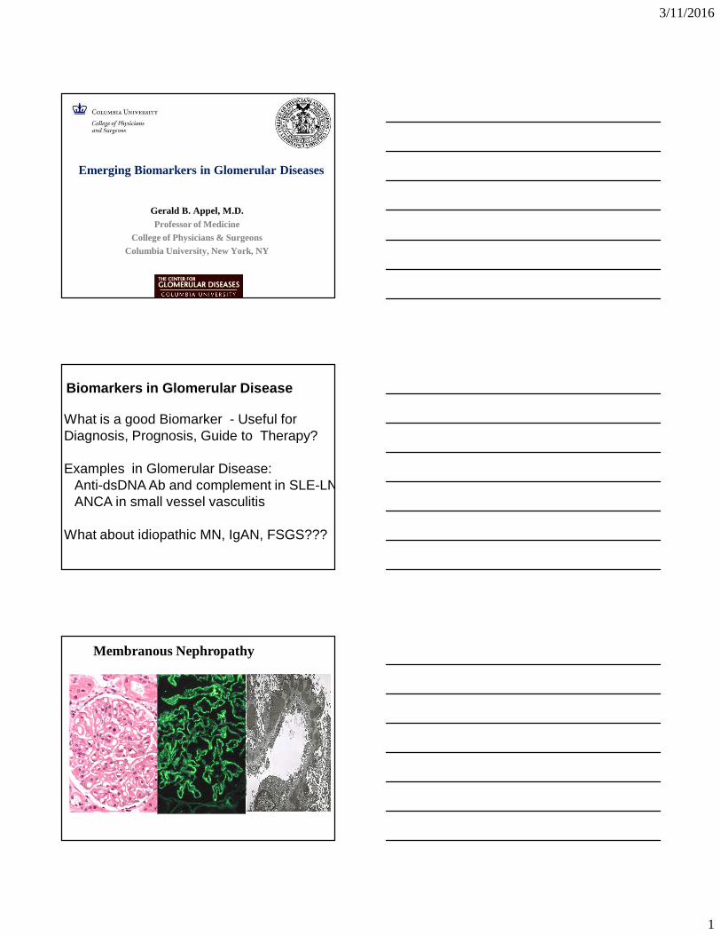
Membranous Nephropathy
3/11/2016
2
Membranous Nephropathy: Overview
• Pathogenesis: subepithelial antigen-antibody immune-complex deposition
− Diffuse granular IgG and complementdeposition along GBM
− Different isotypes depending on etiology ( most common for iMN is igG4 )
− Can also have mesangial immune complex deposition (more typical of secondary forms)
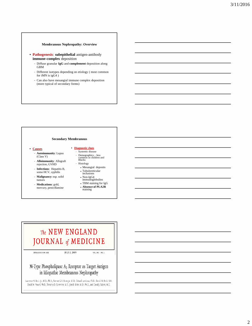
Secondary Membranous
• Causes− Autoimmunity : Lupus
(Class V)
− Alloimmunity : Allograft rejection, GVHD
− Infections: Hepatitis B, some HCV, syphilis
− Malignancy: esp. solid tumors
− Medications: gold, mercury, penicillamine
• Diagnostic clues− Systemic disease− Demographics – less
common in children and Blacks
− Histology
� Mesangial deposits� Tubuloreticular
inclusions� Non-IgG4
immunoglobulins� TBM staining for IgG� Absence of PLA2R
staining
3/11/2016
3
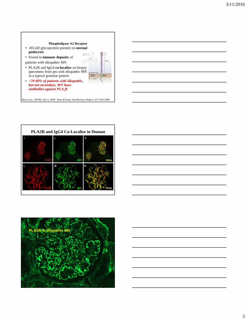
Phospholipase A2 Receptor• 185-kD glycoprotein present on normal
podocytes• Found in immune deposits of
patients with idiopathic MN
• PLA2R and IgG4 co-localizeon biopsy specimens from pts with idiopathic MN in a typical granular pattern
• ~70-80% of patients with idiopathic, but not secondary, MN have antibodies against PLA2R
Beck et al., NEJM, July 2, 2009 Rees & Kain, Nat.Reviews Neph 5, 617-618 2009
IgG4
PLA2R and IgG4 Co-Localize in Human iMGN
Beck et al, NEJM 2009
PLA2R in Idiopathic MN

3/11/2016
4
Genetics independently confirm association of PLA2R with iMN
Stanescu HC et al. New Engl J Med 2011; 364: 616-26
PLA2R
1
HLA-DQA1
GWAS of idiopathic MN
Anti-PLA 2R is sensitive & specific forIdiopathic MN
Ronco, P. & Debiec, H. (2012)Nat. Rev. Nephrol. doi:10.1038/nrneph.2012.35
-OR- Could they have two separate diseases?
Sensitivity ~70%
Specificity ~88%
3/11/2016
5
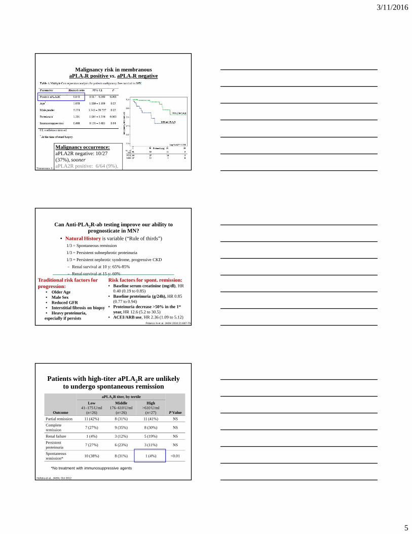
Malignancy risk in membranousaPLA2R positive vs. aPLA2R negative
Timmermans S. et al., AJKD, 62:6, 2013
Malignancy occurrence:aPLA2R negative: 10/27 (37%), sooneraPLA2R positive: 6/64 (9%), later
Can Anti-PLA 2R-ab testing improve our ability to prognosticate in MN?
• Natural History is variable (“Rule of thirds”)1/3 = Spontaneous remission
1/3 = Persistent subnephrotic proteinuria
1/3 = Persistent nephrotic syndrome, progressive CKD
− Renal survival at 10 y: 65%-85%
− Renal survival at 15 y: 60%
Polanco N et al. JASN 2010;21:697-704
Traditional risk factors for progression:
• Older Age• Male Sex• Reduced GFR• Interstitial fibrosis on biopsy• Heavy proteinuria, especially if persists
Risk factors for spont. remission:• Baseline serum creatinine (mg/dl), HR
0.40 (0.19 to 0.85)• Baseline proteinuria (g/24h),HR 0.85
(0.77 to 0.94)• Proteinuria decrease >50% in the 1st
year,HR 12.6 (5.2 to 30.5)• ACEI/ARB use, HR 2.36 (1.09 to 5.12)
Patients with high-titer aPLA2R are unlikely to undergo spontaneous remission
Outcome
aPLA2R titer, by tertile
P Value
Low41–175 U/ml
(n=26)
Middle176–610 U/ml
(n=26)
High>610 U/ml
(n=27)
Partial remission 11 (42%) 8 (31%) 11 (41%) NS
Complete remission
7 (27%) 9 (35%) 8 (30%) NS
Renal failure 1 (4%) 3 (12%) 5 (19%) NS
Persistent proteinuria
7 (27%) 6 (23%) 3 (11%) NS
Spontaneous remission*
10 (38%) 8 (31%) 1 (4%) <0.01
*No treatment with immunosuppressive agents
Hofstra et al., JASN, Oct 2012
3/11/2016
6
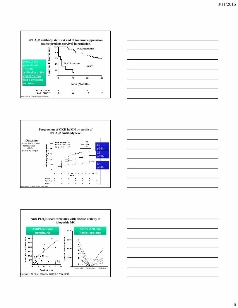
Bech A P et al. CJASN 2014;9:1386-1392
None of the
patients with
+PLA2R
antibodies at the
end of therapy
had a persistent
remission.
aPLA2R antibody status at end of immunosuppression course predicts survival in remission
Hoxha E et al. CJASN 2014;9:1883-1890
Progression of CKD in MN by tertile ofaPLA2R Antibody level
Outcome:≥25% rise in sCreatfrom baseline
-AND-sCreat ≥1.3 mg/dl
1.4 g/24hr
2.5 g/24hr
3.7 g/24hr
Hofstra J M et al. CJASN 2011;6:1286-1291
Anti-PLA 2R level correlates with disease activity in idiopathic MC
AntiPLA2R and proteinuria
AntiPLA2R and Remission status

3/11/2016
7
Disappearance of anti-PLA2Rprecedes that of proteinuria
Beck L H et al. JASN 22:1543-1550, 2011
Anti-PLA2R level Proteinuria
Time-course of anti-PLA2R antibodies and proteinuria
Ronco & Debiec, Nephron Clin Pract, Vol 128, No 3-4, 2014
Suggestions for practical use of anti-PLA 2R
1. If possible, every membranous nephropathy patient should have anti-PLA2R assessed by biopsy and serum
2. In aPLA2R-negative patients, look aggressively for secondary causes
3. For patients aPLA2R positive on biopsy,
− Absence of serum aPLA2R may suggest impending remission
− High-titer serum aPLA2R may suggest low likelihood of remission
4. Assessing aPLA2R at the end of immunosuppressive treatment may be useful in assessing likelihood of maintaining remission
5. Prospective studies using treatment algorithm based on anti-PLA2R level are necessary for proof of concept
3/11/2016
8
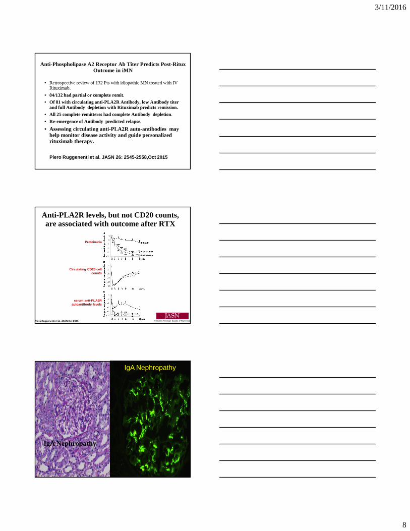
Anti-Phospholipase A2 Receptor Ab Titer Predicts Post-Ritux Outcome in iMN
• Retrospective review of 132 Pts with idiopathic MN treated with IV Rituximab.
• 84/132 had partial or complete remit.
• Of 81 with circulating anti-PLA2R Antibody, low Ant ibody titer and full Antibody depletion with Rituximab predict s remission.
• All 25 complete remitterss had complete Antibody depletion.
• Re-emergence of Antibody predicted relapse.
• Assessing circulating anti-PLA2R auto-antibodies may help monitor disease activity and guide personalized rituximab therapy.
Piero Ruggenenti et al. JASN 26: 2545-2558,Oct 2015
Proteinuria
Circulating CD20 cell counts
serum anti-PLA2R autoantibody levels
Piero Ruggenenti et al. JASN Oct 2015 ©2015 by American Society of Nephrology
Anti-PLA2R levels, but not CD20 counts, are associated with outcome after RTX
IgA NephropathyIgA Nephropathy
IgA Nephropathy
IgA Nephropathy
3/11/2016
9
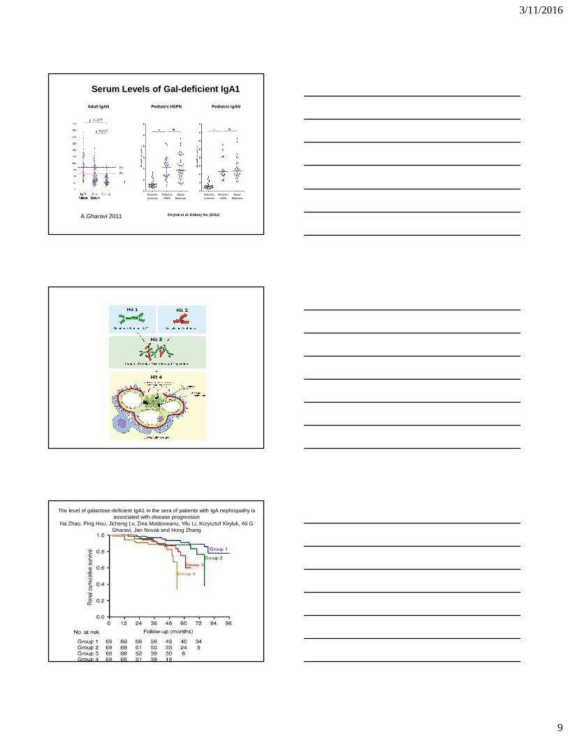
Serum Levels of Gal-deficient IgA1
Adult IgAN
Gharavi et al. JASN (2008)
Blood
Relatives
Pediatric
Controls
Pediatric
HSPN
Pediatric
Controls
Pediatric
IGAN
Kiryluk et al. Kidney Int. (2011)
Pediatric HSPN Pediatric IgAN
Blood
Relatives
A.Gharavi 2011
Multi-hit Pathogenesis Model
The level of galactose-deficient IgA1 in the sera of patients with IgA nephropathy is associated with disease progression
Na Zhao, Ping Hou, Jicheng Lv, Zina Moldoveanu, Yifu Li, Krzysztof Kiryluk, Ali G Gharavi, Jan Novak and Hong Zhang

3/11/2016
10
Kaplan–Meier survival curves without dialysis/death event, with time zero set at diagnosis and elevated serum levels of autoantibodies (IgG >1.33 OD and/or IgA >1.79 U/ml) at diagnosis in IgAN
patients.
Berthoux F et al. JASN 2012;23:1579-1587
©2012 by American Society of Nephrology
GWAS for IgA Nephropathy20,612 individuals
121086420
1 2 3 4 5 6 7 8 9 10 11 12 13 14 15 16 17 18 20 22
Chromosomes
Ob
serv
ed (
-Log
P)
19 21
26242220181614
2830
CFHR3,1-del HORMAD2
DEFA2,3
Multiple signalsHLA-DQ/DR
VAV3CARD9
ITGAM ITGAX
DEFA5,6
TAP1/PSMB8HLA-DP
15 independent risk alleles(10 distinct genomic regions)Disease variance explained:
8% in Asians, 6% in Europeans
Kiryluk et al. Nat Gen (2014)
GWAS and Pathogenesis Model
Defect in allorecognitionMHC alleles
Adaptive Immunity
Tissue inflammation and injury,immune complex clearance
Deletion of CFHR1 and CFHR3ITGAM/ITGAX (CR3 and CR4)
Complement System
Mucosal innate immunity
Dysregulated response to mucosal antigens
TNFSF13, LIF/OSMCARD9, VAV3, DEFA
Magistroni et al. Kid Int 2015 (in press)
3/11/2016
11
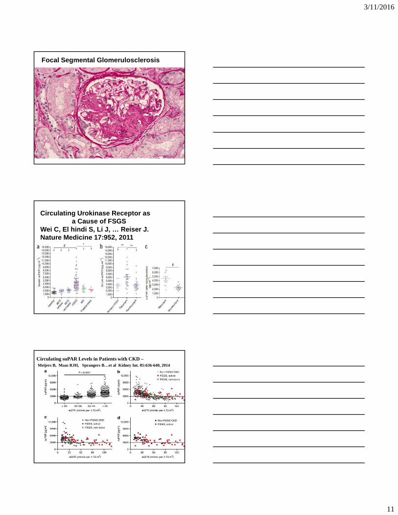
Focal Segmental Glomerulosclerosis
Circulating Urokinase Receptor as a Cause of FSGS
Wei C, El hindi S, Li J, … Reiser J. Nature Medicine 17:952, 2011
Circulating suPAR Levels in Patients with CKD –Meijers B, Maas RJH, Sprangers B…et al Kidney Int. 85:636-640, 2014

3/11/2016
12
suPAR vs GFR suPAR in Primary GN GFR > 60 cc/min/1.73m2
Wada T, Nangaku M, Maruyama S et al.Kidney Int 85:641-646, 2014
Are Serum suPAR Levels by Current ELISA reliable diagnostic biomarkers for
FSGS
1) Elevated suPAR levels occur in ½ children and adults with Neph Synd and other renal disease
2) SuPAR levels correlate inversely with GFR3) suPAR levels correlate with CRP ( inflammation )4) Results were evaluated by same ELISAs as original
results5) Elevated levels do not distinguish primary from
secondary FSGS or other glomerular diseases
Schlondorff D, Kidney Int. 85:499-501,2014.
B7
• B7 is a membrane protein found on activated antigen presenting cells.
• When paired with CD28 or CD152 ( CTLA-4) it produces co-stimulatory or co-inhibitory signals between the APC and the Tcells.
• 2 major B7proteins B7-1 (CD 80) and B7-2 ( CD86 ).

3/11/2016
13
Induction of B7-1 in podocytes is associatedwith Nephrotic Syndrome
Reiser J, von Gersdorff G, Loos M…Mundel P. JCI 113:1390-1397, 2004
Role of B7-1 in podocytes as an inducible modifier of glomerular permselectivity. B7-1 staining is NOT found in normal human podocytes.B7-1 in podocytes was induced in genetic, drug-induced, immune mediated and bacterial toxin induced experimental kidney disease. In human lupus nephritis podocyte expression of B 7-1correlated with the severity of the LN.Mice lacking B7-1 protected form forms of induced nephrotic syndrome.
B7-1 Expression in murine and human podocytes correlates with proteinuriaA) Minimal in normal
mouse – increased in NZB/W and at podocytes.
B) Localizes to glomeruliD-E) Increase with proteinuria in human LNE) Merge with synaptopodin.
Reiser et al JCI 2004
Reiser et al JCI 2004
Induction of B71 in podocytes of nephrin -/- mice vs WT

3/11/2016
14
Original Article: Brief Report
Abatacept in B7-1–Positive Proteinuric Kidney Disease
Chih-Chuan Yu, M.Sc., Alessia Fornoni, M.D., Ph.D., Astrid Weins, M.D., Ph.D., Samy Hakroush, M.D., Dony Maiguel, Ph.D., Junichiro Sageshima, M.D., Linda
Chen, M.D., Gaetano Ciancio, M.D., Mohd. Hafeez Fari di, Ph.D., Daniel Behr, Kirk N. Campbell, M.D., Jer-Ming Chang, M.D., Hung-Chun Che n, M.D., Jun Oh, M.D.,
Christian Faul, Ph.D., M. Amin Arnaout, M.D., Paolo Fiorina, M.D., Ph.D., Vineet Gupta, Ph.D., Anna Greka, M.D., Ph.D., George W. Bu rke, III, M.D., and Peter
Mundel, M.D.
N Engl J Med Volume 369(25):2416-2423 December 19, 2013
Since B 7-1 is induced in podocytes in animal models of proteinuria, and B7-1 immunostaining found in 13/21 human biospies withproteinuric kidney disease including FSGS – concluded B7-1 induced . All transplant recurrent FSGS they found were + for B7-1 podocyte staining. Used abatacept to treat these patients.
Abatacept - a fusion protein of the Fc region of IgG1 fused to the extracellular domain of CTLA-4. Binds to CD80 ( B7-1 ) and CD86 ( B 7-2 ) and prevents second signalof T cell activation.

3/11/2016
15
Abatacept in B7-1 Positive Proteinuric Kidney Disease
• Abatacept ( CTLA4-Ig) ,the co-stimulatory inhibitor targeting B7-1 ( CD 80), to treat FSGS.
• Report of 5 patients ( 4 rituximab resistant recurrent FSGS in the allograft - 2 lost prior kidney txplants due to recurrent FSGS - and one glucocorticoid resistant FSGS in native kidney ) all proteinuria, all +B7-1 immunostaining of podocytes.
• Elegant studies showing role of B7-1 in disrupting beta 1 integrin activation in pododcytes
• Abatacept induced partial or complete remission in all 5 patients “suggesting B7-1 may be a useful biomarker for treatment of some glomerulopathies.”
Yu C-C, Fornoni A, Weins A…Mudel P et al NEJM Nov 8,2013.
Abatacept Therapy for FSGS and GN???
Positive B7-1 staining of Bxs in other glomerular diseases. 3/5 MCD, 1/5 secondary FSGS, 3/3 Lupus GN, strongest staining in MN (PLA2R + or negative )Urinary CD 80 ( B7-1) is a marker of MCD not of
FSGS.Abatacept has failed a number of trials of Lupus
Nephritis a disease where B7-1 (CD80) staining is consistently positive.
FSGS patients with failed results now being reported.
Negative Staining of FSGS Biopsies for B7-1Benigni A… Remuzzi G. NEJM 370:1259 March 27,2014

3/11/2016
16
Clinical Features and Response to B7-1 Blocker Ther apy in FSGS
Alachar N, Carter-Monroe N, Reiser J.N Engl J Med 2014;370:1261-1266.
Figure 1. Immunohistochemical detection of B7-1 in a biopsy specimen from a patient with recurrent nephrotic syndrome after transplantation. (A) Interstitial activated B cells and monocytes show positive B7-1 staining. (B) Interstitial inflammatory cells show strong internal positive control staining for B7-1 while glomeruli are completely negative.
LarsenCP, MessiasNC, Walker PD. American Journal of Kidney Diseases, Volume 64, Issue 6, 2014, 1001–1003, 2014.
B7-1 Immunostaining in Proteinuric Kidney Disease
60 Bxs by IF and immunoperoxidase – 38 FSGS, 19MCD, 3MN
No Glomerular Staining
B7-1Blocakade Does Not Improve Post-Transplant Nephrotic Syndrome Caused by recurrent FSGS
Prospectively treated 9 patients with recurrent FSGS after transplant using either abatacept or belatacept ( a B7-1 blocker with higher affinity,) and did not produce proteinuria remission. Did NOT dectectB7-1 expression by IF in podocytes of biopsy specimens from these or other kidney grafts or podocytes of native kindey biopsies specimens. In Conclusion, B7-1 blockade did not induce FSGS remission after transplantation in our study.
Delville M, Baye E, Durrbach A et al. JASN 2015-6
3/11/2016
17
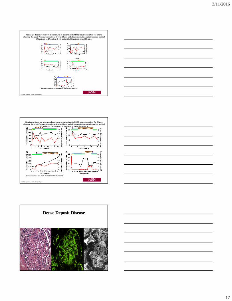
Abatacept does not improve albuminuria in patients with FSGS recurrence after Tx. Charts showing the post–Tx serum creatinine levels (black) and albuminuria-to-creatinine ratios (red) of
(A) patient 1, (B) patient 2, (C) patient 3, (D) pa tient 4, and (E) pa...
Marianne Delville et al. JASN doi:10.1681/ASN.20150 91002
©2015 by American Society of Nephrology
Belatacept does not improve albuminuria in patients with FSGS recurrence after Tx. Charts showing the post–Tx serum creatinine levels (black) and albuminuria-to-creatinine ratios (red) of
(A) patient 6, (B) patient 7, (C) patient 8, and (D ) patient 9.
Marianne Delville et al. JASN doi:10.1681/ASN.20150 91002
©2015 by American Society of Nephrology
Dense Deposit DiseaseDense Deposit Disease
IF C3 EM GBMLM - MPGN
3/11/2016
18
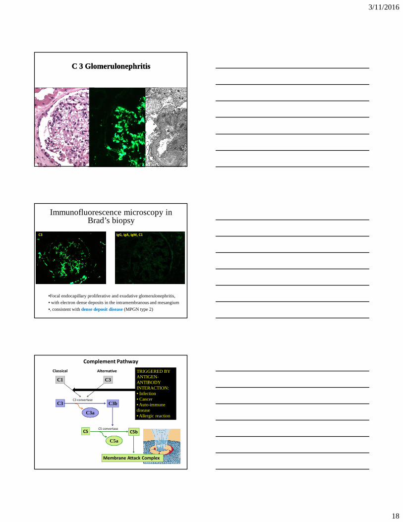
C 3 GlomerulonephritisC 3 Glomerulonephritis
IF C3 EM GBMLM
C3
Immunofluorescence microscopy in Brad’s biopsy
IgG, IgA, IgM, C1
•Focal endocapillary proliferative and exudative glomerulonephritis,
• with electron dense deposits in the intramembranous and mesangium
•, consistent with dense deposit disease (MPGN type 2)
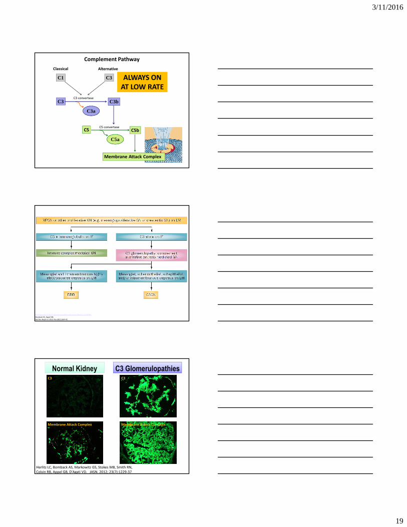
Classical Alternative
C1 C3
Complement Pathway
C3 C3bC3 convertase
C3a
C5 C5bC5 convertase
C5a
Membrane Attack Complex
TRIGGERED BY ANTIGEN-ANTIBODY INTERACTION:• Infection• Cancer• Auto-immune disease• Allergic reaction
3/11/2016
19
Classical Alternative
C1 C3
Complement Pathway
C3 C3bC3 convertase
C3a
C5 C5bC5 convertase
C5a
Membrane Attack Complex
ALWAYS ON
AT LOW RATE
Pathogenesis of the C3 glomerulopathies and reclassification of MPGN.
Bomback AS, Appel GB.
Nat Rev Nephrol. 2012 Nov;8(11):634-42.
Normal KidneyNormal Kidney C3 GlomerulopathiesC3 GlomerulopathiesC3 C3
Membrane Attack Complex Membrane Attack Complex
Herlitz LC, Bomback AS, Markowitz GS, Stokes MB, Smith RN,
Colvin RB, Appel GB, D'Agati VD. JASN. 2012; 23(7):1229-37

3/11/2016
20
C3
C3b
C3a
C5
C5b
C5b-9MEMBRANE ATTACK
COMPLEX
C3 convertase(C3bBb)
ALTERNATIVE PATHWAY
C5 convertase(C3bBbC3b)
C5a
C6 C7 C8 C9
TERMINAL COMPLEMENT CASCADE
Factor HFactor IMCPFactor H-related proteins
Factor BFactor D
Nat Rev Nephrol. 2012 Nov;8(11):634-42.
Pathogenesis of the C3 glomerulopathies and reclassification of MPGN.
Bomback AS, Appel GB.
Principal regulatory/inhibitory proteins of the alternative pathway:1. Complement factor H (CFH)2. Complement factor I (CFI)3. Membrane cofactor protein
(MCP)4. Factor H-related proteins
Patient Brad: Management
• Given minimal proteinuria and no specific therapies for C3 glomerulopathies in 2008, he was treated conservatively with losartan 25 mg daily
Creatinin
e
Proteinuria C3 C4
2008 1.8 230 ↓ nl
2009 1.9 500 ↓ nl
2010 2.2 1000 ↓ nl
C3
2008 Biopsy:
• 0% glomerular scarring
• 0% interstitial scarring
2010 Biopsy
• 22% glomerular scarring
• 15% interstitial scarring

3/11/2016
21
CUMC Eculizumab Pilot Study
ID Native/
Transplant
Age Race Sex Months from
diagnosis
Previous
immunosuppression
DDD1 Native 22 W M 25 None
DDD2 Native 32 W M 332 Steroids
DDD3 Transplant 42 W M150 (native)
0.5 (graft)
Steroids (native);
tacrolimus and
mycophenolate (graft)
C3GN1 Native 25 W M 162Steroids and
mycophenolate
C3GN2 Transplant 22 W M138 (native)
8 (graft)
Steroids (native);
steroids, tacrolimus,
mycophenolate (graft)
C3GN3 Transplant 20 W M114 (native)
2 (graft)
Tacrolimus,
mycophenolate,
rituximab (native);
steroids, tacrolimus,
mycophenolate (graft)
Eculizumab for Dense Deposit Disease and C3 Glomerulonephritis.
Bomback AS, Smith RJ, Barile GR, Zhang Y, Heher EC, Herlitz L, Stokes MB, Markowitz GS, D'Agati VD, Canetta PA, Radhakrishnan J, Appel GB.
Clin J Am Soc Nephrol. 2012 May;7(5):748-756.
CUMC Eculizumab Pilot Study
ID CFH
Mutation
CFI
Mutation
MCP
Mutation
C3 Nephritic
Factor
Factor H
Autoantibodies
Membrane
Attack
Complex
DDD1 Yes No No Negative Negative Elevated
DDD2 No No No Positive Negative Normal
DDD3 No No No Negative Negative --*
C3GN1 No No No Negative Negative Normal
C3GN2 No No No Positive Negative Elevated
C3GN3 No No Yes Positive Negative Elevated
* Pre-treatment sample for sMAC testing was inadequate in this subject; sMAC level at week 4 was normal
Eculizumab for Dense Deposit Disease and C3 Glomerulonephritis.
Bomback AS, Smith RJ, Barile GR, Zhang Y, Heher EC, Herlitz L, Stokes MB, Markowitz GS, D'Agati VD, Canetta PA, Radhakrishnan J, Appel GB.
Clin J Am Soc Nephrol. 2012;7(5):748-756.
ID Treatment
Time
(weeks)
Creatinine Proteinuria Post-treatment
Kidney Biopsy
DDD1 52 ↓ ↓ Less
inflammation
DDD2 40 Worsened Worsened Not performed
DDD3 52 Stable ↓ Less
inflammation
C3GN1 52 Worsened Stable Increased scarring
C3GN2 52 Stable ↓ Less
inflammation
C3GN3 52 ↓ Stable Less
inflammation
Eculizumab for Dense Deposit Disease and C3 Glomerulonephritis.
Bomback AS, Smith RJ, Barile GR, Zhang Y, Heher EC, Herlitz L, Stokes MB, Markowitz GS, D'Agati VD, Canetta PA, Radhakrishnan J, Appel GB.
Clin J Am Soc Nephrol. 2012;7(5):748-756.
CUMC Eculizumab Pilot Study

3/11/2016
22
Resolution of endocapillary proliferation and exudative features
Partial resorption of
electron dense deposits
Post-study follow-up for Brad (DDD1)
Creatinine (mg/dl) Soluble MAC level (nl <0.30 mg/L)
Pre-eculizumab 2.2 1.08
Study completion (1 year) 1.4 0.01
3 months post-study 1.5 1.26
Creatinine (mg/dl) Soluble MAC level (nl <0.30 mg/L)
1 month into 2nd course of
eculizumab
1.3 0.01
1 year into 2nd course of
eculizumab
1.2 0.01
RESTART ECULIZUMAB THERAPY
*** JUST SIGNED A CONTRACT WITH NATIONAL HOCKEY LEAGUE FRANCHISE ***
MMF in C3 GlomerulonephritisRabasco C, Praga M GLOSEN ASN Abstract 2014
• Multicenter, 60 Pts – 57% M mean age 37yo
• Presentation- 50% nephrotic, 33% nephritic, 17% asymptomatic urinary abnrormalities
• 40/60 pts received immune modulatory meds (55% CS + MMF, 22% CS + other immune meds, 22% only CS) -Follow 48 months.
• Treated pts higher response ( 70% vs 30 %), and Less ESRD ( 7% vs 35% )
3/11/2016
23
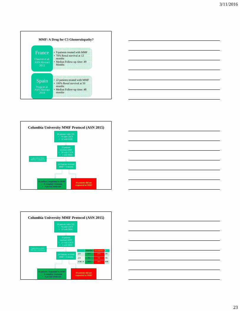
MMF: A Drug for C3 Glomerulopathy?
• 9 patients treated with MMF• 78% Renal survival at 12
months• Median Follow-up time: 49
Months
FranceChauvet et al. ASN Abstract
2012
• 22 patients treated with MMF• 100% Renal survival at 50
months• Median Follow-up time: 48
months
SpainPraga et al.
ASN Abstract 2014
98 patients with C3G• 76 with C3GN• 22 with DDD
25 patients received MMF
• 24 with C3GN• 1 with DDD
24 Patients received MMF > 6 months
14 patients responded to MMF• 8 complete remission• 6 partial remission
1 patient did not tolerate MMF due to lymphopenia
Columbia University MMF Protocol (ASN 2015)
10 patients did not responded to MMF
Responders Non-responders p
↓C3 82% 57% NS
↓C4 9% 0% NS
↑C5b – 9 100% 25% 0.02
3/11/2016
24
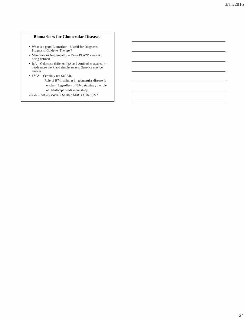
Biomarkers for Glomerular Diseases
• What is a good Biomarker - Useful for Diagnosis, Prognosis, Guide to Therapy?
• Membranous Nephropathy – Yes – PLA2R - role is being defined.
• IgA – Galactose deficient IgA and Antibodies against it -needs more work and simple assays. Genetics may be answer.
• FSGS – Certainly not SuPAR.
Role of B7-1 staining in glomerular disease is
unclear. Regardless of B7-1 staining , the role
of Abatacept needs more study.
C3GN – not C3 levels, ? Soluble MAC ( C5b-9 )???