Emergency Department Thoracotomy (EDT) Clinical Guideline ... · Emergency Department . Medical :...
12
Emergency Department Thoracotomy (EDT) Clinical Guideline Version : 2 Approved by: Service 4 Safety & Quality Committee 03/2018 Date Compiled: 04/2015 Endorsed by: Clinical Practice Committee 04/2018 Document Owner: Trauma Programme Manager Authorised by: Policy Committee 19/04/2018 Revision Due: 04/2021 Page 1 of 12 Emergency Department Thoracotomy (EDT) Clinical Guideline Scope Site Department, Service or Operational Area Applicable to RPH only Trauma Emergency Department Medical Key Alerts The EDT should only be done by experienced clinicians, trained in this procedure. The procedure is always performed in a high-stress environment, with considerable risk to both patient and staff; due care and diligence is required to minimise these risk. Standard precautions to avoid sharps injuries and body fluid exposures should be followed. Related Policy, RPBG Practice Standard, Clinical Guidelines • Emergency Department Guideline: Role of the Nurse Assisting with thoracotomy and Internal Defibrillation • Trauma Service Guideline: Penetrating Abdominal, Thoracoabdominal and Chest Trauma Management • Trauma Service Guideline: Resuscitative Endovascular Balloon Occlusion of the Aorta (REBOA) Related National Standards NSQHS Standard 9: Recognising and Responding to Clinical Deterioration in Acute Health Care Preamble Emergency Department Thoracotomy (EDT) is an advanced resuscitative manoeuvre, which may assist resuscitation of the trauma patient, in physiological extremis. Recent maturation of trauma systems have resulted in increased numbers of such sick patients arriving at hospital; historically these patients succumb at the scene or during transport. Most recently, REBOA (Resuscitative Endovascular Balloon Occlusion of the Aorta) has become a feasible alternative in select patients, where the goal of the EDT has been to clamp the descending thoracic aorta, for the control of non-compressible torso haemorrhage.
Transcript of Emergency Department Thoracotomy (EDT) Clinical Guideline ... · Emergency Department . Medical :...

Emergency Department Thoracotomy (EDT) Clinical Guideline
Version : 2
Approved by: Service 4 Safety & Quality Committee 03/2018 Date Compiled: 04/2015
Endorsed by: Clinical Practice Committee 04/2018 Document Owner: Trauma Programme Manager
Authorised by: Policy Committee 19/04/2018 Revision Due: 04/2021 Page 1 of 12
Emergency Department Thoracotomy (EDT) Clinical Guideline Scope
Site Department, Service or Operational Area
Applicable to
RPH only Trauma
Emergency Department Medical
Key Alerts
The EDT should only be done by experienced clinicians, trained in this procedure. The procedure is always performed in a high-stress environment, with considerable risk to both patient and staff; due care and diligence is required to minimise these risk. Standard precautions to avoid sharps injuries and body fluid exposures should be followed.
Related Policy, RPBG Practice Standard, Clinical Guidelines • Emergency Department Guideline: Role of the Nurse Assisting with thoracotomy and
Internal Defibrillation • Trauma Service Guideline: Penetrating Abdominal, Thoracoabdominal and Chest
Trauma Management • Trauma Service Guideline: Resuscitative Endovascular Balloon Occlusion of the Aorta
(REBOA)
Related National Standards NSQHS Standard 9: Recognising and Responding to Clinical Deterioration in Acute Health Care
Preamble Emergency Department Thoracotomy (EDT) is an advanced resuscitative manoeuvre, which may assist resuscitation of the trauma patient, in physiological extremis. Recent maturation of trauma systems have resulted in increased numbers of such sick patients arriving at hospital; historically these patients succumb at the scene or during transport. Most recently, REBOA (Resuscitative Endovascular Balloon Occlusion of the Aorta) has become a feasible alternative in select patients, where the goal of the EDT has been to clamp the descending thoracic aorta, for the control of non-compressible torso haemorrhage.
Emergency Department Thoracotomy (EDT) Clinical Guideline
Version : 2
Approved by: Service 4 Safety & Quality Committee 03/2018 Date Compiled: 04/2015
Endorsed by Clinical Practice Committee 04/2018 Document Owner: Trauma Programme Manager
Authorised by: Policy Committee 19/04/2018 Revision Due: 04/2021 Page 2 of 12
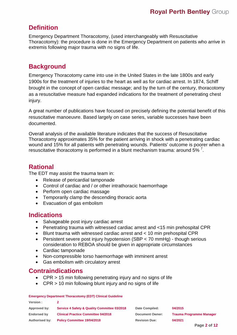
Definition Emergency Department Thoracotomy, (used interchangeably with Resuscitative Thoracotomy): the procedure is done in the Emergency Department on patients who arrive in extremis following major trauma with no signs of life.
Background Emergency Thoracotomy came into use in the United States in the late 1800s and early 1900s for the treatment of injuries to the heart as well as for cardiac arrest. In 1874, Schiff brought in the concept of open cardiac message; and by the turn of the century, thoracotomy as a resuscitative measure had expanded indications for the treatment of penetrating chest injury.
A great number of publications have focused on precisely defining the potential benefit of this resuscitative manoeuvre. Based largely on case series, variable successes have been documented.
Overall analysis of the available literature indicates that the success of Resuscitative Thoracotomy approximates 35% for the patient arriving in shock with a penetrating cardiac wound and 15% for all patients with penetrating wounds. Patients’ outcome is poorer when a resuscitative thoracotomy is performed in a blunt mechanism trauma: around 5% 7.
Rational The EDT may assist the trauma team in:
• Release of pericardial tamponade • Control of cardiac and / or other intrathoracic haemorrhage • Perform open cardiac massage • Temporarily clamp the descending thoracic aorta • Evacuation of gas embolism
Indications • Salvageable post injury cardiac arrest • Penetrating trauma with witnessed cardiac arrest and <15 min prehospital CPR • Blunt trauma with witnessed cardiac arrest and < 10 min prehospital CPR • Persistent severe post injury hypotension (SBP < 70 mmHg) - though serious
consideration to REBOA should be given in appropriate circumstances • Cardiac tamponade • Non-compressible torso haemorrhage with imminent arrest • Gas embolism with circulatory arrest
Contraindications • CPR > 15 min following penetrating injury and no signs of life • CPR > 10 min following blunt injury and no signs of life

Emergency Department Thoracotomy (EDT) Clinical Guideline
Version : 2
Approved by: Service 4 Safety & Quality Committee 03/2018 Date Compiled: 04/2015
Endorsed by Clinical Practice Committee 04/2018 Document Owner: Trauma Programme Manager
Authorised by: Policy Committee 19/04/2018 Revision Due: 04/2021 Page 3 of 12
Algorithm

Emergency Department Thoracotomy (EDT) Clinical Guideline
Version : 2
Approved by: Service 4 Safety & Quality Committee 03/2018 Date Compiled: 04/2015
Endorsed by Clinical Practice Committee 04/2018 Document Owner: Trauma Programme Manager
Authorised by: Policy Committee 19/04/2018 Revision Due: 04/2021 Page 4 of 12
Set up in the Trauma Resuscitation Bay The red drawered rapid intervention trolley contains a starter thoracotomy kit and a disposable scalpel. The internal defibrillation paddles are kept in the bottom drawer of the white drawered emergency resuscitation trolley. A second starter thoracotomy kit, the major thoracotomy tray and sutures are kept on the open trolley at the end of the trauma resuscitation bay. See the photograph below.
Draping packs and sterile gloves are located on the shelf in the back alcove.
An additional procedure trolley is also available at the entry to the trauma resuscitation area.
Emergency Resuscitation Trolley Rapid Intervention Trolley
Location of the Thoracotomy Starter Kit
Location of the spare Thoracotomy Starter Kit, and the Major
Thoracotomy tray and sutures

Emergency Department Thoracotomy (EDT) Clinical Guideline
Version : 2
Approved by: Service 4 Safety & Quality Committee 03/2018 Date Compiled: 04/2015
Endorsed by Clinical Practice Committee 04/2018 Document Owner: Trauma Programme Manager
Authorised by: Policy Committee 19/04/2018 Revision Due: 04/2021 Page 5 of 12
Equipment Contents of the STARTER THORACOTOMY KIT: (2 of these trays are stored in ED Resus)
NB: a disposable preloaded scalpel is taped to the Starter Thoracotomy Kit to negate the need to load the scalpel blade in a hurry.
Contents of the MAJOR THORACTOMY TRAY: (1 of these trays is stored in ED)

Emergency Department Thoracotomy (EDT) Clinical Guideline
Version : 2
Approved by: Service 4 Safety & Quality Committee 03/2018 Date Compiled: 04/2015
Endorsed by Clinical Practice Committee 04/2018 Document Owner: Trauma Programme Manager
Authorised by: Policy Committee 19/04/2018 Revision Due: 04/2021 Page 6 of 12
Technical Considerations The EDT should only be done by experienced clinicians, trained in this procedure. The procedure is always performed in a high-stress environment, with considerable risk to both patient and staff; due care and diligence is required to minimise these risk. Standard precautions to avoid sharps injuries and body fluid exposures should be followed.
Adequate instrumentation (as described above), lighting, personnel, and an appropriate disposition plan (usually theatre), should all be available.
The following description is a guide and not a substitute for surgical technical training. It is intended as a guide only, and should be supplemented with current literature, operative texts, and experience from clinical surgical training. This guide is also not a complete description of technical variations and manoeuvres available to the experienced practitioner.
Usually, a left anterolateral thoracotomy is performed. Where required, for example in cases of cardiac injuries or bilateral thoracic injuries, this may be extended across the sternum, and into a clamshell thoracotomy. (Rarely, the surgeon may commence with a right anterolateral thoracotomy – usually in the case of bleeding in the right chest, from penetrating injury on that side).
The initial thoracic incision is performed through approximately the 5th intercostal space. The incision should start to the right of the sternum and be curve to below the left axilla. In the case of a clamshell thoracotomy, where the sternum is transected, the internal thoracic arteries will be divided, and will require ligation after the circulation is restored. The rib retractor should have the bar placed posteriorly, such that it does not obscure the view medially (and interfere, with clamshell extension of the anterolateral thoracotomy).

Emergency Department Thoracotomy (EDT) Clinical Guideline
Version : 2
Approved by: Service 4 Safety & Quality Committee 03/2018 Date Compiled: 04/2015
Endorsed by Clinical Practice Committee 04/2018 Document Owner: Trauma Programme Manager
Authorised by: Policy Committee 19/04/2018 Revision Due: 04/2021 Page 7 of 12
Pericardiotomy and cardiac haemorrhage control: The pericardium is incised widely, in a north-south direction, a safe distance anterior to the phrenic nerve. If a cardiac injury is diagnosed, a clamshell extension is almost essential. The pericardial clot can be evacuated, and the injury to the heart controlled by simple light digital compression on the epicardium at the injury. Definitive repair may be considered using primary suturing, pledgetted sutures, staples, and haemostatic materials, among other techniques. A Foley catheter may also be useful to occlude the injury. Care should be exercised not to incorporate the coronary vessels into any repair.

Emergency Department Thoracotomy (EDT) Clinical Guideline
Version : 2
Approved by: Service 4 Safety & Quality Committee 03/2018 Date Compiled: 04/2015
Endorsed by Clinical Practice Committee 04/2018 Document Owner: Trauma Programme Manager
Authorised by: Policy Committee 19/04/2018 Revision Due: 04/2021 Page 8 of 12
Thoracic aorta occlusion and pulmonary hilar control: Following thoracotomy and pericardiotomy with evaluation of the heart the descending thoracic aorta should be occluded to maximize coronary perfusion if hypotension persists. The patient will at this stage usually be ventilated using a single lumen endotracheal tube. The lung may need to be compressed and adequate exposure created by elevating the left lung both anteriorly and superiorly. To isolate the hilum, the inferior pulmonary ligament will need to be divided, with care taken not to injury the pulmonary vein found at the inferior aspect of the hilum. The descending thoracic aorta is encountered in its left, paravertebral, position, though unlike in the elective surgical setting, it will often be collapsed, and not under its normal pressure. The parietal pleura should to be divided on either side of the aorta, prior to clamping; the clamp will slide off if the parietal pleura is not incised. Care should be taken to avoid the oesophagus found medial to the aorta in this location; a nasogastric tube is helpful for this.

Emergency Department Thoracotomy (EDT) Clinical Guideline
Version : 2
Approved by: Service 4 Safety & Quality Committee 03/2018 Date Compiled: 04/2015
Endorsed by Clinical Practice Committee 04/2018 Document Owner: Trauma Programme Manager
Authorised by: Policy Committee 19/04/2018 Revision Due: 04/2021 Page 9 of 12
If vascular control of the lung is required, a clamp may be placed across the hilum. Alternatively, a hilar twist may be helpful.

Emergency Department Thoracotomy (EDT) Clinical Guideline
Version : 2
Approved by: Service 4 Safety & Quality Committee 03/2018 Date Compiled: 04/2015
Endorsed by Clinical Practice Committee 04/2018 Document Owner: Trauma Programme Manager
Authorised by: Policy Committee 19/04/2018 Revision Due: 04/2021 Page 10 of 12
Internal cardiac massage: In situations of circulatory arrest e.g. asystole or pulseless electrical activity (PEA), internal cardiac massage may assist the reanimation of the circulation. A two handed technique is strongly recommended, with particular attention to avoid the fingertips “digging in” to the myocardium and resulting in perforation of the heart. The two hands function in a hinged clapping motion, with the wrists apposed, sequentially closing from the palms to the fingers. The ventricular compression should proceed from cardiac apex to the base of the heart.

Emergency Department Thoracotomy (EDT) Clinical Guideline
Version : 2
Approved by: Service 4 Safety & Quality Committee 03/2018 Date Compiled: 04/2015
Endorsed by Clinical Practice Committee 04/2018 Document Owner: Trauma Programme Manager
Authorised by: Policy Committee 19/04/2018 Revision Due: 04/2021 Page 11 of 12
When necessary, the internal defibrillation paddles may be used for defibrillation / cardioversion.

Emergency Department Thoracotomy (EDT) Clinical Guideline
Version : 2
Approved by: Service 4 Safety & Quality Committee 03/2018 Date Compiled: 04/2015
Endorsed by Clinical Practice Committee 04/2018 Document Owner: Trauma Programme Manager
Authorised by: Policy Committee 19/04/2018 Revision Due: 04/2021 Page 12 of 12
Document Version Control Date Version (Author) Amendments
April 2015 v.1 D. Weber Original document
Mar 2018 v.2 D. Weber/M. Burrell Review and Conversion to new guideline template
Authors / Acknowledgements We acknowledge the following previous site endorsed work and/or contributors used to compile this document.
Dr Dieter Weber, Trauma Surgeon, RPH
Review Authors RPH Trauma Committee, comprising representatives from Trauma, Anaesthesia, Emergency Department, Orthopaedics, Neurosurgery, Intensive Care, Nursing, Imaging, Theatres. Kandy Connell, A/CNS, Emergency Department, RPH
Karen White, A/CNS, Emergency Department, RPH
Dr Dieter Weber, Trauma Surgeon, RPH
Dr David McCutcheon, Consultant, Emergency Department, RPH
References 1. Burlew CC, Moore EE, Moore FA, et al. (2012) Western Trauma Association critical decisions in
trauma: resuscitative thoracotomy. J Trauma. 2012; 73: 1359. 2. Burlew CC & Moore EE. (2013) Emergency Department Thoracotomy. In: Mattox KL, Moore EE,
Feliciano DV. Trauma. 7th Ed. 2013. McGraw Hill: New York. 3. Boffard K. Manual of Definitive Surgical Trauma Care. (2007) 2nd Ed. 2007. Hodder Arnold: London. 4. Soreide K, Petrone P, Asensio JA. (2007) Emergency thoracotomy in trauma: rationale, risks and
realities. Scand J Surg. 2007; 96: 4. 5. Powell DW, Moore EE, Cothren CC, et al. (2004) Is emergency department resuscitative thoracotomy
futile care of the critically injured patient requiring pre-hospital cardiopulmonary resuscitation. J Am Coll Surg. 2004; 199: 211.Moore EE, Knudson
6. MM, Burlew CC, et al. (2011) Defining the limits of resuscitative emergency department thoracotomy: a contemporary Western Trauma Association perspective. J Trauma. 2011; 70: 334.
7. Seamon et al. (2015) An evidence-based approach to patient selection for emergency department thoracotomy: A practice management guideline from the Eastern Association for the Surgery of Trauma. J Trauma Acute Care Surg. 2015 Jul;79(1):159-73