Embryology of the Central Nervous System - Springer · 2 Embryology of the Central Nervous System A...
14
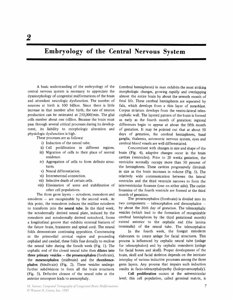
2 Embryology of the Central Nervous System A basic understanding of the embryology of the central nervous system is necessary to appreciate the dysmorphology of congenital malformations of the brain and attendant neurologic dysfunction. The number of neurons at birth is 100 billion. Since there is little increase in that number after birth, the rate of neuron production can be estimated at 2S0,000/min. The glial cells number about one trillion. Because the brain must pass through several critical processes during its develop- ment, its liability to morphologic alteration and physiologic dysfunction is high. These processes are as follows: i) Induction of the neural tube. ii) Cell proliferation in different regions. iii) Migration of cells to their place of normal residence. iv) Aggregation of cells to form definite struc- tures. v) Neural differentiation. vi) Interneuronal connection. vii) Selective death of certain cells. viii) Elimination of some and stabilization of other cell populations. The three germ layers - ectoderm, mesoderm and entoderm - are recognizable by the second week. At this point, the mesoderm induces the midline ectoderm to transform into the neural tube. In the third week, the ectodermally derived neural plate, induced by the mesoderm and ectodermally derived notochord, forms a longitudinal groove that exhibits external features of the future brain, brainstem and spinal cord. The neural folds demonstrate continuing apposition. Commencing in the primordial cervical region and proceeding cephalad and caudad, these folds fuse dorsally to enclose the neural tube during the foruth week (Fig. I). The cephalic end of the closed neural tube then dilates into three primary vesicles - the prosencephalon (forebrain), the mesencephalon (midbrain) and the rhombence- phalon (hindbrain) (Fig. 2). These vesicles undergo further subdivisions to form all the brain structures (Fig. 3). Defective closure of the neural tube at the anterior neuropore leads to anencephaly. (cerebral hemispheres) in man exhibits the most striking morphologic changes, growing rapidly and overlapping almost the entire brain by about the seventh month of fetal life. These cerebral hemispheres are separated by falx, which develops from a thin layer of mesoblast. Corpus striatum develops from the ventro-Iateral telen- cephalic wall. The layered pattern of the brain is formed as early as the fourth month of gestation; regional differences begin to appear at about the fifth month of gestation. It may be pointed out that at about 50 days of gestation, the cerebral hemispheres, basal ganglia, thalamus, autonomic nervous system, eyes and cerebral blood vessels are well-differentiated. Concomitant with changes in size and shape of the brain (Fig. 4), adaptive changes occur in the brain cavities (ventricles). Prior to 20 weeks gestation, the ventricles normally occupy more than 50 percent of the hemispheres. These cavities progressively diminish in size as the brain increases in volume (Fig. 5). The relatively wide communication between the lateral ventricles and the third ventricle narrows to form the interventricular foramen (one on either side). The outlet foramina of the fourth ventricle are formed at the third month of gestation. The prosencephalon (forebrain) is divided into its two components - telencephalon and diencephalon - by about the 36th day of gestation. The telencephalic vesicles (which lead to the formation of recognizable cerebral hemispheres by the third gestational month) extend anterior to the cephalic end (the lamina terminalis) of the neural tube. The telencephalon In the fourth week, the foregut entoderm elaborates to create anlage for facial structures. This process is influenced by cephalic neural tube (anlage for telencephalon) and by cephalic mesoderm (anlage for facial bones and skull). Proper development of the brain, skull and facial skeleton depends on the intricate interplay of various inductive processes among the three germ layers. Any process that impairs such induction results in facio-telencephalopathy (holoprosencephaly). Cell proliferation occurs at the subventricular level; this cell population, called germinal matrix, is 7 M. Sarwar, Computed Tomography of Congenital Brain Malformations © Warren H. Green, Inc. 1985
Transcript of Embryology of the Central Nervous System - Springer · 2 Embryology of the Central Nervous System A...
2
Embryology of the Central Nervous System
A basic understanding of the embryology of the central nervous system is necessary to appreciate the dysmorphology of congenital malformations of the brain and attendant neurologic dysfunction. The number of neurons at birth is 100 billion. Since there is little increase in that number after birth, the rate of neuron production can be estimated at 2S0,000/min. The glial cells number about one trillion. Because the brain must pass through several critical processes during its development, its liability to morphologic alteration and physiologic dysfunction is high.
These processes are as follows: i) Induction of the neural tube.
ii) Cell proliferation in different regions. iii) Migration of cells to their place of normal
residence. iv) Aggregation of cells to form definite struc-
tures. v) Neural differentiation.
vi) Interneuronal connection. vii) Selective death of certain cells.
viii) Elimination of some and stabilization of other cell populations.
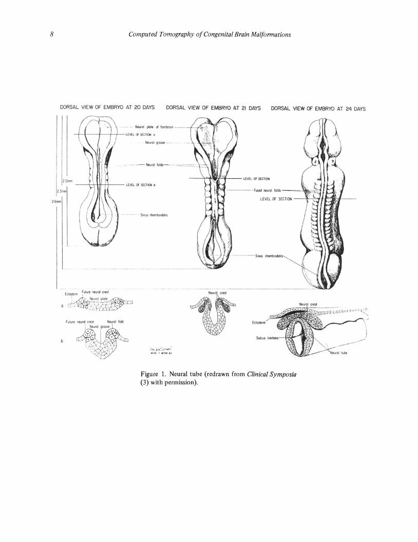
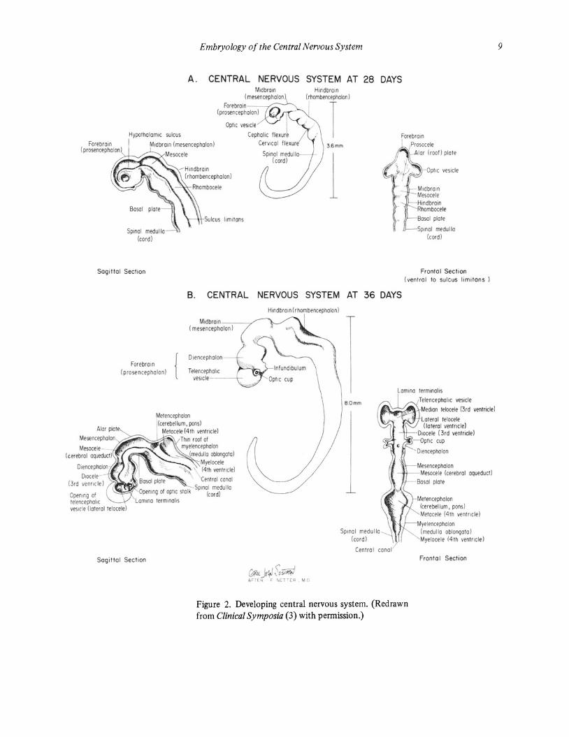
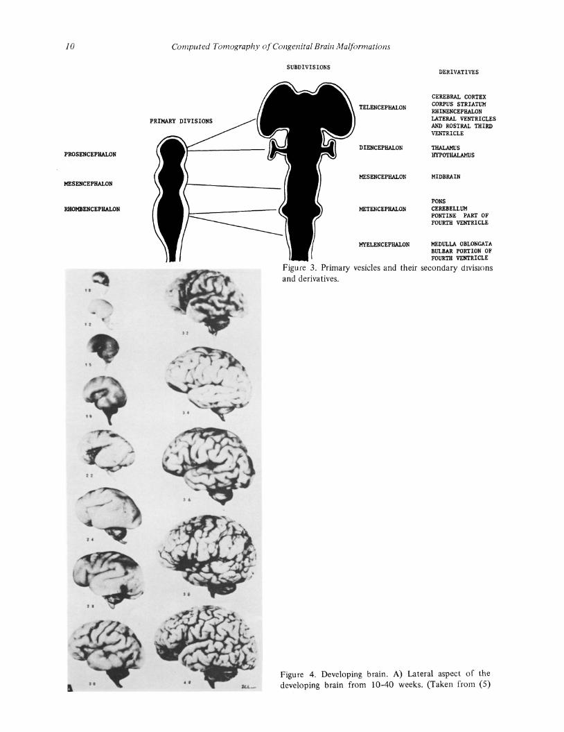
The three germ layers - ectoderm, mesoderm and entoderm - are recognizable by the second week. At this point, the mesoderm induces the midline ectoderm to transform into the neural tube. In the third week, the ectodermally derived neural plate, induced by the mesoderm and ectodermally derived notochord, forms a longitudinal groove that exhibits external features of the future brain, brainstem and spinal cord. The neural folds demonstrate continuing apposition. Commencing in the primordial cervical region and proceeding cephalad and caudad, these folds fuse dorsally to enclose the neural tube during the foruth week (Fig. I). The cephalic end of the closed neural tube then dilates into three primary vesicles - the prosencephalon (forebrain), the mesencephalon (midbrain) and the rhombencephalon (hindbrain) (Fig. 2). These vesicles undergo further subdivisions to form all the brain structures (Fig. 3). Defective closure of the neural tube at the anterior neuropore leads to anencephaly.
(cerebral hemispheres) in man exhibits the most striking morphologic changes, growing rapidly and overlapping almost the entire brain by about the seventh month of fetal life. These cerebral hemispheres are separated by falx, which develops from a thin layer of mesoblast. Corpus striatum develops from the ventro-Iateral telencephalic wall. The layered pattern of the brain is formed as early as the fourth month of gestation; regional differences begin to appear at about the fifth month of gestation. It may be pointed out that at about 50 days of gestation, the cerebral hemispheres, basal ganglia, thalamus, autonomic nervous system, eyes and cerebral blood vessels are well-differentiated.
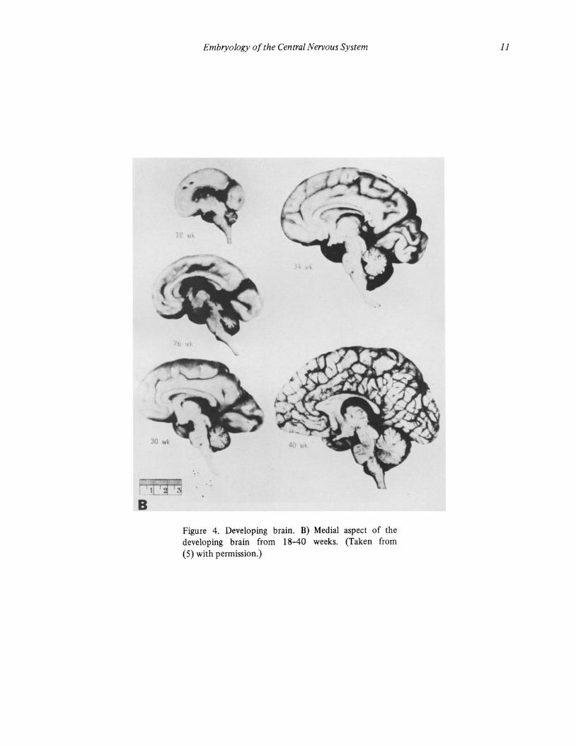
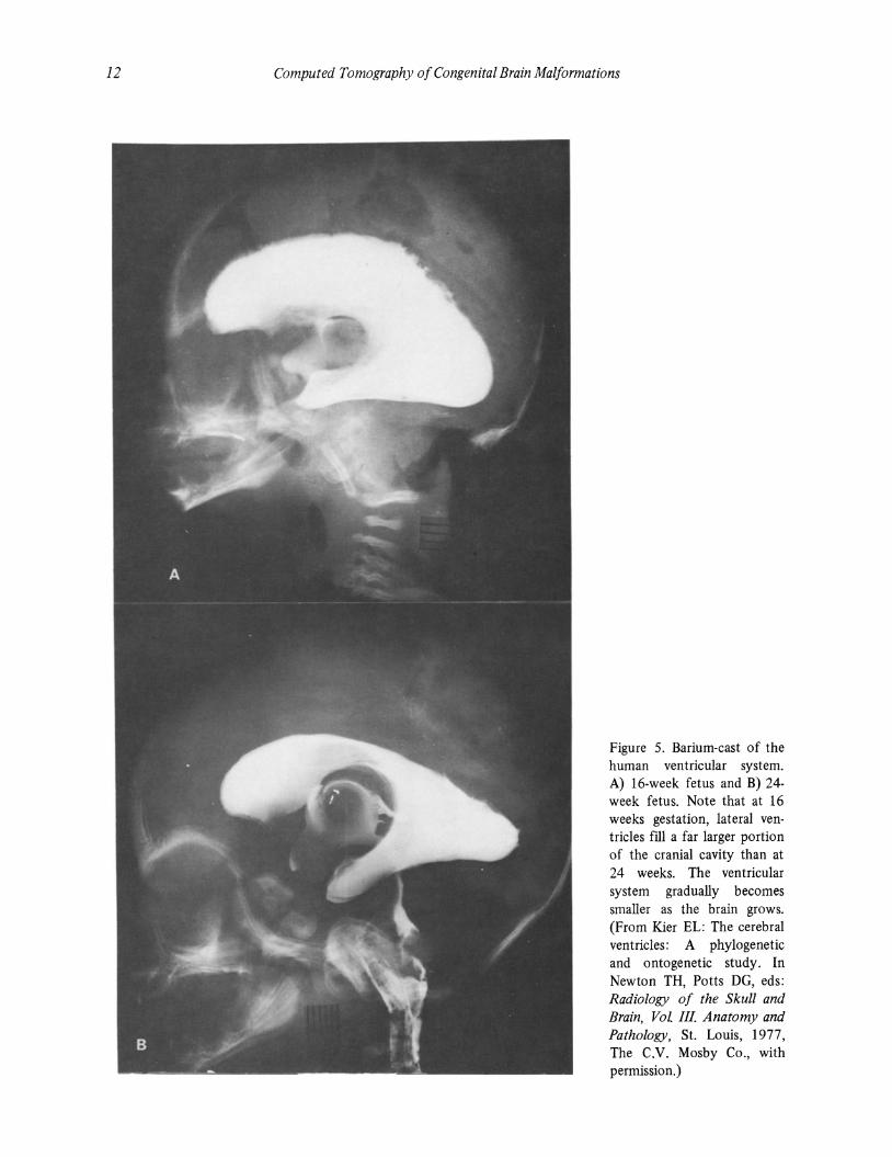
Concomitant with changes in size and shape of the brain (Fig. 4), adaptive changes occur in the brain cavities (ventricles). Prior to 20 weeks gestation, the ventricles normally occupy more than 50 percent of the hemispheres. These cavities progressively diminish in size as the brain increases in volume (Fig. 5). The relatively wide communication between the lateral ventricles and the third ventricle narrows to form the interventricular foramen (one on either side). The outlet foramina of the fourth ventricle are formed at the third month of gestation.
The prosencephalon (forebrain) is divided into its two components - telencephalon and diencephalon -by about the 36th day of gestation. The telencephalic vesicles (which lead to the formation of recognizable cerebral hemispheres by the third gestational month) extend anterior to the cephalic end (the lamina terminalis) of the neural tube. The telencephalon
In the fourth week, the foregut entoderm elaborates to create anlage for facial structures. This process is influenced by cephalic neural tube (anlage for telencephalon) and by cephalic mesoderm (anlage for facial bones and skull). Proper development of the brain, skull and facial skeleton depends on the intricate interplay of various inductive processes among the three germ layers. Any process that impairs such induction results in facio-telencephalopathy (holoprosencephaly).
Cell proliferation occurs at the subventricular level; this cell population, called germinal matrix, is
7 M. Sarwar, Computed Tomography of Congenital Brain Malformations© Warren H. Green, Inc. 1985
8 Computed Tomography of Congenital Brain Malformations
DORSAL VIEW OF EMBRYO AT 20 DAYS DORSAL VIEW OF EMBRYO AT 21 DAYS DORSAL VIEW OF EMBRYO AT 24 DAYS
_.. NlNfol pklf!' 01 fOftbtOln -----1-~~""\\
-i----1f--+ -+-+--'£\O[l srCTION. Nwol qrc:!<M -
- - -N"',ol lold.-
20rTIm ---+"4..~'f--- LIVE, or SlCTHlO
--+-\--\--11-1+-- ,£\0[, rT SlCTIOII •
---- - Fu5Od ""'01 loId'.---=-,,~~~
26 .....
Fulure nalJcl (Ieil NeutOI fok!
b
I ~euiOI 9100Y!'
~. I •• . . ., - . . . . . ' . ,
- ~ -.
----5,"'" ,1Ion>b~d.I,."
~~ ;'f .. ,~.,~.-¥·J H lft f 1Il1l[1. , .
Figure 1. Neural tube (redrawn from Clinical Symposia (3) with permission).
Embryology of the Central Nervous System
A. CENTRAL NERVOUS SYSTEM AT 28 DAYS Midbrain
( mesencephalon Forebraln--_r._
(prosencephalon)
Hindbrain (rtx:robercepholon)
HypothalamIc sulcus I
Sagittal Sectoon
D,ocele (3rd venroc le)
Openong of elencephallc
Forebrain { ( prosencephalon)
vesicle !lateral lelocele)
Sogittol Section
3.6mm
Sulcus iomltons
- OpflC vesIcle
MIdbrain Mesocele Hindbrain Rhombocele
Bosal plate
SPI nol medulla (cord)
Frontal Section (vent ral to sulcus hml tans )
B. CENTRAL NERVOUS SYSTEM AT 36 DAYS
Hind bra I n (r hom benc epha Ion)
Mldbraln- __ -/ (mesencephalon)
DIencephalon
Telencephalic veslCle---\-
ThIn roof of myelencephalon
(rTle()jlo oIlIonga 0)
Myelocele (4th ventricle)
Cenlral conal
Gsr.'"jr' \Si7' .I\~f[~ F' \ ET~(R M I)
8.0mm
SPinO I medulla
L amIno lermlnoll s TelencephalIC veSIcle
~~~@\kMed lan lelocele (3rd venlricle)
Loteral telocele (lateral ventficleJ
-Dlocele (3rd ventflcle) - OpIIC cup
Mesencephalon Mesocele (cerebral aqueducl)
8asal plate
Metencephalon (cere bellum, pons) Melocele (4 h venrrocle)
Myelencephalon
(cord) "/
Cenlro l cona l
(medulla oblongata) Myelocele (4 th ventrocle)
Frontal Section
Figure 2. Developing central nervous system. (Redrawn from Clinical Symposia (3) with permission.)
9
10
PROSENCEPHALON
MESENCEPHALON
RHOMBENCEPHALON
.. • •
Computed Tomography of Congenital Brain Malformations
PRIMARY DIVISIONS
SUBDIVISIONS
TELENCEPHALON
DIENCEPHALON
MESENCEPHALON
METENCEPHALON
DERIVATIVES
CEREBRAL CORTEX CORPUS STRIATUM RHINENCEPHALON LATERAL VENTRICLES AND ROSTRAL THIRD VENTRICLE
THALAMUS HYPOTHALAMUS
MIDBRAIN
PONS CEREBELLUM PONTINE PART OF FOURTH VENTRICLE
MYELENCEPHALON MEDULLA OBLONGATA BULBAR PORTION OF FOURTH VENTRICLE
Figure 3. Primary vesicles and their secondary dIviSIOns and derivatives .
Figure 4. Developing brain. A) Lateral aspect of the developing brain from 10-40 weeks. (Taken from (5)
B
Embryology of the Central Nervous System
..
Figure 4. Developing brain. B) Medial aspect of the developing brain from 18-40 weeks. (Taken from (5) with permission.)
11
12 Computed Tomography of Congenital Brain Malformations
Figure 5. Barium-cast of the human ventricular system. A) 16-week fetus and B) 24-week fetus. Note that at 16 weeks gestation, lateral ventricles fill a far larger portion of the cranial cavity than at 24 weeks. The ventricular system gradually becomes smaller as the brain grows. (From Kier EL: The cerebral ventricles: A phylogenetic and ontogenetic study. In Newton TH, Potts DG, eds: Radiology of the Skull and Brain, Vol III Anatomy and Pathology, St. Louis, 1977, The C.V_ Mosby Co., with permission.)
Embryology of the Central Nervous System 13
particularly well-developed along the frontal horns. These cells migrate to the periphery with each successive generation progressing further than the preceding one. The migration process occurs at a rate of l/lOmm per day and appears to be amoeboid. The complexity of neuronal organization can lead to developmental disorders of the cerebral cortex, which may be manifested in a variety of ways. Brain dysmorphogenesis may be caused by defects in gyration (micro polygyria), cortical organization (disruption of normal cortical lamination), cell migration (heterotopia of gray matter), and in cell proliferation (astrocytosis). Neuronal ectopias, although commonly seen in association with other CNS malformations, can be present in apparently normal brains.
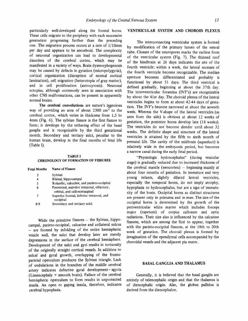
The cerebral convolutions are nature's ingenious way of providing an area of about 2300 cm2 to the cerebral cortex, which varies in thickness from 1.5 to 4mm (Fig. 6). The sylvian fissure is the first fissure to form; it develops by the tethering effect of the basal ganglia and is recognizable by the third gestational month. Secondary and tertiary sulci, peculiar to the human brain, develop in the final months of fetal life (Table I).
TABLE I CHRONOLOGY OF FORMATION OF FISSURES
Fetal Months Name of Fissure
3 Sylvian 4 Rhinal, hippocampal 5 Rolandic, calcarine, and parieto-occipital 6 Precentral, superior temporal, olfactory,
orbital, and callosomarginal 7 Superior frontal, inferior temporal, and
occipital 8-9 Secondary and tertiary sulci
While the primitive fissures - the Sylvian, hippocampal, parieto-occipital, calcarine and collatera~ sulc~s - are formed by infolding of the entire hemIsphenc vesicle wall, the sulci that develop later are merely depressions in the surface of the cerebral hemisphere. Development of the sulci and gyri results in tortuosity of the originally straight cortical vessels. In addition to sulcal and gyral growth, overlapping of the frontoparietal operculum produces the Sylvian triangle. Lack of undulations in the branches of the middle cerebral artery indicates defective gyral development - agyria (Lissencephaly = smooth brain). Failure of the cerebral hemispheric operculum to form results in unprotected insula. An open or gaping insula, therefore, indicates cerebral hypoplasia.
VENTRICULAR SYSTEM AND CHOROID PLEXUS
The interconnecting ventricular system is formed by modification of the primary lumen of the neural tube. Closure of the neuropores marks the earliest form of the ventricular system (Fig. 7). The thinned roof of the hindbrain at 26 days indicates the site of the fourth ventricle; within a week, the lateral recesses of the fourth ventricle become recognizable. The median aperture becomes differentiated and probably is functional by about 51 days. The third ventrical is defined gradually, beginning at about the 37th day. The interventricular foramina (IVF's) are recognizable by about the 41st day. The choroid plexus of the lateral ventricles begins to form at about 42-44 days of gestation. The IVF's become narrowed at about the seventh week. Whereas the V-shape of the lateral ventricles (as seen from the side) is obvious at about 12 weeks of gestation, the posterior horns develop late (18 weeks). The ventricles do not become slender until about 32 weeks. The definite shape and structure of the lateral ventricles is attained by the fifth to sixth month of prenatal life. The cavity of the midbrain (aqueduct) is relatively wide in the embryonic period, but becomes a narrow canal during the early fetal period.
"Physiologic hydrocephalus" (during vesicular stage) is gradually reduced due to increased thickness of the cerebral mantle (neocortex) - beginning mainly at about four months of gestation. In immature and very young infants, slightly dilated lateral ventricles, especially the temporal horns, do not imply atrophy, hypoplasia or hydrocephalus, but are a sign of immaturity of the brain. Occipital horns as distinct structures are present only in primates and in man. The size of the occipital horns is determined by the growth of the periventricular white matter which includes forceps ma,jor (tapetum) of corpus callosum and optic radiations. Their size also is influenced by the calcarine fissures, which are among the first to appear, together with the parieto-occipital fissures, at the 18th to 20th week of gestation. The choroid plexus is formed by invagination of the ependymal cells accompanied by the choroidal vessels and the adjacent pia mater.
BASAL GANGLIA AND THALAMUS
Generally, it is believed that the basal ganglia are entirely of telencephalic origin and that the thalamus is of diencephalic origin. Also, the globus pallidus is derived from the diencephalon.
14 Computed Tomography of Congenital Brain Malformations
Figure 6. Lateral view of fetal cerebral hemispheres demonstrating formation of the fissures and sulci and opercularization of the sylvian fossa. A) 17·week fetus. The external surface of the cerebral hemisphere is smooth. The insula (arrow) can be seen through the wide open sylvian fossa (arrOWheads). B) 24-week fetus. The insula (arrow) is now partially overlapped, mainly by the parietal operculum., The posterior portion of the sylvian fossa 'is now transformed into the sylvian fissure (arrowhead). C = Central Sulcus C) 32-week fetus. The insula is now almost totally covered by the parietotemporal opercula. The frontal operculum develops completely postnatally. (From Kier EL: Development of cerebral vessels. Section 1. Fetal cerebral arteries: a phylogenetic and ontogenetic study. In Newton TH, Potts DG, eds: Radiology of the Skull and Brain. Vol. II. Angiography. St. Louis, 1974, The C.V. Mosby Co., with permission.)

Embryology o/the Central Nervous System
A. FOREBRAIN AT 7 WEEKS !transverse section)
Roo a 3rd _en flcle ~Chorolda l vein and or ery
" " __ I,", Telencep allc vesicle !cerebrat hemisphere, neopat Ilum I
B. TELENCEPHALON AT 7112 WEEKS (transverse sect ion)
Roo a 3rd en flcle
Hippocampus (are lpaillum)
15
Loterot telocele ----I-.LL
Choro:d ple,us
(lateral ventncle)
Mantle loyer
Marginal layer -
Median lelacele (3rd ventric le)
Anterior lobe a hypophySIS (pltullory gland)
C. FOREBRAIN AT 2 MONTHS (secliQned coronolly and viewed from anterior)
D. TELENCEPHALON AT 2112 MONTHS (Viewed from right and ontenor)
Epophysls (pmeol body)
Choroid plexus-
holomus
3rd 'Ientllcle
OptiC (nerve) salk
E. RIGHT CEREBRAL HEMISPHERE AT 3 MONTHS (medial ospecll
edlCl surface of right cerebral hemisphere (neopallium)
Corpus callosum~
Commissure of the lorn" ( rnppocompol
commlssure)1
\ For",x~
AnleMr commissu re
3rd venilicle
ChorOidal vessels passmg to lie cholOld plexus which pro ludes
Into the fight loterol ventllcle along Ihe chorOid fissure
HI ppocompus (archipallium)
StliO termmolls
ChorOId ple.us prolrudmg IOta IIghl lateral ven/llcle olong chorOid fissure
F. CEREBRAL HEMISPHERES AT 3 MONTHS !coronal section}
Duro moler $operlQ( sagil 01 SIOllS Fol. cerebll Inlellar s agittal smus
Ependymol loyer =-+-:::~I Loterol ventllcle Mon Ie loye(c./-;::;--~~ Ependymol- piO I
MorgIOol layer covellng 01
Hlppocompol cor extl--+1~;'%
chorOid plexus
Corpus Siliotum {Caudate nucleus (basal Lenticular nucleus ganglial
,' r.
Lommo lermlnolls
Olloctory lobe (poleopolhum) AFTER r ETTER. !II 0
Figure 7. Developing telencephalon. (Redrawn from Clinical Symposia (3) with permission.)

16 Computed Tomography of Congenital Brain Malformations
BRAINSTEM AND CEREBELLUM
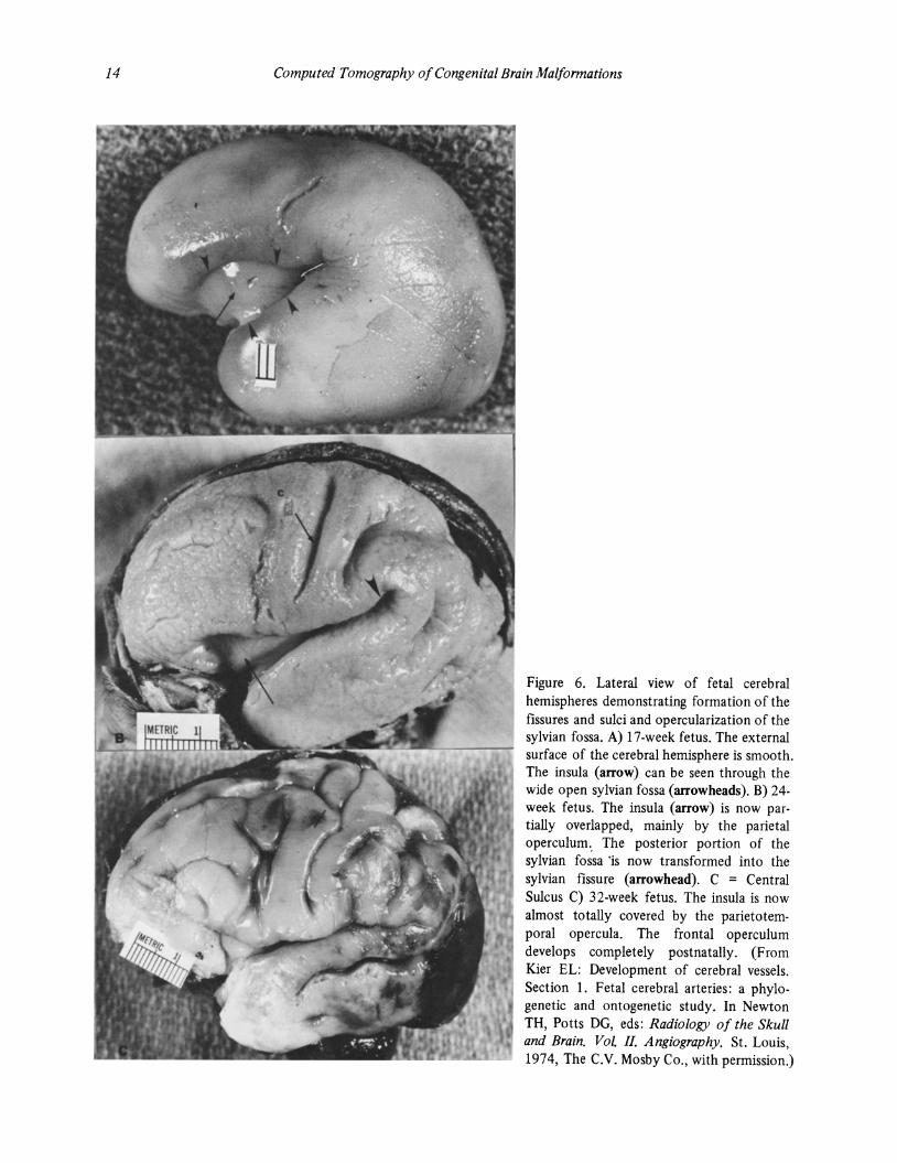
The brainstem and the spinal cord grow much more rapidly in the first half of prenatal life; the converse is true of the cerebellum. Growth of the cerebellum is completed by about 1.5-2 years of life after birth (cf: maturation of the cerebral cortex is relatively delayed, extending well into the postnatal period). Cerebellar primordia are paired structures; they soon coalesce at the midline: The medial portions form the vermis, and the cerebellar hemispheres expand rapidly on either side of the vermis. During the fourth to fifth month of gestation, the superficial parts of the cerebellum grow profusely, accounting for the numerous lobules and fissures (Fig. 8). The horizontal fissure (at fourth month of gestation) divides the cerebellar hemispheres transversely. The pons is intimately related structurally and functionally to these structures; it develops secondarily in the ventrolateral margins of the original rhombencephalon. Early in the embryo stage, its location is indicated by the pontine flexors.
Table II summarizes the main events of brain development.
TABLE II OUTLINE OF NEUROEMBRYLOGIC DEVELOPMENT
Weeks
3.5
4
5
6
7
B
12
16
24
2B
Length Body Weight (in grams)
2.5mm Optic vesicles and auditory placodes present.
5mm
Bmm
12mm
27mm
23mm
56mm
112mm
32cm
3B.Scm
BOO
1100
1) Neural tube closed. 2) Three primary brain
vesicles present. 3) Olfactory placodes
present.
Five brain vesicles.
Three primary flexors present
1) Cerebral hemispheres enlarging.
2) Prominent basal ganglia and thalami.
3) Choroid plexi forming.
Meninges distinct.
1) Confluence of sinuses attains normal location.
2) Cerebellum developed.
Myelination begins.
Typical layers in cerebral cortex.
Cerebral fissures and convolutions.
RATE OF BRAIN GROWTH
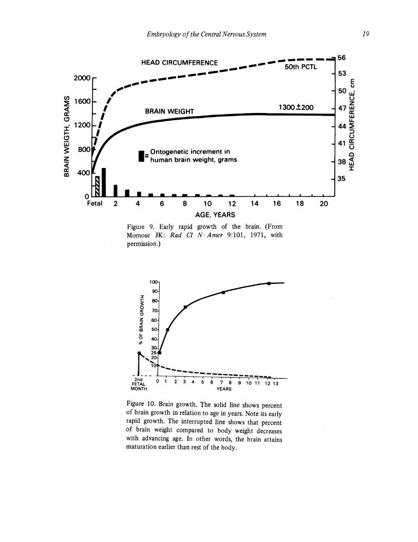
At birth, the brain weighs 400-450 gm. It doubles its weight in one year and triples it at three to five years of age. After age five, the brain growth slows considerably; the adult weight is attained by about 7·12 years of age (Fig. 9 and 10).
DEVELOPMENTAL HISTOLOGY AND CELLULAR PROLIFERATION AND MIGRATION
The nerve cells, which themselves do not divide, originate from the periventricular (neuroepithelial) germinal cells (these cells form the loosely arranged periventricular germinal matrix). Germinal cells may be considered the precursors of all types of cells. Located at the ventricular surface, germinal cells give rise to neurons, while the more externally placed spongioblasts give rise to neuroglia. It is believed that germinal cells and spongioblasts are the same type of cells but that they have moved different distances from the ventricular wall during the various phases of the mitotic cycle.
An intricate system of mutually inductive processes between the three germ layers leads to formation of the brain. The major inductive processes are complete by about 30 days of gestation when cellular differentiation begins. The young neurons, which arise from the germinal cells, migrate peripherally from the ventricular surface in a radial pattern using the glial fibers as guides. Each successive neuron moves to the outside of its predecessor ("inside-out" rule). Cell migration produces white and gray matter. It follows, therefore, that CNS malformations may originate at the time of migration or during any process that impairs the migratory pattern or development of the synaptic mechanisms.
By about 25 weeks of gestation, the adult number of neurons is achieved; from thereon there is minimal, if any, further cellular proliferation. At the same time, neuronal electrical activity becomes identifiable. The process of formation of neuronal (synaptic) processes -dendrites and axons - rapidly ensues. The synaptic development continues into the second year after birth. At the time of birth, the oligodendroglia show great activity presaging postnatal myelination. By about the 12th week, the cerebral cortex is highly cellular, and programmed cell death occurs in those structures that are destined to disappear.
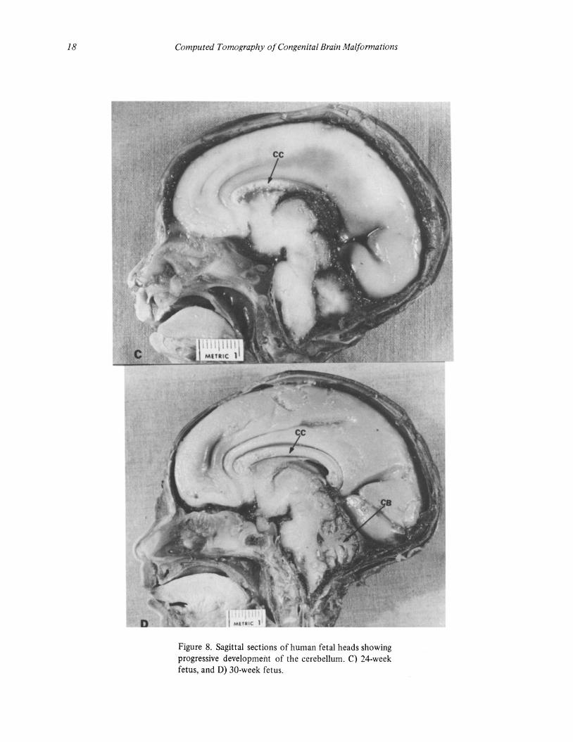
Figure 8. Sagittal sections of human fetal heads showing progressive development of the cerebellum. A) II-week fetus; B) IS-week fetus; C) 24-week fetus; D) 30-week fetus. From its rudimentary status in A, the cerebellum (CB) attains considerable size and folial formation by 30 weeks as seen in D. The brainstem is relatively larger than the cerebrum and cerebellum in the early fetal period. Note how the tectum (TC) overlaps the cerebellum at 11 weeks and 15 weeks of gestation (A & B). The corpus callosum (CC) is recognizable at 24 and 30 weeks of gestation (C & D). Note also the progressive growth of the cerebrum (From Kier EL: The cerebral ventricles: a phylogenetic and ontogenetic study. In Newton TH, Potts DG, eds: Radiology of the Skull and Brain. Vol. III. Anatomy and Pathology, St. Louis, 1977, The C.V. Mosby Co., with permission.)
Embryology of the Central Nervous System 17
18 Computed Tomography of Congenital Brain Malformations
Figure 8. Sagittal sections of human fetal heads showing progressive development of the cerebellum. C) 24-week fetus, and D) 30-week fetus.
CJ)
~ « a: (!)
:::c ~ (!)
w ~ z « a: a:l
2000
1600
1200
," I
I I I I
Embryology of the Central Nervous System
-----HEAD CIRCUMFERENCE ______ .- -- 50th PCTl --------... --... -.".""
BRAIN WEIGHT 1300.:!:200
1_ Ontogenetic increment in - human brain weight, grams
O~~~~~~~LA~~~----~~~~~~~~ Fetal 2 4 6 8 10 12 14 16 18 20
AGE, YEARS
Figure 9. Early rapid growth of the brain. (From Momose JK: Rad Cl N· Amer 9:101, 1971, with permission.)
100
90 :z: ~ 80
0 70 II:
C,!)
z 60 < II: m IL. 0 *-
3
J~~ ~-~-~-------_______ _ 2nd 0 1 2 3 4 5 6 7 8 9 10 11 12 13 FETAL
MONTH YEARS
Figure 10. Brain growth. The solid line shows percent of brain growth in relation to age in years. Note its early rapid growth. The interrupted line shows that percent of brain weight compared to body weight decreases with advancing age. In other words, the brain attains maturation earlier than rest of the body.
19
56
53 E u
50 w' u z
47 w a: w u.
44 ~ :::> u
41 !!: u 0
38 ~ :::c
35
20 Computed Tomography of Congenital Brain Malformations
MYELINA nON
At birth and in the perinatal period, most of the white matter of the cerebral hemispheres of the fuD term newborn is deficient in myelin. The exceptions are a small number of fibers of the primary afferent projection system - e.g., somatic sensory fibers from the ventral posterolateral nucleus of the thalamus to the postcentral· and precentral cortex and the visual and auditory fibers from the geniculate bodies to their respective cortical areas. At the end of the first month in the full term infant, nascent deposition of myelin in the corticospinal tract is recognizable. Simultaneously, a considerable number of commissural fibers of the corpus callosum become myelinated. The frontotemporal pontine fibers and the long association tracts, such as the superior occipital frontal fasciculum and cingulum, contain only a small trace of myelin sheaths at birth and in early infancy. The process of myelination, although more pronounced in the first two years after birth, continues into the early adult life. The chronology and pace of myelination of different fiber tracts is closely related to their function - i.e., the ability of the infant to acquire progressive control of its muscular movement in the first and second year of life bears close relationship to the relatively rapid pace
of myelination in the latter part of the first and early part of the second postnatal year.
REFERENCES
1. Arey JB: Developmental Neuroanatomy. Philadelphia, Saunders, 1954.
2. Cowan WM: The development of the brain. Scientif icAmerican 241:113-133,1979.
3. Crelin ED: Development of the nervous system. A logical approach to neuroanatomy. CIBA Clinical Symposium 26:No. 2, 1974.
4. Kier EL: The cerebral ventricles: A phylogenetic and ontogenetic study. In: Newton TH, Potts DG, eds: Radiology of the Skull and Brain. Anatomy and Pathology. p.2787. St. Louis, CV Mosby Co, 1977.
5. Larroche J-e: Developmental Pathology of the Neonate. Amsterdam, Excerpta Medica, 1977.
6. Lemire RJ, Loeser JD, Leech RW, Alvord EC, Jr: Normal and Abnormal Development of the Human Nervous System. Hagerstown, Harper and Row, 1975.
7. Patten BM: Human Embryology. 3d ed. New York, Blakiston DiVision, McGraw - Hill, 1968.