Embryology and Desmosomes - dermpathmd.comdermpathmd.com/Clinical Dermatology/Embryology and...
76
Transcript of Embryology and Desmosomes - dermpathmd.comdermpathmd.com/Clinical Dermatology/Embryology and...


Embryology Refresher
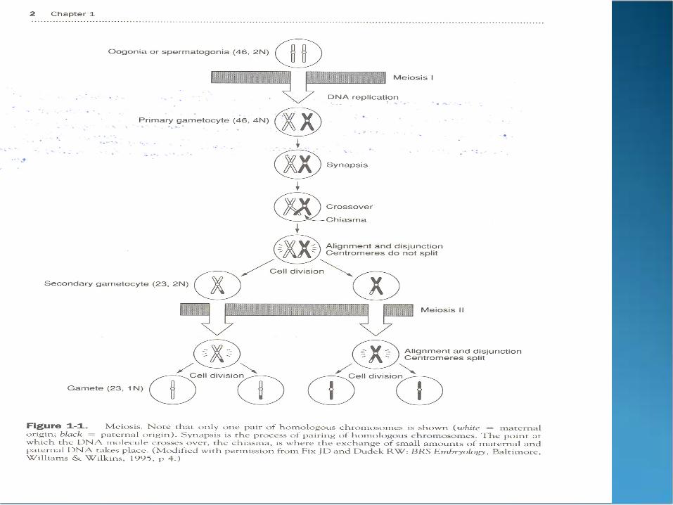
Gametes (oocytes and spermatozoa) Descendents of primordial germ cells (46, 2N) Meiosis (oogenesis or spermatogenesis)
2 divisions (I and II) Results in 23, 1N Allows genetic variability Maintain # of chromosomes
Embryology Refresher
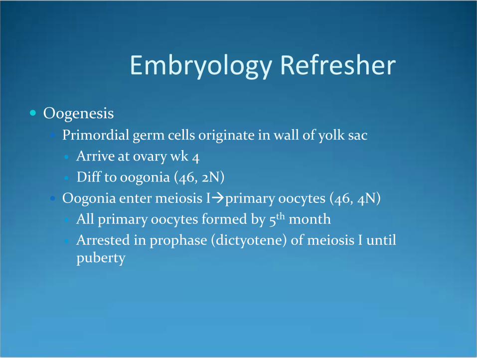
Oogenesis Primordial germ cells originate in wall of yolk sac
Arrive at ovary wk 4 Diff to oogonia (46, 2N)
Oogonia enter meiosis Iprimary oocytes (46, 4N) All primary oocytes formed by 5th month Arrested in prophase (dictyotene) of meiosis I until
puberty
Embryology Refresher
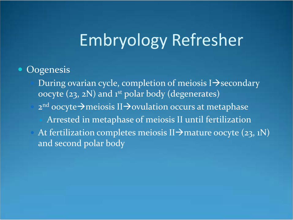
Oogenesis During ovarian cycle, completion of meiosis Isecondary
oocyte (23, 2N) and 1st polar body (degenerates) 2nd oocytemeiosis IIovulation occurs at metaphase
Arrested in metaphase of meiosis II until fertilization At fertilization completes meiosis IImature oocyte (23, 1N)
and second polar body
Embryology Refresher
Spermatogenesis Primordial germ cells (46, 2N) arrive at testes wk 4 Dormant until puberty At puberty diff. to type A spermatogonia (46, 2N)

Embryology Refresher
Type A spermatogonia undergo mitosismore type A or type B
Type Bmeiosis Iprimary spermatocytes (46, 4N)
Primary spermatocytes complete meiosis I2 secondary spermatocytes (23, 2N)
Secondary spermatocytes complete meiosis II4 spermatids (23, 1N)

Embryology Refresher
Human genome has 23 different chromosomes of which each cell has 2 copies (=46 total)
1 copy from mom 1 copy from dad 2 nonidentical copies of a chromosome are called? Different versions of genes are?

Embryology Refresher
Fertilization occurs where? Male and female pronuclei fuse to form? Zygote is successively cleaved to form? At 32 cell stage blastomeres form? What forms when fluid is secreted within morula? Inner cell mass becomes? Outer cell mass becomes?

Embryology Refresher
Embryonic period occurs? What is the process that establishes the 3 primary
germ layers (ectoderm, mesoderm, and endoderm)? The formation of what marks the initiation of
gastrulation?


Embryology Refresher
Ectoderm Surface ectoderm
Epithelial lining of ant 2/3 of tongue, hard palate, sides of mouth, ameloblasts (teeth), and parotid glands and ducts
Mammary glands Epithelial lining of lower anal canal Epithelial lining of distal penile urethra Epidermis, hair, nails, sweat and sebaceous glands

Embryology Refresher
Ectoderm Neuroectoderm
CNS stuff
Neural Crest Melanocytes
Embryology Refresher
Mesoderm Lateral
Lymphatic system CVS
Intermediate Paraxial
Dermis Extraocular muscles Skeletal muscles of trunk and head and neck Intrinsic muscles of the tongue
Embryology Refresher
Endoderm Epithelial lining of:
Post 1/3 of tongue Floor of mouth Palatoglossal and palatopharyngeal folds Soft palate Vagina Female urethra and most of male urethra Auditory tube

Embryology Refresher
Fetal erythropoiesis occurs in (order)? What does a persistent cervical sinus lead to? What does aberrant development of the 3rd and 4th
pharyngeal pouches lead to? 3rd pouch dorsal—inf parathyroids 3rd pouch ventral—thymus 4th pouch sup parathyroids

Embryology Refresher
Most common site of ectopic thyroid tissue? Tongue innervation: Taste? CN 7, 9, 10 Pain? CN V3, 9, 10 Motor? CN 12

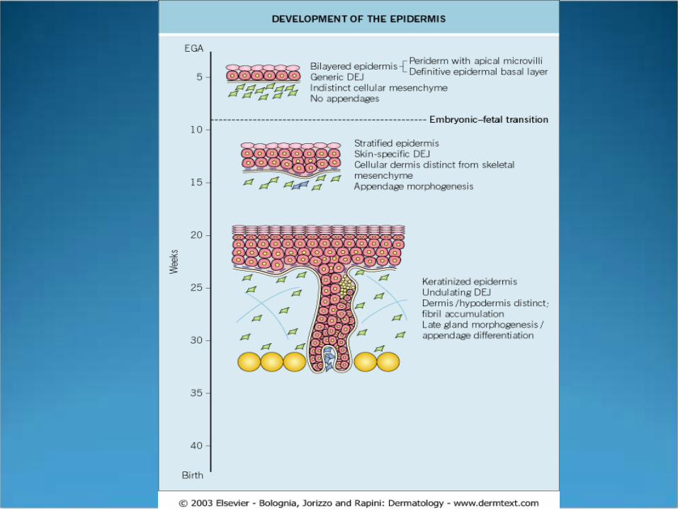
Embryology of the Skin
High Points Morphogenesis of all skin components (except non-
volar swear glands) is underway by end of 1st trimester Differentiation of epidermis and appendages occurs
primarily in 2nd trimester EGA—Fertilization occurs day 1, lags by 2 weeks (used in
Fitz and Bolognia) LMP—1st day of last menstrual period
OB/GYNs, fertilization day 14


Epidermis
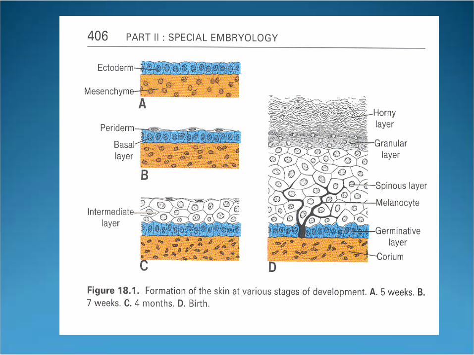
1st month primitive single layer epidermis creates periderm
Periderm Specialized embryonic structure Covers epidermis until keratinization occurs Then degenerates Cells attached by tight junctions Studded with microvilli
Large single blebsmultiple blebs
Epidermis
Periderm Cells attached by tight junctions Studded with microvilli
Large single blebsmultiple blebs
Possible role in diffusion or exchange of substances across fetal skin

Epidermis
Periderm Cells detach from underlying epidermissloughed
into amniotic fluid Becomes part of vernix caseosa
Prevents maceration from amniotic fluid
Sloughing via apoptosis DNA fragmentation TGase 1 and 3 detected


Epidermis
Organogenesis wks 3-8 Bone marrow hematopoiesis begins at 8 wks EGA
(transition from embryo to fetus) Epidermal Stratification begins 8 wks EGA Completed by 2nd trimester
Epidermis
Epidermal stratification begins with formation of intermediate layer Int. layer between basal and periderm layer Highly proliferative p63 required for epidermal stratification Several new layers added over next few wks
By 22-24 wks, 4-5 layers plus periderm
Epidermis
Keratinization Begins 2nd trimester Matures by mid 3rd trimester Keratinization of appendages begins 11-15 Keratinization of epidermis begins 22-24 wks
Begins on head, face, palms, soles 24 wks s. corneum few layers

Epidermis
Progression of keratinization # of keratohyalin and lamellar body granules increases Increase in # of organelle-depleted cornified cells Neonate’s skin barrier not completely mature until a few
weeks after birth Full barrier function 3 wks of age

Defects of Epidermal Maturation X-linked ichthyosis Steroid sulfatase Lamellar ichthyosis TGase 1 Responsible for cross-linking precursor proteins to
form insoluble cornified envelope Born with collodion membraneshedlarge
polygonal platelike hyperpigmented scales


Specialized Cells in Epidermis Melanocytes
Neural crest along dorsal neural tube Migrate to epidermis and hair follicles via
mesenchyme Also migrate to uveal tract, leptomeninges, and cochlea
Present in epidermis by mid 1st tri (50 days) Fully functional 2nd tri Melanin production 3-4 months Melanosome transfer 5 months Newborn skin not fully pigmented at birth

Specialized Cells of the Epidermis Melanocytes (Receptor-Ligands)
Steel factor binds to KIT receptor on melanocytes and melanoblasts
Mutations in KIT gene? Endothelin B receptor, endothelin 3 Pax3 critical in migration from neural crest and
activation of melanocyte proliferation Piebaldism and 4 types of Waardenburg’s syndrome
caused by failure of adequate number of melanoblasts to reach distal sites


Specialized Cells of Epidermis Langerhans cells
Appear 40 days Express HLA DR High levels of ATPase Expression of CD1a and production of Birbeck granules
(mature) begins at 8 wks

Specialized Cells of Epidermis Merkel Cells
Highly innervated neuroendocrine cells; mechanoreceptors
Detected at 8-12 wks in palmoplantar epidermislater in interfollicular skin
Identified by cytoplasmic dense core granules, cytokeratin 20, and neuropeptide expression
Dense on volar skin Probably derived from pluripotent keratinocytes,
not neural crest
Dermis
Dermis origin varies by body site Face and anterior scalpNeural crest (facial dysmorphia
in Waardenburg’s) BackDermomyotome of embryonic somite Extremities and ventral trunklateral plate mesoderm

Dermis
Embryonic fibroblasts are pluripotent cellsadipocytes, fibroblasts, and cartilage-producing cells
Dermal cells situated under epidermis by 6-8 wks At this stage:
Able to synthesize collagen, but ratio of collagen III to I is 3:1 (reverse in adults)
No demarcation b/w cellsdermis and cellsmusculoskeletal components

Dermis
At 60 days (embryonic-fetal transition), superficial mesenchyme becomes distinct from skeletal components
12-15 wks: distinguish fine weave pattern of papillary dermis from reticular dermis
Large collagen fibers accumulate in 2nd and 3rd tri 22-24 wks: elastic fibers detected

Dermis
Differences b/w embryonic and adult dermis Embryo: watery, cellularAdult: more fibrous and
acellular Embryo: extracellular gel-like matrix of large well-
hydrated proteoglycansAdult: rigid fibrous End of 2nd trimester shift from non-scarring to scarring At birth thick and well organized but still thinner and
more cellular than adults

Why important?
Goltz syndrome—focal dermal hypoplasia X-linked dominant Boys die in utero Girls, functional mosaicism Islands of dermal hypoplasia follow Blaschko’s lines;
bordered by normal dermis


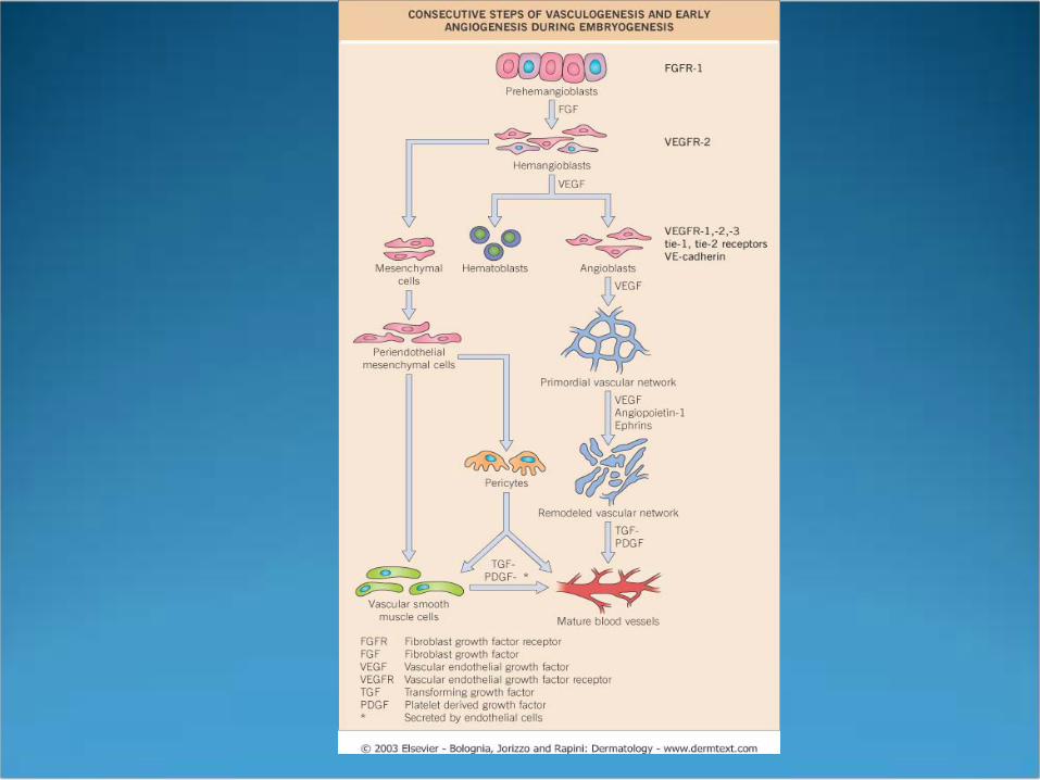
Vasculature
Vasculogenesis Differentiation of angioblasts into endothelial cells that
form a primitive vascular network Angioblasts originate in extraembryonic mesoderm of
yolk sac Hemangioblasts differentiate into hematopoietic cells
and angioblasts

Vasculature
Angioblasts express receptors: VEGFR-1,-2,-3, tie-1, tie-2, VE-cadherin
VEGF stimulation Angioblasts coalesce to form dorsal aorta and large
vessel primordia Angioblasts form a lumenendothelial cells

Vasculature
Angioblasts form a lumenendothelial cells Formation of primordial vascular plexus
Sinusoidal capillaries Polygonal honeycomb pattern
Establishment of interendothelial adherence jcts requires VE-cadherin

Vasculature
Primordial vascular plexus surrounded by mesenchymal cellspericytes and vascular smooth muscle cells
Then remodeling under influence of VEGF, angiopoietins, and ephrins
Major mechanism for new blood vessel formation—angiogenesis (sprouting)
Other mechanism—intussusception: division of vascular lumennew vascular space

Vasculature
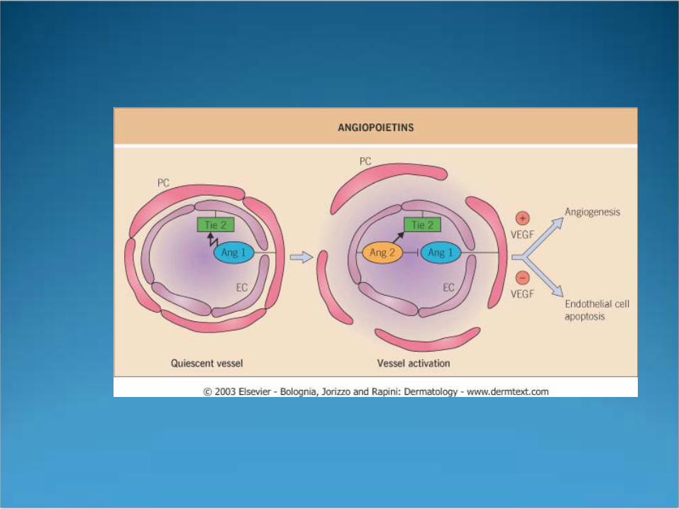
Tie-2 tyrosine kinase R is expressed on endothelial cells
Angiopoietin-1—vessel maturation Activates tie-2vascular sprouting and remodeling
Angiopoietin-2 Can inhibit Ang-1 Promotes angiogenesis in presence of VEGF Vessel regression in absence of VEGF
Vasculature
Ephrin-ephrin interactions determine vascular identity
Ephrin B2 is on arterial endothelium Ephrin B4 (B2s receptor) is on venous
endothelium Both define boundaries b/w arterial and venous
endothelial cells

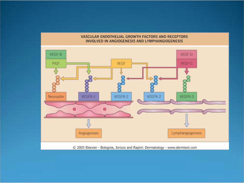
Lymphatics Primitive lymph sacs through to originate from
venous endothelial cell buds Peripheral lymphatics originate from lymph sacs
and sproud into tissues with capillaries Homeobox gene Prox1 specific marker for
lymphatic endothelial cells Prox1 deficiencyabsence of lymphatic system in mice
Lymphatics Markers of lymphatic system
VEGFR-3—receptor for VEGF-C and VEGF-D Podoplanin LYVE-1


DEJ
Components first appear 8 wks (with epidermal stratification)
Almost all structures in place by 12 wks BM proteins derived from basal keratinocytes
(ectoderm) HD proteins (BPAG1, plectin), BPAG2, integrin subunits
α6β4, types IV and VII collagen, laminins 5 and 6 and HSPGs

DEJ
Dermal fibroblasts (mesoderm) produce Nidogen (entactin), types IV and VII collagen and other
proteins translocated to the plasma membrane of basal keratinocytes
Plasma membrane of basal keratinocytes—localizational and organizational cues for fibroblast-derived proteins Cues provided by integrins


Hair
No new hair follicles form after birth 10 wks follicle formation begins on head (esp.
eyebrows and upper and lower lips 12-14 wks base of presumptive hair follicle invaginates,
enveloping presumptive dermal papilla cells forming bulbous hair peg
16 wks follicles develop cephalocaudally and ventrally 19-21 wks hair canal fully formed and scalp hairs visible
above skin surface

Hair
Follicle formation initiated by signals from dermisformation of follicular placode or anlage
Placodes seen wk 10 on scalp and face Placodes instruct dermis to condensedermal papilla Dermal papilla instruct placode cells to proliferate and
extend deeper into dermispeg stage hair Invagination of base of hair follicle (wks 12-14)
envelopes dermal papilla cellsbulbous hair peg
Hair
19-21 wks hair canal fully formed 24-28 wks go from anagencatagentelogen Telogen hairs shed in amniotic fluid Enter 2nd cycle Most hairs become thicker and coarser with
subsequent growth cycles (vellusterminal)
Hair
Bulbous Peg Phase Hair follicles differentiate in 2nd tri7 concentric layers
ORS, IRS (Henley’s, Huxley’s, cuticle), hair shaft cuticle, cortex, and medulla
Melanocytes interspersed among keratinocytes 3 distinct bulges
Upper bulgeapocrine gland Middle bulgesebaceous gland Deeper bulgeinsertion point of arrector pili muscle (hair bulge)

Hair Fodder
Undifferentiated epithelium? B-catenin Germ phase? Sonic Hedgehog Bulbous Peg Phase? NOTCH1

Sebaceous Glands
Parallel follicular development First 13-16 wks Middle bulge (aka superficial) Maternal hormones cause hypertrophy Unless stimulated by maternal hormones or
endogenous (tumor) become quiescent

Eccrine Glands
Palmoplantar eccrine development begins with formation of large mesenchymal bulges or pads (paw pads) Pads regress by 3rd trimester
Parallel ectodermal ridges overly pads Curves form fingerprints
Like hair and nails Begin to develop 1st trimester Fully developed by 2nd trimester
Eccrine Glands Eccrine gland primordia bud along ectodermal ridges at
wks 14-16 Buds elongate Dermal component canalized by wk 16 Epidermal component canalized by wk 22 Apocrine and interfollicular eccrine glands do not begin
to form until 5th month Apocrine glands function transiently in 3rd trimester,
quiescent in neonate Eccrine glands do not function in utero, only function
after birth
Nails Begins 8-10 wks from same primitive epidermis that
gives rise to hair, sweat glands, and s. corneum Complete 5th month Future nail bed demarcated by folds visible by 8-10
wks Ectoderm invaginates along proximal endproximal
nail fold Nail bed on dorsal digit—first skin structure to
keratinize at 11 wks Keratinization begins distallycontinues to proximal
nail fold

Nails
First preliminary nail is shed Replaced by hard differentiated nail plate
Emerges 4th month from under proximal nail fold Completely covers nail bed by 5th month
Predictable and constant time course of nail developmentused to estimate gestational age at term